Identification and tracking of problematic host cell proteins removed by a synthetic, highly functionalized nonwoven media in downstream bioprocessing of monoclonal antibodies
Bạn đang xem bản rút gọn của tài liệu. Xem và tải ngay bản đầy đủ của tài liệu tại đây (1.46 MB, 11 trang )
Journal of Chromatography A, 1595 (2019) 28–38
Contents lists available at ScienceDirect
Journal of Chromatography A
journal homepage: www.elsevier.com/locate/chroma
Identification and tracking of problematic host cell proteins removed
by a synthetic, highly functionalized nonwoven media in downstream
bioprocessing of monoclonal antibodies
S. Gilgunn a,1 , H. El-Sabbahy b,1 , S. Albrecht a , M. Gaikwad a , K. Corrigan a , L. Deakin b ,
G. Jellum c , J. Bones a,d,∗
a
Characterisation and Comparability Laboratory, The National Institute for Bioprocessing Research and Training, Foster Avenue, Mount Merrion, Blackrock,
Co. Dublin, A94 X099, Ireland
b
Separation and Purification Sciences Division, 3M United Kingdom PLC, 3M Centre, Cain Road, Bracknell, RG12 8HT, UK
c
Separation and Purification Sciences Division, 3M Centre, Building 236-1C-14, St. Paul, MN, 55144, United States
d
School of Chemical and Bioprocess Engineering, University College Dublin, Belfield, Dublin 4, D04 V1W8, Ireland
a r t i c l e
i n f o
Article history:
Received 8 January 2019
Received in revised form 15 February 2019
Accepted 24 February 2019
Available online 25 February 2019
Keywords:
Monoclonal antibodies (mAbs)
Protein A affinity chromatography
Host cell proteins (HCPs)
Downstream bioprocessing
Emphaze AEX Hybrid Purifier
a b s t r a c t
The repertoire of complex proteins produced by the host cell during monoclonal antibody (mAb) production has generated a bottleneck in downstream bioprocessing. Low ppm levels of host cell proteins (HCPs)
must be achieved at the downstream purification process stage to generate an end product suitable for use
in humans. The increased demand for mAb drug products globally has driven research to focus on affordability of mAb production platforms. This has fuelled advancements in manufacturing R&D to deliver
higher product titres with better economics without sacrificing product quality. This study highlights the
beneficial effects of inclusion of the EmphazeTM AEX Hybrid Purifier, compared to a conventional clarification process, for removal problematic HCPs during downstream bioprocessing of mAbs. Advanced
proteomic methods were used to track and identify known ‘problematic’ HCPs through a multi-cycle
Protein A purification process. Removal of histone proteins was observed, along with an average total
HCP reduction of 38-fold and an average reduction of 2.3 log in HCDNA concentration. Chromatographic
clarification using the EmphazeTM AEX Hybrid Purifier in conjunction with Protein A chromatography
resulted in the removal of problematic HCPs including 78 kD glucose-regulated protein, nidogen-1, heat
shock proteins, actin, serine protease HTRA1 and matrix metalloproteinase-19. It is shown herein that
the EmphazeTM AEX Hybrid Purifier, which is readily incorporated into a mAb purification process during
the clarification stage, has the potential to increase Protein A resin lifetime and potentially reduce the
number of subsequent polishing chromatographic steps needed to remove HCPs that have a tendency to
co-purify with mAb products.
© 2019 The Authors. Published by Elsevier B.V. This is an open access article under the CC BY license
( />
1. Introduction
Since the commercialisation of the first therapeutic monoclonal
antibody (mAb) product in 1986, the success story of mAbs as
therapeutic drugs continues to be truly remarkable. The “Purple
Book” list of licensed biological products, including biosimilar and
interchangeable biological products, regulated by the Centre for
∗ Corresponding author at: Characterisation and Comparability Laboratory, The
National Institute for Bioprocessing Research and Training, Foster Avenue, Mount
Merrion, Blackrock, Co. Dublin, A94 X099, Ireland.
E-mail address: (J. Bones).
1
Joint 1st Authors.
Drug Evaluation and Research (CDER), now stands at a lengthy
143 approved biological drugs. Over half (52%) of these are mAbs,
with 17 approved in 2017 alone (including Fc-fusion proteins, antibody fragments, and antibody-drug conjugates) [1]. The growing
approval and sales of these products also means there is a need to
increase the total quantities of mAb products produced annually to
meet the demands of the market [2].
MAb therapeutics must be manufactured in living cells or organisms unlike conventional pharmaceuticals which are developed
through chemical synthesis. Consequently, the species origin, the
choice of cell line, and culture conditions all affect the final product characteristics [3]. Mammalian cell lines, such as those derived
from Chinese hamster ovary (CHO) cells are long established as
the standard production platforms for such recombinant proteins
/>0021-9673/© 2019 The Authors. Published by Elsevier B.V. This is an open access article under the CC BY license ( />
S. Gilgunn et al. / J. Chromatogr. A 1595 (2019) 28–38
[4]. A major issue with using biological systems for mAb production is that the product itself must be purified from any cell-based
impurities that may co-purify with the drug substance. If not sufficiently removed, these process-related host cell impurities can
potentially become components of the final drug product. The protein impurities are more commonly known as host cell proteins
(HCPs) [5].
Over the past two decades, masses of biological medicines have
dominated the pharmaceutical armamentarium and the increased
demand for mAbs now drives research to focus on well-designed
upstream cell culture platforms for scaling up mAb production
[6,7]. Naturally, an increase in product titre also brings an increase
in process-related HCPs, challenging downstream bioprocessing
even further. HCP composition can be influenced at all stages of
upstream bioprocessing which, in turn, will impact the number
and type of chromatography steps required to ensure they are adequately removed prior to final drug product formulation [8,9].
HCPs are a highly diverse range of proteins, with considerable
differences in properties such as molecular mass, isoelectric point,
hydrophobicity, and structure [10]. This diverse pool of proteins
contained in the HCP profile generates various challenges for the
final drug product; many are enzymes that may catalyse degradation or comparable undesired alterations to the product [11,12].
Other HCPs may induce an unwanted immune response compromising the overall safety and efficacy of the therapeutic biologic
[13]. In some instances, HCPs can be both potentially degradative and immunogenic as was evident in initial phase III studies
of Lebrikizumab, a humanized immunoglobulin IgG4. The drug
product was found to contain a process-related impurity which
was identified as CHO phospholipase B-like 2 (PLBL2), a 66 kDa
mannose-6-phosphate glycosylated lysosomal enzyme. This a nonhuman protein with both unknown enzymatic activity and the
potential to induce an immune response [14].
The International Conference on Harmonisation (ICH) guideline
Q11 establishes HCPs as a Critical Quality Attribute (CQA) [15] and
regulatory guidelines (ICH guideline Q6B) stating that HCP levels
must be monitored and managed to acceptable levels. Exact limits are not specified in the regulations [16], however, they must
be established using risk-based approaches for each filing and take
into consideration manufacturing capability. A target limit of less
than 100 ppm in final drug product is commonly employed across
the industry, with the objective of lower levels for all commercial processes [17,18]. In order to meet these low ppm HCP target
levels, the downstream purification process must be robust to generate an end product suitable for use in humans. The vast majority
of purification processes for mAbs involve Protein A affinity chromatography following cell culture harvest. Subsequently, two or
three steps such as anion exchange, cation exchange and hydrophobic interaction chromatography are included as polishing steps
to remove problematic, co-purifying HCPs [19]. Owing to its high
selectivity for mAbs, Protein A affinity chromatography dominates
the capture technologies, however, it is also the biggest economical
challenge in downstream bioprocessing – attributing for 50–80%
of total purification costs [20,21]. Cost effective mechanisms to
improve Protein A performance and resin lifetime are now at the
forefront downstream process R&D [21].
Typically, ELISA is the most common method for the monitoring, detection and measurement of total HCP concentration during
mAb bioprocessing and in final biotherapeutic protein formulations. These assays utilise polyclonal antibodies generated from
immunised animals with a HCP pool from a null cell line [22].
There are numerous issues with using conventional ELISA including
low sensitivity, preferential detection of highly immunogenic HCPs,
laborious workflows and lack of dilution linearity [18]. Recently,
a move towards analytical methods such as liquid chromatography (LC) coupled with mass spectrometry (MS)-based methods are
29
being developed for identification and characterisation of specific
HCPs [13,23,24].
This body of research focuses on the evaluation of a novel, synthetic, highly functionalized media – 3 M’s EmphazeTM AEX Hybrid
Purifier – for removal of problematic HCP’s during clarification
of a mAb producing CHO cell culture. Previous work has shown
that cell culture and clarification conditions can have a significant
impact on the HCP profile which, in turn, will impact the number
and type of chromatography steps required to clear them [25,26].
The EmphazeTM AEX Hybrid Purifier enables reduction of soluble
and insoluble bioprocess-related contaminants, during clarification, using a Q-functional nonwoven matrix. This complex matrix
is formed with four layers of quaternary ammonium functionalised
nonwoven material and an asymmetric polyamide membrane with
a final pore size of 0.2 m [27]. The analysis and tracking of
HCPs removed by EmphazeTM AEX Hybrid Purifier and following
multi-cycle Protein A chromatography was carried out through
the use of highly sensitive and quantitative LC-MS approaches,
combined with immuno-PCR quantitation. These methods overcome the issues associated with conventional ELISA, allowing for
the detection, identification and monitoring of specific HCPs during mAb downstream bioprocessing. The inclusion of EmphazeTM
AEX Hybrid Purifier during clarification of mAb containing conditioned media has the potential to increase Protein A resin lifetime
and reduce the number of chromatographic steps in downstream
bioprocessing of mAbs.
2. Materials and methods
2.1. Reagents and consumables
All chemicals and reagents used during this study were purchased from Sigma-Aldrich and were ACS reagent grade or better
(Wicklow, Ireland). Water and solvents used were LC − MS Optima
grade and were obtained from Fisher Scientific (Dublin, Ireland).
2.2. Clarified cell culture material
Recombinant tocilizumab biosimilar IgG1 monoclonal antibody
was expressed by mammalian cell culture in a CHO cell line
as previously described [27]. Briefly, antibody was produced in
two 50 L disposable stirred tank bioreactors (Eppendorf, Hamburg,
Germany) in fed-batch cultures. Cells were harvested at day 14
with cell densities of 5.7 × 10⁶ cells/mL and 6.6 × 10⁶ cells/mL, and
final viabilities of 64% and 74%, respectively. Initial clarification of
harvest cell culture fluid (HCCF) was performed with a 30SP02 A
primary Zeta PlusTM depth filter (3M, St Paul, MN, USA) at throughputs of 75 L/m2 and 78 L/m2 , respectively, and a flux of 261 litres
per meter square per hour (LMH). The clarified material from each
bioreactor was pooled and divided for further clarification. The
product titre of the pool was 3.5 g/L. The first aliquot was clarified through a 90ZB08 A Zeta PlusTM polishing grade depth filter
(herein referred to as depth filter clarified material) at a throughput of 243 L/m2 and flux of 197 LMH, and the second was further
clarified using the EmphazeTM AEX Hybrid Purifier (herein referred
to as flow through anion exchange (FT-AEX) clarified material) at
a throughput of 262 L/m2 and a flux of 197 LMH. All material was
then sterile filtered using a 0.2 m LifeASSURETM PDA membrane
filter (3M, St Paul, MN, USA), aliquoted and frozen at −80 ◦ C.
2.3. Protein A chromatography
An ÄKTA Avant (GE Healthcare, Uppsala, Sweden) was used
for chromatographic experiments, monitored with Unicorn 7.0
software. A 1 mL MabSelectTM SuReTM HiTrap column (GE Healthcare, Uppsala, Sweden) was equilibrated with equilibration buffer
30
S. Gilgunn et al. / J. Chromatogr. A 1595 (2019) 28–38
(20 mM sodium phosphate, 0.15 M NaCl, pH 7.0) for 10 CV at a flow
rate of 0.5 mL/min. Eight CV of clarified cell culture fluid was applied
to the column at a flow rate of 0.25 mL/min. Following this, the column was washed with 10 CV of equilibration buffer (0.25 mL/min
for the first column volume and 0.5 mL/min thereafter) and the mAb
was then eluted with 0.1 M sodium citrate, pH 3.2 in 8 CVs and the
elution peak was automatically collected (when the UV 280 nm
signal rose above 50 mAU) into 15 mL tubes containing 250 L and
300 L of neutralising buffer (1 M Tris–HCl, pH 9) for FT-AEX clarified material and depth filter clarified material, respectively. The
column was regenerated with 2 CV 0.5 M HAc. Column sanitisation
was varied depending on the clarified load material as described
below.
2.3.1. Protein A cycling studies
Initially, 20 cycles of Protein A chromatography was carried out
with depth filtered material and FT-AEX clarified material, with a
column sanitisation (5 CV of 0.1 M NaOH at 0.3 mL/min) at cycle 21.
These cycling studies were then extended with a further 100 cycles
with no sanitisation between cycles and final column sanitisation (5
CV of 0.1 M NaOH at 0.3 mL/min) at cycle 121. Two further sanitisation strategies were investigated for depth filter clarified material;
a harsh sanitisation regime which consisted of sanitisation with
0.5 M NaOH every 3rd cycle and a mild sanitisation regime which
was carried out with 0.1 M NaOH every 5th cycle. A new column
was used for each set of cycling experiments.
2.3.2. Breakthrough curves
Breakthrough curves were generated as previously described
[27]. Overloading of the column was carried out for the initial cycle
with 42 CV clarified material (and every 20th cycle thereafter) at
flow rate of 0.25 mL/min. The load flow through was collected in
0.5 mL fractions in a 96-deep well plate. An Agilent 1200 series LC,
equipped with a quaternary pump, an auto sampler and variable
wavelength detector, was used to determine the mAb concentration in the flow though. A protein G affinity column – 1 mL HiTrap
Protein G HP (GE Healthcare, Uppsala, Sweden) was used with
20 mM sodium phosphate pH 7.0 as buffer A and 20 mM GlycineHCL, pH 2.8 as buffer B. Gradient conditions for the 10 min method
were as follows; 100% A for 3.5 min. followed by 100% B for 4 min.
and finally 100% A for 2.5 min., at a constant flow rate of 2 mL/ min.
Sample injection volume was 100 L. Elution profiles were monitored at 280 nm. Data acquisition and analysis of results was carried
out using ChemStation software (version B04.01). Protein concentration was determined using the Beer Lambert law from peak area
at 280 nm based on a theoretical antibody extinction coefficient of
1.462 mL mg−1 cm−1 .
2.4. Host cell protein quantification
HCPs were quantified from the eluate of approximately every
20th cycle using a ProteinSEQTM CHO HCP Quantitation Kit (ThermoFisher Scientific, Paisley, UK). Analysis was carried out according
to the manufacturer’s protocol. Sample preparation and magnetic bead processing was performed on a ThermoFisher Scientific
Kingfisher Flex instrument, and qPCR reaction and signal readout
performed on an Applied Biosystems 7500 FAST real time qPCR
instrument.
2.5. Host cell DNA quantification
HCDNA was quantified using the resDNASEQ® Quantitative CHO
DNA System (ThermoFisher Scientific, Paisley, UK). DNA was recovered from the Protein A eluates from approximately every 20th
cycle using a ThermoFisher Scientific Kingfisher Flex. Subsequent
TaqMan® -based quantitation of residual DNA was carried out on
an Applied Biosystems 7500 FAST real time qPCR instrument. Sample preparation and analysis were carried out per manufacturer’s
instructions.
2.6. Sample preparation using tryptic digestion
Approximately every 20th cycle, Protein A eluates were concentrated and buffer exchanged into 1X PBS using 3 K Vivaspin® 500
concentrators (Sartorius Stedum Biotech, Gottingen, Germany).
Quantification of the concentrated protein was carried out using
a NanoDrop 2000 spectrophotometer (ThermoFisher Scientific,
Waltham, MA, USA) at 280 nm and a BCA assay kit (Pierce Biotechnology, Rockford, IL, USA). RapigestTM SF Surfactant (Waters,
Milford, MA, USA) was suspended in 100 L of 0.5 M TEAB (Sigma
Aldrich, Wicklow, Ireland) to obtain a solution of 1%. The Rapigest
solution was added to sample volume aliquots containing 1 mg
of concentrated protein to a final Rapigest concentration of 0.1%.
The samples were reduced in 5 mM DTT (Sigma Aldrich, Wicklow,
Ireland) for 60 min. at room temperature and mixed at 400 rpm.
Subsequently, alkylation was performed in 15 mM IAA (Sigma
Aldrich, Wicklow, Ireland) for 30 min. at room temperature in
the dark (without mixing). Proteins were digested using 20 g
sequencing grade trypsin (Promega, Madison, WI, USA) for 18 h
at 37 ◦ C at 400 rpm mixing. Following digestion, the Rapigest was
hydrolysed with 20 L of 10% v/v formic acid solution in 10% v/v
acetonitrile (40 L was used for sanitisation samples) and incubated at 37 ◦ C for 30 min. To remove the cloudy white precipitate
formed sample was centrifuged at maximum speed for 10 min.
The supernatant was vacuum dried using a SpeedVAc concentrator (Thermo Scientific, Waltham, MA, USA). Samples were stored at
−30 ◦ C. Peptides were cleaned up using C18 column chromatography [28].
2.7. LC–MS/MS analysis of tryptic digests
Data-dependent (DDA) LC–MS/MS analysis of the tryptic digests
was performed using a Thermo Vanquish Flex Binary UHPLC system coupled to a Q ExactiveTM Plus Hybrid Quadrupole-OrbitrapTM
Mass Spectrometer. Peptide samples were dissolved in 0.1% formic
acid at a concentration of 10 g/mL and spiked with Waters Hi3
PhosB (composed of E. coli ClpB and rabbit. Phosphorylase B protein) to a final concentration of 10 pmol/L. A total of 10 L of
sample/standard mixture was injected onto a Thermo Acclaim 120
C18 column (2.2 m, 2.1 mm × 250 mm). Analytical separation of
the peptides was performed at 0.3 mL/min and column temperature of 25 ◦ C using a gradient from 98% A to 60% A in 45 min. (buffer
A, 0.1% formic acid in water; buffer B, 0.1% formic acid in acetonitrile), followed by a column cleaning step at 20% A (5 min.) and
column equilibration at 98% A (15 min.).
The mass spectrometer was operated in positive ion mode at
a spray voltage of 3.8 kV and capillary temperature of 320 ◦ C. MS1
spectra were collected in the range of 200–2000 m/z. The n = 5 most
intense precursors were selected for MS/MS, collected in the range
of 50–2000 m/z for 200 ms.
Proteomic data analysis was performed using Progenesis
QI for Proteomics V 3.2 (Nonlinear Dynamics, Newcastle, UK)
after performing database search in PEAKS (Bioinformatics Solutions, Waterloo, ON, Canada) against the Cricetulus griseus
NCBI FASTA database ( />GCF 000419365.1/, downloaded 12th June 2015). The error tolerance for precursor mass was set to 10 ppm using monisotopic mass
and 0.01 Da for the fragment ion. The maximum number of missed
cleavage was set to one. Carbamidomethyl C was specified as fixed
modification and oxidation M and deamidation N and Q were specified as variable modifications. False discovery rate (FDR) was set
to ≤ 1%.
S. Gilgunn et al. / J. Chromatogr. A 1595 (2019) 28–38
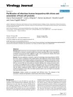
Fig. 1. Dynamic Binding Capacity (DBC) at 10% breakthrough for Protein A cycling
studies with FT-AEX clarified feed material with no sanitisation (white squares),
depth filter clarified material with no sanitisation (black squares), depth filter clarified material with mild sanitisation (black circles) and depth filter clarified material
with harsh sanitisation (black triangles).
3. Results and discussion
3.1. Dynamic binding capacity
Dynamic binding capacity (DBC) is a key measure of Protein
A process economics [29]. DBC at 10% breakthrough was determined to assess the impact of differing clarification and sanitisation
methods over 100 cycles. Cycling studies were carried out with
depth filtered cell culture material under three different sanitisation regimes and compared to FT-AEX clarified material with no
sanitisation between cycles.
From Fig. 1 it can be seen that there is no significant difference
in DBC between the different clarified cell culture fluids and the
sanitisation approaches examined, with no notable loss in capacity observed over 100 cycles. Mechanisms that contribute to a loss
in Protein A capacity include resin ligand hydrolysis and buildup of HCPs and mAb aggregates leading to resin fouling [30]. Mab
Select SuRe is an alkali stable Protein A affinity resin and the results
presented here show that it is capable of withstanding harsh sanitations of 0.5 M NaOH every 3rd cycle. Zhang and colleagues recently
described similar findings, highlighting the effectiveness of sodium
hydroxide-based cleaning in preventing resin fouling of Mab Select
SuRe and showed that it maintained a binding capacity of 95% following exposure to 0.1 M NaOH over 50 h. [30].
3.2. Host cell protein quantification
HCP concentration in the eluates of approximately every 20th
cycle was measured for each set of cycling conditions using a ProteinSEQ CHO HCP Quantitation Kit. The use of proximity ligation
assay (PLA) immunoassay for protein detection and quantification
increases specificity and sensitivity compared to standard ELISA
methods. PLA combines antibody–protein binding with detection
of the reporter nucleic acid using real-time quantitative PCR (qPCR)
[31]. The quantity of CHO HCP was determined using AccuSEQTM
software. Cycle threshold (Ct) was set to 0.2 and the standard curve
fitted using a 5 Parameter Logistic (5 PL) curve fitting. Each sample was prepared in triplicate and acceptance criteria for precision
was set to % CV ≤ 20% throughout the curve and ≤ 25% at the lower
limit of quantification (LLOQ). Random samples were spiked with
31
stock from the standard curve and recovery efficiency determined
to ensure accuracy of a quantitation assay the sample matrices. A
back-calculation acceptance value was set to 75–125%.
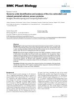
The concentration of HCPs in the Protein A eluate versus cycle
number was plotted for the four sets of cycling investigated
(Fig. 2A). Protein A eluates from FT-AEX clarified material contained
significantly less HCPs throughout cycling compared to all sets of
depth filter clarified material (Fig. 2A). The average Protein A eluate from FT-AEX clarified cycling contained almost 60 times less
HCPs than depth filter clarified material where both sets of feed
material were conducted with no sanitisation. For the depth filter
clarified material investigated with sanitisation between cycling
with NaOH, sanitisation slightly reduced the number of HCPs but
still contained significant more than the FT-AEX clarified material. The best HCP reduction with the conventional depth filtered
material was seen for harsh sanitisation cycling (0.5 M NaOH every
3rd cycle) - which still contained 38-fold higher HCP concentration
compared to FT-AEX clarified material. (Fig. 2C). In a previous study,
Castro-Forero et al. noted a 19-fold reduction in the level of HCPs in
Protein A eluates from FT-AEX clarified material compared to depth
filter clarified material [32].
From Fig. 2B we can see the HCP concentration following Protein
A is consistent over 100 cycles and very close to the consensus target limit of less than 100 ppm in final drug product [17,18] after one
chromatography step, highlighting the impact of chromatographic
clarification on post Protein A purity and its potential in the drive
towards a more compressed downstream process. The downward
trend in HCP concentration with cycle number observed in Fig. 2B
was also seen in a recent similar study [27].
3.3. Host cell DNA quantification
Host Cell DNA (HCDNA) concentration in the eluates of approximately every 20th cycle was measured for each set of cycling
conditions using a resDNASEQ® Quantitative CHO DNA kit. A standard curve (3 ng – 0.03 pg) was generated to quantify the DNA in
the Protein A eluate samples. AccuSEQTM software was used to set
the Ct to 0.2 (with a 3–15 cycle baseline) and a linear standard
curve with an R2 value of 0.999 was generated. Each sample was
prepared in triplicate and acceptance criteria for precision was set
to % CV ≤ 20% throughout the curve and ≤ 25% at the LLOQ. Random
samples were spiked with stock from the standard curve and recovery efficiency determined to ensure accuracy of a quantitation assay
the sample matrices. A back-calculation acceptance value was set
to 75–125%.
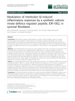
From Fig. 3A a dramatic reduction (2.3 log reduction) in HCDNA
in the Protein A eluates from the FT-AEX clarified material compared to depth filter clarified material was observed. These results
are consistent with Castro-Forero, et al. who showed post Protein
A HCDNA was 3.5 log lower for FT-AEX clarified material compared
to depth filter clarified material [32].
Host cell DNA concentration in the depth filter clarified material
Protein A eluates appears to follow a downward trend (Fig. 3B). Aa
recent study investigating the inclusion of EmphazeTM AEX Hybrid
Purifier on Protein A Periodic Counter-Current Chromatography
(PCC) carried out with the same depth filtered and FT-AEX clarified
cell culture fluid as used in this study showed comparable results.
It is possible that residual HCDNA can encode or harbour oncogenes or infectious agents, and if carried through to the final drug
product, could lead to undesirable oncogenic or infective events
in patients. Both the World Health Organization (WHO) and U.S.
Food and Drug Administration (FDA) guideline recommendations
state that residual HCDNA is limited to 10 ng/dose in the final product dose [33]. The average HCDNA levels for the FT-AEX clarified
material is less than 200 pg/mL (Fig. 3C, supplementary table 1).
Typically, following Protein A chromatography, additional polish-
32
S. Gilgunn et al. / J. Chromatogr. A 1595 (2019) 28–38
Fig. 2. Eluate host cell protein (HCP) concentration during Protein A cycling studies for FT-AEX clarified material with no sanitisation (white squares), depth filter clarified
feed material with no sanitisation (black squares), depth filter clarified feed material with mild sanitisation strategy (black circles) and depth filter clarified feed material
with harsh sanitisation (black triangles) shown on a log scale (A) and a linear scale (B). The average eluate HCP concentration across 100 cycles of Protein A chromatography
is shown in (C) where FT-AEX clarified feed material with no sanitisation is depicted by the black bar, depth filter clarified feed material with no sanitisation is shown by
the white bar, depth filter clarified material with a mild sanitisation strategy is depicted by the horizontal hashed bar and depth filter clarified feed material with a harsh
sanitisation strategy is shown as the diagonally hashed bar.
Fig. 3. Eluate host cell DNA (HCDNA) concentration during Protein A cycling studies for FT-AEX clarified material with no sanitisation (white squares), depth filter clarified
feed material with no sanitisation (black squares), depth filter clarified feed material with mild sanitisation strategy (black circles) and depth filter clarified feed material with
harsh sanitisation (black triangles) shown on a log scale (A) and a linear scale (B). The average eluate HCDNA concentration across 100 cycles of Protein A chromatography
is shown in (C) where FT-AEX clarified feed material with no sanitisation is depicted by the black bar, depth filter clarified feed material with no sanitisation is shown by
the white bar, depth filter clarified material with a mild sanitisation strategy is depicted by the horizontal hashed bar and depth filter clarified feed material with a harsh
sanitisation strategy is shown as the diagonally hashed bar.
ing steps are carried out to provide additional clearance of virus,
HCP, HCDNA and other product related contaminants [19]. The data
generated from this body of research suggests that the use of the
EmphazeTM AEX Hybrid Purifier may reduce the number of additional polishing steps as the levels of both HCP and HCDNA are
significantly reduced following Protein A chromatography.
S. Gilgunn et al. / J. Chromatogr. A 1595 (2019) 28–38
33
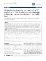
Fig. 4. concentration of specific problematic HCPs, in ppm, identified following depth filtration (white bars) and in the subsequent Protein A eluate (light hash bars) compared
to FT-AEX clarified material (black bars) and subsequent Protein A eluates obtained with FT-AEX clarified material (dark hashed bars).
3.4. Analysis of problematic HCPs
The diverse portfolio of HCP proteins that make their way from
upstream bioprocessing through downstream purification and into
final drug product remains a focal point in discussion of mAb
bioprocessing. HCPs are identified as a CQA of mAb formulations
and can threaten patient safety and product quality through (1)
potential immunogenicity; (2) catalytic activity for product fragmentation and (3) involvement in product aggregation [7]. In the
numerous HCP profiling studies to date some commonly observed,
problematic HCPs are frequently identified as ‘difficult to remove’.
Proteases and other degradative enzymes previously reported in
the literature include cathepsin A and D, matrix metalloproteinase19, serine protease HTRA1 and protein disulphide-isomerase A6.
Similarly, considerable attention has been drawn to potentially
immunogenic CHO HCPs such as Protein S100-A6, 60 s ribosomal
protein L30, Annexin A5, C-X-C motif chemokine 3, Putative phospholipase B-like 2 and various histones [13,24,34–36]
It is evident that FT-AEX clarified material contains significantly
less HCPs, hence, LC–MS analysis was carried out in order to track
where they were removed and identify if any of these commonly
observed problematic HCPs remained following Protein A chromatography.
Isoelectric point, molecular weight and grand average of
hydropathy (GRAVY) scores, for HCPs removed by FT-AEX clarification were compared to those remaining, however, no significant
trend in isoelectric point or enrichment of GRAVY scores was
observed suggesting the retention of HCPs cannot be predicted by
theoretical hydrophobicity, molecular weight and isoelectric point
(supplementary Fig. 1). Levy, et al. found similar results when modelling co-elution of impurities on polishing columns [37].
3.4.1. Removal of histones
In this study we used LC–MS/MS to determine the levels of histone proteins present before and after Protein A chromatography
in the tryptic sample digests. The MS data were searched against
a CHO database for protein identification and HCP quantification
(ppm) was performed against the residual mAb using Hi3 relative
quantitation of the three most intense peptides of each protein
[13,24]. From Fig. 4 we can see EmphazeTM AEX Hybrid Purifier
removes the histone proteins H2B and H3 below detectable levels prior to Protein A purification. Conventional HCP ELISA kits are
unable to detect histone proteins. Gagnon, et al. used generation
3 CHO HCP kit from Cygnus Technologies and using a calibration
standard containing histone H3 and showed it made an underestimation of more than 20,000-fold [38]. The use of LC–MS/MS
analysis in this study provides confidence that EmphazeTM AEX
Hybrid Purifier is capable of removing histones during the clarification stage of the bioprocess.
Chromatin released from dead cells during upstream bioprocessing of mAbs exists predominantly as complex heteroaggregates consisting of nucleosomal arrays, individual nucleosomes, histone proteins and DNA. Chromatin can be considered
as a vehicle for “smuggling” a range of HCPs through Protein A
chromatography. The DNA component of chromatin is negatively
charged (pKa ± 2.6) and the histone component is hydrophobic and
positively charged (pI ± 11.5), giving rise to a chemical surface ideal
for non-specific HCP binding. DNA in cell culture harvests binds Protein A indirectly through the histones with which it is associated,
reducing dynamic capacity for IgG to bind [7].
Following a series of publications from Gagnon, et al., investigating the role of chromatin in mAb purification, it is now well
established that removing chromatin hetero-aggregates before
Protein A chromatography can significantly reduce the level of
residual HCPs and HCDNA, while increasing DBC of a Protein A column [29,38–40]. Gagnon, et al. pre-treated crude mAb supernatant
with allantoin and ethacridine to precipitate out the chromatin
hetero-aggregates [38]. While successful, the implementation of
this method for large scale mAb bioprocessing may be difficult to
implement. Alternatively, the EmphazeTM AEX Hybrid Purifier can
be easily scaled into an industrial bioprocess, for clarification of
cell culture harvest, to remove problematic histone proteins via
binding to the Q-function matrix without adding additional process
steps.
3.4.2. Degradative host cell proteins
The safety, quality and efficacy of mAb molecules is threatened by proteases and other degradative HCPs. If the protein has
enzymatic activity then the risk is from the direct action of the
HCP impurity. HCP activities have been observed that resulted in
direct biological action in patients or in degradation, fragmentation,
aggregation, or particle formation in the final mAb product [41]. The
molecular susceptibility of mAbs to fragmentation by proteolytic
enzymes is broadly recognised. Hence, polishing chromatography steps such as anion and cation exchange chromatography are
carried out to remove residual impurities including proteolytic
enzymes that could potentially cause fragmentation of the final
drug product or its excipients [41,42].
34
S. Gilgunn et al. / J. Chromatogr. A 1595 (2019) 28–38
Fig. 5. Heat maps of concentration of problematic HCPs (red) and commonly occurring HCPs (green) in the Protein A eluates during the cycling experiments. Graph (A)
shows cycling experiments with depth filter clarified material with no sanitisation during the 100 cycles (B) shows 100 cycles with mild sanitisation conditions (C) shows
100 cycles with harsh sanitisation regime and (D) shows 100 cycles with FT-AEX clarified material and no sanitization. (For interpretation of the references to colour in this
figure legend, the reader is referred to the web version of this article).
Some of the more commonly observed degradative HCPs
such as protein disulfide-isomerase A6, cathepsin D, matrix
metalloproteinase-19 and serine protease HTRA1 were decreased
by chromatographic clarification and subsequently removed following Protein A purification (Fig. 4); whereas all of these
degradative HCPs, bar cathepsins B and D, where still present in
Protein A eluates arising from the depth filter clarified material.
Proteases, particularly cathepsins B and D, have been implicated in
the degradation of some antibodies and have been shown to cause
heavy chain C-terminal fragmentation of a mAb resulting in particle
formation [43–45]. This is thought to be due to HCP:mAb interactions driven by direct hydrophobic interactions of mAbs with a
common motif (LLY) and the hydrophobic cleft surrounding the
cathepsin D active site [36].
3.4.3. Immunogenic host cell proteins
While degradative HCPs can affect the product which, in turn,
can affect the patient, other HCPs can be immunogenic, with the
patient eliciting an antibody response against the specific HCP
impurity. It is possible for these immune responses to be benign,
however, they serve no benefit to the patient and, hence, still
carry risk [41]. In-silico tools and proteomic database are continually being developed to aid in risk assessment and help identify
the immunogenicity potential of CHO proteins. CHOPPI, a web
tool specifically developed for determination of immunogenicity risk of HCPs in CHO-based protein therapeutics investigated
35 transcribed, secreted CHO proteins and identified C-X-C motif
chemokine 3 (CXCC3) as highly immunogenic. It was ranked with
an immunogenicity score of 92 as it contains 23 epitopes, one of
which is cross-reactive with numerous (637) human epitopes, with
potential capabilities of inducing a regulatory immune response in
humans [46]. From Fig. 4 we can see that this highly immunogenic
HCP is removed by chromatographic clarification prior to Protein A
purification, whilst it persists following depth filtration. In investigating the impact of different elution buffers on HCP profile, CXCC3
was shown to co-elute with a mAb under two of the four elution conditions assessed [13]. In this study, while CXCC3 was not
detected post Protein A for either the depth filter or FT-AEX clarified
material, it is likely that the buffer conditions used did not result
in the coelution of CXCC3 with the mAb. In circumstances where
CXCC3 coelutes with the mAb, chromatographic clarification could
be an effective way to remove this protein.
Another notable immunogenic HCPs, protein S100-A6 [13,34] is
present in the Protein A eluate of the depth filtered material but was
removed to below a detectable level in the Protein A eluate of the
FT-AEX clarified material (Fig. 4). Using the CHOPPI tool, proteins
with an immunogenicity score of >20 are considered to be high
risk proteins. S100-A6 proteins was previously classified as highly
immunogenic with an immunogenicity score of 52.84 [13].
PLBL2 has attracted attention as a highly immunogenic HCP and
was also previously ranked with a CHOPPI score of 32.89 [13]. This
HCP binds to humanized mAbs, in particular the IgG4 isotype, and is
not detected in some widely used anti-CHOP immunoassays [41].
The amino acid sequence of CHO PLBL2 is 80% similar to human
PLBL2, however, many surface exposed residues are different which
has resulted in the generation of anti-PLBL2 antibodies in clinical
trials [7,14]. In this study, PLBL2 was not detected in the Protein A
eluate of both clarified materials. It is suspected that in this study,
PLBL2 did not bind to the mAb product, hence, was cleared during
Protein A chromatography for both feed materials. Aboulaich, et al.
looked at the association of HCPs and 4 different mAb products and
noted PLBL2 only bound 3 out of 4 mAbs [34].
S. Gilgunn et al. / J. Chromatogr. A 1595 (2019) 28–38
35
Fig. 6. Graph showing presence (black shading) or absence (white shading) of host cell proteins identified in the samples taken from the sanitisation following 100 cycles of
Protein A chromatography with depth filter clarified feed, using the no sanitisation, mild sanitisation and harsh sanitisation regimes compared to FT-AEX clarified feed with
no sanitisation during the cycling.
36
S. Gilgunn et al. / J. Chromatogr. A 1595 (2019) 28–38
The overall reduction in the level of histone proteins, degradative and immunogenic HCP contaminants prior to Protein A has the
potential to aid in product quality and safety. Ultimately, this could
also increase the overall performance of the Protein A column. The
clearance of the majority of these problematic HCPs post Protein A,
as highlighted in Fig. 4, demonstrates the importance of the clarification stage in removing problematic HCPs prior to and during
Protein A chromatography. This reduces the HCP burden on subsequent polishing steps offering the potential of downstream process
simplification.
3.5. Tracking problematic and commonly occurring HCPs found
in Protein A eluate
Fig. 5 tracks some of the more commonly observed and problematic HCPs found in the Protein A eluate over 100 cycles of
chromatography. There is a difference in number of HCPs across
the depth filtered material and the various sanitisation conditions
investigated. The majority of these difficult to remove HCPs are
considered to interact with mAbs and/or the Protein A resin and it
is evident that even stringent sanitisation with NaOH is not significantly efficient to remove them throughout the lifetime of these
cycling studies.
The number of problematic HCPs detected in the eluates from
the cycling experiments performed without sanitisation or when
using mild sanitisation conditions was lower than that found in the
eluates wherein harsh sanitisation was employed. These observations suggest retention of HCPs under the no and mild sanitisation
conditions and insufficient removal from the Protein A resin. The
harsh sanitisation conditions proved appropriate for the efficient
cleaning of the Protein A resin as reflected by the associated higher
levels of HCPs detected in the corresponding eluates when harsh
sanitisation was implemented.
Protein A eluates from the FT-AEX clarified material show
removal of all problematic HCPs and 7 commonly observed HCPs.
Various studies have suggested that 78 kD glucose-regulated protein, nidogen-1, heat shock proteins and actin interact with the Fc
and constant regions of IgG molecules [23,34,35,37]. These HCPs
were reduced following chromatographic clarification (data not
shown) and then entirely cleared following Protein A purification. There are a number of possible reasons that the removal of
these HCPs in EmphazeTM AEX Hybrid Purifier Protein A eluate was
observed yet carried through to the Protein A eluate in the depth
filter clarified material. Firstly, as they were present in limited
quantities this could reduce the possible interactions available with
the mAb product itself. Secondly, it is likely that mAb:HCP interactions are promoted by binding interactions with other HCPs such as
histones, probably in the form of chromatin. Since chromatin was
depleted following chromatographic clarification, carry-though of
the problematic HCPs was not observed in the Protein A eluate.
Zhang, et al. noted particularly poor clearance during Protein A
chromatography for clusterin and actin [23] – in Fig. 5D removal of
actin can be seen along with a reduction in the presence of clusterin.
Some HCPs including lipoprotein lipase and nidogen continue to
pervade and are particularly difficult to remove even after polishing
steps such as anion/cation exchange or hydrophobic interaction
chromatography through resin association or co-elution with mAbs
[7,13,34,37]. Clarification with the EmphazeTM AEX Hybrid Purifier
was able to remove nidogen-1 to below the limit of detection and
reduce the quantity of lipoprotein lipase in the Protein A eluate by
an order of magnitude.
3.6. Resin fouling
In this study we have highlighted the positive effect of cell
culture clarification with the EmphazeTM AEX Hybrid Purifier in
reducing the number of HCPs in the Protein A eluate across 100
cycles of Protein A chromatography. This notable reduction of HCPs
can benefit the purification process by offering the potential to
reduce the number of polishing steps. A reduction in column fouling was also noted. A column sanitisation in the final cycle for all
four sanitisation strategies examined was carried out using 0.1 M
sodium hydroxide for the no sanitisation and mild sanitisation
regimes and 0.5 M sodium hydroxide for the harsh sanitisation
strategy. Each sanitisation fraction was collected and analysed by
LC–MS/MS to determine the number of HCPs present. From Fig. 6
a difference in the number of HCPs identified for the depth filter
clarified material that underwent no sanitisation (74 HCPs), mild
sanitisation (61 HCPs) and harsh sanitisation (96 HCPs) is observed.
The number of HCPs present in the final sanitisation fraction
for the harsh sanitisation condition is the highest. This is thought
to be due to the higher concentration of sodium hydroxide used
in the final sanitisation. The final sanitisation fraction for the mild
sanitisation condition contained fewer host cell proteins than the
no sanitisation condition which is due to the regular sanitisation
during the cycling which acts to reduce the accumulation of HCPs
on the resin during the cycling.
More notably, the number of HCPs present in this final sanitisation fraction for the FT-AEX clarified material is over 4 times less
than the depth filter material with no sanitisation, and 3.5 times less
than the mild sanitisation, indicating there is less over-all fouling
of the Protein A column over 100 cycles.
Recently, a study by Pathak, et al. showed the feed material
composition is correlated to the rate and mode of resin aging, and
emphasized negative effect the nuclear material present in HCCF
has on overall column performance and product quality. Chromatin
hetero-aggregates were shown to accumulate on the Protein A particle surfaces, obstructing IgG access to bind to the particle pores
[47]. Clarification using the EmphazeTM AEX Hybrid Purifier can
deplete chromatin from the HCCF, prior to Protein A chromatography, resulting in less fouling of the Protein A column.
4. Conclusions
Protein A affinity chromatography is currently the industry gold
standard for initial capture and purification of the vast majority of
commercial mAbs produced in CHO cell lines. Innovative mechanisms upstream that led to the much sought after increased product
titres shifted bioprocessing concerns downstream due to a parallel
increase in expression of unwanted CHO HCPs. The implementation
of the EmphazeTM AEX Hybrid Purifier during clarification of HCCF
has potential to overcome some of these issue through a significant
reduction in HCP and HCDNA.
Over 100 cycles of Protein A chromatography, without any standard sodium hydroxide cleaning, was carried on FT-AEX clarified
material for purification of a recombinant biosimilar IgG1 monoclonal antibody. An average HCP reduction of 38-fold and an
average HCDNA concentration reduction of 2.3 log was achieved
in the FT-AEX clarified material compared to standard depth filter
clarified material with the harsh sanitisation conditions of 0.5 M
NaOH every 3 cycles.
FT-AEX clarification in conjunction with Protein A chromatography resulted in the removal of problematic HCPs,
including 78 kD glucose-regulated protein, nidogen-1, heat shock
proteins, actin, histones, serine protease HTRA1 and matrix
metalloproteinase-19, which were tracked through the purification process using LC–MS/MS. The EmphazeTM AEX Hybrid Purifier
is readily incorporated into a process, in a scalable fashion, by
substituting the standard polishing depth filter during cell culture
harvest, and can, ultimately, lead to a reduction in subsequent polishing steps downstream.
S. Gilgunn et al. / J. Chromatogr. A 1595 (2019) 28–38
Disclosure of potential conflicts of interest
The authors declare the following competing financial interest(s): H. El-Sabbahy, L. Deakin and G. Jellum are employees of 3 M,
the corporation that develops and produces the EmphazeTM AEX
Hybrid Purifier. Beyond this, the authors are not aware of any affiliations, memberships, funding, or financial holdings that might be
perceived as affecting the objectivity of this article.
[18]
[19]
[20]
Acknowledgements
The authors acknowledge the generous financial support provided by 3M. HCP and HCDNA kits were kindly provided by Thermo
Fischer Scientific.
[21]
[22]
Appendix A. Supplementary data
Supplementary material related to this article can be found, in
the online version, at doi: />02.056.
[23]
[24]
References
[1] FDA, U.S Food And Drug Administration, 2019 [Online]. Available:https://
www.fda.gov/downloads/Drugs/DevelopmentApprovalProcess/
HowDrugsareDevelopedandApproved/ApprovalApplications/
TherapeuticBiologicApplications/Biosimilars/UCM412398.pdf[Accessed: 20
August 2018].
[2] D.M. Ecker, S.D. Jones, H.L. Levine, The therapeutic monoclonal antibody
market, MAbs 7 (1) (2015) 9–14, />989042.
[3] H.A.D. Lagassé, A. Alexaki, V. Simhadri, N. Katagiri, W. Jankowski, Z. Sauna, C.
Kimchi-Sarfaty, Recent advances in (therapeutic protein) drug development,
F1000Research 6 (2017) 113, />1.
[4] A. Farrell, N. McLoughlin, J.J. Milne, I.W. Marison, J. Bones, Application of
multi-omics techniques for bioprocess design and optimisation in chinese
Hamster ovary cells, J. Proteome Res. 13 (7) (2014) 3144–3159, .
org/10.1021/pr500219b.
[5] C.E.M. Hogwood, D.G. Bracewell, C.M. Smales, Measurement and control of
host cell proteins (HCPs) in CHO cell bioprocesses, Curr. Opin. Biotechnol. 30
(2014) 153–160, />[6] C. Rader, Chemical biology: how to minimalize antibodies, Nature 518 (7537)
(2015) 38–39, />[7] C.H. Goey, S. Alhuthali, C. Kontoravdi, Host cell protein removal from
biopharmaceutical preparations: Towards the implementation of quality by
design, Biotechnol. Adv. 36 (4) (2019) 1223–1237, />biotechadv.2018.03.021.
[8] P. Gronemeyer, R. Ditz, J. Strube, Trends in upstream and downstream process
development for antibody manufacturing, Bioengineering 1 (4) (2014)
188–212, />[9] K.N. Valente, A.M. Lenhoff, K.H. Lee, Expression of difficult-to-remove host
cell protein impurities during extended Chinese hamster ovary cell culture
and their impact on continuous bioprocessing, Biotechnol. Bioeng. 112 (6)
(2015) 1232–1242, />[10] A.L. Tscheliessnig, J. Konrath, R. Bates, A. Jungbauer, Host cell protein analysis
in therapeutic protein bioprocessing - methods and applications, Biotechnol.
J. 8 (6) (2013) 655–670, />[11] Y.H. Kao, D.P. Hewitt, M. Trexler-Schmidt, M.W. Laird, Mechanism of antibody
reduction in cell culture production processes, Biotechnol. Bioeng. 107 (4)
(2010) 622–632, />[12] M. Trexler-Schmidt, S. Sargis, J. Chiu, S. Sze-Khoo, M. Mun, Y.H. Kao, M.W.
Laird, Identification and prevention of antibody disulfide bond reduction
during cell culture manufacturing, Biotechnol. Bioeng. 106 (3) (2010)
452–461, />[13] A. Farrell, S. Mittermayr, B. Morrissey, N. Mc Loughlin, N.N. Iglesias, I.W.
Marison, J. Bones, Quantitative host cell protein analysis using two
dimensional data independent LC–MSE, Anal. Chem. 87 (18) (2015)
9186–9193, />[14] S.K. Fischer, M. Cheu, K. Peng, J. Lowe, J. Araujo, E. Murray, D. McClintock, J.
Matthews, P. Siguenza, A. Song, Specific immune response to phospholipase
B-Like 2 protein, a host cell impurity in Lebrikizumab Clinical Material, AAPS
19 (1) (2017) 254–263, />[15] IHC, Development and Manufcature of Drug Substances (Chemical Entities
and Biotechnological/biological Entities) Q11., 2012.
[16] ICH, Guidance for Industry Q6B Specifications: Test Procedures and
Acceptance Criteria for Biotechnological/Biotechnology Products, 1999.
[17] C.L.Z. de Zafra, V. Quarmby, K. Francissen, M. Vanderlaan, J. Zhu-Shimoni, Host
cell proteins in biotechnology-derived products: a risk assessment
[25]
[26]
[27]
[28]
[29]
[30]
[31]
[32]
[33]
[34]
[35]
[36]
[37]
[38]
37
framework, Biotechnol. Bioeng. 112 (11) (2015) 2284–2291, />10.1002/bit.25647.
K.N. Valente, N.E. Levy, K.H. Lee, A.M. Lenhoff, Applications of proteomic
methods for CHO host cell protein characterization in biopharmaceutical
manufacturing, Curr. Opin. Biotechnol. 53 (2018) 144–150, />10.1016/j.copbio.2018.01.004.
H.F. Liu, J. Ma, C. Winter, R. Bayer, Recovery and purification process
development for monoclonal antibody production, MAbs 2 (5) (2010)
480–499, />M. Grom, M. Kozorog, S. Caserman, A. Pohar, B. Likozar, Protein A affinity
chromatography of Chinese hamster ovary (CHO) cell culture broths
containing biopharmaceutical monoclonal antibody (mAb): Experiments and
mechanistic transport, binding and equilibrium modeling, J. Chromatogr. B
Anal. Technol. Biomed. Life Sci. 1083 (2018) 44–56, />j.jchromb.2018.02.032.
A.L. Grilo, M. Mateus, M.R. Aires-Barros, A.M. Azevedo, Monoclonal antibodies
production platforms: an opportunity study of a Non-Protein-A
chromatographic platform based on process economics, Biotechnol. J. 12 (12)
(2017) 1–10, />D.G. Bracewell, R. Francis, C.M. Smales, The future of host cell protein (HCP)
identification during process development and manufacturing linked to a
risk-based management for their control, Biotechnol. Bioeng. 112 (9) (2015)
1727–1737, />Q. Zhang, A.M. Goetze, H. Cui, J. Wylie, S. Trimble, A. Hewig, G.C. Flynn,
Comprehensive tracking of host cell proteins during monoclonal antibody
purifications using mass spectrometry, mAbs 6 (3) (2014) 659–670, http://dx.
doi.org/10.4161/mabs.28120.
C.E. Doneanu, A. Xenopoulos, K. Fadgen, J. Murphy, St.J. Skilton, H. Prentice, M.
Stapels, W. Chen, Analysis of host-cell proteins in biotherapeutic proteins by
comprehensive online two-dimensional liquid chromatography/mass
spectrometry, MAbs 4 (1) (2012) 24–44, />18748.
C.E.M. Hogwood, A.S. Tait, N. Koloteva-Levine, D.G. Bracewell, C.M. Smales,
The dynamics of the CHO host cell protein profile during clarification and
protein A capture in a platform antibody purification process, Biotechnol.
Bioeng. 110 (1) (2013) 240–251, />C.H. Goey, D. Bell, C. Kontoravdi, Mild hypothermic culture conditions affect
residual host cell protein composition post-protein A chromatography, MAbs
10 (3) (2018) 476–487, />H. El-Sabbahy, D. Ward, O. Ogonah, L. Deakin, G.M. Jellum, D.G. Bracewell, The
effect of feed quality due to clarification strategy on the design and
performance of protein A periodic counter-current chromatography,
Biotechnol. Prog. (October 3) (2018), doi:10.1002/btpr.2709. [Epub ahead of
print].
S. Albrecht, C. Kaisermayer, C. Gallagher, A. Farrell, A. Lindeberg, J. Bones,
Proteomics in biomanufacturing control: protein dynamics of CHO-K1 cells
and conditioned media during apoptosis and necrosis, Biotechnol. Bioeng.
115 (6) (2018) 1509–1520, />P. Gagnon, R. Nian, Y. Yang, Q. Yang, C.L. Lim, Non-immunospecific association
of immunoglobulin g withchromatin during elution from protein a inflates
host contamination, aggregate content, and antibody loss, J. Chromatogr. A
1408 (2015) 151–160, />J. Zhang, S. Siva, R. Caple, S. Ghose, R. Gronke, Maximizing the functional
lifetime of Protein A resins, Biotechnol. Prog. 33 (3) (2017) 708–715, http://
dx.doi.org/10.1002/btpr.2448.
N. Liu, M. Brevnov, M. Furtado, J. Liu, Host cellular protein quantification,
Bioprocess Int. 10 (2) (2012) 44.
A.A. Castro-Forero, Z. Jokondo, A. Voloshin, J.F. Hester, Anion-Exchange
Chromatographic Clarification: Bringing Simplification, Robustness, and
Savings to MAb Purification, Bioprocess Int, 2015, Available at: http://www.
bioprocessintl.com/downstream-processing/chromatography/anionexchange-chromatographic-clarification-bringing-simplification-robustnessand-savings-to-mab-purification/ (Accessed 29 November 2018).
H. Yang, Establishing acceptable limits of residual DNA, PDA J. Pharm. Sci.
Technol. 67 (2) (2013) 155–163, />00910.
N. Aboulaich, W.K. Chung, J.H. Thompson, C. Larkin, D. Robbins, M. Zhu, A
novel approach to monitor clearance of host cell proteins associated with
monoclonal antibodies, Biotechnol. Prog. 30 (5) (2014) 1114–1124, http://dx.
doi.org/10.1002/btpr.1948.
N.E. Levy, K.N. Valente, L.H. Choe, K.H. Lee, A.M. Lenhoff, Identification and
characterization of host cell protein product-associated impurities in
monoclonal antibody, Bioprocessing 111 (5) (2014) 904–912, .
org/10.1002/bit.25158.
J.S. Bee, L.M. Machiesky, L. Peng, K.C. Jusino, M. Dickson, J. Gill, D. Johnson,
H.Y. Lin, K. Miller, J. Heidbrink Thompson, R.L. Remmele Jr., Identification of
an IgG CDR sequence contributing to co-purification of the host cell protease
cathepsin D, Biotechnol. Prog. 33 (1) (2017) 140–145, />1002/btpr.2397.
N.E. Levy, K.N. Valente, K.H. Lee, A.M. Lenhoff, Host cell protein impurities in
chromatographic polishing steps for monoclonal antibody purification,
Biotechnol. Bioeng. 113 (6) (2016) 1260–1272, />25882.
P. Gagnon, R. Nian, J. Lee, L. Tan, S.M. Abdul Latiff, C. Ling Lim, C. Chuah, X. Bi,
Y. Yang, W. Zhang, H. Theng Gan, Nonspecific interactions of chromatin with
immunoglobulin G and protein A, and their impact on purification
38
[39]
[40]
[41]
[42]
[43]
S. Gilgunn et al. / J. Chromatogr. A 1595 (2019) 28–38
performance, J. Chromatogr. A 1340 (2014) 68–78, />j.chroma.2014.03.010.
R. Nian, W. Zhang, L. Tan, J. Lee, X. Bi, Y. Yang, H. Theng Gan, P. Gagnon,
Advance chromatin extraction improves capture performance of protein A
affinity chromatography, J. Chromatogr. A (2016) 1–7, />1016/j.chroma.2015.12.044, 1431 92016).
R. Nian, P. Gagnon, Advance chromatin extraction enhances performance and
productivity of cation exchange chromatography-based capture of
Immunoglobulin G monoclonal antibodies, J. Chromatogr. A 1453 (2016)
54–61, />M. Vanderlaan, J. Zhu-Shimoni, S. Lin, F. Gunawan, T. Waerner, K. van Cott,
Experience with host cell protein impurities in biopharmaceuticals,
Biotechnol. Prog. (2018), [Epub ahead of
print].
S.X. Gao, Y. Zhang, K. Stansberry-Perkins, A. Buko, S. Bai, V. Nguyen, M.L.
Brader, Fragmentation of a highly purified monoclonal antibody attributed to
residual CHO cell protease activity, Biotechnol. Bioeng. 108 (4) (2011)
977–982, />F. Robert, H. Bierau, M. Rossi, D. Agugiaro, T. Soranzo, H. Broly, C.
Mitchell-Logean, Degradation of an Fc-fusion recombinant protein by host
[44]
[45]
[46]
[47]
cell proteases: identification of a CHO cathepsin D protease, Biotechnol.
Bioeng. 104 (6) (2009) 1132–1141, />Q. Zhang, A.M. Goetze, H. Cui, J. Wylie, B. Tillotson, Hewig A, M.P. Hall, G.C.
Flynn, Characterization of the Co-elution of host cell proteins with
monoclonal antibodies during protein a purification, Biotechnol. Prog. 32 (3)
(2016) 708–717, />J.S. Bee, L. Tie, D. Johnson, M.N. Dimitrova, K.C. Jusino, C.D. Afdahl, Trace levels
of the CHO host cell protease cathepsin D caused particle formation in a
monoclonal antibody product, Biotechnol. Prog. 31 (5) (2015) 1360–1369,
/>C. Bailey-Kellogg, A.H. Gutiérrez, L. Moise, F. Terry, W.D. Martin, A.S. De Groot,
CHOPPI: a web tool for the analysis of immunogenicity risk from host cell
proteins in CHO-based protein production, Biotechnol. Bioeng. 111 (11)
(2014) 2170–2182, />M. Pathak, A.S. Rathore, K. Lintern, D.G. Bracewell, Protein A chromatography
resin lifetime —impact of feed composition, Biotechnol. Prog. 34 (2) (2018)
412–419, />