báo cáo khoa học: "Identification and analysis of phosphorylation status of proteins in dormant terminal buds of poplar" pptx
Bạn đang xem bản rút gọn của tài liệu. Xem và tải ngay bản đầy đủ của tài liệu tại đây (4.89 MB, 16 trang )
RESEARCH ARTIC LE Open Access
Identification and analysis of phosphorylation status
of proteins in dormant terminal buds of poplar
Chang-Cai Liu
1,2†
, Chang-Fu Liu
3†
, Hong-Xia Wang
4
, Zhi-Ying Shen
5
, Chuan-Ping Yang
1*
and Zhi-Gang Wei
1*
Abstract
Background: Although there has been considerable progress made towards understanding the molecular
mechanisms of bud dormancy, the roles of protein phosphorylation in the process of dormancy regulation in
woody plants remain unclear.
Results: We used mass spectrometry combined with TiO
2
phosphopeptide-enrichment strategies to investigate the
phosphoproteome of dormant terminal buds (DTBs) in poplar (Populus simonii × P. nigra). There were 161 unique
phosphorylated sites in 161 phosphopeptides from 151 proteins; 141 proteins have orthologs in Arabidopsis, and
10 proteins are unique to poplar. Only 34 sites in proteins in poplar did not match well with the equivalent
phosphorylation sites of their orthologs in Arabidopsis, indicating that regulatory mechanisms are well conserved
between poplar and Arabidop sis. Further functional classifications showed that most of these phosphoproteins
were involved in binding and catalytic activity. Extraction of the phosphorylation motif using Motif-X indicated that
proline-directed kinases are a major kinase group involved in protein phosphorylation in dormant poplar tissues.
Conclusions: This study pro vides evidence about the significance of protein phosphorylation during dormancy,
and will be useful for similar studies on other woody plants.
Background
Dormancy is a key feature of perennial plants. During dor-
mancy the meristem becomes insensitive to growth-
promoting signals for a period of time, before it is released
and growth r esumes [1,2]. Bud dormancy is a critical devel-
opmental process that allows perennial plants to survive
extreme seasonal variations in climate. The regulation of
dormancy is a complex process that is necessary for plant
survival, development, and architecture [3,4]. A thorough
understanding of regulation mechanisms controlling dor-
mancy in woody perennials would have a variety of appli-
cations for genetic improvement of woody trees [3,5,6].
Considerable progress has been made in understanding the
molecular mechanisms and regulatory pathways involved
in bud dormancy [2]. However, until recently such studies
focused on regulation at the levels of transcription, post-
transcription, and translation [1,7-12]. Despite the impor-
tance of dormancy reg ulation for perennial behavior [3],
the roles of post-translational modifications, especially
protein phosphorylation, remain poorly understood.
The identification of phosphorylation sites within a cer-
tain protein can not provide a comprehensive view of the
regulatory role of protein phosphorylation [13-17]. Instead,
the simultaneous identification of the phosphorylation sta-
tus of numerous proteins at a certain developmental stage
is required to decode regulatory mechanisms. Large-scale
mapping of phosphorylations that occur in response to
diverse environmental signals has become an indispensa-
ble method for unraveling plant regulatory networks
[17-22]. In recent years, advances in mass spectrometry
(MS)-based protein analysis technologies, combined with
phosphopeptide enrichment methods, paved the way for
large-scale mapping of phosphorylation sites in vivo
[13,18,23]. Specifically, the titanium dioxide (TiO
2
)micro-
column is an effective method to selectively enrich phos-
phopeptides [17,24-28]. There have been several stu dies
on plant phosphoproteomes. These studies have provided
large datasets that allow new insights into phosphorylation
events; however , they have been carried out only on her-
baceous plants, such as Arabidopsis [22,29-40], oilseed
rape [41], rice [42], barley [43], and maize [44]. To date,
* Correspondence: ;
† Contributed equally
1
State Key Laboratory of Forest Genetics and Tree Breeding (Northeast
Forestry University), 26 Hexing Road, Harbin 150040, China
Full list of author information is available at the end of the article
Liu et al. BMC Plant Biology 2011, 11:158
/>© 2011 Liu et al; licensee BioMed Central Ltd. This is an Open Access article distribut ed under the terms of the Creative Commons
Attribution License ( which permits unrestricted use, distri bution, and reproduction in
any medium, provided the original work is properly cited.
there have been no reports on the phosphoproteomes of
woody plant species, except for the identification of eight
phosphorylated poplar P-proteins [45].
Numerous cellular signaling pathways are based on the
sequential phosphorylation of an array of proteins
[15,33,46]. Therefore, the analysis of signaling pathways in
plants has often focused on protein kinases. Kinases show
catalytic preferences for specific phosphorylation motifs
with certain amino acid context sequences [33,47,48].
Therefore, identification of in vivo phosphorylation sites
can provide important information about the activity of
protein kinases in their cellular context.
To better understand the regulation mechanism of
phosphoproteins and cellular signali ng networks during
dormancy, we investigated the phosphoproteome of dor-
mant terminal buds (DTBs) of hybrid poplar (Populus
simonii × P. nigra)usingaMSmethodcombinedwitha
TiO
2
phosphopeptide enrichment strategy. We identified
161 phosphorylation sites in 161 phosphopeptides from
151 proteins, most of which are associated with binding
and catalytic activity. The information gained from this
study provides a wealth of resources and novel insights to
decode the comp licated mechanisms of phosphorylation
modifications in poplar. As far as we know, this is the first
phosphoproteomic analysis of woody plants.
Results
Identification and characterization of the
phosphoproteome of DTBs
Total proteins were isolated from DTBs of poplar, and
then digested with trypsin in solution. The resulting tryp-
tic peptides were subjected to nanoUPLC-ESI-MS/MS to
identify phosphorylation modifications after TiO
2
enrich-
ment. In total, 161 unique phosphorylation site s were
identified in 161 phosphopeptides from 151 proteins
(Table 1, Additional file 1, Additional file 2 and Additional
file 3).
Among th ese phosphorylation sites, 81.3% (131) of
phosphorylation events occurred on Ser and 17.4% (28) on
Thr (Table 1). This finding is consistent with previously
reported phosphorylation patterns: 85% pSer and 10.6%
pThr [22] and 88% pSer and 11% pThr [33] in Arabidop-
sis; and 86% pSe r and 12.7% pThr in M. truncatula [49].
Only 1.2% (2) of the phosphorylation events of these phos-
phopeptides occurred on Tyr residue. This is lower than
the pTyr values reported for Arabidopsis (4.2%) and rice
(2.9%) [22,50], but comparable to that reported for Medi-
cago truncatula (1.3%) [49]. The results of these studies
indicate that Tyr phosphorylation in plants is more abun-
dant than once thought [51]. The spectra representing all
phosphopeptides and the original detailed data are shown
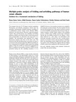
in Additional file 4. As examples, the spectra of phospho-
peptides with single pSer, pThr, and pTyr are shown in
Figure 1a, c, and 1d, respectively. The spectrum of a phos-
phopeptide containing two phosphorylated Ser residues is
shown in Figure 1b.
The majority (93.8%) o f the 161 phosphopeptides were
phosphorylated at a single residue. This value is higher
than that reported for Arabidopsis (80.9%) [22] and M.
truncatula (66.4%) [49]. Only 6.2% of the phosphopeptides
from poplar contained two phosphorylated residues, and
none were phosphorylated at multiple sites. In Arabidopsis
and M. truncatula, 19.1 and 27.1% of phosphopeptides,
respectively, were doubly phosphorylate d [22,49] (Addi-
tional file 5). This may be a result of different enrichment
strategies that show selective or preferred affinity for single
or multiple phosphopeptides [52,53].
In a recent phosphorylation mapping study in Arabidop-
sis, the phosphorylation sites were concentrated outside
conserved domains [22,30]. To evaluate whether this pat-
tern also occurred among poplar phosphopeptides, we
conducted Pfam searches [54] to obtain domain informa-
tion for the 151 phosphoproteins. We acquired domain
information of 134 phosphoproteins (Additional file 1).
These data showed that 81.9% of the phosphorylation sites
were located outside of conserved domains (Additional file
6), consistent with previous results [22,30]. Protein phos-
phorylation often leads to structural changes in proteins,
and such changes can directly modulate protein activity
and reflect changes in interaction partners or subcellular
localization [14]. Thus, phosphorylations outside con-
served domains can be expected to alter protein confor-
mation and functions.
Conservation of phosphoproteins and phosphosites
between poplar and Arabidopsis
We compared phosphorylation patterns of ort hologous
proteins between poplar and Arabidopsis to analyze con-
servation between their phosphoproteomes. Additional
file 7 s hows orthologous proteins in poplar and Arabi-
dopsis. Phosphorylation sites in poplar that were absent
from their equivalent sites in proteins from other plant
species were considered to be novel phosphorylation sites
(Additional file 2).
We found only 10 phosphoproteins that were unique
to poplar, and the rest had ortholog(s) in Arabidopsis.
Among these ortholog(s), more than 75% (110) were
Table 1 Characterization of identified phosphopeptides,
phosphoproteins, and phosphosites
Items Number
Phosphopeptides
1
161
Phosphoproteins 151
Phosphorylation sites 161
Phosphorylated residues (Ser: Thr: Tyr) 131: 28: 2
(81.3%) (17.4%) (1.2%)
1
Number of phosphopeptides counted according to unique sequences
containing oxidized methionine or acetylated/phosphorylated residues.
Liu et al. BMC Plant Biology 2011, 11:158
/>Page 2 of 16
phosphoproteins, and almost half of them were phos-
phorylated at equivalent site(s) or neighboring site(s) in
poplar and Arabidopsis (Table 2; Table 3). Among the
identified phosphosites, 127 (84.1%) were conserved
across the two species. The proteins containing these
sites were involved in various physiological processes
(see Additional file 8). Of the 127 conserved sites, only
62 were phosphorylated in the Arabidopsis ortholog(s),
and the remaining 65 were novel phosphorylation sites
in poplar (Additional files 8 and 9). Note that the resi-
dues at the equivalent sites of ortholog(s) are potential
phosphorylation sites, as shown in Additi onal file 8. For
example, two different poplar plasma membrane H
+-ATPase isoforms (PtrAHA10, 826518 and PtrAHA11,
422528) and their Arabidopsis homologs (At1g17260
and A t5g62670) were phosphorylated at their well-con-
served C-terminal domain (Figure 2a). In Populus tricho-
carpa, the Lhcb1 protein exists as three distinct
isoforms; Lhcb1.1 (568456), Lhcb1.2 (652073) and
Lhcb1.3 (715463). In the present study, we identified
two previously unknown phosphorylation sites at the N-
terminus; Thr38, which is well conserved across the
Lhcb1 isoforms of several plants, and Thr39, which is
not conserved across Lhcb1 isoforms of other plants,
but is present as a non-phosphorylated residue in the
Lhcb1 isoforms of Arabidopsis and spinach (Figure 2b).
Figure 1 MS/MS spectra of poplar phosphopeptides wit h single or double phosphorylation s. ESI-QUAD-TOF tandem MS spectra of
doubly charged parent molecular ions with 780.30 m/z. b-type and y-type ions, including H
3
PO
4
neutral loss ions (indicated as -H
3
PO
4
and # in
spectra), were labeled to determine peptide sequences and to localize phosphorylation sites. Asterisks denote phosphorylated serine, threonine,
or tyrosine residues. (a) Phosphopeptide spectrum of EAVADMS*EDLSEGEKGDTVGDLSAHGDSVR with a single pSer, corresponding to
glycosyltransferase (578888). (b) Phosphopeptide spectrum of EAVADMS*EDLS*EGEKGDTVGDLSAHGDSVR containing two phosphorylated Ser
residues, corresponding to glycosyltransferase (578888). (c) Phosphopeptide spectrum of FGIIEGLMTTVHSITAT*QK with a single pThr,
corresponding to glyceraldehyde 3-phosphate dehydrogenase (728998). (d) Phosphopeptide spectrum of MSFEDKDLTGDVSGLGPFELEALQDWEY*K
with a single pTyr, corresponding to cytochrome b5 domain-containing proteins (662371 and 666994).
Table 2 Conservation of phosphosites and
phosphoproteins between poplar and Arabidopsis
Phosphoproteins Number
1) Proteins unique to poplar 10
2) Proteins with ortholog(s) in Arabidopsis 141
3) Proteins whose ortholog(s) are not phosphorylated 31
4) Proteins whose ortholog(s) are phosphorylated 110
5) Equivalent site(s) are phosphorylated in ortholog(s) 62
6) Other site(s) are phosphorylated in ortholog(s) 48
Liu et al. BMC Plant Biology 2011, 11:158
/>Page 3 of 16
Table 3 Similarities of phosphoproteins/phosphosites conserved between poplar and Arabidopsis
Similarity with closest homologs in Arabidopsis Number of phosphoproteins Number of phosphosites Conservation of phosphosites Phosphosites in Arabidopsis counterparts
Unconserved Conserved Undescribed Described
70-100% 124 132 18 114 53 61
50-70% 17 19 6 13 12 1
<50% 8 7 7 0 7 0
No similarity 2 3 3 0 3 0
Total 151 161 34 127 75 62
Liu et al. BMC Plant Biology 2011, 11:158
/>Page 4 of 16
Recently, overlaps among Medicago, rice, and Arabidopsis
phosphoproteomes suggested that the phosphoproteomes
are similarly conserved among various herbaceous plant
species, and that overlaps are not specifically dependent
on experimental conditions [50]. In this work, we observed
overlaps between the poplar and Arabidopsis phosphopro-
teomes, providing additional evidence that phosphopro-
teomes overlap across plant kingdoms.
Unique phosphorylation sites of poplar proteins,
compared with orthologs in other plants
Many physiological features of woody plants are not
reflected in herbaceous models, e.g., Arabidopsis or rice. In
our study, several poplar phosphoproteins were highly con-
served with their Arabidopsis ortholog(s), but their corre-
sponding phosphorylation sites were not conserved
(Additional file 9). For example, the poplar 20S proteasome
subunit protein (PtrPBA1) shared high sequence similarity
with its orthologs in Arabidopsis (AtPBA1), Medicago trun-
catula (MtPBA1), and rice (OsPBA1). In PtrPBA1 (673509
and 819127), there is a C-terminal motif tha t includes a
pSer residue at position 231. This motif is conserved across
two other PtrPBA1 isoforms (Figure 3a), but the equivalent
sites are substituted with a non-phosphorylatable residue
in the homologs in the other three species (Figure 3a). The
poplar glucose-6-phosphate 1-dehydrogenase isoforms
(PtrG6PD, 736146 and 641721) are another good example;
they share high sequence similarity with their homologs in
Arabidopsis (AtG6PD), M. truncatula (MtG6PD), and rice
(OsG6PD). However, PtrG6PD (736146) is phosphorylated
at the N-terminus at residue Thr25 (Figure 3b), which is
conserved across poplar G6PD isoforms, but the residues
at the equivalent position in G6PD isoforms of Arabidopsis,
Medicago, and rice are non-phosphorylatable. Interestingly,
Figure 2 Conservation of phosphorylation sites bet ween poplar proteins and homologs in other plants.Sequencealignmentswere
conducted to determine conservation of phosphorylation sites among homologs. Gaps were introduced to ensure maximum identity. Fine red
boxes represent phosphopeptides identified in this study. Phosphorylation sites identified in our study are shown in red bold font. Previously
identified phosphorylation sites in Arabidopsis are indicated blue bold font. Well-conserved phosphorylation sites are shown within blue box in
bold. Phosphorylation site is marked with an asterisk. (a) Phosphorylation sites conserved across plant plasma membrane H+-ATPases (AHA)
orthologs. (b) Phosphorylation sites conserved across plant chlorophyll-a/b-binding protein 1 (Lhcb1) orthologs.
Liu et al. BMC Plant Biology 2011, 11:158
/>Page 5 of 16
Figure 3 Sequence alignment of poplar phosphoproteins and their closest Arabidopsis homologs to identify unique phosphosites in
poplar. Asterisk indicates phosphorylation site. Fine red boxes show phosphopeptides identified in this study. Phosphorylation sites identified
from poplar in our study are shown in red bold font. Blue bold boxes show non-conserved phosphorylation sites. (a) Sequence alignment with
all PBA1 orthologs. (b) Sequence alignment with all G6PD orthologs.
Liu et al. BMC Plant Biology 2011, 11:158
/>Page 6 of 16
pSer16 is conserved acr oss rice G6PD orthologs, but it is
substituted with a non-phosphorylatable Asn residue in its
Arabidopsis and Medicago orthologs (Figure 3b). These
findings suggest that there are unique mechanisms regulat-
ing phosphorylation in poplar.
In summary, identification of new phosphorylation sites
can provide significant biological insights about the cellu-
lar mechanisms of signaling activation and inhibition.
Although many phosphorylation sites have been identified
in Arabidopsis from the PhosPhAt database [55], we iden-
tified 99 novel phosphosites and 41 novel phosphoproteins
in popl ar in the prese nt study. These nov el phosphopr o-
teins and phosphorylation sites could provide useful data
to identify components of phosphorylation-dependent
signal cascades, and to determine the function of phos-
phorylation events in responses to specific envi ronment
signals.
Classification of the DTB phosphoproteome
Figure 4a shows the result s of a euKaryotic Orthologous
Groups (KOG) classification analysis [56] of the 151
phosphoproteins. The KOG classification of the identi-
fied phosphoproteins and all proteins encoded in the
P. trichocarpa genome are shown in Additional files 10
and 11, respectively. Of the 151 phosphoproteins, 129
were assigned a KOG ID according to the KOG classifi-
cation. The remaining phosphoproteins were poorly
annotated and could not be assigned to any KOG group.
The classified proteins were further divided into various
subgroups: the largest functional subgroup consisted of
19 phosphoproteins, which were assigned to the J sub-
group (translation, ribosomal structure, and biogenesis),
16 phosphoproteins were assigned to the G subgroup
(carbohydrate transport and metabolism), and 15 phos-
phoproteins were assigned to the O subgroup (post-
translational modification, protein turnover, chap erones)
(Figure 4a and Additional file 11).
Functional annotation of phosphoproteins was also con-
ducted using the Blast2Go program [57]. Sequences were
searched against the non-redundant (NR) protein database
at NCBI. T hese identified phosphoproteins were categor-
ized into seven major classes with diverse functions
(Figure 4b): 80.6% were related to binding affini ty (45.3%
to binding affinity associated with regulation of gene
expression and catalytic activity, and 35.3% to binding affi-
nity related to carbohydrate transport, biosynthesis, and
metabolism). The rest were categorized as having struc-
tural molecule activity (7.1%), translation (5.3%) or tran-
scription regulator act ivity (2.9%), membrane proteins
with transporter activity (2.9%), and enzyme regulator
activity (1.2%) (Figure 4b). In this study, most of the iden-
tified phosphoproteins were involved in binding and cata-
lytic activity, consistent with previous studies [22,32,33].
Potential protein kinases involved in signal transduction
during dormancy in poplar
Confirmed phosphorylation sites are footprint s of kinase
activities. To date, several kinases have been documented
in Arabidopsis, and their substrate spectra and functional
interactions have mainly been deciphered by large-scal e
investigations of phosphoproteins [22,33]. However, little
is known about the kinases involved in regulating
dormancy in plants. To identify the protein kinases
responsible for phosphorylation of the phosphosites iden-
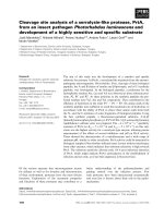
tified in this study, we obtained putativ e phosphorylation
motifs from the phosphopeptide dataset using the Motif-X
software tool (Figure 5). This tool extracts overrepresented
patterns from any sequence dataset by c omparing it to a
dynamic statistical background [58]. Four significantly
enriched phosphorylation motifs were extracted from the
identified DTB phosphopeptides dataset (Figure 5b). One
of the enriched phosphorylation site motifs resembled a
known motif in proline-directed kinases (pS/pTP). This
was also supported by the alignment of all the identified
DTB phosphorylation sites (Figure 5a). The identity of the
second enriched motif was unknown, and had no counter-
parts in any known kinases. The third enriched phosphor-
ylation motif showed high similarity to a motif found in
members of the casein kinase II subfamily (pS/pTXXE/D).
Members of this family can phosphorylate a wide variety
of plant proteins in vitro. The fourth enriched motif was
similar to the 14-3-3 binding motif (RXXpS/pT). Kinases
with this motif regulate the activities of the vacuolar potas-
sium channel KCO1 and the vacuolar ATPase [59] (Figure
5b). These results suggest that proline-directed kinases
could be the major kinase group involved phosphorylation
of these identified proteins during dormancy in poplar
(Figure 5).
Discussion
A series of differential expression profiling analyses of the
induction, maintenance, and release of bud dormancy
made it possible to identify a large set of dormancy-related
candidate genes [1,9-12,60-66]. These genes were mainly
involved in ABA signaling pathways, cold and oxidative
responses, flavonoid biosynthesis, flowering time, and cir-
cadian regulation [66,67]. A lthough there is increasing
information available about the roles of genes and their
products in dormancy, very little is known about the rele-
vance of protein phosphorylation in dormancy. To address
this, in this work, we identified the phosphorylation status
of proteins in dormant terminal buds of poplar using mass
spectrometry combined with TiO
2
phosphopeptide-
enrichment strategies. However, it remains unknown
whether these phos phoproteins identified i n dormant
buds in this study actually participate in dormancy-related
processes. To interpret the significance of the presence of
Liu et al. BMC Plant Biology 2011, 11:158
/>Page 7 of 16
these phosphoproteins in dormant buds, we compared the
identified phosphoproteins with previously reported dor-
mancy-related genes and their products. Notably, some of
these phosphoproteins were well matched to homologs of
known dormancy-related candidate gene-products identi-
fied in previous studies of various species. Some of these
common proteins of interest are briefly disc ussed in the
context of dormancy.
Phosphoproteins involved in dormancy-related signal
transduction
Abscisic acid (ABA) is the major plant hormone involved
in growth, dormancy, and cold acclimation [68]. The
ABA signaling pathway is regulat ed by rever sible protein
phosphorylation mediated by protein kinases and phos-
phatases [68]. Genet ic evidence demonstrat ed that
sucrose non-fermenting (SNF)-like protein kinase, recep-
tor-like protein kinase (LRK), and protein phosphatases
2C (PP2Cs) encoded by ABI1 and ABI2 are important
regulators of the ABA signaling pathway, which plays an
important role in the induction or release of bud dor-
mancy [5,6,10,63,68-72]. In this work, three SNF1-type
kinases in poplar (299214, 818055, and 828986) contain-
ing the phosphopeptide “DGHFLKTSCGpSPNYAAPE-
VISGK” , and one leucine-rich repeat receptor-like
protein kinase (LRK, 422370) were phosphorylated
Figure 4 KOG and molecular functional classificatio n of phosphoproteins identified from poplar DTBs with verified phosphopeptides
(n = 151). (a) KOG classification of phosphoproteins identified from poplar DTBs; X represents phosphoproteins without KOG classification; (b)
Molecular functional classification of identified phosphoproteins.
Liu et al. BMC Plant Biology 2011, 11:158
/>Page 8 of 16
(Additional files 12 and 13). These phosphorylation sites
were all well conserved, and corresponding phos phosi tes
were identified in Arabidopsis (Additional file 12). In the
case of PP2C, the Ser131 in the phosphopeptide
“VSGMIEGLIWpSPR” from PP2C (554898, 587195) was
identified as a novel phos phorylation site (Additional file
14). Calmodulin (CaM) and the CaM-binding protein
play an important role in Ca
2+
signaling, which is related
to bud dormancy [ 61,64,70,73,74]. In this study, two
CaM family proteins (729432 and 823453) were phos-
phorylated (Additional file 3 and Additional file 13); how-
ever, the corresponding site has not been identified as a
phosphorylation site in their respective Arabidopsis
counterparts, AT1G56340.1 and AT5G61790.1.
Phosphoproteins involved in auxin responses and growth
development related to dormancy
The auxin-sensitive Dormancy-associated/auxin-
repressed (DAAR) gene is associated with bud dormancy
[66,75,76]. In this study, one DAAR protein (647948)
showed three isoforms with respect to phosphorylation
status, the three forms respectively phosphorylated at
Thr61, Thr63, and Thr70 (Additional file 3 and Addi-
tional file 13). These corresponding sites have not been
identified as phosphorylation sites in its homolog in
Arabidopsis, the DAAR protein (AT1G28330.1). Inter-
estingly, the Arabidopsis DAAR protein is phosphory-
lated at its conserved Thr28 and Thr29 residues [33].
Vernali zation independence 4 (VIP4) interacts with the
FLOWERING LOCUS C-LIKE MADS-BOX PROTEIN
(FLC) to activate FLC, leading to inhibition of flower
development [77-79]. They are key components in the
regulatory pat hway of col d-mediated bud dorm ancy
induction and release [4,77]. In our study, we observed
tha t poplar VIP4 (569930) was phosphorylated at Ser225
(Additional file 3 and Additional file 13); the correspond-
ing site in its Arabidopsis homolog (AT5G61150.2) is
also known to be phosphorylated [50]. The mei2-Like
(ML) genes, which play roles in plant meiosis and deve l-
opment [80], were preferentially expressed in dormant
buds of leafy spurge [66]. In this study, two phosphoryla-
tion sites were respectively identified on the N- and C-
terminus of two i soforms of poplar m ei2-like proteins
(714870 and 41 0877), which are homologous to Arabi-
dopsis ML (AT1G29400.2) (Additional file 3 and Addi-
tional file 13). The corresponding site at the N-terminus
in Arabidopsis ML is known to be phosphorylated [ 50],
while the C-terminal phosphorylation site was novel.
Phosphoproteins involved in dormancy-related cold stress
response
Dehydrins (DHNs) are Group II (D-11 family), lat e
embryogenesis abundant (LEA) proteins that accumulate
in response to water deficit induced by drought, low tem-
perature, or salinity [81-84]. Certain DHNs play a vital
role in bud dormancy and cold acclimation of trees
Figure 5 Sequence alignment of phosphorylation sites and extraction of significantly enriched phosphorylation motifs. (a) Amino acid
sequence around the phosphorylated amino acid based on alignment of all phosphorylation sites from the identified DTBs phosphopeptide
dataset using Weblogo. (b) Motif-X-extracted motifs from entire phosphopeptide dataset. JGI Populus trichocarpa v1.1 protein database was used
as the background database to normalize the score against a random distribution of amino acids. Note that only those phosphorylated amino
acids that were confidently identified as the exact site of phosphorylation were used for the analysis (see “Materials and Methods” for detailed
description). Motif 1, Pro-directed kinase motif (n = 40); Motif 2, Unknown phosphorylation motifs (n = 20); Motif 3, CKII motif (n = 17); Motif 4,
14-3-3 binding motif (n = 13).
Liu et al. BMC Plant Biology 2011, 11:158
/>Page 9 of 16
[1,12,66,85-88]. Phosphorylation of their S-segment is
required for targeting to the nucleus [89-91]. In this
study, three DHN proteins were phosphorylated in
regions outside of the S-segment, one (663123) belongs
to the K
n
type of DHNs, one (571250) belongs to the K
n
S
type of DHNs, and the other (818850) belongs to the SK
n
type of DHNs (Additional file 3 and Additional file 13).
Heat shock proteins (HSP) function as molecular chaper-
ones, and are induced by various environmental stress,
such as cold, salinity, and oxidative stress [92]. Recent
data suggested that they are also involved in the process
of bud dormancy [12,93,94]. A phosphorylation event on
an HSP was identified in Arabidopsis [22,40]. Here, two
HSP70s (657150 and 769322), one HSP90 (652330), and
one HSP26 (832078) were phosphorylated in poplar
(Additional file 3 and Additional file 13).
Phosphoprotein associated with dormancy-related
flavonoid biosynthesis
Many genes related to flavonoid biosynthesis are signifi-
cantly regulated during the release of dormancy, such as
acetyl-CoA carboxylase (ACCase), chalcone synthase,
chalcone isomerase, and f lavonol syn thase [12,65- 67].
Acetyl-CoA carboxylase (ACCase) catalyzes the formation
of malonyl-CoA, which is the substrate for biosynthesis of
fatty acids and secondary metabolites, such as flavonoids
and anthocyanins [67]. In this work, one putative ACCase
(736443) was phosphorylated at Ser94 and Ser95 (Addi-
tional file 3 a nd Additional file 13). There have b een no
reports of phosphorylation of its homolog in Arabidopsis
(AT5G16390.1). Interestingly, we also found another
phosphorylation event related to flavonoid biosynthesis;
polyphenol oxidase (PPO) (275859) was phosphorylated at
Ser452 (Additional file 3 and Additional file 13). The
poplar PPO has no counterpart s in Arabidopsis,butit
shows homology to aureusidin synthase (AS) in Antirrhi-
num majus, a flavonoid synthase enzyme that catalyzes
the formation of aurones from chalcones [95]. To our
knowledge, this is the first report of a specific phosphory-
lation site in a plant flavonoid synthase. The existence of
this site suggests that phosphorylation may regulate its
functions.
Phosphoproteins involved in transport related to
dormancy
The plasma membrane H+-ATPase (AHA) is responsible
for the transport of protons out of the cell through the
membrane [96]. The AHA gene is strongly expressed dur-
ing dormancy transition, and contributes to changes in the
plasma membrane [12]. The regulation of AHA is con-
trolled by phosphorylation of one Thr residue in the well-
conserved C-terminal domain [97,98]. In the AHA family
in Arabidopsis, the well-conserved Thr residue is
phosphorylated in response to stress [37,42,97]. Here, the
exact Thr site (Thr949) in the C-terminus of poplar
AHA10 (826518), and its corresponding site in AHA11 of
poplar (422528) were both phosphorylated (Figure 2a).
Another example of a transport protein is ATP-binding
cassette (ABC) transporters, which are integral membrane
proteins that transport a wide variety of substrates, such as
ABA, auxin, and some plant secondary metabolites across
cellular membranes [99,100]. Genes encoding ABC trans-
porters are regulated during dormancy transition
[11,12,66], suggesting that they are linked with dormancy.
Here, two ABC transporter family proteins (554850 and
8001 53) were phosphorylated at Thr55 (Additional file 3
and Additional file 13). The corresponding site is phos-
phorylated in its homologs in rice, Arabidopsis,andMedi-
cago [42,49,50].
Phosphoproteins involved in protein synthesis related to
dormancy
Some genes and proteins involved in protein biosynthesis
play a role in the mechanism of bud dormancy release
[12,60,101]. Phosphorylation of ribosomal proteins can
affect protein synthesis by altering ribosome structure
[45]. In the present work, six 60S acidic ribosomal proteins
including P0-, P1-, P2-, and P3-types were phosphorylated
close to their conserved C terminus, consistent with
results reported elsewhere [45]. However, the pSer at posi-
tion 2 on the 40S ribosomal protein S12 of poplar (RPS12,
714910) was novel (Additional file 15). Recent evidence
suggests that phosphorylation of Ser2 plays an important
role in regulating nucleocytoplasmic shuttling of eukaryo-
tic translation initiation factor 5A (eIF5A) in plant cells
[102-104]. Here, four poplar eIF5A proteins (717121,
832646, 835953, and 724093) were phosphorylated at their
well-conserved serine residue and ace tyla ted at their N-
terminus (Additional file 16). Phosphorylation regulates
the function and/or location of translation elongation fac-
tor 1A (eEF1A), which is involved in protein biosynthesis
and signal transduction [105-107]. Here, five eEF1A iso-
forms (256777, 655943, 675976, 655949, and 720367)
from poplar, all containing the phosphopeptide pSVEMH-
HEALQEALPGDNVGFNVK (Ser279) were novel phos-
phoproteins (Additional file 17).
Phosphoproteins involved in electron transport or energy
pathways
There are increases in expressions of some genes involved
in energy pathways during bud release, including glyceral-
dehyde-3-phosphate dehydrogenase (GAPC) and phos-
phoenolpyruvate carboxy lase (PEPC) [11,12,60,93]. Here,
three GAPC isoforms (821843, 575307 and 728998) and
three PEPC isoforms (552645, 745223, and 728315) were
phosphorylated (Additional file 13 and Additional file 3).
Liu et al. BMC Plant Biology 2011, 11:158
/>Page 10 of 16
The light harvesting com plex protein Lhcb1, which is
essential for light electron transport, is sign ificantly regu-
lated during bud release [11,63,66]. Reversible phosphory-
lation of Lhcb1 is important for distributing absorbed light
energy between the two photosystems [108,109]. As
reported in other experiments on Arabidopsis [33,110]
and spinach, Lhcb1 proteins are phosphorylated at several
Thr and Ser residues in their amino terminus [108]. Here,
we identified two previously unknown phosphorylation
sites on the poplar Lhcb1 protein; the conserved Thr38
phosphosite and the unconserved Thr39 phosphosite (Fig-
ure 2b).
In summary, this information on phospho proteins in
dormant poplar provides a useful dataset, and provides
new insights for exploring the relevanc e of ph osphoryla-
tion for dormancy. However, further r esearch, e.g ., com-
paring proteomes between dormant/non-dormant tissues,
is required to clarify the roles of phosphorylation in the
dormancy process.
Conclusions
Many physiological features of woody plants are not
reflected in the herbaceous model Arabidopsis or in rice.
Therefore, it is important to determine phosphorylation
sites in poplar proteins, and to determine the roles of these
phosphorylations in modifying protein function during
growth and development. To date, there have been no
extensive studies on the poplar phosphoproteome. In this
work, we conducted a detailed analysis of the phosphopro-
teom e of dor mant poplar buds using an MS method and
TiO
2
phosphopeptide-enrichment strategies. We found
161 unique phosphorylated sites in 161 phosphopeptides
from 151 proteins, most of which are associated with bind-
ing and catalytic activity. Most of the poplar phosphopro-
teins have orthologs in Arabidopsis, suggesting that there
are similar signaling pathways mediated by phosphoryla-
tion in poplar and Arabidopsis. However, some phospho-
proteins and phosphorylated sites were unique to poplar,
thus confirming the need to obtain phosphoproteome data
from poplar. Se veral phosphorylation motifs were e xtracted
from the dataset by Motif-X. This could provide evidence
for the involvement of kinases in phosphorylation of these
identified proteins during dormancy in poplar. Further
experiments are now required to confirm that these speci-
fic kinases interact with the identified phosphoproteins in
vivo. A promising way forward is to comprehensively char-
acterize and analyze the dynamics of pho sphorylation of
poplar proteins in response to environmental changes,
using specialized targeted quantitative proteomics tools.
Methods
Plant materials and chemicals
Dormant terminal buds were collected from hybrid
poplar (Populus simonii × P. nigra) in Harbin, China,
(E126°37’ , N45°42’ ) at the end of December, 2009.
Samples were frozen in liquid nitrogen and stored at
-80°C until use.
Iodoacetamide (IAA) and dithiothreitol (DTT) were
purchased from Acros Organics (Morris Plains, NJ, USA).
HPLC-grade acetonitrile (ACN) was obtained from JT
Baker (Thomas Scientific, Swedesboro, NJ, USA). HPLC-
grade water was prepared using a Milli-Q A10 system
from Millipore (Billerica, MA, USA). ModiWed sequen-
cing-grade trypsin was supplied by Promega (Madison,
WI, USA). Prot ease-inhibitor cocktail and the 2-D Quant
kitwereobtainedfromAmershamPharmaciaBiotech
(Uppsala, Sweden). All other reagents were purchased
from Sigma (St Louis, MO, USA).
Preparation of total proteins
The dormant terminal buds were crushed into a fine pow-
der in liquid nitrogen and resuspended at -20°C in 1 0%
(w/v) trichloroacetic acid (TCA) in cold acetone contain-
ing 0.07% (v/v) 2-mercaptoethanol for at least 2 h. The
mixture was centrifuged at 10000 g at 4°C for 1 h, and the
precipitates were washed with cold acetone containing
0.07% (v/v) 2-mercaptoethanol. The pellets were dried by
vacuum centrifugation and dissolved in 7 M urea, 2 M
thiourea, 20 mM dithiothreitol, 1% (v/v) protease-inhibitor
cocktail, 0.2 mM Na
2
VO
3
, and 1 mM NaF at room tem-
perat ure for 2 h, before centrifugation at 40000 g at 10°C
for 1 h. The resulting supernatant was collected and kept
at -80°C until further use. The total protein content of the
samples was quantified using a 2-D Quant kit.
In-solution protein digestion
Total proteins were digested as described elsewhere
[111,112]. Briefly, the total protein solution was adjusted
to pH 8.5 with 1 M ammonium bicarbonate. Then, the
sample was reduced for 45 min at 55°C by adding DTT to
a final concentration of 10 mM, and then carboxyamido-
methylated by incubation with 55 mM IAA for 30 min in
the dark at room temperature. After this step, CaCl
2
was
added to a final concentration of 20 mM. Then, endopro-
tease Lys-C was added to a final substrate-to-enzyme ratio
of 100:1, and this reaction was incubated for 12 h at 37°C.
The Lys-C digest was added to 1 M urea containing 100
mM ammonium bicarbonate, and modified trypsin was
added to a final substrate-to-enzyme ratio of 50:1. The
trypsin digest w as also incubated at 37°C for 12 h. After
digestion, the peptide mixture was enriched using TiO
2
microcolumns for further MS analysis.
Enrichment of phosphorylated peptides using TiO
2
microcolumns
The TiO
2
microcolumns were packed as described else-
where [25]. A small plug of C8 material was stamped out
of a 3M Empore C8 extraction disk with a HPLC syringe
Liu et al. BMC Plant Biology 2011, 11:158
/>Page 11 of 16
needle and placed to form a frit at the small end of the
GELoader tip. The TiO
2
beads were suspended in 100%
ACN, and an appropriate volume of this suspension
(depending on the size of the column) was loaded into
the GELoader tip. Gentle air pressure produced by a plas-
tic syringe was applied to pack the column. The TiO
2
microc olumn was equilibrated with loading buffer (40 μl;
80% ACN/5% TFA/saturated phthalic acid solution).
Immediately, the trypsin-digested peptide mixture diluted
in loading buffer was added to the TiO
2
microcolumn.
Then, the column was washed once with loading buffer
(40 μl) and three times with washing buffer (40 μl; 80%
ACN/2% TFA). The washing and loading buffer con-
tained 80% ACN organic solvent in order to abrogate the
adsorption of peptides to the C8 material [28]. The
bound peptides were eluted twice with 40 μlammonium
bicarbonate (pH > 10.5), and then with 10 μl30%ACN.
The eluted phosphopeptides were lyophilized and then
dissolved in 1% formic acid before MS analysis.
NanoUPLC-ESI-MS/MS
NanoUPLC-ESI-MS/MS was performed with a splitless
nanoUPLC (10 kpsi nanoAcquity; Waters) in combination
with a Synapt high-definition mass spectrometer with a
nanospray ion source (Waters). A symmetric C
18
5-μm,
180-μm × 20-mm pre-column and a BEH C
18
1.7-μm, 75-
μm × 250-mm analytical reversed-phase column (Waters)
were used. The MassLyn x (versio n 4.1; Waters) program
was used for instrument control and data acquisition. The
mobile phases were (A) 100% H
2
O/0.1% formic acid and
(B) 100% ACN/0.1% formic acid. The samples were dis-
solved in aqueous 0.1% formic acid solution and loaded
onto the pre-column at a flow rate of 5 μl/min for 3 min.
The phosphopeptides were separated by a gradient of 5-
40% mobile phase B for 90 min at a flow rate of 2 00 nl/
min, followed by a 10-min rinse with 90% mobile phase B.
The column was re-equilibrated with the initial conditions
for 20 min. The lock mass was delivered from the auxiliary
pump of the NanoAcquity pump at a constant flow rate of
400 nl/min at a concentration of 100 fmol/μlof(Glu1)
fibrinopeptide B to the reference sprayer of the NanoLock-
Spray source from the mass spectrometer. In this study,
every sample was analyzed in triplicate. Data-dependent
acquisition was carried out in positive ion mode. MS spec-
tra were acquired for 1 s from mass-to-charge ratios of
(m/z) 350 to 1990. Two of the most intense precursor ions
that were doubly or triply charged were selected from m/z
350 to 1990. MS/MS spectra produced by collision-
induced dissociation (CID) were acquired for 2 s from m/z
50 to 1990. The collision energy was automatically calcu-
lated according to peptide charge and m/z; a dynamic
exclusion window was applied to prevent the same m/z
from being selected for 2 min after its acquisition. The
candidate phosphopeptides were initially assigned by ESI-
MS/MS using 79.96-Da mass increments per phosphate
moiety relative to the unmodified peptides. To detect the
phosphopeptides, we utilized the preferred loss of the
phosphate group upon collision-induced dissociation. In
positive ion tandem MS, an intense neutral loss of 98 Da,
corresponding to H
3
PO
4
, was observed for peptides con-
taining phosphorylated Ser, Thr, and Tyr residues.
Data analysis and Mascot database search
The MS/MS data were processed and converted to a pkl
file format with ProteinLynx software (Waters), and the
resulting pkl file was used to search against the JGI Popu-
lus trichocarpa v1.1 ( optr1_1/
Poptr1_1.home.html) protein sequence database using an
in-house Mascot server (version 1.8) with acetylation in
the N-terminus of the protein, carbamido methylation,
methionine oxidation, and phosphorylation of serine/
threonine/tyrosine residues as variable modifications. Two
missed cleavage sites were allowed. The search was per-
formed with a peptide mass tolerance of 15 ppm in the
MS and 50 ppm in the MS/MS modes. The false discovery
rate (FDR) was 0.00% for peptide matches above the iden-
tity threshold and 0.36-0.85% for peptide matches above
the homology or identity threshold.
Bioinformatics
Using a cu stom Perl program, all the phosphop rotein
sequences were extracted from protein databases (http://
genome.jgi-psf.org/Poptr1_ 1/Poptr1_1.home.html) b y
their protein ID. The Blast2Go program [57] was used to
obtain descriptions of protein sequences by a BlastP search
against a non-redundant protein database (http://blast.
ncbi.nlm.nih.gov/Blast.cgi) with default parameter settings.
Protein functions, annotations, and classifications were
also examined using gene ontology (GO), GO-Enzyme-
Code, and InterPro databases and search tools.
The Batch sequence search tool (.
uk/search) was applied to obtain Pfam information for
identified phosphoproteins. The significantly enriched
phosphorylation motifs set was extracted from our phos-
phopeptide data using the Motif-X algorithm [58]. All
phosphorylated peptides with confidently identified phos-
phorylation sites were used as the data set to extract sig-
nificantly enriched phosphorylation motifs. The
phosphopeptides were centered at the phosphorylated
amino acid residues and aligned, and ten positions
upstream and downstream of the phosphor ylation site
were included. In the case of C- and N-terminal peptides,
the sequence was completed to 21 amino acids with the
required number of “X"s, where X represents any amino
acid. As the background data set, protein sequences of the
entire genome poplar database Populus trichocarpa v1.1 in
Fasta format (in a shortened version due to upload restric-
tions of 10 MB) were used. The occurrence threshold was
Liu et al. BMC Plant Biology 2011, 11:158
/>Page 12 of 16
set to 5% of the input data set at a minimum of three pep-
tides, and the probability threshold was set to P <10
-5
.
Amino acid sequences around the phosphorylated amino
acid based on the alignment of all the phosphorylation
sites were completed by the Weblogo program [ 113] in
the entire identified DTBs data set.
Additional material
Additional file 1: Nine sheets as follows: Sheet 1: Contents. Sheet 2:
Phosphopeptide identification list. Sheet 3: Phosphorylation site list.
Sheet 4: Blast results. Sheet 5:Annotation_of_phosphoproteins. Sheet 6:
KOG classifications. Sheet 7: Pfam_domain_information. Sheet 8:
Source_for_motif_analysis. Sheet 9: pS_motifs.
Additional file 2: Phosphopeptides and phosphorylation sites
identified in dormant terminal buds of poplar.
Additional file 3: Detailed information for phosphopeptides and
phosphoproteins identified in dormant terminal buds of poplar.
Additional file 4: MS/MS spectra (in a separate file). File contains all
the original MS/MS spectra of 161 phosphopeptides identified in
this study.
Additional file 5: Comparison of singly and doubly phosphorylated
peptides.
Additional file 6: Location of phosphorylation sites in characterized
conserved domains.
Additional file 7: Flowchart for analyzing the conservation of
phosphoproteins and phosp hosites between poplar and Arabidopsis.
Additional file 8: Conserved phosphorylation sites within
orthologous proteins. (a) Phosphosites conserved in orthologous
proteins. (b) Phosphosites that were not conserved in orthologous
proteins.
Additional file 9: Unconserved phosphorylation sites within
orthologous proteins.
Additional file 10: KOG analysis of identified phosphoproteins and
all proteins encoded in Populus trichocarpa genome. (a) Percentage
of KOG functional group categories from the identified phosphoproteins
and all proteins encoded in Populus trichocarpa genome. (b) Percentage
of KOG functional subgroup categories from the identified
phosphoproteins and all proteins encoded in Populus trichocarpa
genome.
Additional file 11: Complete list of KOG analysis of
phosphoproteins and all proteins encoded in Populus trichocarpa
genome.
Additional file 12: Sequence alignment of phosphorylated sites in
protein kinases between poplar and Arabidopsis.
Additional file 13: Detailed information for identified
phosphoproteins referred to in discussion section.
Additional file 14: Sequence alignment of phosphorylated sites in
protein phosphatases between poplar and Arabidopsis.
Additional file 15: Sequence alignment of RPS12 between poplar
and Arabidopsis.
Additional file 16: Sequence alignment of conserved N-terminus of
eIF5A between poplar and Arabidopsis.
Additional file 17: Sequence alignment of conserved C-terminus of
EF-1-alpha between poplar and Arabidopsis.
List of abbreviations
DTB: Dormant terminal buds; NanoUPLC: Nano ultra-performance liquid
chromatography; Ser: Serine; Thr: Threonine; Tyr: Tyrosine; PTM: Post-
translational modification.
Acknowledgements
We thank Prof. Bai-Chen Wang for helpful discussions. This work was
partially supported by the National Basic Research Priorities Program (Grant
No. 2009CB119102), the State Key Program of National Natural Science of
China (Grant No. 31030017), doctoral funding from Northeast Forestry
University (Grant No. 140-602055), and the Fund for Key Projects and
Innovation Teams from Northeast Forestry University (Grant No. DL09EA01-
2). The authors have no conflicts of interest to declare.
Author details
1
State Key Laboratory of Forest Genetics and Tree Breeding (Northeast
Forestry University), 26 Hexing Road, Harbin 150040, China.
2
Laboratory for
Chemical Defence and Microscale Analysis, P.O. Box 3, Zhijiang 443200,
China.
3
Shenyang Agricultural University, Dongling Road 120, Shenyang,
Liaoning 110866, China.
4
Institute of Basic Medical Sciences, National Center
for Biomedical Analysis, 27 Taiping Road, Beijing 100850, China.
5
Daqing
Branch, Harbin Medical University, Daqing 163319, China.
Authors’ contributions
The study was conceived by CPY and ZGW. CCL and HXW carried out
experimental work, participated in data analyses, and drafted the manuscript.
CFL and ZYS participated in the design of the study and performed in silico
analyses. All authors read and approved the final manuscript.
Competing interests
The authors declare that they have no competing interests.
Received: 22 April 2011 Accepted: 11 November 2011
Published: 11 November 2011
References
1. Rohde A, Ruttink T, Hostyn V, Sterck L, Van Driessche K, Boerjan W: Gene
expression during the induction, maintenance, and release of dormancy
in apical buds of poplar. J Exp Bot 2007, 58:4047-4060.
2. Rajeev Arora LJR, Tanino Karen: Induction and Release of Bud Dormancy
in Woody perennials: A science Comes of Age. Hortscience 2003,
38:911-921.
3. Allona I, Ramos A, Ibá ez C, Contreras R, Casado R, Aragoncillo C: Review.
Molecular control of winter dormancy establishment in trees. J Agric Res
2008, 6:201-210.
4. Rohde A, Bhalerao R: Plant dormancy in the perennial context. Trends
Plant Sci 2007, 12:217-223.
5. David P Horvath JVA, Chao SWun, Foley EMichael: Knowing when to grow:
signals regulating bud dormancy. Trends Plant Sci 2003, 8:534-540.
6. Anderson J, Chao W, Horvath D, USDA A: A current review on the
regulation of dormancy in vegetative buds. Weed Sci 2001, 49:581-589.
7. Druart N, Johansson A, Baba K, Schrader J, Sj din A, Bhalerao R, Resman L,
Trygg J, Moritz T, Bhalerao R: Environmental and hormonal regulation of
the activity-dormancy cycle in the cambial meristem involves stage-
specific modulation of transcriptional and metabolic networks. Plant J
2007, 50:557-573.
8. Campbell M, Segear E, Beers L, Knauber D, Suttle J: Dormancy in potato
tuber meristems: chemically induced cessation in dormancy matches
the natural process based on transcript profiles. Funct Integr Genomics
2008, 8:317-328.
9. Schrader J, Moyle R, Bhalerao R, Hertzberg M, Lundeberg J, Nilsson P,
Bhalerao R: Cambial meristem dormancy in trees involves extensive
remodelling of the transcriptome. Plant J 2004, 40:173-187.
10. Ruttink T, Arend M, Morreel K, Storme V, Rombauts S, Fromm J, Bhalerao R,
Boerjan W, Rohde A: A molecular timetable for apical bud formation and
dormancy induction in poplar. Plant Cell 2007, 19:2370-2390.
11. Mathiason K, He D, Grimplet J, Venkateswari J, Galbraith D, Or E, Fennell A:
Transcript profiling in Vitis riparia during chilling requirement fulfillment
reveals coordination of gene expression patterns with optimized bud
break. Funct Integr Genomics 2009, 9:81-96.
12. Mazzitelli L, Hancock R, Haupt S, Walker P, Pont S, McNicol J, Cardle L,
Morris J, Viola R, Brennan R: Co-ordinated gene expression during phases
of dormancy release in raspberry (Rubus idaeus L.) buds. J Exp Bot 2007,
58:1035-1045.
13. Paradela A, Albar J: Advances in the Analysis of Protein Phosphorylation.
J Proteome Res 2008, 7:1809-1818.
Liu et al. BMC Plant Biology 2011, 11:158
/>Page 13 of 16
14. Schulze W: Proteomics approaches to understand protein
phosphorylation in pathway modulation. Curr Opin Plant Biol 2010,
13:280-287.
15. White FM: Quantitative phosphoproteomic analysis of signaling network
dynamics. Curr Opin Biotechnol 2008, 19:404-409.
16. Linding R, Jensen L, Ostheimer G, van Vugt M, Jorgensen C, Miron I,
Diella F, Colwill K, Taylor L, Elder K: Systematic discovery of in vivo
phosphorylation networks. Cell 2007, 129:1415-1426.
17. Mann M, Ong S, Grønborg M, Steen H, Jensen O, Pandey A: Analysis of
protein phosphorylation using mass spectrometry: deciphering the
phosphoproteome. Trends Biotechnol 2002, 20:261-268.
18. Schmelzle K, White FM: Phosphoproteomic approaches to elucidate
cellular signaling networks. Curr Opin Biotechnol 2006, 17:406-414.
19. van Bentem S, Roitinger E, Anrather D, Csaszar E, Hirt H:
Phosphoproteomics as a tool to unravel plant regulatory mechanisms.
Physiol Plant 2006, 126:110-119.
20. de la Fuente van Bentem S, Hirt H: Using phosphoproteomics to reveal
signalling dynamics in plants. Trends Plant Sci 2007, 12:404-411.
21. Peck S: Phosphoproteomics in Arabidopsis: moving from empirical to
predictive science. J Exp Bot 2006, 57:1523-1527.
22. Sugiyama N, Nakagami H, Mochida K, Daudi A, Tomita M, Shirasu K,
Ishihama Y: Large-scale phosphorylation mapping reveals the extent of
tyrosine phosphorylation in Arabidopsis. Mol Syst Biol 2008, 4:1-7.
23. Aebersold R, Mann M: Mass spectrometry-based proteomics. Nature 2003,
422:198-207.
24. Zhou H, Watts J, Aebersold R: A systematic approach to the analysis of
protein phosphorylation. Nat Biotechnol 2001, 19:375-378.
25. Larsen M, Thingholm T, Jensen O, Roepstorff P, Jorgensen T: Highly
Selective Enrichment of Phosphorylated Peptides from Peptide Mixtures
Using Titanium Dioxide Microcolumns*. Mol Cell Proteomics 2005,
4:873-886.
26. Klemm C, Otto S, Wolf C, Haseloff R, Beyermann M, Krause E: Evaluation of
the titanium dioxide approach for MS analysis of phosphopeptides. J
Mass Spectrom 2006, 41:1623-1632.
27. Areces LB, Matafora V, Bachi A: Analysis of protein phosphorylation by
mass spectrometry. Eur J Mass Spectrom 2004, 10
:383-392.
28.
Thingholm TE, Jorgensen TJD, Jensen ON, Larsen MR: Highly selective
enrichment of phosphorylated peptides using titanium dioxide. Nat
Protoc 2006, 1:1929-1935.
29. Nühse T, Stensballe A, Jensen O, Peck S: Large-scale analysis of in vivo
phosphorylated membrane proteins by immobilized metal ion affinity
chromatography and mass spectrometry. Mol Cell Proteomics 2003,
2:1234-1243.
30. Nühse T, Stensballe A, Jensen O, Peck S: Phosphoproteomics of the
Arabidopsis Plasma Membrane and a New Phosphorylation Site
Database. Plant Cell 2004, 16:2394-2405.
31. Nühse T, Bottrill A, Jones A, Peck S: Quantitative phosphoproteomic
analysis of plasma membrane proteins reveals regulatory mechanisms
of plant innate immune responses. Plant J 2007, 51:931-940.
32. de la Fuente van Bentem S, Anrather D, Roitinger E, Djamei A, Hufnagl T,
Barta A, Csaszar E, Dohnal I, Lecourieux D, Hirt H: Phosphoproteomics
reveals extensive in vivo phosphorylation of Arabidopsis proteins
involved in RNA metabolism. Nucleic Acids Res 2006, 34:3267-3278.
33. Reiland S, Messerli G, Baerenfaller K, Gerrits B, Endler A, Grossmann J,
Gruissem W, Baginsky S: Large-scale Arabidopsis phosphoproteome
profiling reveals novel chloroplast kinase substrates and
phosphorylation networks. Plant Physiol 2009, 150:889-903.
34. Li H, Wong WS, Zhu L, Guo HW, Ecker J, Li N: Phosphoproteomic analysis
of ethylene-regulated protein phosphorylation in etiolated seedlings of
Arabidopsis mutant ein2 using two-dimensional separations coupled
with a hybrid quadrupole time-of-flight mass spectrometer. Proteomics
2009, 9:1646-1661.
35. de la Fuente van Bentem S, Anrather D, Dohnal I, Roitinger E, Csaszar E,
Joore J, Buijnink J, Carreri A, Forzani C, Lorkovic ZJ, et al: Site-specific
phosphorylation profiling of Arabidopsis proteins by mass spectrometry
and peptide chip analysis. J Proteome Res 2008, 7:2458-2470.
36. Carroll AJ, Heazlewood JL, Ito J, Millar AH: Analysis of the Arabidopsis
cytosolic ribosome proteome provides detailed insights into its
components and their post-translational modification. Mol Cell Proteomics
2008, 7:347-369.
37. Niittyla T, Fuglsang AT, Palmgren MG, Frommer WB, Schulze WX: Temporal
analysis of sucrose-induced phosphorylation changes in plasma
membrane proteins of Arabidopsis. Mol Cell Proteomics 2007, 6:1711-1726.
38. Wolschin F, Weckwerth W: Combining metal oxide affinity
chromatography (MOAC) and selective mass spectrometry for robust
identification of in vivo protein phosphorylation sites. Plant Methods
2005, 1:1-9.
39. Benschop J, Mohammed S, O’Flaherty
M, Heck A, Slijper M, Menke F:
Quantitative phosphoproteomics of early elicitor signaling in
Arabidopsis. Mol Cell Proteomics 2007, 6:1198-1214.
40. Jones A, MacLean D, Studholme D, Serna-Sanz A, Andreasson E, Rathjen J,
Peck S: Phosphoproteomic analysis of nuclei-enriched fractions from
Arabidopsis thaliana. J Proteomics 2009, 72:439-451.
41. Agrawal G, Thelen J: Large scale identification and quantitative profiling
of phosphoproteins expressed during seed filling in oilseed rape. Mol
Cell Proteomics 2006, 5 :2044-2059.
42. Whiteman SA, Nuhse TS, Ashford DA, Sanders D, Maathuis FJ: A proteomic
and phosphoproteomic analysis of Oryza sativa plasma membrane and
vacuolar membrane. Plant J 2008, 56:146-156.
43. Endler A, Reiland S, Gerrits B, Schmidt UG, Baginsky S, Martinoia E: In vivo
phosphorylation sites of barley tonoplast proteins identified by a
phosphoproteomic approach. Proteomics 2009, 9:310-321.
44. Lu T, Meng L, Yang C, Liu G, Liu G, Ma W, Wang B: A shotgun
phosphoproteomics analysis of embryos in germinated maize seeds.
Planta 2008, 228:1029-1041.
45. Liu C-C, Lu T-C, Li H-H, Wang H-X, Liu G-F, Ma L, Yang C-P, Wang B-C:
Phosphoproteomic identification and phylogenetic analysis of ribosomal
P-proteins in Populus dormant terminal buds. Planta 2010, 231:571-581.
46. Stone J, Walker J: Plant protein kinase families and signal transduction.
Plant Physiol 1995, 108:451-457.
47. Mackintosh R, Davies S, Clarke P, Weekes J, GIllespie J, Gibb B, Hardie D:
Evidence for a protein kinase cascade in higher plants. Eur J Biochem
2005, 209:923-931.
48. Baginsky S, Gruissem W: The chloroplast kinase network: new insights
from large-scale phosphoproteome profiling. Mol Plant 2009, 2:1141-1153.
49. Grimsrud PA, den Os D, Wenger CD, Swaney DL, Schwartz D, Sussman MR,
Ane JM, Coon JJ: Large-scale phosphoprotein analysis in Medicago
truncatula roots provides insight into in vivo kinase activity in legumes.
Plant Physiol 2010, 152:19-28.
50. Nakagami H, Sugiyama N, Mochida K, Daudi A, Yoshida Y, Toyoda T,
Tomita M, Ishihama Y, Shirasu K: Large-scale comparative
phosphoproteomics identifies conserved phosphorylation sites in plants.
Plant Physiol 2010, 153:1161-1174.
51. van Bentem SD, Hirt H: Protein tyrosine phosphorylation in plants: more
abundant than expected? Trends Plant Sci 2009, 14:71-76.
52. Han G, Ye M, Zou H: Development of phosphopeptide enrichment
techniques for phosphoproteome analysis. Analyst 2008, 133:1128-1138.
53.
Dunn JD, Reid GE, Bruening ML: Techniques for phosphopeptide
enrichment prior to analysis by mass spectrometry. Mass Spectrom Rev
2010, 29:29-54.
54. Bateman A, Birney E, Cerruti L, Durbin R, Etwiller L, Eddy S, Griffiths-Jones S,
Howe K, Marshall M, Sonnhammer E: The Pfam protein families database.
Nucleic Acids Res 2002, 30:276-280.
55. Durek P, Schmidt R, Heazlewood JL, Jones A, MacLean D, Nagel A,
Kersten B, Schulze WX: PhosPhAt: the Arabidopsis thaliana
phosphorylation site database. An update. Nucleic Acids Res 2010, 38:
D828-834.
56. Tuskan G, Difazio S, Jansson S, Bohlmann J, Grigoriev I, Hellsten U,
Putnam N, Ralph S, Rombauts S, Salamov A: The genome of black
cottonwood, Populus trichocarpa (Torr. & Gray). Science 2006,
313:1596-1604.
57. Conesa A, Gotz S, Garcia-Gomez JM, Terol J, Talon M, Robles M: Blast2GO: a
universal tool for annotation, visualization and analysis in functional
genomics research. Bioinformatics 2005, 21:3674-3676.
58. Schwartz D, Gygi S: An iterative statistical approach to the identification
of protein phosphorylation motifs from large-scale data sets. Nat
Biotechnol 2005, 23:1391-1398.
59. Bunney T, van Walraven H, de Boer A: 14-3-3 protein is a regulator of the
mitochondrial and chloroplast ATP synthase. Proc Natl Acad Sci 2001,
98:4249-4254.
Liu et al. BMC Plant Biology 2011, 11:158
/>Page 14 of 16
60. Derory J, Leger P, Garcia V, Schaeffer J, Hauser MT, Salin F, Luschnig C,
Plomion C, Glossl J, Kremer A: Transcriptome analysis of bud burst in
sessile oak (Quercus petraea). New Phytol 2006, 170:723-738.
61. Jimenez S, Li Z, Reighard G, Bielenberg D: Identification of genes
associated with growth cessation and bud dormancy entrance using a
dormancy-incapable tree mutant. BMC Plant Biol 2010, 10:25.
62. Park S, Keathley DE, Han KH: Transcriptional profiles of the annual growth
cycle in Populus deltoides. Tree Physiol 2008, 28:321-329.
63. Jia Y, Anderson JV, Horvath DP, Gu YQ, Lym RG, Chao WS: Subtractive
cDNA libraries identify differentially expressed genes in dormant and
growing buds of leafy spurge (Euphorbia esula). Plant Mol Biol 2006,
61:329-344.
64. Yakovlev IA, Fossdal CG, Junttila O, Skr ppa T: Analysis of gene expression
during bud burst initiation in Norway spruce via ESTs from subtracted
cDNA libraries. Tree Genet Genomes 2006, 2:39-52.
65. Horvath DP, Anderson JV, Soto-Suarez M, Chao WS: Transcriptome analysis
of leafy spurge (Euphorbia esula) crown buds during shifts in well-
defined phases of dormancy. Weed Sci 2006, 821-827.
66. Horvath D, Chao W, Suttle J, Thimmapuram J, Anderson J: Transcriptome
analysis identifies novel responses and potential regulatory genes
involved in seasonal dormancy transitions of leafy spurge (Euphorbia
esula L.). BMC Genomics 2008, 9:536.
67. Hedley P, Russell J, Jorgensen L, Gordon S, Morris J, Hackett C, Cardle L,
Brennan R: Candidate genes associated with bud dormancy release in
blackcurrant (Ribes nigrum L.). BMC Plant Biol 2010, 10:202.
68. Vlad F, Rubio S, Rodrigues A, Sirichandra C, Belin C, Robert N, Leung J,
Rodriguez PL, Lauriere C, Merlot S: Protein Phosphatases 2C Regulate the
Activation of the Snf1-Related Kinase OST1 by Abscisic Acid in
Arabidopsis. Plant Cell 2009, 21:3170-3184.
69. Meyer K, Leube MP, Grill E: A protein phosphatase 2C involved in ABA
signal transduction in Arabidopsis thaliana. Science 1994, 264:1452-1455.
70. Pang X, Halaly T, Crane O, Keilin T, Keren-Keiserman A, Ogrodovitch A,
Galbraith D, Or E: Involvement of calcium signalling in dormancy release
of grape buds. J Exp Bot 2007, 58:3249-3262.
71. Osakabe Y, Maruyama K, Seki M, Satou M, Shinozaki K, Yamaguchi-
Shinozaki K: Leucine-rich
repeat receptor-like kinase1 is a key
membrane-bound regulator of abscisic acid early signaling in
Arabidopsis. Plant Cell 2005, 17:1105-1119.
72. Leung J, Merlot S, Giraudat J: The Arabidopsis ABSCISIC ACID-INSENSITIVE2
(ABI2) and ABI1 genes encode homologous protein phosphatases 2C
involved in abscisic acid signal transduction. Plant Cell 1997, 9:759-771.
73. Kim MC, Chung WS, Yun DJ, Cho MJ: Calcium and calmodulin-mediated
regulation of gene expression in plants. Mol Plant 2009, 2:13-21.
74. Brennan R, Jorgensen L, Hackett C, Woodhead M, Gordon S, Russell J: The
development of a genetic linkage map of blackcurrant (Ribes nigrum L.)
and the identification of regions associated with key fruit quality and
agronomic traits. Euphytica 2008, 161:19-34.
75. Huang X, Xue T, Dai S, Gai S, Zheng C, Zheng G: Genes associated with
the release of dormant buds in tree peonies (Paeonia suffruticosa ). Acta
Physiol Plant 2008, 30 :797-806.
76. Anderson JV, Gesch RW, Jia Y, Chao WS, Horvath DP: Seasonal shifts in
dormancy status, carbohydrate metabolism, and related gene
expression in crown buds of leafy spurge. Plant Cell Environ 2005,
28:1567-1578.
77. Sreekantan L, Mathiason K, Grimplet J, Schlauch K, Dickerson JA, Fennell AY:
Differential floral development and gene expression in grapevines
during long and short photoperiods suggests a role for floral genes in
dormancy transitioning. Plant Mol Biol 2010, 73:191-205.
78. Sheldon C, Hills M, Lister C, Dean C, Dennis E, Peacock W: Resetting of
FLOWERING LOCUS C expression after epigenetic repression by
vernalization. Proc Natl Acad Sci 2008, 105:2214-2219.
79. Zhang H, Van Nocker S: The VERNALIZATION INDEPENDENCE 4 gene
encodes a novel regulator of FLOWERING LOCUS C. Plant J 2002,
31:663-673.
80. Kaur J, Sebastian J, Siddiqi I: The Arabidopsis-mei2-like genes play a role
in meiosis and vegetative growth in Arabidopsis. Plant Cell 2006,
18:545-559.
81. Close T: Dehydrins: emergence of a biochemical role of a family of plant
dehydration proteins. Physiol Plant 1996, 97:795-803.
82. Allagulova C, Gimalov F, Shakirova F, Vakhitov V: The plant dehydrins:
structure and putative functions. Biochemistry (Mosc) 2003, 68:945-951.
83. Kosova K, Vitamvas P, Prásil I: The role of dehydrins in plant response to
cold. Biol
Plant 2007, 51:601-617.
84. Tunnacliffe A, Wise M: The continuing conundrum of the LEA proteins.
Naturwissenschaften 2007, 94:791-812.
85. Rorat T: Plant dehydrins-tissue location, structure and function. Cell Mol
Biol Lett 2006, 11:536-556.
86. Rinne P, Welling A, Kaikuranta P: Onset of freezing tolerance in birch
(Betula pubescens Ehrh.) involves LEA proteins and osmoregulation and
is impaired in an ABA-deficient genotype. Plant Cell Environ 1998,
21:601-611.
87. Rinne PLH, Kaikuranta PLM, van der Plas LHW, van der Schoot C: Dehydrins
in cold-acclimated apices of birch (Betula pubescens Ehrh.): production,
localization and potential role in rescuing enzyme function during
dehydration. Planta 1999, 209:377-388.
88. Puhakainen T, Li C, Boije-Malm M, Kangasj rvi J, Heino P, Palva ET: Short-
day potentiation of low temperature-induced gene expression of a C-
repeat-binding factor-controlled gene during cold acclimation in silver
birch. Plant Physiol 2004, 136:4299-4307.
89. Goday A, Jensen AB, Culianez-Macia FA, Alba MM, Figueras M, Serratosa J,
Torrent M, Pages M: The Maize Abscisic Acid-Responsive Protein Rab17 Is
Located in the Nucleus and Interacts with Nuclear Localization Signals.
Plant Cell 1994, , :: 351-360.
90. Jensen AB, Goday A, Figueras M, Jessop AC, Pages M: Phosphorylation
mediates the nuclear targeting of the maize Rab17 protein. Plant J 1998,
13:691-697.
91. Riera M, Figueras M, Lopez C, Goday A, Pages M: Protein kinase CK2
modulates developmental functions of the abscisic acid responsive
protein Rab17 from maize. Proc Natl Acad Sci 2004, 101:9879-9884.
92. Wang W, Vinocur B, Shoseyov O, Altman A: Role of plant heat-shock
proteins and molecular chaperones in the abiotic stress response. Trends
Plant Sci 2004, 9:244-252.
93. Santamaria ME, Rodriguez R, Ca al MJ, Toorop PE: Transcriptome analysis
of chestnut (Castanea sativa) tree buds suggests a putative role for
epigenetic control of bud dormancy. Ann Bot 2011, 108:485-498.
94. Wisniewski M, Sauter J, Fuchigami L, Stepien V: Effects of near-lethal heat
stress on bud break, heat-shock proteins and ubiquitin in dormant
poplar (Populus nigra Charkowiensis × P. nigra incrassata). Tree Physiol
1997, 17:453-460.
95. Ono E, Hatayama M, Isono Y, Sato T, Watanabe R, Yonekura-Sakakibara K,
Fukuchi-Mizutani M, Tanaka Y, Kusumi T, Nishino T: Localization of a
flavonoid biosynthetic polyphenol oxidase in vacuoles. Plant J
2006,
45:133-143.
96.
Jefferies KC, Cipriano DJ, Forgac M: Function, structure and regulation of
the vacuolar (H+)-ATPases. Arch Biochem Biophys 2008, 476:33-42.
97. Duby G, Boutry M: The plant plasma membrane proton pump ATPase: a
highly regulated P-type ATPase with multiple physiological roles. Pflugers
Arch 2009, 457:645-655.
98. Palmgren MG: Plant plasma membrane H+-ATPases: powerhouses for
nutrient uptake. Annu Rev Plant Biol 2001, 52:817-845.
99. Kuromori T, Miyaji T, Yabuuchi H, Shimizu H, Sugimoto E, Kamiya A,
Moriyama Y, Shinozaki K: ABC transporter AtABCG25 is involved in
abscisic acid transport and responses. Proc Natl Acad Sci 2010,
107:2361-2366.
100. Pighin JA, Zheng H, Balakshin LJ, Goodman IP, Western TL, Jetter R, Kunst L,
Samuels AL: Plant cuticular lipid export requires an ABC transporter.
Science 2004, 306:702-704.
101. Paw owski TA: Proteomic approach to analyze dormancy breaking of
tree seeds. Plant Mol Biol 2010, 73:15-25.
102. Hopkins M, Lampi Y, Wang T, Liu Z, Thompson J: Eukaryotic translation
initiation factor 5A is involved in pathogen-induced cell death and
development of disease symptoms in Arabidopsis. Plant Physiol 2008,
148:479-489.
103. Feng H, Chen Q, Feng J, Zhang J, Yang X, Zuo J: Functional
characterization of the Arabidopsis eukaryotic translation initiation factor
5A-2 that plays a crucial role in plant growth and development by
regulating cell division, cell growth, and cell death. Plant Physiol 2007,
144:1531-1545.
104. Lebska M, Ciesielski A, Szymona L, Godecka L, Lewandowska-Gnatowska E,
Szczegielniak J, Muszyńska G: Phosphorylation of Maize Eukaryotic
Translation Initiation Factor 5A (eIF5A) by Casein Kinase 2. J Biol Chem
2010, 285:6217-6226.
Liu et al. BMC Plant Biology 2011, 11:158
/>Page 15 of 16
105. Greganova E, Heller M, Bütikofer P: A Structural Domain Mediates
Attachment of Ethanolamine Phosphoglycerol to Eukaryotic Elongation
Factor 1A in Trypanosoma brucei. PLoS ONE 2010, 5:e9486.
106. Gross S, Kinzy T: Translation elongation factor 1A is essential for
regulation of the actin cytoskeleton and cell morphology. Nat Struct Mol
Biol 2005, 12:772-778.
107. Peters H, Chang Y, Traugh J: Phosphorylation of elongation factor 1 (EF-1)
by protein kinase C stimulates GDP/GTP-exchange activity. Eur J Biochem
2004, 234:550-556.
108. Rinalducci S, Larsen M, Mohammed S, Zolla L: Novel protein
phosphorylation site identification in spinach stroma membranes by
titanium dioxide microcolumns and tandem mass spectrometry. J
Proteome Res 2006, 5:973-982.
109. Klimmek F, Sjodin A, Noutsos C, Leister D, Jansson S: Abundantly and
rarely expressed Lhc protein genes exhibit distinct regulation patterns
in plants. Plant Physiol 2006, 140:793-804.
110. Hansson M, Vener A: Identification of three previously unknown in vivo
protein phosphorylation sites in thylakoid membranes of Arabidopsis
thaliana. Mol Cell Proteomics 2003, 2:550-559.
111. Link AJ, Eng J, Schieltz DM, Carmack E, Mize GJ, Morris DR, Garvik BM,
Yates JR: Direct analysis of protein complexes using mass spectrometry.
Nat Biotechnol 1999, 17:676-682.
112. Washburn MP, Wolters D, Yates JR: Large-scale analysis of the yeast
proteome by multidimensional protein identification technology. Nat
Biotechnol 2001, 19:242-247.
113. Crooks G, Hon G, Chandonia J, Brenner S: WebLogo: a sequence logo
generator. Genome Res 2004, 14:1188-1190.
doi:10.1186/1471-2229-11-158
Cite this article as: Liu et al .: Identification and analysis of
phosphorylation status of proteins in dormant terminal buds of poplar.
BMC Plant Biology 2011 11 :158.
Submit your next manuscript to BioMed Central
and take full advantage of:
• Convenient online submission
• Thorough peer review
• No space constraints or color figure charges
• Immediate publication on acceptance
• Inclusion in PubMed, CAS, Scopus and Google Scholar
• Research which is freely available for redistribution
Submit your manuscript at
www.biomedcentral.com/submit
Liu et al. BMC Plant Biology 2011, 11:158
/>Page 16 of 16