Báo cáo Y học: Activation of transcription through the ligand-binding pocket of the orphan nuclear receptor ultraspiracle docx
Bạn đang xem bản rút gọn của tài liệu. Xem và tải ngay bản đầy đủ của tài liệu tại đây (351.72 KB, 11 trang )
Activation of transcription through the ligand-binding pocket
of the orphan nuclear receptor ultraspiracle
Yong Xu
1
, Fang Fang
1
, YanXia Chu
1
, Davy Jones
2,
* and Grace Jones
1,
*
1
Molecular and Cellular Biology Section, Department of Biology, and
2
Graduate Center for Toxicology, Chandler Medical Center,
University of Kentucky Lexington, USA
The invertebrate nuclear receptor, ultraspiracle (USP), an
ortholog of the vertebrate RXR, is typically modelled as
an orphan receptor that functions without a ligand-binding
activity. The identification of a ligand that can transcrip-
tionally activate USP would provide heuristic leads to the
structure of potentially high affinity activating compounds,
with which to detect unknown regulatory pathways in
which this nuclear receptor participates. We show here that
the application of the sesquiterpenoid methyl epoxyfarne-
soate (juvenile hormone III) to Sf9 cells induces tran-
scription from a transfected heterologous core promoter,
through a 5¢-placed DR12 enhancer to which the receptor
ultraspiracle (USP) binds. Isolated, recombinant USP from
Drosophila melanogaster specifically binds methyl epoxy-
farnesoate, whereupon the receptor homodimerizes and
changes tertiary conformation, including the movement of
the ligand-binding domain a-helix 12. Ligand-binding
pocket point mutants of USP that do not bind methyl
epoxyfarnesoate act as dominant negative suppressors of
methyl epoxyfarnesoate-activation of the reporter promo-
ter, and addition of wild-type USP rescues this activation.
These data establish a paradigm in which the USP ligand-
binding pocket can productively bind ligand with a func-
tional outcome of enhanced promoter activity, the first
such demonstration for an invertebrate orphan nuclear
receptor. USP thus establishes the precedent that inver-
tebrate orphan receptors are viable targets for development
of agonists and antagonists with which to discern and
manipulate transcriptional pathways dependent on USP or
other orphan receptors. The demonstration here of these
functional capacities of USP in a transcriptonal activation
pathway has significant implications for current paradigms
of USP action that do not include for USP a ligand-
binding activity.
Keywords: ultraspiracle; retinoic acid receptor; juvenile
hormone; ligand; methyl epoxyfarnesoate.
Nuclear hormone receptors are a primary tranduction
mechanism through which extracellular hormonal signals
are transduced into genetic regulation of metabolic path-
ways and developmental programs. The past two decades
have seen the steady identification of mammalian receptors
of well-known ligands (steroids, thyroid hormone, all-trans
retinoic acid (RA) [1,2]), as well as the identification of
endogenous ligands for initially orphaned receptors [3–5].
Similarly, steroid nuclear receptors in invertebrate models of
transcriptional regulation, such as the Drosophila melano-
gaster ecdysteroid receptor (dEcR), were isolated a decade
ago and used to develop important concepts in hormone
action [6–12].
In parallel to the search for receptors that can be
activated by known ligands, has been the search for ligands
of orphan receptors, which are members of the steroid
nuclear receptor superfamily whose natural ligands are
unknown [13]. The biological relevance of identification of
agonistic or antagonistic ligands for orphan receptors is
several fold. First, the ability of a chemical structure to fit
into the ligand-binding pocket of an orphan receptor and
thereby transcriptionally activate the orphan receptor would
raise the possibility that the orphan receptor ligand-binding
pocket has a conformation enabling it to bind with and be
activated by a natural ligand of similar structure. Second,
the identification of ligands that transcriptionally activate or
antagonize an orphan receptor would aid the discovery of
regulatory pathways in which the receptor participates.
Finally, transcriptional agonists and antagonists of orphan
receptors provide leads to pharmacologically significant
structures that, through the orphan receptor, can selectively
intercede in disease pathways or that can disrupt disease-
causing or disease-transmitting organisms, and not affect
related receptors in humans or other nontarget organisms
[14].
Identification of chemical compounds that bind to the
ligand-binding pocket of ultraspiracle, the Drosophila RXR
ortholog [15–17], has been stymied in part by difficulty in
demonstrating specific binding of a test compound to the
purified receptor and that such binding then induces
conformational changes in the receptor. Indeed, the current
paradigm expressed in most published models for USP
function is that USP does not bind to any ligand in exerting
its regulatory functions [18, Fig. 1; 19, Fig. 8; 20, Fig. 8; 21,
Fig. 3B; 22, Fig. 4; 6, 23, Fig. 8; 24, 25, 26, Fig. 8]. A
demonstration that endogenous USP can become tran-
scriptionally activated upon binding to an agonist would
Correspondence to G. Jones, Molecular and Cellular Biology Section,
Department of Biology, University of Kentucky Lexington, KY
40506, USA. Fax: + 1 859 257 7505, Tel.: + 1 859 257 2105,
E-mail:
Abbreviations: core, a reporter core promoter from the JHE gene;
DR, direct repeat; RA, retinoic acid; hRAR, human retinoic acid
receptor; hRXR, human retinoid X receptor; USP, ultraspiracle.
Note: *These authors contributed equally to this work.
(Received 5 August 2002, accepted 4 October 2002)
Eur. J. Biochem. 269, 6026–6036 (2002) Ó FEBS 2002 doi:10.1046/j.1432-1033.2002.03293.x
have major implications for the current paradigms of
hormone action in invertebrates.
The orthology between invertebrate USP and vertebrate
RXR offers the possibility that the USP ligand-binding
pocket may be conformed so as to be susceptible to binding
and transcriptional activation by a terpenoid-related ligand
[27]. In a previous report, we observed that methyl
epoxyfarnesoate (juvenile hormone III) appeared to bind
to USP in biochemical assay, and application of methyl
epoxyfarnesoate to cells activated a transfected reporter
construct containing direct repeat elements to which
recombinant USP bound in gel shift assay [28]. However,
these indirect experiments did not address whether methyl
epoxyfarnesoate actually binds to the ligand-binding pocket
of the receptor, nor whether endogenous USP in the
transfected cells actually binds to the direct repeat elements,
nor do they address whether methyl epoxyfarnesoate-
activation of the reporter is dependent upon liganded
USP, all of which are crucial underpinnings to the concept
of the USP ligand-binding pocket as a viable target for
experimental or practical agonistic or antagonistic ligands.
In the present report we demonstrate a functional tran-
scriptional outcome of occupancy of the ligand-binding
pocket of the nuclear receptor ultraspiracle.
MATERIALS AND METHODS
Cell culture and transfections
Spodoptera frugiperda cell line, Sf9, was maintained and
transfected as described previously [29,30]. As an internal
control to compare activities of different constructs, 0.3 lg
of a constituitive heat-shock promoter-driven b-galactosi-
dase gene was cotransfected. To study the role of USP in
activation of the reporter promoter in methyl epoxyfarne-
soate-treated cells, cloned D. melangaster USP (dUSP)
cDNA and its derivatives containing mutations in the
ligand-binding pocket were cotransfected with the reporter
and internal control plasmids. At 36 h after the transfection,
the cells were treated with 75 l
M
methyl epoxyfarnesoate
(Sigma) in ethanol carrier (1% final ethanol concentration)
or just ethanol carrier only (previous studies demonstrated
methyl epoxyfarnesoate effects were dose dependent, with
maximum near 75 l
M
[28]). After 48 h of the treatment,
the cells were harvested and the activity of the luci-
ferase reporter was measured using a luciferase assay kit
(Promega) in a multipurpose scintillation counter (Beck-
man, Fullerton, CA). b-Galactosidase activity was meas-
ured using chlorophenol red-a-
D
-galactopyranoside
monosodium (CPRG; Roche Molecular Biochemicals) as
a colorimetric substrate.
Plasmid constructs
The sequences and characteristics of the core promoter ()61
to +28) of the JHE gene were described by Jones et al.
[28,29], and it was previously verified to respond to methyl
epoxyfarnesoate through a heterologous 5¢ flanking direct
repeat motif in cell transfection assay [28]. This Core
promoter reporter was cloned into KpnI/BglII sites of
pGL3. An NheI site was then placed immediately 5¢ to the
KpnI site, and multiple direct repeat (DR) sequences were
cloned into the NheI site by the following method.
Complementary oligonucleotides encoding the particular
DR motif were synthesized, with each oligonucleotide
possessing at its 5¢ end a four base overhang of an NheI
restriction site (CTAG). Upon annealing, the double
stranded oligonucleotides would then have a CTAG
overhang at each 5¢ end. The annealed oligonucleotides
were then ligated into concatamers, fractionated by native
PAGE and the gel fractions corresponding to higher
concatamer forms recovered and ligated into the NheIsite.
Specific DR sequences for the oligonucleotides were (upper
strand) for DR1: 5¢-CA
AGGTCAAAGGTCAG-3¢,for
DR4: 5¢-CA
AGGTCAAGAAAGGTCAG-3¢,forDR
12: 5¢-CA
AGGTCAAGAAGGCCAAAGAGGTCAG-3¢
(repeat motif underlined; CTAG on 5¢ ends not shown).
The recovered YDRXCore constructs (X representing 1, 4
or 12 intervening bases; Y representing the number of
tandem pairs of direct repeats) were verified by sequencing.
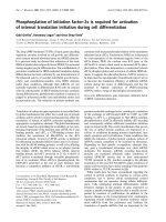
Fig. 1. Activation of transfected Core promoter reporter through DR12 enhancer. (A)ThesequencesofsinglecopiesofDR1,DR4,DR12and
mutant DR12 enhancer motifs used in the promoter constructs. Each half site is dashed underlined. Mutated residues are shown in lower case
letters. (B) On the left are the designs of the vector construct encoding the luciferase reporter enzyme, of the vector construct containing the Core
promoter reporter, and of the three vector constructs in which the Core promoter is preceded by four tandem copies either a DR1-, a DR4- or a
DR12-based enhancer, with the orientation of each motif shown by the small arrows. On the right are the activations of the indicated promoter
reporter construct in response to treatment of transfected cells with 75 l
M
methyl epoxyfarnesoate.
Ó FEBS 2002 Ligand activation of orphan receptor ultraspiracle (Eur. J. Biochem. 269) 6027
The intervening sequences in the DR1 and DR4 motifs were
randomly chosen, while the DR12 sequence used is found in
the ecdysteroid-sensitive ng-1 and ng-2 genes that are
expressed during metamorphosis of D. melanogaster,and
can serve in vitro as a binding site for the various receptor
dimers involving USP (ecdysteroid receptor (EcR)/USP
heterodimer, USP/DHR38 heterodimer and USP/USP
homodimer [28,31,32]).
Point mutations in the ligand-binding domain of dUSP
were made with a Chameleon
TM
double-stranded site-
directed mutagenesis kit (Stratagene) according to the
manufacturer’s instructions. The selection primer used to
change the unique NdeI (underlined) site in the pIE1-4
vector was CGGTATTTCACACCG
CAcATGGTGCACT
CTCAGTACAATC. The primer to mutate Q288 to alanine
in the ligand-binding pocket was: GTGCCAAGTGGTCA
ACAAA
gcGCTCTTCCAGATGGTCGAATAC. A pri-
mer that targeted two amino acids was used to make the
double mutation in C473A and H476L because of their
adjacent locations, with the sequence: GCGATCGATCAG
CCTGAAG
gcCCAGGATCtCCTGTTCCTCTTCCGCA
TTAC. A primer that replaced two proline residues (P498,
P499) at the end of a-helix 12 with tryptophan residues was:
5¢-CTTTCTCGAGCAGCTGGAGGCG
tgGtgGCCACC
CGGCCTGGCGATGAAACT-3¢. All mutant constructs
were confirmed by DNA sequencing.
For expressing dUSP in Sf9 cells, PCR-generated full-
length wild-type and point-mutated dUSP coding sequences
were cloned into PmeIandNotI sites of the pIE1-4 vector
(Novagen) and confirmed by sequencing, and for bacterial
overexpression were cloned into pET32EK (Novagen).
Nuclear extracts and electrophoretic mobility
shift assay
Nuclear extracts were isolated from Sf9 cells as previously
described [28,29]. For the DR12 probe, the double stranded
DR12 oligonucleotide (sequence as shown above) was 5¢
end-labelled with
32
P by T4 polynucleotide kinase (New
England Biolabs Inc.), and then purified from a 20% native
polyacrylamide gel. The same double stranded DR12
oligonucleotide was used in 100-fold excess as a self
competitor. For the 4DR12Core probe, the 4DR12Core
sequence was liberated from the vector as a 148-bp ClaI/
HindIII fragment, and was 5¢ end-labelled with
32
Pand
purified. The same, unlabelled fragment was used at 100-
fold excess as a self competitor. As a negative control for
specificity in gel shifts, the 36 bp BglII/KpnI polylinker
region fragment of the pGL3 vector was liberated and
recovered from low melting point agarose gels and used
as a 100· nonself competitor (sequence: GGTAC
CGAGCTCTTACGCGTGCTAGCCCGGGCTCGA).
Either a final concentration of 500 n
M
of His-tagged wild-
type USP or His-tagged mutant Cys472Ala/His475Leu
(¼ C472A/H475L), or five micrograms of nuclear proteins,
were incubated with the given probe on ice for 30 min in
binding buffer (10 m
M
Tris/HCl, pH 7.5; 50 m
M
NaCl,
0.5 m
M
EDTA, 5% glycerol, 1 m
M
MgCl
2
,and1m
M
dithiothreitol). In some experiments, nuclear proteins were
preincubated with the probe for 30 min followed by
incubation with anti-USP mAb (a gift from F. Kafatos,
EMBL, Heidelberg), or monoclonal Elav antibody (Devel-
opmental Studies Hybridoma Bank, University of Iowa),
for an additional 1 h on ice. Samples were then subjected to
4% (w/v) polyacrylamide gel electrophoresis in 0.5 · Tris/
borate/EDTA buffer. After electrophoresis, the gels were
dried and exposed to Kodak film at )70 °C for 12–48 h.
Extraction of total proteins and immunoblotting
analysis
Total Sf9 cell protein extracts from transfected Sf9 cells were
fractionated by SDS/PAGE, 8% (w/v) polyacrylamide gel,
and then transferred onto a nitrocellulose membrane. USP
was detected using a primary USP AB11 monoclonal
antibody and with an anti-mouse IgG-AP secondary Ig
(Bio-Rad) by a BCIP/NBT color development solution
(Bio-Rad). The USP signals were normalized by an internal
control, a-actin, which was detected by a primary polyclonal
a-actin antibody (Sigma) and with an anti-rabbit IgG-AP
secondary Ig (Southern Biotechnology Associates, Inc.).
Purification of the His–USP fusion protein
and ligand-binding assay
The homodimer-enriched fraction of bacterial recombinant
His–dUSP fusion protein was purified by nickel resin
selection, elution with imidazole, centrifugal concentration,
and then gel permeation chromatography (Superdex 200)
with procedures and chemical sources exactly as already
described previously [27]. The homodimer-enriched fraction
of the purified His–USP fusion protein was raised to 2 mL
of NaCl/P
i
and a final concentration of 0.5 l
M
.Fora
fluorescence-based ligand-binding assay based on intrinsic
tryptophan fluorescence [28,33], ligand or ethanol carrier
was added and the receptor preparation excited at 290 nm
and monitored for emission at 340 nm, until the signal from
the receptor had stabilized. Fluorescence was measured
three times for each sample, with standard deviation
typically smaller than the graphical plotted datum
point. Each fluorescence experiment was replicated on three
or more independent occasions, each time with similar
results.
Modelling of hRXRa and
D. melanogaster
USP
Tertiary conformation of human RXRa and D. melano-
gaster USP was analyzed by
RASMOL
software, using the
coordinates reported into the Protein Data Bank by
Bourguet and Moras (deposition number 1LB) and by
Schwabe and Clayton (deposition number 1HG4), respect-
ively. Using a minimum energy conformation of farnesol as
a scaffold, a conformation of epoxyfarnesoic acid was
prepared and placed by hand into the ligand-binding pocket
of USP along a generally similar trace as was reported for
the (more bent) 9-cis retinoic acid ligand when the latter was
cocrystalized with hRXRa (Egea et al. [52]).
RESULTS
Placement of four tandem copies of a DR12 motif
(CAAGGTCA(N)
12
AGGTCAG, Fig. 1A) at 5¢ to the
Core promoter reporter (4DR12Core construct, Fig. 1B)
yielded a 10-fold induction in promoter activity in response
to treatment of the transfected Sf9 cells with methyl
epoxyfarnesoate (Fig. 1B). In contrast, insertion of a
6028 Y. Xu et al.(Eur. J. Biochem. 269) Ó FEBS 2002
cassette containing four tandem copies of either a DR1 or
DR4 motif yielded only a 2.5- and 3.5-fold induction,
respectively (Fig. 1B). This differential result confirms that
the 10-fold activation observed with the 4DR12Core
construct was caused by the sequence of the inserted
DR12 cassette itself, and was not due to either insertional
disruption or creation of a putative cryptic regulatory
element at the vector multiple cloning site. Due to the
highest reporter activity being obtained with the DR12
motif, we focussed on the DR12 repeat construct, towards
the goal of the study of ligand activation of USP.
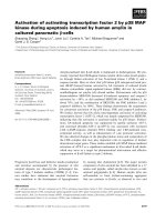
We then confirmed that sequences in the AGGTCA half
sites themselves of the DR12 motif were necessary for
transducing the methyl epoxyfarnesoate signalling. We took
advantage of the previous report that mutation of each half
site abrogated the ability of DR12 motif to enhance
ecdysteroid transcriptional activation [31]. When we
mutated here each half site of the DR12 motif (in a construct
containing a single DR12 in order to simplify mutational
analysis; 1DR12mutCore), the responsiveness of the
1DR12mutCore to methyl epoxyfarnesoate was no greater
than the background of a Core promoter with no enhancer
(Fig. 2B); in contrast to the responsiveness of the Core
promoter in the presence of a wild-type DR12 (1DR12Core,
Fig. 2B). As an independent confirmation of the important
role of the two direct repeat half sites in the DR12 motif, we
demonstrated that in a gel mobility shift assay with Sf9
nuclear extracts, the DR12 motif probe yielded a shifted
probe band that could be competed with excess, unlabelled
wild-type DR12. However, the same DR12 mutated in its
two half sites that had failed to support methyl epoxyfar-
nesoate-enhanced transcription in the cell transfection assay
also correspondingly failed to compete with the wild-type
DR12 probe in the gel shift assay (Fig. 2A), confirming the
functional necessity of the two half sites for interaction with
a nuclear component(s). Thus, the lack of binding to the
mutant DR12 combined with the lack of a transcriptional
effect of that same mutant DR12 suggests that the specific
binding to the wild-type DR12 observed here relates to its
positive action to transduce the methyl epoxyfarnesoate
signalling observed in the transfection assay. The gel
mobility shift assay using Sf9 nuclear extracts detected a
single major complex binding to the DR12 probe (Fig. 2C).
An anti-dUSP mAb (AB11, epitope on DNA binding
domain) displaced the endogenous USP in the major
complex binding to the DR12 probe (Fig. 2C). The
specificity of the AB11 monoclonal antibody effect on
USP binding was further confirmed in that no such effect
was produced by a negative control monoclonal antibody
against the transcription factor Elav.
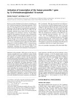
In many vertebrate nuclear receptors, residues in a
narrow region of a-helix 11 make contact with the
endogenous ligand of the given receptor [27]. In hRXRa,
Cys432 and His435 on a-helix 11 make contact with the
distal end of the 9-cis RAligandattwomethylbranches
(C16, C17) and also at the terpene backbone (Fig. 3A,B).
The homologous two residues on a-helix 11 of dUSP
(Cys472 and His475) are highly conserved in other USPs
[27], and point into the ligand-binding pocket of USP crystal
structures (Fig. 3C [34,35]). Other residues that contact 9-cis
RA in hRXRa are also conserved in identity and similar
location in the ligand-binding pocket of dUSP, such as
Gln288, Trp318 and Leu367 in dUSP corresponding to
Gln275, Try305 and Leu326 of hRXRa (Fig. 3B–D). If an
epoxy farnesoid-like ligand were to reside in the dUSP
ligand-binding pocket along a similar trace as does 9-cis RA
in hRXRa, then the terpene backbone and the methyl
branches C12 and C15 at the distal end of the epoxy
farnesoid ligand might be similarly placed to interact with
His475 and Cys472 in dUSP, as does 9-cis RA interact with
Cys432 and His435 in hRXRa (Fig. 3B–D).
We tested this model by overexpressing the His-tagged
dUSP double mutant Cys472Ala/His475Leu (C472A/
H475L) in methyl epoxyfarnesoate-treated Sf9 cells that
were cotransfected with the 4DR12Core reporter plasmid.
Cells transfected with either empty pIE1-4 vector, or that
vector expressing wild-type dUSP, responded to methyl
epoxyfarnesoate application with a similar induction of the
Fig. 2. Functional analysis of the DR12 motif. (A) Gel mobility shift assay, using Sf9 nuclear extracts, of the same single DR12 motif that was used
as an enhancer in the cell transfection assay in (B). The shifted probe band was competitively displaced by 100· of the unlabelled DR12 motif (self),
but was not competed with either by the same mutant DR12 motif as failed to act as an enhancer in cell transfection assay in B (mutDR12) or by the
negative control polylinker sequence (nonself). (B) Activations of the indicated promoter reporter constructs in response to treatment of transfected
cells with 75 l
M
methyl epoxyfarnesoate. (C) Intracellular USP binds to DR12 hormone response element. Gel mobility shift assay using Sf9
nuclear extracts (N.E.) and a
32
P-labelled probe that is the four tandem DR12 motifs (Ô4DR12Õ) shown in Fig. 1, performed as described in [28]. The
USP in the Sf9 nuclear extract that is the major binding complex (small arrow) is displaced by the AB11 monoclonal antibody, just as we have
previously shown is the effect of this antibody on recombinant dUSP binding to a DR12 probe [28]. The lack of similar effect by monoclonal
antibody against the negative control nerve transcription factor (Elav) shows the specificity of the AB11 result.
Ó FEBS 2002 Ligand activation of orphan receptor ultraspiracle (Eur. J. Biochem. 269) 6029
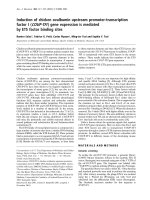
4DR12Core promoter (Fig. 4A). However, cells transfected
with the plasmid expressing the C472A/H475L mutant
exhibited a distinct suppression in the level of methyl
epoxyfarnesoate-induced activation, as compared with the
activation observed for cells transfected with either the
empty plasmid or plasmid expressing wild-type dUSP
(Fig. 4A). In addition, cotransfection of the empty vector,
or vector expressing either wild-type dUSP or the C472A/
H475L mutant, did not affect the basal activation exhibited
when the Core promoter without DR12 motifs was used.
Together, these data demonstrate that the suppression in
methyl epoxyfarnesoate-induced activation caused by over-
expression of the C472A/H475L double mutant was not
due to nonspecific titration of coactivators required by a
receptor other than USP and not due to disruption of Core-
binding basal transcription components independent of
action through the DR12 enhancer. In addition, overex-
pression of either the C472A/H475L double mutant or the
Fig. 3. Comparision of dUSP and hRXR ligand binding domains. (A) Selected contacts made between 9-cis RA and residues in the hRXRa ligand-
binding pocket as determined from cocrystals (4.2 A
˚
or less, from Doyle et al. [55] and Egea et al. [53]). On the left is also shown a conformation of
epoxyfarnesoic acid, exhibiting similarities between its structure and that of the terpenoid backbone and carboxyl group of 9-cis retinoic acid. (B
and C)
RASMOL
-generated ribbon diagrams for the ligand-binding domains of the hRXRa and dUSP, respectively. (B) This shows in the hRXRa
ligand-binding pocket the structure of the ligand 9-cis retinoic acid (carbon backbone in light blue, terminal carboxylate oxygens in dark blue,
adapted from Egea et al. [52]). (C) This shows methyl epoxyfarnesoic acid (yellow carbon backbone and blue terminal carboxylate oxygens) lain
manually in the dUSP ligand-binding pocket with the carboxy and distal (epoxy) ends, respectively, situated in similar regions of the pocket as the
carboxyl end and distal end of 9-cis RA in hRXRa. (D) An overlay of the dUSP and hRXRa ribbon diagrams of B and C, with emphasis (white
arrows) on the similar placement of Gln275,Trp305, Leu326, Cys432 and His435 in hRXRa as compared to Gln288, Trp318, Leu367, Cys472 and
His475 in dUSP.
6030 Y. Xu et al.(Eur. J. Biochem. 269) Ó FEBS 2002
wild-type dUSP did not change the level of endogenous
USP (Fig. 4A), confirming that overexpression of exogen-
ous dUSP did not indirectly affect the methyl epoxyfarne-
soate-activation pathway by disruption of endogenous USP
expression.
Concerning the proximal end of the hRXRa ligand,
cocrystals of 9-cis RA and hRXRa have also established
that a glutamine residue on a-helix 3 (Gln275) makes
contact with both the carbonyl carbon and a carboxylate
oxygen (Figs 3A,B and 5). This glutamine residue is
conserved in all reported USPs (Fig. 5C [27]). Therefore,
we mutated this Gln288 in dUSP to alanine (Gln288A), and
found that this mutant dUSP also acted as a dominant
negative suppressor of activation of the DR12Core reporter
promoter in methyl epoxyfarnesoate-treated Sf9 cells
(Fig. 4A).
Under the model that overexpression of the C472A/
H475L double mutant competed with endogenous USP in
the pathways for transduction of the exogenous methyl
epoxyfarnesoate signal, the level of effect of the double
mutant ought to be dependent on its dose. Indeed, we
determined that a progressive increase in the intracellular
concentration of this double mutant (with endogenous USP
level remaining unchanged) caused progressive suppression
in the methyl epoxyfarnesoate-activation of the DR12Core
promoter, down to the transcriptional level observed for the
Core promoter without DR12 enhancers (Fig. 4B). Over
the range of the progressive suppression of the methyl
epoxyfarnesoate-activated transcription there was no effect
of the double mutant on the basal level of transcription in
EtOH-treated controls. We then used this background of
the blocked activation pathway to test whether activation by
methyl epoxyfarnesoate treatment was actually dependent
on the presence of wild-type USP. As shown in Fig. 4C, the
activation of the 4DR12Core promoter in methyl epoxy-
farnesoate-treated cells was monotonically restored in a
manner dependent on the increasing dose of the added wild-
type dUSP. Again, over the range of the monotonic
restoration of methyl epoxyfarnesoate-activated transcrip-
tion, there was no effect of the transfected wild-type dUSP
on the basal level of transcription in EtOH-treated controls.
We examined the ability of the C472A/H475L mutant to
bind DNA and to homodimerize to confirm that the
mutations to the ligand-binding pocket did not generally
deform receptor structure. As shown in Fig. 5A, under
electrophoretic mobility shift assay conditions, both the
wild-type dUSP and the C472A/H475L mutant dUSP
similarly bound to a DR12 motif. In addition, both receptor
preparations bound to the probe similarly in part as
monomer and in part as homodimer. The homodimeriza-
tion of RXR and other steroid receptor superfamily
members is primarily due to contacts in the ligand-binding
domain that are outside of the ligand-binding pocket (in
addition to some contacts also in the DNA-binding
domain). The similar DNA binding and homodimerization
capacities of the wild-type dUSP and mutant C472A/
Fig. 4. Dominant negative activity of USP ligand-binding pocket mutants. (A) Histogram (shaded boxes) shows the dominant negative effect of
transfected dUSP mutant and the double mutant (C472A/H475L) on methyl epoxyfarnesoate-activation of 4DR12Core reporter promoters,
whereas transfected wild-type dUSP shows no such suppression of methyl epoxyfarnesoate activation, in comparison with transfection of Core
reporter vector (reporter and expression plasmids transfected at 1 : 1 ratio). Transfection of neither the wild-type USP nor either mutant had any
effect on the minimal basal activation of the Core promoter in the absence of the DR12 motif (clear boxes). Immunoblot of transfected cellular
extracts with anti-(a-actin) and anti-dUSP (AB11) mAbs verified that the overexpression of mutant and wild-type dUSP did not affect the level of
expression of endogenous USP, and that the transfected mutant and transfected wild-type dUSP were expressed at similar levels to each other. The
molecular weights of the transfected and endogenous USPs detected by immunoblotting were 50 and 52 kDa, respectively, as estimated by
molecular size standards run in parallel lanes (not shown). (B) Progressive increase in ratio of transfected dominant negative plasmid DNA relative
to 4DR12Core reporter plasmid DNA yielded an increasing dominant negative suppression of methyl epoxyfarnesoate activation of reporter
plasmid. Immunoblot verifies that the progressively higher overexpression of the mutant dUSP (C472A/H475L) did not affect the level of
expression of endogenous USP. Inset above shows calculation of transcriptional activation ratio of reporter promoter activity in methyl epoxy-
farnesoate- treated cells relative to EtOH treated cells, as a function of the ratio of the amount of transfected mutant dUSP plasmid relative to
amount of transfected reporter plasmid. (C) Transfection of plasmid expressing wild-type USP rescues the dominant negative-suppression of methyl
epoxyfarnesoate-activation of the reporter promoter. Open circle, methyl epoxyfarnesoate activation of 4DR12Core in the absence of USP
expressing plasmid. Hashed circle, methyl epoxyfarnesoate activation is suppressed by transfection with the C472A/H475L dominant negative
mutant. Filled circles, methyl epoxyfarnesoate activation is progressively restored by increasing doses of plasmid expressing wild-type dUSP. In
A–C, hormone-treated cells received 75 l
M
of methyl epoxyfarnesoate.
Ó FEBS 2002 Ligand activation of orphan receptor ultraspiracle (Eur. J. Biochem. 269) 6031
H475L dUSP is strongly indicative that the DNA-binding
domain, and the parts of the ligand-binding domain that are
outside of the ligand-binding pocket, are in a functionally
similar conformation for both the wild-type and mutant
receptors. Thus, any difference detected in ligand binding of
the two receptors is most reasonably inferred as arising from
differences in the architecture inside the cavity of the ligand-
binding pocket due to the C472A/H475L point mutations.
We then tested the ability of the wild-type dUSP and
dominant negative, ligand-binding pocket mutant dUSP to
bind methyl epoxyfarnesoate. In a ligand-binding assay that
detects methyl epoxyfarnesoate binding through its effects
to suppress intrinsic fluorescence of dUSP [28,33], the
bacterially overexpressed His-tagged wild-type dUSP in-
deed exhibited suppressed the fluorescence due to the
binding of methyl epoxyfarnesoate (Fig. 5B). However, the
C472A/H475L mutant dUSP did not exhibit a significant
response to epoxymethyl farnsoate (Fig. 5B). This result
was reproduced with independent preparations of the wild-
type dUSP and C472/H475L dUSP. These results strongly
support the inference that the behavior of C472A/H475L as
a dominant negative mutant in the pathway for methyl
epoxyfarnesoate activation of the 4DR12Core promoter is
due to the effect of the C472A/H475L mutations on the
ligand-binding activity of USP.
Some models of nuclear hormone receptor action include
the component that binding of ligand to the ligand-binding
pocket induces a tertiary conformational change involving
the movement of a-helix 12 to a new position [36]. However,
the two published crystal structures of USP in complex with
a phospholipid located at the opening of the ligand-binding
pocket show a-helix 12 in a position that the investigators
described as so firmly ÔlockedÕ against other residues of the
ligand-binding domain that a-helix 12 would not be able to
move even if the phospholipid were not present [33,34]. We
therefore tested the hypothesis that a-helix 12 is so firmly
locked in position that it does not move, by replacing two of
the four continuous proline residues at the end of a-helix 12
with tryptophan residues. Under the model that USP a-helix
12 does not move upon binding of methyl epoxyfarnesoate
in the ligand-binding pocket, these two tryptophan residues
would only raise the constant background intrinsic fluores-
cence of the receptor, but, on account of the fact that they (as
part of the fixed a-helix 12) do not move in position, their
level of fluorescence would not change upon binding of
methyl epoxyfarnesoate into the pocket. Therefore, their
constant background fluorescence would not enhance or
disguise the suppression in fluorescence exhibited by the two
other natural tryptophan residues (on a-helix 5) upon
binding of methyl epoxyfarnesoate. Alternatively, if a-helix
12 does move in position upon binding of methyl epoxyfar-
nesoate, then the change in the local environment of the two
added tryptophan residues on a-helix 12 may change their
fluorescence in a way that yields a markedly different overall
fluorescence pattern for the receptor. Indeed, as Fig. 6B
shows, in this test the wild-type USP with only two natural
tryptophan resides on a-helix 5 exhibits a distinct suppres-
sion in fluorescence upon binding of methyl epoxyfarneso-
ate. In contrast, the mutant USP containing two additional
tryptophan residues at the end of a-helix 12 showed a much
different profile, instead sharply increasing in fluorescence
before then decreasing (Panel C). Collectively, these mark-
edly different patterns of fluorescent response are most easily
explained by a model in which a-helix 12 does move in
relative position, upon the binding of methyl epoxyfarneso-
ate into the ligand-binding pocket of USP.
Fig. 5. Bacterially overexpressed double mutant dUSP (C472A/H475L) and wild-type dUSP analyzed for binding to DNA or to ligand. (A) The wild-
type dUSP and the C472AH475L mutant both similarly bound in part as a homodimer (upper band) and in part as a monomer (lower band) to a
4DR12 motif probe (identification of monomer and homodimer bands was made by comparative analysis of binding by other dimer-enriched vs.
monomer-enriched fractions obtained from Superdex 200 chromatography, not shown). Control competitions with self and nonself unlabelled
excess probes confirmed the specificity of binding. The similar formation of the homodimer form by the wild-type USP and mutant USP, along with
the similar binding to DNA of the wild-type USP and mutant USP, confirm that the mutation to the ligand-binding pocket in C475A/H475L did
not generally disrupt the structure of the receptor. (B) The homodimer-enriched fraction of each receptor preparation was then analyzed for binding
to 75 l
M
methyl epoxyfarnesoate, using an intrinsic fluorescence assay method that tracks ligand binding (by suppression in receptor fluorescence)
[27,28]. The wild-type dUSP exhibited binding to methyl epoxyfarnesoate in this assay. However, the double mutant dUSP exhibited no binding
activity. Arrows show time of addition of methyl epoxyfarnesoate or EtOH carrier.
6032 Y. Xu et al.(Eur. J. Biochem. 269) Ó FEBS 2002
DISCUSSION
With the inception of the original model by Ashburner on
hierarchical, steroid-driven genetic programs for inverte-
brate development [37], the sophistication of the models
has progressively increased as more transcription factors
have been discovered to participate in these complex
developmental programs [38]. However, despite the inclu-
sion of ultraspiracle in these conceptual models since its
discovery over 10 years ago, there has been much angst
over whether this receptor possesses a ligand-binding
activity. In the absence of an experimental demonstration
that ultraspiracle can bind ligand and transduce that
binding into transcriptional modulation, models of genetic
programs that include ultraspiracle have not overtly
included a ligand-binding role for ultraspiracle [18–
26,39,40]. While there is genetic evidence that the ligand-
binding domain of USP globally contributes to function of
the EcR/USP heterodimer [41], other models expressly
envision that ultraspiracle does not have any ligand-
binding role in certain pathways [23,42].
We have previously demonstrated [28,33] that dUSP can
specifically bind to small terpenoid-derived compounds
such as epoxy methyl farnesoate and bisepoxy methylfar-
nesoate, in a saturable, dose-dependent manner, causing a
conformational change to the receptor that suppresses its
intrinsic fluorescence, while compounds such as farnesol
and epoxyfarnesoic acid, and the steroid 20-OH ecdysone
do not have this effect. We have also shown elsewhere [28]
that the marked increase in transcription of the model
DR12Core reporter promoter, with methyl epoxyfarnesoate
(Fig. 1), is dose-dependent, but that neither retinoic acid nor
T3 yield this effect. However, these previous results do not
demonstrate whether methyl epoxyfarnesoate binds to the
receptor in its ligand-binding pocket, nor whether such
binding induces movement in a-helix 12, nor whether
endogenous USP in the transfected cells can bind to the
direct repeat motifs that 5¢ flank the reporter promoter, nor
do they address whether methyl epoxyfarnesoate-activation
of the reporter is dependent upon liganded USP, all of
which are crucial underpinnings to the concept that the USP
ligand-binding pocket is a viable target for experimental or
practical agonistic or antagonistic ligands.
In the present report, we have demonstrated that methyl
epoxyfarnesoate does indeed bind to the ligand-binding
pocket, and that point mutations to the dUSP ligand-
binding pocket that disrupt methyl epoxyfarnesoate binding
cause the mutant receptor to act as a dominant negative in a
model transcription pathway that is activated by methyl
epoxyfarnesoate treatment. These data suggest further
inquiry is warranted into farnesoid-derived ligands as
agonists for USP. Our demonstration here that the USP
ligand-binding pocket is conformed such that it can bind
methyl epoxyfarnesoate-like compounds, with a resultant
change in USP conformation, including the movement of
a-helix 12, and with an effect to activate transcription in
methyl epoxyfarnesoate-treated cells is the first such iden-
tification of the activating binding of any compound,
natural or synthetic, to the ligand-binding pocket of an
invertebrate orphan receptor. This precedent establishes
that invertebrate orphan receptors are not qualitatively
different from the situation for vertebrate orphan receptors
for which a number have now been shown to have ligand-
binding pockets with the functional capacity to bind and be
transcriptionally activated by appropriately structured
compounds.
Fig. 6. Fluorescence response of wild-type and P498W/P499W mutant USP to farnesoid ligands. (A) The location of the mutational placement of the
two tryptophan residues at the end of (red colored) a-helix 12. USP also possesses two natural tryptophan residues on helix 5 (W318, shown in green
extending into pocket; W328, not shown, extending out of pocket). (B) Methyl epoxyfarnesoate binding to wild-type USP results in suppression of
receptor fluorescence, while farnesol and ethanol carrier do not have that effect. (C) Methyl epoxyfarnesoate binding to P498W/P499W mutant
results in a very different pattern of fluorescence response than wild-type USP in B, evidencing that a-helix 12 moves in its relative location upon
USP binding of methyl epoxyfarnesoate. The wild-type USP and P498W/P499W similarly bound in part as monomer and in part as dimer to a
DR12 probe in gel shift assay, evidencing that the P498W/P499W mutations did not affect receptor structure globally (not shown).
Ó FEBS 2002 Ligand activation of orphan receptor ultraspiracle (Eur. J. Biochem. 269) 6033
USPs, which compared to RXR are unusual for their
stretch of additional amino acids inserted after a-helix 5,
have recently been cocrystalized with fortuitous phospho-
lipid pseudoligands [34,35]. These cocrystals had a relat-
ively large total van der Waals volume of the USP ligand-
binding pocket ( 1300 A
˚
3
), compared to the volume of
JH III (259 A
˚
3
[43]). However, the volume of the PPARc
ligand-binding pocket (similar to that of USP, 1300 A
˚
3
[44]) is also much larger than that of its natural ligand 15-
deoxy-D
12,14
-prostaglandin J
2
(which has a volume similar
to that of JH III, at 301 A
˚
3
[43]), yet this prostaglandin
ligand is able to bind and transcriptionally activate the
PPARc [45]. In addition, the volume of b-estradiol (which
at 245–251 A
˚
3
is smaller than methyl epoxyfarnesoate
[43]), is approximately half the volume of the ligand-
binding pocket of the estrogen receptor (450–500 A
˚
3
[46,47]), Yet, b-estradiol is nonetheless able to bind to
and activate the estrogen receptor. Thus, PPARc and the
estrogen receptor demonstrate that endogenous com-
pounds much smaller than the total ligand-binding pocket
volume of a nuclear hormone receptor can and do serve as
natural activating ligands. The recently crystallized PXR,
which binds with, and is activated by, a variety of small
and large ligands, also possesses a large 1300 A
˚
3
ligand-
binding pocket [48], and possesses an unusual additional
stretch of amino acids that the authors postulated enables
what would otherwise be a smaller PXR ligand-binding
pocket to enlarge to accommodate a large ligand. Import-
ant in these considerations is whether there is a subregion
in the ligand-binding pocket in which the local conforma-
tion corresponds well to the conformation of a particular
small ligand. Although the overall volume of the ligand-
binding pocket observed in the cocrystals of
USP ( 1300 A
˚
3
)ismuchlargerthanthatofhRXRa
( 500 A
˚
3
), the proximal subregion of the ligand-binding
pocket of hRXRa and USP are much more similar in
volume and shape [34]. The proximal subregion of each of
the two receptors also has a similar placement of conserved
amino acids that in hRXRa interact with the terpenoid
backbone of 9-cis RA (Fig. 4A–D). In addition, 9-cis RA
and methyl epoxyfarnesoate have similar van der Waals
volumes of 291 and 258 A
˚
3
, respectively [43]. These
considerations suggest that methyl epoxyfarnesoate-like
metabolites cannot be dismissed apriorias potential USP
agonists, merely on the basis of comparison of the volume
of methyl epoxyfarnesoate vs. the reported total volume of
the USP ligand-binding pocket.
Our combined use of an equilibrium, fluorescence
binding assay and a transfection transcriptional assay that
is activated by treatment with methyl epoxyfarnesoate will
be very useful in identifying new, higher-affinity ligands for
USP. The molecular interactions between a receptor and a
synthetic activating ligand have previously provided insight
to the molecular basis by which agonist ligand(s) activates
the receptor. Crystal structures of the vitamin D receptor
in complex with natural activating ligand vs. with synthetic
agonists revealed that both induced the same intramole-
cular conformational changes in the receptor [49]. Cocrys-
tal structure analysis showed that human RARa was
induced to undergo similar intramolecular conformational
changes by either natural 9-cis RA or a synthetic agonist
[50]. We have shown that binding of methyl epoxyfarne-
soate by dUSP promotes not only an intramolecular
conformational change of movement of a-helix 12, but
also homodimerization [28], which together appears remi-
niscent of the way in which 9-cis RA induces an activating
intramolecular conformational change in human RXRa
(e.g. movement of a-helix 12) as well as that receptor’s
homodimerization [51–53]. These results have considerable
significance for current popular models of USP function as
a heterodimeric partner with EcR, because most of these
models do not envision the binding effect of an agonist by
USP.
It is becoming increasingly appreciated that not all core
promoters are alike in their ability to respond to the same
transcriptional enhancer, as additional DNA sequence in
and around the TATA box and initiator motifs confer
selectivity in the nature of the components that nucleate to
form the basal transcription apparatus at the core promo-
ter. Indeed, a number of different parameters have been
identified under which different EcR/USP heterodimer
DNA binding sites exert very different levels of effect in
transducing ecdysteroid signalling [54–57]. Therefore, we
do not anticipate that the DR12 motif used here will
necessarily function to enhance the activity of all model
core promoters in response to methyl epoxyfarnesoate-like
molecules. However, it is clear that this model system of
the DR12Core promoter in Sf9 cells will be appropriate
and useful as a tool in exploring the functional structure of
the ligand-binding pocket of USP with respect to USP
activation upon binding of methyl epoxyfarnesoate and
other agonistic compounds.
ACKNOWLEDGEMENTS
The research reported herein was supported, in part, by NIH grants
462795 and 463713. We express our appreciation to Drs David
Mangelsdorf, Carl Thummel and Mietek Wozniak for helpful
discussions on the framing of hypotheses on the functional structure
of ligand-activated nuclear receptors.
REFERENCES
1. Evans, R.M. (1988) The steroid and thyroid hormone receptor
superfamily. Science 240, 889–895.
2. McKenna, H.J. & O’Malley, B.W. (2000) From ligand to
response: generating diversity in nuclear receptor coregulator
function. J. Steroid Biochem. Mol. Biol. 74, 351–356.
3. Heyman, R.A., Mangelsdorf, D.J., Dyck, J.A., Stein, R.B.,
Eichele, G., Evans, R.M. & Thaller, C. (1992) 9-cis retinoic acid is
a high affinity ligand for the retinoid X receptor. Cell 68, 397–406.
4. Repa, J.J. & Mangelsdorf, D.J. (2000) The role of orphan nuclear
receptors in the regulation of cholesterol homeostasis. Annu. Rev.
Cell Dev. Biol. 16, 459–481.
5. Chawla, A., Repa, J.J., Evans, R.M. & Mangelsdorf, D.J. (2001)
Nuclear receptors and lipid physiology: opening the X-files.
Science 294, 1866–1870.
6. Koelle, M.R., Talbot, W.S., Segraves, W.A., Bender, M.T.,
Cherbas, P. & Hogness, D.S. (1991) The Drosophila EcR gene
encodes an ecdysone receptor, a new member of the steroid
receptor superfamily. Cell 67, 59–77.
7. Oro, A.E., McKeown, M. & Evans, R.M. (1992) The Drosophila
nuclear receptors: new insight into the actions of nuclear receptors
in development. Curr. Opin. Genet. Dev. 2, 269–474.
8. Yao, T.P., Forman, B.M., Jiang, Z., Cherbas, L., Chen, J.D.,
McKeown,M.,Cherbas,P.&Evans,R.M.(1993)Functional
ecdysone receptor is the product of EcR and Ultraspiracle genes.
Nature 366, 476–479.
6034 Y. Xu et al.(Eur. J. Biochem. 269) Ó FEBS 2002
9. Henrich, V.C. & Brown, N.E. (1995) Insect nuclear receptors: a
developmental and comparative perspective. Insect Biochem. Mol.
Biol. 25, 881–897.
10. Arbeitman, M. & Hogness, D.S. (2000) Molecular chaperones
activate the Drosophila ecdysone receptor, an RXR heterodimer.
Cell 101, 67–77.
11. Yao, T.P., Segraves, W.A., Oro, A.E., McKeown, M. & Evans,
R.M. (1992) Drosophila ultraspiracle modulates ecdysone receptor
function via heterodimer formation. Cell 71, 63–72.
12. Thummel, C. (1995) From embryogenesis to metamorphosis: the
regulation and function of Drosophila nuclear receptor super-
family members. Cell 83, 871–877.
13. Mangelsdorf, D.J. & Evans, R.M. (1995) The RXR heterodimers
andorphanreceptors.Cell 83, 841–850.
14. Harmon, M.A., Boehm, M.F., Heyman, R.A. & Mangelsdorf,
D.J. (1995) Activation of mammalian retinoid X receptors by the
insect growth regulator methoprene. Proc. Natl Acad. Sci. USA
92, 6157–6160.
15. Henrich, V.C., Sliter, T.J., Lubahn, D.B., MacIntyre, A. &
Gilbert, L.I. (1990) A steroid/thyroid hormone receptor super-
family member in Drosophila melanogaster that shares extensive
sequence similarity with a mammalian homologue. Nucleic Acids
Res. 18, 4143–4148.
16. Shea, M.J., King, D.L, Conboy, M.J., Mariani, B.D.& Kafatos,
F.C. (1990) Proteins that bind to Drosophila chorion cis-regulatory
elements: a new C2H2 zinc finger protein and a C2C2 steroid
receptor-like component. Genes Dev. 4, 1128–1140.
17. Oro, A.E., McKeown, M. & Evans, R.M. (1990) Relationship
between the product of the Drosophila ultraspiracle locus and the
vertebrate retinoid X receptor. Nature 347, 298–301.
18. White, K.P., Hurban, P., Watanabe, T. & Hogness, D.S. (1997)
Coordination of Drosophila metamorphosis by two ecdysone-
induced nuclear receptors. Science 276, 114–117.
19. Jiang, C., Lamblin, A.F., Steller, H. & Thummel, C.S. (2000)
A steroid-triggered transcriptional hierarchy controls salivary
gland cell death during Drosophila metamorphosis. Mol. Cell 5,
45–55.
20. D’Avino, P.P. & Thummel, C. (2000) The ecdysone regulatory
pathway controls wing morphogenesis and integrin expression
during Drosophila metamorphosis. Dev. Biol. 220, 211–224.
21. Riddiford, L.M., Hiruma, K., Lan, Q. & Zhou, B.H. (1999)
Regulation and role of nuclear receptors during larval molting and
metamorphosis of Lepidoptera. Am. Zool. 39, 736–746.
22. Henrich, V.C., Rybczynski, R. & Gilbert, L.I. (1999) Peptide
hormones, steroid hormones, and puffs: mechanisms and models
in insect development. Vitam. Hormones 55, 73–125.
23. Schubiger, M. & Truman, J.W. (2000) The RXR ortholog USP
suppresses early metamorphic processes in Drosophila in the
absence of ecdysteroids. Development 127, 1151–1159.
24. Baehrecke, E. (2000) Steroid regulation of programmed cell death
during Drosophila development. Cell Death Differ. 7, 1057–1062.
25. Buszczak, M. & Segraves, W.A. (2000) Insect metamorphosis: out
with the old, in with the new. Curr. Biol. 10, R830–R833.
26. Huet, F., Ruiz, C. & Richards, G. (1995) Sequential gene activa-
tion by ecdysone in Drosophila melanogaster: the hierarchical
equivalence of early and early late genes. Development 121, 1195–
1204.
27. Jones, G. & Jones D. (2000) Considerations on the structural
evidence of a ligand-binding function of ultraspiracle, an insect
homolog of vertebrate RXR. Insect Biochem. Mol. Biol. 30,
671–679.
28. Jones, G., Wozniak, M., Chu, Y X., Dhar, S. & Jones, D. (2001)
Juvenile hormone III-dependent conformational changes of
the nuclear receptor ultraspiracle. Insect Biochem. Mol. Biol. 32,
33–49.
29. Jones, G., Manczak, M., Schelling, D., Turner, H. & Jones, D.
(1998) Transcription of the juvenile hormone esterase gene under
the control of both an initiator and AT-rich motif. Biochem. J. 335,
79–84.
30. Jones, G., Chu, Y X., Schelling, D. & Jones, D. (2000) Regulation
of the juvenile hormone esterase gene by a composite core pro-
moter. Biochem. J. 346, 233–240.
31. D’Avino, P.P., Crispi, S., Cherbas, L., Cherbas, P. & Furia, M.
(1995) The moulting hormone ecdysone is able to recognize
target elements composed of direct repeats. Mol. Cell. Endo. 113,
1–9.
32. Crispi, S., Giordano, E., D’Avino, P.P. & Furia, M. (1998) Cross-
talking among Drosophila nuclear receptors at the promiscuous
response element of the ng-1 and ng-2 intermolt genes. J. Mol.
Biol. 275, 561–574.
33. Jones, G. & Sharp, P.A. (1997) Ultraspiracle: an invertebrate
nuclear receptor for juvenile hormones. Proc. Natl Acad. Sci. USA
94, 13499–13503.
34. Billas, I.M., Moulinier, L., Rochel, N. & Moras, D. (2000) Crystal
structure of the ligand-binding domain of the ultraspiracle protein
USP, the ortholog of retinoid X receptors in insects. J. Biol. Chem.
276, 7465–7474.
35. Clayton, G.M., Peak-Chew, S.Y., Evans, R.M. & Schwabe,
J.W.R. (2000) The structure of the ultraspiracle ligand-binding
domainrevealsanuclearreceptorlockedinaninactivecon-
formation. Proc. Natl Acad. Sci. USA 98, 1549–1554.
36. Steinmetz, A.C., Renaud, J.P. & Moras, D. (2001) Binding of
ligands and activation of transcription by nuclear receptors. Annu.
Rev. Biophys. Biomol. Struct. 30, 329–359.
37. Ashburner, M., Chihara, C., Meltzer, P. & Richards, G. (1974)
Temporal control of puffing activity in polytene chromosomes.
Cold Spring Harb. Symp. Quant. Biol. 38, 655–662.
38. Richards,G.,DaLage,J.L.,Huet,F.&Ruiz,C.(1999)The
acquisition of competence to respond to ecdysone in Drosophila is
transcript specific. Mech. Dev. 82, 131139.
39. Thummel, C.S. (1997) Dueling orphans – interacting nuclear
receptors coordinate Drosophila metamorphosis. Bioessays 19,
669–672.
40. Thummel, C.S. (2002) Ecdysone-regulated puff genes 2000. Insect
Biochem. Mol. Biol. 32, 113–120.
41. Henrich, V.C., Vogtli, M.E., Antoniewski, C., Spindler-Barth, M.,
Przibilla, S., Noureddine, M. & Lezzi, M. (2000) Developmental
effects of a chimeric ultraspiracle gene derived from Drosophila
and Chironomus. Genesis 28, 125–133.
42. Kapitskaya, M., Wang, S., Cress, D.E., Dhadialla, T.S. & Raikel
A.S. (1996) The mosquito ultraspiracle homologue, a partner of
ecdysteroid receptor heterodimer: cloning and characterization of
isoforms expressed during vitellogenesis. Mol. Cell. Endocrinol.
121, 119–132.
43. Bogan, A.A., Cohen, F.E. & Scanlan, T.S. (1998) Natural ligands
of nuclear receptors have conserved volumes. Nat. Struct. Biol. 5,
679–681.
44. Nolte, R.T., Wisely, G.B., Westin, S., Cobb, J.E., Lambert, M.H.,
Kurokawa, R., Rosenfeld, M.G., Willson, T.M., Glass, C.K. &
Milburn, M.V. (1998) Ligand binding and co-activator assembly
of the peroxisome proliferator-activated receptor-gamma. Nature
395, 137–143.
45. Kliewer, S.A., Lenhard, J.M., Willson, T.M., Patel, I., Morris,
D.C. & Lehmann, J.M. (1995) A prostaglandin J2 metabolite
binds peroxisome proliferator-activated receptor gamma and
promotes adipocyte differentiation. Cell 83, 813–819.
46. Brzozowski, A.M., Pike, A.C., Dauter, Z., Hubbard, R.E., Bonn,
T.,Engstrom,O.,Ohman,L.,Greene,G.L.,Gustafsson,J.A.&
Carlquist, M. (1997) Molecular basis of agonism and antagonism
in the oestrogen receptor. Nature 389, 753–758.
47. Shiau, A.K., Barstad, D., Loria, P.M., Cheng, L., Kushner, P.J.,
Agard, D.A. & Greene, G.L. (1998) The structural basis of
estrogen receptor/coactivator recognition and the antagonism of
this interaction by tamoxifen. Cell 95, 927–937.
Ó FEBS 2002 Ligand activation of orphan receptor ultraspiracle (Eur. J. Biochem. 269) 6035
48. Watkins, R.E., Wisely, G.B., Moore, L.B., Collins, J.L., Lambert,
M.H., Williams, S.P., Willson, T.M., Kliewer, S.A. & Redinbo,
M.R. (2001) The human nuclear xenobiotic receptor PXR:
structural determinants of directed promiscuity. Science 292,
2329–2333.
49. Tocchini-Valentini, G., Rochel, N., Wurtz, J.M., Mitschler, A. &
Moras, D. (2001) Crystal structures of the vitamin D receptor
complexed to superagonist 20-epi ligands. Proc. Natl Acad. Sci.
USA 98, 5491–5496.
50. Klaholz,B.P.,Renaud,J.P.,Mitschler,A.,Zusi,C.,Chambon,P.,
Gronemeyer, H. & Moras, D. (1998) Conformational adaptation
of agonists to the human nuclear receptor RAR gamma. Nat.
Struct. Biol. 5, 199–202.
51. Bourgeut, W., Ruff, N., Chambon, P., Gronemeyer, H. &
Moras, D. (1995) Crystal structure of the ligand-binding
domain of the human nuclear receptor RXR-alpha. Nature 375,
377–382.
52. Egea, P.F., Mitschler, A., Rochel, N., Ruff, M., Chambon, P. &
Moras, D. (2000) Crystal structure of the human RXRalpha
ligand-binding domain bound to its natural ligand: 9-cis retinoic
acid. EMBO J. 19, 2592–2601.
53. Zhang, X.K., Lehmann, J., Hoffmann, B., Dawson, M.I.,
Cameron,J.,Graupner,G.,Hermann,T.,Tran,P.&Pfahl,M.
(1992) Homodimer formation of retinoid X receptor induced by
9-cis retinoic acid. Nature 358, 587–591.
54. Grad, I., Niedziela-Majka, A., Kochman, M. & Ozyhar, A. (2001)
Analysis of Usp DNA binding domain targeting reveals critical
determinants of the ecdysone receptor complex interaction with
the response element. Eur. J. Biochem. 268, 3751–3758.
55. Wang, S.F., Miura K., Miksicek, R.J., Segraves, W.A. & Raikhel,
A.S. (1998) DNA binding and transactivation characteristics of
the mosquito ecdysone receptor-Ultraspiracle complex. J. Biol.
Chem. 273, 27531–27540.
56. Antoniewski, C., Laval M., Dahan, A. & Lepesant, J.A. (1994)
The ecdysone response enhancer of the Fbp1 gene of Drosophila
melanogaster is a direct target for the EcR/USP nuclear receptor.
Mol. Cell Biol. 14, 4465–4474.
57. Lezzi, M., Bergman T., Henrich, V.C., Vogtli, M., Fromel, C.,
Grebe, M., Przibilla, S. & Spindler-Barth, M. (2002) Ligand-
induced heterodimerization between the ligand-binding domains
of the Drosophila ecdysteroid receptor and ultraspiracle. Eur. J.
Biochem. 269, 3237–3245.
6036 Y. Xu et al.(Eur. J. Biochem. 269) Ó FEBS 2002