Báo cáo Y học: Heterologous expression and folding analysis of a b-tubulin isotype from the Antarctic ciliate Euplotes focardii ppt
Bạn đang xem bản rút gọn của tài liệu. Xem và tải ngay bản đầy đủ của tài liệu tại đây (196.5 KB, 7 trang )
Heterologous expression and folding analysis of a b-tubulin isotype
from the Antarctic ciliate
Euplotes focardii
Sandra Pucciarelli
1,2
, Cristina Miceli
1
and Ronald Melki
2
1
Dipartimento di Biologia Molecolare, Cellulare e Animale, Universita
`
di Camerino, Camerino, Italy;
2
Laboratoire d’Enzymologie
et Biochimie Structurales, Centre National de la Recherche Scientifique, Gif-sur-Yvette, France
Mammalian tubulins and actins attain their native con-
formation following interactions with CCT (the cytosolic
chaperonin containing t-complex polypeptide 1). To study
the b-tubulin folding in lower eukaryotes, an isotype of
b-tubulin (b-T1) from the Antarctic ciliate Euplotes
focardii, was expressed in Escherichia coli. Folding analysis
was performed by incubation of the
35
S-labeled, denatured
b-T1 in the presence, or absence, of purified rabbit CCT
and cofactor A, a polypeptide that stabilizes folded
monomeric b-tubulin. We show for the first time in
protozoa that b-tubulin folding is assisted by CCT
and requires cofactor A. In addition, we observed that
E. focardii b-T1 competes with human b5 tubulin isotype
for binding to CCT. The affinity of CCT to E. focardii
b-T1 and b5 tubulin are compared. Finally, the mito-
chondrial chaperonin mt-cpn60 binds to b-T1 but is
unable to release it in a native or quasi-native state.
Keywords: protein folding; chaperonin; CCT; cpn60;
protozoa.
Tubulins are highly conserved proteins of eukaryotic cells
involved in many essential cellular processes including
intracellular transport, cell division and motility. Together
with actin filaments, microtubules are the major components
of the cytoskeletal lattice [1]. Microtubules are cylindrical
polymers assembled from a-andb-tubulin heterodimers.
In pluricellular organisms, tubulin primary sequences
are encoded by a multigenic family whose products are
heterogeneous proteins that can be classified as different
isotypes [2]. Compelling evidence indicated that the different
tubulin isotypes affect the structure and function of micro-
tubules as well as their dynamic properties [3].
Tubulin heterodimers of psychrophilic organisms are able
to assemble into microtubules at temperatures below 4 °C
[4], while non cold-adapted microtubules disassemble.
Compared with those of homeothermic animals, tubulin
sequences from evolutionary distant psychrophilic organ-
isms (as protozoa and fishes) revealed the presence of
unique substitutions, some of which are common to all a-or
b-tubulin chains with others variably distributed in the
different isotypes. These substitutions, with particular post-
translational modifications, may be responsible of the
peculiar dynamic properties of microtubules from cold-
living organisms [4–6, S. Pucciarelli and C. Miceli, unpub-
lished results].
The efficient biogenesis of tubulins, as well as of actins,
depends on the eukaryotic chaperonin referred to as CCT
(cytosolic chaperonin containing TCP-1), TCP-1 complex,
TRiC or Ct-cpn60 [7,8]. Similar to its prokaryotic counter-
part GroEL, CCT has a double ring structure. However
CCT has an eightfold symmetry and is composed of seven
to nine distinct polypeptides (designated as a, b, c, etc.) [8]
whereas GroEL has a sevenfold symmetry and is made of a
single polypeptide chain. In addition, and in contrast to
GroEL, CCT binds and folds a small range of misfolded
polypeptides, most of which are components of the
eukaryotic cytoskeleton. Misfolded actin and tubulin
appear to bind to the apical domain of the CCT ring
[9,10] in a nearly native conformation [10,11]. Substrate
binding to CCT is accompanied by a change in the
conformation of CCT [9,10,12]. Nucleotide exchange and
hydrolysis also induce a change in the conformation of
CCT, which increases the affinity for misfolded target
proteins (as reviewed in Melki [13]). Once released from
CCT, a-andb-tubulin chains interact with additional
cofactors (five were discovered in mammals, denoted A–E)
to constitute the native tubulin heterodimer, i.e. the
functional component of microtubules [14–16]. Cofactor
A (Cof A) has been shown to stabilize the b-tubulin
polypeptide chain [15]. Homologues of the mammalian
postchaperonin tubulin folding cofactors have been identi-
fied in two species of yeast, and their function is correlated
with the biogenesis of a-andb-tubulin and the constitution
of functional microtubules [17,18].
In protozoa, CCT has been characterized at the gene
sequence level in the ciliate Tetrahymena pyriformis [19,20],
in the diplomonadide Giardia lamblia and the parabasilide
Trichomonas vaginalis [21]. From these analyses the genes
encoding CCT subunits in protozoa appeared homologous
to those known in higher organisms. The evidence that even
in early divergent eukaryotes CCT is encoded by a
multigenic family suggests an ancient paralogy within
eukaryotes. The predilection of CCT for cytoskeletal
Correspondence to Sandra Pucciarelli, Dipartimento di Biologia
M.C.A., Universita
`
di Camerino, Via Camerini 2,
62032 Camerino (MC), Italy.
Fax: + 39 0737 636216, Tel.: + 39 0737 413276,
E-mail:
Abbreviations: CCT, cytosolic chaperonin containing TCP-1; Cof A,
cofactor A; mt-cpn60, mitochondrial chaperonin 60 (the number
referring to the approximate kilodalton molecular mass of the sub-
units); TCP-1, t-complex polypeptide 1; TriC, TCP-1 ring complex.
(Received 10 September 2002, revised 28 October 2002,
accepted 4 November 2002)
Eur. J. Biochem. 269, 6271–6277 (2002) Ó FEBS 2002 doi:10.1046/j.1432-1033.2002.03346.x
proteins has led to the hypothesis of a possible coevolution
between CCT and actins and tubulins [22].
Euplotes focardii is a ciliated protozoan endemic to
Antarctic coastal seawater, which shows optimal survival
and multiplication rates at 4–5 °C [23]. In contrast to other
unicellular eukaryotes, in which the tubulin pool is
generally represented by a single isotype, in E. focardii,
three distinct isotypes of b-tubulin have been identified,
named b-T1, b-T3 and b-T4 [15,24,25]. b-T1 appears to be
the most conserved as it shows the highest degree of amino
acid identity (96%) with the Euplotes b-tubulin consensus
sequence, identified by the alignment of tubulin sequences
of non cold-adapted congeneric species available in the
GenBank database. By contrast, b-T3 and b-T4 appeared
quite divergent, as the percentage of identity of both
isotypes compared to the Euplotes consensus sequence is
86% (S. Pucciarelli and C. Miceli, unpublished results.).
Thesynthesisofthreedifferentb-tubulin isotypes may be
an adaptive strategy of E. focardii to generate an hetero-
geneous pool of molecules, each one with peculiar
properties, to allow microtubule polymerization at the
stringent temperature conditions of the Antarctic environ-
ment [25,26, S. Pucciarelli and C. Miceli, unpublished
results]. Whether tubulin folding assisted by CCT plays a
role in microtubular cold-adaptation has not been inves-
tigated so far.
Here we present the first in vitro characterization of the
folding mechanism of E. focardii b-tubulin. In this study we
use the most conserved isotype, b-T1, with the long-term
objective to compare the folding of all the E. focardii
b-tubulin isotypes with those of non cold-adapted organ-
isms. We show for the first time that b-tubulin from lower
eukaryotes needs the assistance of CCT complex to attain its
native tridimensional structure, in a manner similar to
mammalian b-tubulin. Moreover, Cof A is required to
stabilize folded b-T1. These results support the hypothesis
that the CCT-mediated folding evolved early in the
eukaryotic lineage.
MATERIALS AND METHODS
Cells
Cell cultures of the E. focardii strain TN1 [23] were used.
They were isolated from sediment and sea water samples
collected from the coastal waters of Terra Nova Bay
(temperature, ) 1.8 °C; salinity, 35%; pH, 8.1–8.2) and were
grown in a cold room at 4 °C, using the green alga
Dunaliella tertiolecta, or the bacterium Escherichia coli,asa
nutrient source.
E. focardii
cytoplasmic extract preparation, SDS/PAGE
and immunoblotting
Cytoplasmic extracts were obtained from E. focardii cells
deciliated by vigorous shaking in 4% EtOH. Cell bodies
were recovered by centrifugation at 800 g,4°C, suspended
in PHEM buffer (60 m
M
Pipes-NaOH, 25 m
M
Hepes,
10 m
M
EGTA, 2 m
M
MgCl
2
, pH 6.9) containing protease
inhibitors (5 m
M
EGTA, 5 m
M
EDTA, 2 m
M
phenyl-
methanesulfonyl fluoride, 2 m
M
o-phenanthroline,
10 mgÆmL
)1
pepstatin A, 5 mgÆmL
)1
leupeptin), and soni-
cated twice for 5 s each (14/30 amplitude microns, Soniprep
150). Each suspension was centrifuged at 14 100 g and the
supernatant used as cytoplasmic extract.
SDS/PAGE was performed according to the method of
Laemmli [26]. After electrophoresis, the gels (10% acryl-
amide) were blotted as described previously [25]. Immuno-
blotting was performed using polyclonal antibodies directed
against the a subunit of hamster CCT (anti-TCP-1a [12]),
peroxidase–conjugated secondary antibodies (Bio-Rad) and
then detected by enhanced chemiluminescence (ECL,
Amersham).
Subcloning of
E. focardii
b-T1 into the expression
vector pET 11a
The complete b-T1 gene sequence is available on Gene-
Bank
TM
with accession number S72098. Due to the
deviation of ciliates from the universal genetic code [27]
there is still uncertainty about the amino acid at position 21
encoded by a TAG codon, that may specify glutamine or
tryptophan. Therefore, in order to subclone into the
expression vector pET11a (NdeI/BamHI cut), the pUC18
vector containing the b-T1 coding region was amplified by
inverted PCR with primers identical to the b-T1 sequence,
except for the TAG codon that was mutated to TGG for
tryptophan, as this amino acid is conserved at position 21 in
most tubulins from protozoa to mammals. Then, the
pUC18 containing the mutated b-T1 was used as a template
in a PCR with the oligonucleotides 5¢-CGAGCTCGG
TACATATGAGAG-3¢, as forward primer, and
5¢-TCGACTCTAGAGGATCCCC-3¢, as reverse primer.
These primers were properly designed to add restriction sites
for NdeIandBamHI just before the initiation of translation
codon ATG, and after the stop codon TAA, respectively.
The expression vector pET-b-T1 (pET11a containing the
b-T1 coding region) was automatically sequenced on an
ABI Prism sequence analyzer Model 373A (PE Applied
Biosystems) using the Big Dye Terminator Methodology
(PE Applied Biosystems), to verify the correct frame for
b-tubulin expression, and the mutated TGG codon.
Purification of CCT, mt-cpn60 and Cof A
Rabbit reticulocyte lysate was prepared as described previ-
ously [12]. CCT was purified from rabbit reticulocyte lysate
by the chromatographic methods described by Gao et al.
[7]. Fractions containing CCT that emerged from the ATP-
agarose were pooled and concentrated by ultrafiltration
(Centricon 30, Amicon Inc., Beverly, MA, USA). Approxi-
mately 250 lL of the concentrated material was applied to a
Superose 6 column (HR 10/30, Pharmacia) equilibrated in
80 m
M
Mes, pH 6.8, 1 m
M
EGTA, 1 m
M
MgCl
2
and 1 m
M
dithiothreitol. Mt-cpn60 and Cof A purifications were
performed as described previously [15,28].
In vitro
folding assays and competition experiments
In vitro folding assays and analysis of the reaction products
on nondenaturing PAGE were performed in folding buffer
(80 m
M
Mes, 1 m
M
EGTA, 1 m
M
MgCl
2
,1m
M
GTP and
1m
M
ATP). Labeled, denatured b-T1 or human b5
(8 mgÆmL
)1
) was diluted 100-fold in 20 lL folding buffer
alone, or containing 2.5 lg of CCT (or mt-cpn60), and/or
Cof A (0.6–1 mgÆmL
)1
). Folding assays with rabbit
6272 S. Pucciarelli et al.(Eur. J. Biochem. 269) Ó FEBS 2002
reticulocyte lysate on E. focardii tubulin were performed by
diluting the labeled, denatured b-T1 or human b5inthe
presence of rabbit reticulocyte lysate diluted twofold in
folding buffer. The reactions were incubated at 30 °Cfor
90 min and the products analyzed on 4.5% nondenaturing
PAGE. Following staining in Coomassie blue and destain-
ing, the gels were soaked in Amplify solution (Amersham),
dried and autoradiographed.
For competition experiments, labeled denatured b-T1
and b5 tubulins were added to increasing amounts of
unlabeled b5 tubulin. The mixtures were then diluted 100
times in folding buffer containing nucleotide-free CCT
(2.5 lg). After incubation for 10 min at 30 °C to allow
binary complex formation, the reaction products were
analyzed on nondenaturing PAGE, as described above. All
reaction products were quantified by the use of a phos-
phorimager.
RESULTS AND DISCUSSION
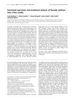
CCT is in the cytoplasm of
E. focardii
The presence of the CCT among the cellular components of
E. focardii was shown by cellular fractionation and subse-
quent immunoblotting analysis using anti-(TCP-1a) poly-
clonal Igs [12]. Two bands with apparent molecular weight
close to that of the rabbit CCT a-subunit were recognized
by the antibodies in the cytosolic fraction of E. focardii
(Fig. 1). One of the polypeptides presumably corresponds
to the a-subunit of E. focardii CCT because it has a
molecular weight identical to that of the a-subunit of rabbit
CCT; the second polypeptide may correspond to another
CCT subunit of E. focardii that contains epitope(s) similar
to that/those of the a-subunit. These results indicate that a
chaperonin containing two vertebrate TCP-1a related
polypeptide chains is present in the cytosol of E. focardii.
Properties of recombinant
E. focardii
b-T1
A number of tubulin polypeptide chains from various
exotic origins [29,30] have been successfully expressed in
E. coli as soluble proteins. In contrast, recombinant tubulin
polypeptide chains from various vertebrates [31–33] form
inclusion bodies within E. coli presumably because they are
misfolded.
To determine whether E. focardii b-T1 is soluble when
expressed in E. coli
35
S-labeled b-T1 was synthesized as
reported by Gao et al. [31] using the construct described in
the Materials and methods section and analysed by SDS/
PAGE. [
35
S]b-T1 has an apparent molecular mass indistin-
guishable from that of native b-tubulin from brain or
recombinant human b5 tubulin. After centrifugation at
20 000 g for 15 min at 4 °C, 96% of the [
35
S]b-T1 was
recovered in the pellet of the bacterial lysate. We conclude
from these observations that b-T1 forms inclusion bodies as
do mice or human b5 tubulins. This result strongly suggests
that b-T1 is misfolded upon expression in E. coli.
To further document b-T1 folding, pelleted [
35
S]b-T1 was
recovered, dissolved in 7.5
M
urea as described [31] to a final
concentration of 5 mgÆmL
)1
and diluted 100 times from
denaturant in folding buffer (0.1
M
Mes, pH 6.8, 1 m
M
EGTA, 1 m
M
MgCl
2
,1m
M
GTP, 1 m
M
ATP). The
reaction products were analyzed on nondenaturing PAGE
as described [31], immediately after dilution or after 1 h of
incubation at 30 °C. b-T1 behaviour was similar to that of
b5 tubulin [33]. At early times, the bulk of the radioactivity
migrates as a broad band with a slower mobility than folded
b5 tubulin run under identical conditions. After 1 h,
virtually all of the input radioactivity was found at the
origin, i.e. at the top of the gel (data not shown). We
conclude that b-T1 aggregates rapidly in solution under the
conditions used in our folding assays, as does b5 tubulin
[33]. We further conclude that b-T1 folding properties differ
significantly from that of Giardia duodenalis and Reticu-
lomyxa filosa tubulins [29,30] and are instead similar to that
of higher vertebrate b-tubulin.
Folding of
E. focardii
b-T1 requires CCT and Cof A
Up to now, the folding pathway of protozoan tubulins has
never been established. In Tetrahymena, coexpression of
genes encoding CCT subunits and tubulin during cilia
recovery has been demonstrated [34,35]. However, no direct
evidence for the participation of the CCT complex and
Cof A with b-tubulin folding process has been documented.
As a first analysis of the protozoan tubulin folding,
labeled, urea-denatured E. focardii b-T1 was diluted in
rabbit reticulocyte lysate, that is known to be enriched in the
tubulin folding machinery [7,12]. A parallel reaction,
consisting in the dilution of labeled, urea-denatured human
b5 tubulin in rabbit reticulocyte lysate, was performed as a
reference. The reaction products were then analyzed in two
separate lanes of a nondenaturing PAGE. Unlabeled native
pig tubulin was run in a third slot to locate the position of
native dimeric tubulin. The autoradiogram of the gel
(Fig. 2A) revealed two intense bands in the lane containing
the E. focardii b-T1 folding reaction (empty arrowheads).
The upper band corresponds to the b-T1/CCT complex.
This band is also observed in the b5 tubulin folding reaction
(lane 2). The lower band migrates at the level of pig a/b-
tubulin heterodimer, while folded b5 tubulin in complex
with Cof A migrates at a lower position [15]. This indicates
that folded, labeled b-T1 is either incorporated in the
tubulin heterodimer [36–38] or that the folded b-T1/Cof A
complex has a slight slower mobility than b5 tubulin/Cof A
complex. This result suggests that labeled, denatured,
recombinant E. focardii b-T1 is properly folded by the
mammalian tubulin folding machinery as it forms either a
stable complex with Cof A or exchanges against rabbit
b-tubulin in the tubulin heterodimer.
To determine whether the abnormal mobility of labeled,
folded b-T1 is due to its incorporation into a mixed rabbit
Fig. 1. Immunodetection of CCT a-subunit in the cytoplasm of E. fo-
cardii. SDS/PAGE of an E. focardii cytoplasmic fraction (20 lg, lane
1) and of CCT purified from rabbit reticulocyte lysate (2 lgand6lg
in lanes 2 and 3, respectively). Lanes 1 and 2 were Western blotted and
immunostained by an anti-(TCP-1a) polyclonal Ig. Lane 3 was Coo-
massie stained. Molecular size markers are indicated on the right.
Ó FEBS 2002 Folding of b-tubulin from Euplotes by CCT (Eur. J. Biochem. 269) 6273
a-tubulin/b-T1 tubulin heterodimer or to a slower mobility
of the b-T1/Cof A complex, labeled denatured b-T1 was
folded in vitro by purified rabbit CCT in the presence or in
the absence of Cof A, and the reaction products were
analyzed on nondenaturing PAGE. Similar reactions where
human b5 tubulin was substituted to b-T1 were run in
parallel, as a control. The results are presented in Fig. 2B. In
the absence of Cof A, b-T1 binds to CCT (upper arrow).
No folded products are generated as described [14]. In the
folding reaction containing both CCT and Cof A, an
additional band is generated (middle arrow). A similar band
(lower arrow) corresponding to b5/Cof A complex [15] is
generated in the equivalent b5 tubulin control folding
reaction. Therefore, the middle band probably corresponds
to folded b-T1 in complex with Cof A. The slightly lower
mobility of the b-T1/Cof A complex as compared to that of
the b5/Cof A complex is probably due to the difference in
the isoelectric point of b-T1 and human b5, calculated from
their respective primary sequences (4.55 and 3.91, respect-
ively, determined using the
WINPEP
software [39]). Finally, in
a manner similar to that observed for b5 tubulin, no folded
b-T1 tubulin is generated in the folding reaction containing
Cof A but lacking CCT. We conclude from these data that
CCT is required for the folding of b-T1. We further
conclude that folded b-T1isstabilizedbyCofA,asisb5
tubulin.
E. focardii
b-T1 does not fold in the presence
of the mt-cpn60
Mammalian unfolded tubulin polypeptide chains bind with
high affinity to the prokaryotic homologues of CCT,
GroEL and the mitochondrial chaperonin cpn60. How-
ever, folding was demonstrated not to proceed any further,
with tubulin chains being trapped on GroEL and cpn60
[15,40]. More recently, the a-andb-tubulin chains from the
giant amoeba R. filosa were shown to remain soluble when
expressed in E. coli following their interaction with the
prokaryotic chaperonin GroEL/ES [29]; the resulting
soluble tubulin is not necessarily native, however, as no
assembly reaction was performed. To test whether b-T1,
which shares about 85% sequence identity with mamma-
lian b-tubulin, folds into a stable soluble state in the
presence of GroEL or cpn60, denatured b-T1 was incuba-
ted at 30 °C for 90 min in folding buffer containing
GroEL or cpn60 in the absence or in the presence of
Cof A. The reaction products were analyzed on nondena-
turing PAGE. Figure 2C shows that b-T1 binds to cpn60,
as does human b5 tubulin incubated under identical
conditions. No additional bands are observed on the gel
which indicates that no stable b-T1, neither in the absence
nor in the presence of Cof A, is generated following the
interaction of b-T1 with cpn60. We conclude from this
observation that the folding of b-T1 either does not
proceed, or that the released folded product is misfolded
and unstable and therefore unable to interact with Cof A.
Thus, b-T1 does not acquire a native-like conformation
following its interaction with cpn60, in a manner similar to
mammalian actin and tubulins [40].
b-T1 and human b5 tubulin compete for
the same site on CCT
The results obtained by the folding assay described above
clearly show that b-T1 and CCT form a complex. To
determine whether b-T1 binds to CCT in a manner similar
to b5 tubulin despite the difference in the primary structure,
we performed a competition experiment using rabbit CCT,
labeled b-T1 and unlabeled b5, as described in Melki et al.
[32]. In a control reaction, a competition experiment was
performed using labeled and unlabeled b5. The reaction
products were analyzed on nondenaturing PAGE and
quantified using a phosphorimager. As shown in Fig. 3, the
radioactive signal in the CCT/tubulin complex (arrow)
decreases with increasing amount of unlabeled b-tubulin.
This result suggests that the binding of b5 to CCT particles
hinders that of b-T1 and vice versa (not shown). A
quantitative analysis of the relative yields of the CCT/
b-tubulin complex formed revealed that CCT has a similar
affinity for b-T1 and b5 tubulin, although slightly lower for
b-T1 (compare the intensities at given unlabeled b5 : labeled
b-T1 and unlabeled b5 : labeled b5 ratios). The slight
difference in affinity is more apparent when the amount of
bound labeled tubulin is plotted as a function of the
unlabeled : labeled b-tubulin ratio, assuming that the higher
value obtained is 100% binary complex. The comparison of
the slopes of unlabeled b5 : labeled b-T1 and unlabeled
b5 : labeled b5 competition experiments (Fig. 3b) reveals
that b-T1 binds to CCT with a twofold lower affinity than
b5 tubulin. This behavior may be the consequence of
Fig. 2. Folding analysis of E. focardii b-T1. (A) In vitro folding reac-
tions of labeled denatured b-T1 (lane 1) and b5 (lane 2) tubulins in
rabbit reticulocyte lysate. The upper and lower open arrowheads in
lane 1 indicate the location of b-T1/CCT complex, and the b-T1/Cof A
complex, respectively. The upper, middle and lower solid arrowheads
in lane 2 indicate the location of b-5/CCT complex, the a/b5 tubulin
heterodimer and b5/Cof A complex, respectively. (B) In vitro folding
reactions of labeled denatured b-T1 and b5 tubulins in the presence
(+) or the absence (–) of purified CCT and Cof A. The upper, middle
and lower arrows indicate the location of b-tubulins/CCT complex,
b-T1/Cof A complex and b5/Cof A complex, respectively. (C) In vitro
folding reactions of labeled denatured b-T1 and b5 tubulins, in the
presence (+) or the absence (–) of cpn60 and Cof A. The arrow
indicates the location of b-tubulin/cpn60 complex.
6274 S. Pucciarelli et al.(Eur. J. Biochem. 269) Ó FEBS 2002
differences in the affinity of CCT for b-T1 and b5 folding
intermediates. Alternatively, this may be due to the differ-
ences in the primary structures of b-T1 and b5 tubulin as
CCT binds unfolded actins, tubulins and other cytosolic
proteins with different affinities [33].
Regions of actin and tubulins that are preferentially
recognized by CCT have been extensively investigated [41–
44]. In b-tubulin, the peptide that comprises amino acids
251–287 and the partially overlapping peptide spanning
amino acids 263–384 define tubulin surface area that is most
likely to interact with CCT [41–44]. A more recent analysis
of the interaction between CCT and b-tubulin by cryoelec-
tron microscopy allowed the identification of three regions
from the N-terminal, and five from the C-terminal domains
of b-tubulin, involved in its binding to CCT [22]. Polypep-
tide P259–T372 is among these regions. It is considered the
most important as it binds to CCT with the strongest
affinity [22]. Based on the tubulin 3D model [45,46], this
polypeptide includes strand S7, that has been demonstrated
to be involved in the stabilization of the native tubulin
monomer by establishing an intramolecular interaction with
the N-terminus, and helix H10, that is implicated in a/b
interdimeric longitudinal contacts (underlined by L, in
Fig. 4) [45]. It also includes amino acid residues involved in
lateral contacts between tubulin heterodimers in microtu-
bule walls (underlined by M, in Fig. 4): the helix H9, the
H10–S9 loop, and the S7–H9 loop, defined as the ÔM loopÕ
[45,46]. Comparison of the primary structure of E. focardii
b-T1 polypeptide P259–T372 with the same region of the
Euplotes consensus sequence, obtained from the alignment
of the known b-tubulin sequences for Euplotes species
(accession numbers P20365 and Q08115, and S. Pucciarelli
and C. Miceli, unpublished results) and three vertebrate
tubulins (including human b5) (Fig. 4), revealed 18
amino acid positions where substitutions occur. The P268I
substitution (shown in bold italics) is unique to E. focardii
and can be correlated with microtubule cold-stability. It
affects a residue in a proline-rich motif. Site-directed
mutagenesis in this tubulin motif results in a weaker affinity
of the mutant tubulin for CCT [43]. Additional substitutions
in b-T1 that are specific to protozoa are indicated by
asterisks in Fig. 4. The substitution A352S is located before
the VCDIP motif (boxed in Fig. 4) that is involved in the
interaction between b-tubulin and CCT [22]. This and other
substitutions (labeled in pale gray in Fig. 4) reduce the
hydrophobicity of tubulin 259–372 region and may be
Fig. 4. Primary structure alignment of the putative domain that interacts with CCT from four b-tubulin isotypes. Tubulin polypeptides 259–372 from
E. focardii b-T1 (accession number S72098) and Euplotes b-tubulin consensus sequence (Eupl. consensus, see text) were aligned with those from
human b5, mouse b5 and chicken b3 tubulins (accession numbers P04450, P05218 and P09206, respectively), and mapped on tubulin secondary
structure as determined by Incla
`
n and Nogales [47]. Arrows and rectangles indicate b-sheets and a-helices, respectively. The labels ÔLÕ and ÔMÕ
indicate residues involved in longitudinal and lateral contacts between tubulin molecules in the microtubule, respectively [47]. Amino acid
substitutions in b-T1 relative to vertebrate b-tubulins are marked by asterisks and the substitution P268I, unique to E. focardii tubulin, is shown in
bold italic. Apolar amino acids are shown in grey. The intensity of the grey color corresponds to the degree of amino acid side chain hydrophobicity
resulting from the comparison of the residues at a specific position (paler gray ¼ less hydrophobic). Boxed areas delineate the CCT-binding motifs
of tubulin characterized by Llorca et al. [22].
Fig. 3. Competition experiments between b-T1 and b5 tubulins for
bindingtoCCT.(A) Labeled denatured b-T1 tubulin (b-T1*) or b5
tubulin (b5*) were mixed with increasing concentrations of unlabeled
b5 and the mixture incubated with CCT. The reaction products were
analyzed by nondenaturing PAGE; the ratios of labeled to unlabeled
tubulins are indicated on the top of each lane and the arrow indicates
the position of the b-tubulin/CCT complex. (B) The amounts of the
tubulin bound to CCT were quantitated by the use of a phosphori-
mager and expressed as a fraction of the maximum amount of bound
labeled tubulin and plotted as a function of the ratio of labeled : un-
labeled tubulin.
Ó FEBS 2002 Folding of b-tubulin from Euplotes by CCT (Eur. J. Biochem. 269) 6275
responsible for the weaker binding of b-T1 to CCT, given
that CCT binds target proteins in their quasi-native
conformation by interaction with exposed hydrophobic
amino acid residues [42].
As mentioned in the introduction, two additional b-tub-
ulin isotypes were identified in E. focardii. They are denoted
as b-T3 and b-T4. Comparison of the primary structure of
b-tubulin isotypes from E. focardii to those of non-Antarc-
tic organisms, revealed numerous amino acid substitutions
that probably accumulated in order to allow microtubules
polymerization and stability at the low temperature of the
Antarctic sea water ([6], S. Pucciarelli and C. Miceli,
unpublished results). An analysis of the putative role of
these amino acid substitutions in cold adaptation by
mapping them on the 3D structure of tubulin (carried out
in collaboration with E. Nogales, University of California,
Berkeley, USA) will be presented elsewhere. The substitu-
tions located in b-T1, b-T3 and b-T4 regions implicated in
the binding to CCT [22,42,44] (Fig. 4) may have coevolved
with CCT surface areas, allowing the interaction of
Antarctic b-tubulin isotypes with CCT in the adverse
energetic conditions of the Antarctic habitat. The role of
these amino acid substitutions in the folding process of
E. focardii b-tubulin isotypes should therefore be investi-
gated further.
ACKNOWLEDGEMENTS
This work was supported by the Italian ÔProgramma Nazionale di
Ricerca in AntartideÕ,bytheÔCentre National de la Recherche
ScientifiqueÕ and by the ÔAssociation pour la Recherche sur le CancerÕ.
REFERENCES
1. Hyams, J.S. & Lloyd, C.W. (1993) Microtubules. In Modern Cell
Biology (Harford, J.B., ed.). Wiley-Liss, New York, USA.
2. Luduena, R.F. (1998) Multiple forms of tubulin: different gene
products and covalent modifications. Int. Rev. Cytol. 178,207–
275.
3. Panda,D.,Miller,H.P.,Banerjee,A.,Luduena,R.F.&Wilson,
L. (1994) Microtubule dynamics in vitro are regulated by the
tubulin isotype composition. Proc. Natl Acad. Sci. USA 91,
11358–11362.
4. Detrich, H.W., Parker, S.K., Williams, R.C., Nogales, E. &
Downing, K.H. (2000) Cold adaptation of microtubule assembly
and dynamics: structural interpretation of primary sequence
changes present in the a and b-tubulins of Antarctic fishes. J. Biol.
Chem. 275, 37038–37048.
5. Miceli, C., Ballarini, P., Di Giuseppe, G., Valbonesi, A. &
Luporini, P. (1994) Identification of the tubulin gene family
and sequence determination of one b-tubulin gene in a cold-
poikilotherm protozoan, the Antarctic ciliate Euplotes focardii. J.
Euk Microbiol. 42, 430–437.
6. Pucciarelli, S. & Miceli, C. (2002) Characterization of the cold-
adapted a-tubulin from the psychrophilic ciliate Euplotes focardii.
Extremophiles 5, 385–389.
7. Gao, Y., Thomas, J.O., Chow, R.L., Lee, G H. & Cowan, N.J.
(1992) A cytoplasmic chaperonin that catalyzes b-actin folding.
Cell 69, 1044–1050.
8. Kubota, H., Hynes, G. & Willison, K. (1995) The chaperonin
containing t-complex polypeptide 1 (TCP-1). Multisubunit
machinery assisting in protein folding and assembly in the
eukaryotic cytosol. Eur. J. Biochem. 230, 3–16.
9. Llorca, O.E., McCormack, A., Hynes, G., Grantham, J., Cordell,
J., Carrascosa, J.L., Willison, K.R., Fernandez, J.J. & Valpuesta,
J.M. (1999) Eukaryotic type II chaperonin CCT interacts with
actin through specific subunits. Nature 412, 693–696.
10. Llorca, O., Martin-Benito, J., Ritco-Vonsovici, M., Willison, K.R.,
Carrascosa, J.L. & Valpuesta, J.M. (2000) Eukaryotic chaperonin
CCT stabilizes actin and tubulin folding intermediates in open
quasi-native conformations. EMBO J. 15, 5971–5979.
11. Llorca, O., Martin-Benito, J., Ritco-Vonsovici, M., Grantham, J.,
Hynes, G.M., Willison, K.R., Carrascosa, J.L. & Valpuesta, J.M.
(2001) The Ôsequential allosteric ringÕ mechanism in the eukaryotic
chaperonin-assisted folding of actin and tubulin. EMBO J. 20,
4165–4175.
12. Melki, R., Batelier, G., Soulie, S. & Williams, R.C. Jr (1997)
Cytoplasmic chaperonin containing TCP-1: structural and func-
tional characterization. Biochemistry 36, 5817–5826.
13. Melki, R. (2001) Nucleotide-dependent conformational changes
of the chaperonin containing TCP-1. J. Struct. Biol. 135, 170–175.
14.Gao,Y.,Melki,R.,Walden,P.D.,Lewis,S.A.,Ampe,C.,
Rommelaere, H., Vandekerckhove, J. & Cowan, N.J. (1994) A
novel cochaperonin that modulates the ATPase activity of cyto-
plasmic chaperonin. J. Cell Biol. 125, 989–996.
15. Melki, R., Rommelaere, H., Leguy, R., Vandekerckhove, J. &
Ampe, C. (1996) Cofactor A is a molecular chaperone required for
b-tubulin folding: functional and structural characterization.
Biochemistry 35, 10432–10445.
16. Tian, G., Bhamidipati, A., Cowan, N.J. & Lewis, S.A. (1999)
Tubulin folding cofactors as GTPase-activating proteins. GTP
hydrolysis and the assembly of the a/b-tubulin heterodimer. J. Biol.
Chem. 274, 24154–24158.
17. Lopez-Fanarraga, M., Avila, J., Guasch, A., Coll, M. & Zabala,
J.C. (2001) Review: postchaperonin tubulin folding cofactors and
their role in microtubule dynamics. J. Struct. Biol. 135, 219–229.
18. Radcliffe, P.A., Hirata, D., Vardy, L. & Toda, T. (1999) Func-
tional dissection and hierarchy of tubulin-folding cofactor
homologues in fission yeast. Mol. Biol. Cell. 10, 2987–3001.
19. Soares, H., Cyrne, L., Casalou, C., Ehmann, B. & Rodrigues-
Pousada, C. (1997) The third member of the Tetrahymena CCT
subunit gene family, TpCCTa, encodes a component of the
hetero-oligomeric chaperonin complex. Biochem. J. 326, 21–29.
20. Domingues, C., Soares, H., Rodrigues-Pousada, C. & Cyrne, L.
(1999) Structure of Tetrahymena CCT theta gene and its expres-
sion under colchicine treatment. Biochim. Biophys. Acta 1457,
453–459.
21. Archibald, J.M., Logsdon, J.M. Jr & Doolittle, W.F. (2000) Origin
and evolution of eukaryotic chaperonins: phylogenetic evidence
for ancient duplications in CCT genes. Mol. Biol. Evol. 17, 1466–
1476.
22. Llorca, O., Martin-Benito, J., Gomez-Puertas, P., Ritco-Vonso-
vici, M., Willison, K.R., Carrascosa, J.L. & Valpuesta, J.M. (2001)
Analysis of the interaction between the eukaryotic chaperonin
CCT and its substrates actin and tubulin. J. Struct. Biol. 135,205–
218.
23. Valbonesi, A. & Luporini. P. (1990) A new marine species of
Euplotes (Ciliophora, Hypotrichida) from Antarctica. Bull. Br.
Mus. Nat. His. (Zool.) 56, 57–61.
24. Miceli, C., Pucciarelli, S., Ballarini, P., Valbonesi, A. & Luporini,
P. (1996) Cold-stable microtubules of the Antarctic ciliate Euplotes
focardii.InAntarctic Communities (B.Battaglia & J.Walton, eds),
pp. 300–306. Cambridge University Press, London, UK.
25. Pucciarelli, S., Ballarini, P. & Miceli, C. (1997) Cold-adapted
microtubules: characterization of tubulin posttranslational mod-
ifications in the Antarctic ciliate Euplotes focardii. Cell Motil.
Cytoskeleton 38, 329–341.
26. Laemmli, U.K. (1970) Cleavage of structural proteins during the
assembly of the head of bacteriophage T4. Nature 15, 680–685.
27. Lozupone, C.A., Knight, R.D. & Landweber, L.F. (2001) The
molecular basis of nuclear genetic code change in ciliates. Curr.
Biol. 11, 65–74.
6276 S. Pucciarelli et al.(Eur. J. Biochem. 269) Ó FEBS 2002
28. Viitanen, P.V., Lorimer, G.H., Seetharam, R., Gupta, R.S.,
Oppenheim, J., Thomas, J.O. & Cowan, N.J. (1992) Mammalian
mitochondrial chaperonin 60 functions as a single toroidal ring.
J. Biol. Chem. 267, 695–698.
29. Linder, S., Schliwa, M. & Kube-Granderath, E. (1998) Expression
of Reticulomyxa filosa a-andb-tubulins in Escherichia coli yields
soluble and partially correctly folded material. Gene 212, 87–94.
30. MacDonald, L.M., Armson, A., Thompson, R.C. & Reynoldson,
J.A. (2001) Expression of Giardia duodenalis b-tubulin as a soluble
protein in Escherichia coli. Protein Expr Purif 22, 25–30.
31. Gao, Y., Vainberg, I.E., Chow, R.L. & Cowan, N.J. (1993) Two
cofactors and cytoplasmic chaperonin are required for the folding
of a and b-tubulin. Mol. Cell. Biol. 13, 2488–2485.
32. Melki, R., Vainberg, I., Chow, R.L. & Cowan, N. (1993) Cha-
peronin-mediated folding of vertebrate actin-related protein and
c-tubulin. J. Cell Biol. 122, 1301–1310.
33. Melki, R. & Cowan, N. (1994) Facilitated folding of actins and
tubulins occurs via a nucleotide–dependent interaction between
cytoplasmic chaperonin and distinctive folding intermediates.
Mol. Cell. Biol. 14, 2895–2904.
34. Soares, H., Penque, D., Mouta, C. & Rodrigues-Pousada, C.
(1994) A Tetrahymena orthologue of the mouse chaperonin sub-
unit CCTc and its coexpression with tubulin during cilia recovery.
J. Biol. Chem. 269, 29299–29307.
35. Cyrne, L., Guerreiro, P., Cardoso, A.C., Rodrigues-Pousada, C.
& Soares, H. (1996) The Tetrahymena chaperonin subunit CCT
eta gene is coexpressed with CCTc gene during cilia biogenesis and
cell sexual reproduction. FEBS Lett. 383, 277–283.
36. Yaffe, M.B., Levison, B.S., Szasz, J. & Sternlicht, H. (1988)
Expression of a human a-tubulin: properties of the isolated sub-
unit. Biochemistry 27, 1869–1880.
37. Zabala, J.C. & Cowan, N.J. (1992) Tubulin dimer formation via
the release of a-andb-tubulin monomers from multimolecular
complexes. Cell. Motil. Cytoskeleton. 23, 222–230.
38. Fontalba, A., Paciucci, R., Avila, J. & Zabala, J.C. (1993)
Incorporation of tubulin subunits into dimers requires GTP
hydrolysis. J. Cell. Sci. 106, 627–632.
39. Hennig, L. (1999) WinGene/WinPep: User-friendly software
for the analysis of aminoacid sequences. Biotechniques 26,
1170–1172.
40. Tian, G., Vainberg, I.E., Tap, W.D., Lewis, S. & Cowan, N.J.
(1995) Specificity in chaperonin-mediated protein folding. Nature
375, 250–253.
41. Dobrzynski, J.K., Sternlicht, M.L., Farr, G.W. & Sternlicht, H.
(1996) Newly-synthesized b-tubulin demonstrates domain–specific
interactions with the cytosolic chaperonin. Biochemistry 35,
15870–15882.
42. Rommelaere, H., De Neve, M., Melki, R., Vandekerckhove, J. &
Ampe, C. (1999) The cytosolic class II chaperonin CCT recognizes
delineated hydrophobic sequences in its target proteins.
Biochemistry 38, 3247–3257.
43. Dobrzynski, J.K., Sternlicht, M.L., Peng, I., Farr, G.W. &
Sternlicht, H. (2000) Evidence that b-tubulin induces a confor-
mation change in the cytosolic chaperonin which stabilizes bind-
ing: implications for the mechanism of action. Biochemistry 39,
3988–4103.
44. Ritco-Vonsovici, M. & Willison, K.R. (2000) Defining the
eukaryotic cytosolic chaperonin-binding sites in human tubulins.
J. Mol. Biol. 304, 81–98.
45. Lowe, J., Li, H., Downing, K.H. & Nogales, E. (2001) Refined
structure of a b-tubulin at 3.5 A
˚
resolution. J. Mol Biol. 313, 1046–
1057.
46. Meurer-Grob, P., Kasparian, J. & Wade, R.H. (2001) Micro-
tubule structure at improved resolution. Biochemistry 41, 8000–
8008.
47. Incla
`
n, Y.F. & Nogales, E. (2001) Structural models for the
self-assembly and microtubule interactions of c, d and e-tubulin.
J. Cell. Sci. 114, 423–432.
Ó FEBS 2002 Folding of b-tubulin from Euplotes by CCT (Eur. J. Biochem. 269) 6277