Báo cáo Y học: Processing, stability, and kinetic parameters of C5a peptidase from Streptococcus pyogenes pptx
Bạn đang xem bản rút gọn của tài liệu. Xem và tải ngay bản đầy đủ của tài liệu tại đây (629.05 KB, 13 trang )
Processing, stability, and kinetic parameters of C5a peptidase
from
Streptococcus pyogenes
Elizabeth T. Anderson
1
, Michael G. Wetherell
1
, Laurie A. Winter
1
, Stephen B. Olmsted
1
,
Patrick P. Cleary
2
and Yury V. Matsuka
1
1
Wyeth Research, West Henrietta, NY, USA;
2
Microbiology Department, University of Minnesota, Minneapolis, MN, USA
A recombinant streptococcal C5a peptidase was expressed in
Escherichia coli and its catalytic properties and thermal sta-
bility were subjected to examination. It was shown that the
NH
2
-terminal region of C5a peptidase (Asn32–Asp79/
Lys90) forms the pro-sequence segment. Upon maturation
the propeptide is hydrolyzed either via an autocatalytic
intramolecular cleavage or by exogenous protease strepto-
pain. At pH 7.4 the enzyme exhibited maximum activity in
the narrow range of temperatures between 40 and 43 °C.
The process of heat denaturation of C5a peptidase investi-
gated by fluorescence and circular dichroism spectroscopy
revealed that the protein undergoes biphasic unfolding
transition with T
m
of 50 and 70 °C suggesting melting of
different parts of the molecule with different stability.
Unfolding of the less stable structures was accompanied by
the loss of proteolytic activity. Using synthetic peptides
corresponding to the COOH-terminus of human comple-
ment C5a we demonstrated that in vitro peptidase catalyzes
hydrolysis of two His67-Lys68 and Ala58-Ser59 peptide
bonds. The high catalytic efficiency obtained for the
SQLRANISHKDMQLGR extended peptide compared to
the poor hydrolysis of its derivative Ac-SQLRANISH-pNA
that lacks residues at P2¢–P7¢ positions, suggest the import-
ance of C5a peptidase interactions with the P¢ side of the
substrate.
Keywords: maturation; propeptide; streptopain; denatura-
tion; substrate binding.
Group A streptococcus (Streptococcus pyogenes)isa
common human pathogen causing a wide variety of
diseases. These include relatively mild pathological condi-
tions such as pharyngitis and impetigo, more serious
nonsuppurative sequelae, acute rheumatic fever, glomerulo-
nephritis, deadly toxic shock syndrome and necrotizing
fasciitis. S. pyogenes has developed complex and sophisti-
cated molecular mechanisms that allow it to avoid human
defenses. One of the important virulence factors of strep-
tococci involved in such activity is an extracellular C5a
peptidase [2]. Streptococcal C5a peptidase is a surface-
associated subtilisin-like serine protease with an unusually
restricted substrate specificity. The only known protein
substrate hydrolyzed by C5a peptidase is human comple-
ment fragment C5a [3,4]. C5a peptidase-generated cleavage
within the COOH-terminal region of human C5a drastically
reduces the ability of this anaphylatoxin to bind receptors
on the surface of polymorphonuclear neutrophil leukocytes
(PMNLs) and therefore abolishes its chemotactic activity
[3]. It is believed that C5a peptidase plays an important role
in bacterial colonization of the host by inhibiting the influx
of PMNLs and impeding initial clearance of the strepto-
cocci.
C5a peptidase from S. pyogenes is encoded by the
chromosomal scpA gene and consists of 1167 amino
residues (Fig. 1A). C5a peptidase is first produced as a
precursor, with an NH
2
-terminal 31 amino acid residue
signal peptide responsible for exporting the protein across
the membrane [2]. The length of C5a peptidase pro-
sequence segment and the mechanism of its cleavage remain
unknown. Homology modeling has shown that the catalytic
domain of C5a peptidase contains the structurally con-
served core typical of subtilases, but in addition contains a
number of extra segments corresponding to various size
inserts located in external loops. All inserts found in the C5a
peptidase catalytic domain form a total of 216 additional
amino acid residues relative to subtilisin BPN¢ [5]. The active
site of C5a peptidase is located within the NH
2
-terminal half
of its polypeptide chain and formed by catalytic residues
Asp130, His193 and Ser512 (corresponding to Asp32,
His64, and Ser221 in subtilisin BPN¢). Asn294 (Asn155 in
subtilisin BPN¢) is involved in the formation of an
oxyanion-hole and is critical for the catalytic activity of
C5a peptidase [2,5]. The function of the COOH-terminal
region of C5a peptidase, starting at residue 583 and
representing half of the total polypeptide, is not known.
The COOH-terminal extension of C5a peptidase is involved
in association with the surface of S. pyogenes. This segment
is comprised of four R1–R4 hydrophilic 17 amino acid
Correspondence to Y. V. Matsuka, Department of Protein Chemistry,
Wyeth Research, WV, 211 Bailey Road, West Henrietta,
NY 14586-9728, USA.
Fax: + 1 585 273 7515, Tel.: + 1 585 273 7565,
E-mail:
Abbreviations: PMNL, polymorphonuclear neutrophil leukocytes;
pNA, p-nitroanilide; T
m
, transition midpoint of denaturation; Pn,P2,
P1, P1¢,P2¢,Pn¢, protease substrate residues accommodated by cor-
responding Sn,S2,S1,S1¢,S2¢,Sn¢ subsites of the enzyme. The scissile
peptide bond is located between the P1 and P1¢. Protease substrate
residues and subsites of the enzyme substrate-binding site are desig-
nated using the nomenclature of Schechter and Berger [1].
(Received 28 May 2002, revised 8 August 2002,
accepted 15 August 2002)
Eur. J. Biochem. 269, 4839–4851 (2002) Ó FEBS 2002 doi:10.1046/j.1432-1033.2002.03183.x
residues cell wall repeats, followed by the LPTTN motif,
hydrophobic membrane-spanning region and a cytoplasmic
tail [2]. The presence of the LPTTN motif that precedes the
hydrophobic membrane-spanning region and cytoplasmic
charged tail suggests covalent linkage of C5a peptidase to
the peptidoglycan [6,7].
Despite the recognized role of C5a peptidase as an
important streptococcal virulence factor [8,9], there is a lack
of data on its biochemical and catalytic properties. Studies
with synthetic peptides corresponding to the COOH-
terminus of human C5a suggested that C5a peptidase
generates a single cleavage site between histidine 67 and
lysine 68 residues, resulting in the release of the
KDMQLGR heptapeptide from the C5a fragment [4].
The identified His67-Lys68 peptide bond within human C5a
represented the only known cleavage site for streptococcal
C5a peptidase. Several proteins including human comple-
ment C5, C3, human serum albumin, myosin, ovalbumin,
and cytochrome c were tested as substrates for the C5a
peptidase, but none of them underwent hydrolysis [3,4].
Such a highly restricted substrate specificity of C5a pepti-
dase is in striking contrast to the broad specificity of well-
studied bacterial serine proteases of the subtilisin family.
Thus, it is of great interest to investigate the biochemical and
catalytic properties of C5a peptidase. In the present study,
we have focused on the mode of C5a peptidase maturation
and determination of the exact borders of its pro-sequence
region. We have also evaluated the thermal stability and
kinetic parameters of C5a peptidase and the results are
discussed with regard to the structural organization and
biological activity.
EXPERIMENTAL PROCEDURES
Construction of expression vectors
Wild-type C5a peptidase. The region of the scpA gene
(bases 94–3112) encoding C5a peptidase amino acid
residues 32–1038 (Fig. 1A) was produced by PCR ampli-
fication using chromosomal DNA from S. pyogenes M1
strain 90–226 as a template. To clone the scpA gene, we
designed the following forward 5¢-CCC
GAA TTC AAT
ACT GTG ACA GAA GAC ACT CCT GC-3¢ and reverse
5¢-CCC
GGA TCC TTA TTG TTC TGG TTT ATT AGA
GTG GCC-3¢ PCR primers. The forward primer incorpor-
ated the EcoRI restriction site, while the reverse primer
included a BamHI site (underlined). The reverse primer also
incorporated a TAA stop codon immediately after the
coding segment. The amplified PCR product was first
ligated into TA cloning vector pCR2.1 (Invitrogen Corp.)
and then subcloned into pTrc99a expression vector (Amer-
sham Pharmacia Biotech) using the EcoRI and BamHI
restriction sites. Incorporation of the EcoRI restriction site
within the forward primer for subsequent ligation of the
amplified scpA gene into pTrc99a expression vector yielded
three NH
2
-terminal extra residues MEF that are not part of
the natural protein. The resulting plasmid pTrc99a (wild-
type C5a peptidase) was transformed into E. coli DH5a
host cells for protein expression.
S512A C5a peptidase mutant. Site-directed mutagenesis
was performed using inverse PCR amplification [10] using
expression plasmid pTrc99a (wild-type C5a peptidase) as a
template. For this purpose we designed two PCR primers,
so that they would abut each other in opposite orientations:
5¢-ACT
GCT ATG TCT GCG CCA TTA G-3¢ (forward)
and 5¢-TCC AGA AAG TTT GGC ATA CTT GTT GTT
AGC C-3¢ (reverse). The forward primer contains
GCT
codon that replaced AGT to produce the desired SerfiAla
mutation at position 512. The GCTfiAGT mutation also
resulted in elimination of a SpeI restriction site. The inverse
PCR was performed using Expand
TM
Long Template PCR
System (Boehringer Mannheim Corp.). The resulting blunt
ended PCR product was self-ligated and transformed into
TOP10F¢ E. coli cells (Invitrogen Corp.). Clones were
screened and selected for the presence of the desired
mutation by loss of a SpeI restriction site. The presence of
SerfiAla mutation at position 512 was also confirmed by
sequencing the scpA gene. The resultant plasmid was
digested with NcoIandBamHI restriction enzymes and
isolated scpA gene containing GCTfiAGT mutation was
then subcloned into a pTrc99a expression vector where a
kanamycin resistance cassette had been inserted into the
ampicillin gene. The plasmid pTrc99a (S512A C5a pepti-
dase) was transformed into E. coli DH5a host cells for
protein expression.
Expression and purification of recombinant
C5a peptidase proteins
DH5a cells were grown overnight at 37 °C in HSY medium
(10 m
M
potassium phosphate, pH 7.2, 20 gÆL
)1
HySoy,
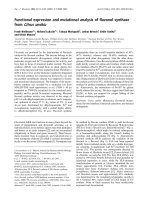
Fig. 1. Schematic representation of C5a peptidase (panel A) and SDS/
PAGE analysis of purified recombinant C5a peptidase species (panel B).
Panel A depicts location of the major regions of C5a peptidase. The
signal sequence (presequence), catalytic triad residues, cell wall,
membrane, and cytoplasmic segments are indicated. The recombinant
C5a peptidase (residues Asn32 through Gln1038) expressed in E. coli is
boxed. Panel B shows the relative mobility of isolated recombinant
S512A mutant (lane 2) and the wild-type (lane 3) C5a peptidase species
on 10–20% gradient gel. The outer lanes 1 and 4 in the gel contain
molecular mass standards as indicated.
4840 E. T. Anderson et al. (Eur. J. Biochem. 269) Ó FEBS 2002
5gÆL
)1
yeast extract, 10 m
M
NaCl). For expression of the
wild-type C5a peptidase or S512A mutant, 100 lgÆmL
)1
ampicillin or 50 lgÆmL
)1
kanamycin, respectively, was
incorporated into HSY medium. Overnight cultures were
diluted 1 : 100 with fresh HSY medium, grown to
D
600
¼ 1.5, and induced with 3 m
M
isopropyl thio-b-
D
-galactoside. After 3 h of induction, DH5a cells were
harvested by centrifugation and lysed by the freeze-thaw
method. Isolated soluble fractions of bacterial lysate were
sequentially fractionated with 50% and then 70% of
ammonium sulfate. Material precipitated with 70% ammo-
nium sulfate was collected by centrifugation, dissolved in
20 m
M
Tris, pH 8.5, 25 m
M
NaCl and dialyzed overnight at
4 °C against the same buffer. Dialyzed samples were diluted
1 : 1 (v/v) with 20 m
M
Tris, pH 8.5, 2
M
urea, and applied
to a Q-Sepharose ion exchange column (Amersham Phar-
macia Biotech), equilibrated with 20 m
M
Tris, pH 8.5, 1
M
urea. Material bound to the anion exchange resin was eluted
with a linear gradient of NaCl and pH using 20 m
M
Tris,
pH 7.0, 1
M
Urea, 1
M
NaCl buffer. Fractions containing
C5a peptidase species were collected, pooled, and dialyzed
against NaCl/Tris, pH 7.4. Purified recombinant C5a
peptidase samples (wild-type enzyme and S512A mutant)
were aliquoted and stored frozen at )20 °C.
Protein concentration determination
Protein concentrations were determined spectrophotomet-
rically, using extinction coefficients (E
280, 0.1%
)calculated
from the amino acid composition. The extinction coefficients
were estimated using the equation: E
280, 0.1%
¼ (5690 W +
1280Y + 120S-S)/M, where W, Y, and S-S represent the
number of Trp and Tyr residues and disulfide bonds,
respectively, and M represents the molecular mass [11,12].
Molecular masses of the proteins were calculated on the
basis of their amino acid composition. The following
molecularmassesandE
280, 0.1%
values were obtained:
wild-type C5a peptidase, 104.2 kDa and 0.92; S512A C5a
peptidase, 109.9 kDa and 0.87. Alternatively, protein con-
centrations were estimated using bicinchoninic acid assay
[13] according to BCA protein assay kit instructions (Pierce
Chemical Company). Concentration of streptococcal cys-
teine protease was also determined using active-site titration
with E64 (Roche Molecular Biochemicals) [14]. Titration of
the cysteine protease active-sites was performed in NaCl/
Tris, pH 7.4, 10 m
M
dithiothreitol, using resorufin-labeled
casein (Roche Molecular Biochemicals) as a substrate.
SDS/PAGE
SDS/PAGE was performed with the Bio-Rad electrophor-
esis system (Bio-Rad Laboratories) using precast 10–20%
gradient gels. All SDS-polyacrylamide gels in this study
were stained with Coomassie Brilliant Blue R (Bio-Rad
Laboratories) or Coomassie R-350 (Amersham Pharmacia
Biotech) solutions.
Amino terminal sequence analysis
NH
2
-terminal sequence analysis was performed with an
Applied Biosystems model 490 sequenator. The NH
2
-
termini of the proteins and peptides were determined by
direct sequencing for 10 or more cycles.
Synthetic peptides
Peptides corresponding to the COOH-terminal region of the
human C5a fragment (VVASQLRANISHKDMQLGR,
SQLRANISHKDMQLGR, and VVASQLRANISH) and
NH
2
-terminal segment of the C5a peptidase (QTPDEAAE
ETI and AEETIADDANDL) were synthesized as C-ter-
minal amides on a Gilson AMS422 Multiple Peptide
Synthesizer using Fmoc chemistry with pentrafluorophenyl
amino acid active esters and a polyethylene glycol polysty-
rene support with a 5-(4¢-Fmoc-aminomethyl-3¢-5¢-dimeth-
oxyphenoxy)valeric acid linker. After synthesis, peptides
were purified by reverse-phase HPLC and lyophilized.
Homogeneity of synthesized peptides was assessed by NH
2
-
terminal sequence and mass spectral analysis. Peptide
solutions of known concentration were prepared by weigh-
ing and dissolving purified lyophilized peptide in a known
volume of distilled water to give concentrated stock
solutions. Two chromogenic p-nitroanilide (pNA) peptide
derivatives were used in this study: the Ac-SQLRANISH-
pNA was custom synthesized by New England Peptide, Inc.
and the Suc-AAPF-pNA was obtained from Sigma. To
prepare concentrated stock solutions, the Ac-SQLRAN
ISH-pNA was dissolved in distilled water and Suc-AAPF-
pNA was dissolved in dimethylsulfoxide.
Mass spectral analysis
Determination of the molecular masses of proteins and
peptides was performed using MALDI-TOF mass spectro-
meter Voyager DE-STR (Perseptive Biosystems). Ions
formed by laser desorption at 337 nm (N
2
laser) were
recorded at an acceleration voltage of 20 kV in the linear
mode for proteins and 25 kV in the reflector mode for
peptides. In general, 200 single spectra were accumulated for
improving the signal/noise ratio and analyzed by use of the
DATA EXPLORER
software supplied with the spectrometer.
Sinapinic acid and a-cyano-4-hydroxycinnamic acid were
used as ultraviolet-absorbing matrices for proteins and
peptides, respectively. 1 lLofa10-mgÆmL
)1
solution of the
matrix compounds in 70% acetonitrile/0.1% trifluoroacetic
acid was mixed with 1 lL analyte solution (5–10 pmolÆ
lL
)1
). For MALDI-TOF MS, 1 lL of this mixture was
spotted on a stainless steel sample target and dried at room
temperature. The mass spectra were calibrated using
external standards: serum albumin (bovine), Glu1-fibrino-
peptide B (human), angiotensin I (human), and des-Arg1-
bradykinin (synthetic). The mass accuracy was in the range
of 0.1%.
Hydrolysis of protein and peptide substrates
Treatment of the S512A C5a peptidase precursor with C5a
peptidase was performed at 25 °C in NaCl/Tris, pH 7.4,
5m
M
CaCl
2
. For this purpose 33 l
M
of the S512A C5a
peptidase precursor was incubated with 3.3 l
M
of wild-type
C5a peptidase resulting in an enzyme/substrate ratio of
1:10(
M
/
M
). Proteolysis of S512A C5a peptidase precursor
(50 l
M
) with streptococcal cysteine protease (1.8 l
M
)was
performed at 25 °C in NaCl/Tris, pH 7.4, 10 m
M
dithio-
threitol at enzyme/substrate ratio of 1 : 25 (
M
/
M
). Samples
from each reaction mixture were removed at 15, 30, 45, 60,
120, 240, 360, and 1320 min, mixed with SDS, heated and
Ó FEBS 2002 Enzymology of C5a peptidase (Eur. J. Biochem. 269) 4841
later analyzed by SDS/PAGE using 10–20% gradient gel.
Streptococcal cysteine protease or streptopain (EC
3.4.422.10) was prepared as described elsewhere [15].
Operational molarity of cysteine protease preparations used
in this study corresponded to 98% of that expected on a
protein concentration basis. The specificity of streptopain
catalyzed cleavage was confirmed using the specific cysteine
protease inhibitor E64 (Roche Molecular Biochemicals).
The NH
2
-terminal truncation of S512A C5a peptidase
precursor by streptococcal cysteine protease was completely
blocked in the presence of 20 l
M
E64.
Caseinolytic activity of C5a peptidase was evaluated
using resorufin-labeled casein (Roche Molecular Biochemi-
cals). Briefly, increasing amounts of wild-type C5a pepti-
dase, S512A C5a peptidase mutant, and subtilisin from
Bacillus subtilis (EC 3.4.21.14) (Fluka) ranging from 0 to
10 lg were incubated for 60 min at 37 °C in the presence of
0.4% resorufin-labeled casein in NaCl/Tris, pH 7.8, 5 m
M
CaCl
2
. Undigested substrate was removed by 5% trichlo-
roacetic acid precipitation, and followed by centrifugation
the absorbance of released resorufin-labeled peptides in the
supernatant fractions was measured spectrophotometrically
at 574 nm.
The C5a peptidase-catalyzed hydrolysis of the 19-mer
synthetic peptide VVASQLRANISHKDMQLGR was
performed at 25 °C in NaCl/Tris, pH 7.4, 5 m
M
CaCl
2
.
Incubation of 345 l
M
VVASQLRANISHKDMQLGR
peptide with 0.28 l
M
of C5a peptidase was carried out for
5, 10, 15, 20, 30, 40, 60, 80, 100, 120, and 140 min and
reactions were terminated by the addition of trifluoroacetic
acid to 0.05%. At each time point, the presence of specific
peptide in reaction mixture was monitored at 210 nm by
reverse-phase HPLC (Hewlett Packard model 1090 Liquid
Chromatograph) using C4 reverse-phase column (Vydac).
Buffer A was 0.1% trifluoroacetic acid in distilled water,
and buffer B was 0.1% trifluoroacetic acid in 100%
acetonitrile. Peptides were eluted with 0–40% linear gradi-
ent of Buffer B during a 20-min interval. The relative
amount of each peptide in the reaction mixture was
determined using the area beneath the peak corresponding
to this peptide and then plotted as a function of time. The
identity of peptides was determined by NH
2
-terminal
sequence- and mass spectral analysis.
Treatment of the QTPDEAAEETI and AEETIADD
ANDL synthetic peptides with C5a peptidase was per-
formed either at 25 or 37 °C in NaCl/Tris, pH 7.4, 5 m
M
CaCl
2
and in 100 m
M
Tris, pH 8.6, 5 m
M
CaCl
2
.Eachof
these peptides (100 or 200 l
M
) was incubated with 0.1 or
1 l
M
of C5a peptidase for 1, 18, and 71 h. Reaction
mixtures were analyzed using reverse-phase HPLC as
described above. At tested conditions, cleavage of the
QTPDEAAEETI and AEETIADDANDL peptides was
not detected.
Effect of temperature on the proteolytic activity of C5a
peptidase was evaluated using 19-mer synthetic peptide
VVASQLRANISHKDMQLGR. At each tested tempera-
ture, 0.1 l
M
of the C5a peptidase in NaCl/Tris, pH 7.4 was
preincubated for 3 min. The reactions were started by
addition of 200 l
M
of the 19-mer peptide to the preincu-
bated solution of C5a peptidase followed by another 10-min
incubation at the same temperature. After termination of
hydrolysis with 0.05% trifluoroacetic acid, the reaction
mixtures were analyzed using HPLC as described above.
The percentage of hydrolyzed peptide substrate (S
hydr
%)
was determined using the equation: S
hydr
% ¼ P/(P +S),
where P represents area of the product peaks and S
represents area of the uncleaved substrate peak.
Assays revealing the pH dependence of the hydrolysis of
VVASQLRANISHKDMQLGR peptide were performed
at 25 °Cin100m
M
NaAc (pH 4.5–5.0), 100 m
M
Mes
(pH 5.5–6.5), 100 m
M
Hepes (pH 7.0–8.0), 100 m
M
Tris
(pH 7.0–9.0), 100 m
M
Ampso (pH 8.5–9.5), 100 m
M
Caps
(pH 10.0–11.0). At each tested pH, 0.1 l
M
of C5a peptidase
was incubated with 200 l
M
of the 19-mer peptide for 10 min
followed by HPLC analysis. The percentage of hydrolyzed
peptide was determined and plotted as a function of pH.
Hydrolysis of the peptide substrate in both temperature and
pH dependence experiments did not exceed 25%. The effect
of pH on hydrolysis of pNA substrate Ac-SQLRANISH-
pNA was determined at 25 °C in 100 m
M
Mes (pH 6.0–6.5),
100 m
M
Hepes (pH 7.0–8.0), 100 m
M
Tris (pH 7.0–9.0),
100 m
M
Ampso (pH 8.5–9.5), 100 m
M
Caps (pH 10.0–
11.0). At each tested pH, 200 l
M
of the pNA peptide
substrate was incubated in quartz cell for 60 min either
alone or in the presence of 1 l
M
of C5a peptidase while
monitoring hydrolysis by measurement of absorbance at
405 nm using Spectronic Genesis 2 Spectrophotometer
(Spectronic Instruments, Inc.).
Kinetic measurements
All kinetic data were obtained by incubating various
concentrations of peptide with a constant enzyme concen-
tration to achieve between 5 and 20% cleavage of the
substrate in each reaction. The concentration of C5a
peptidaseineachreactionwas0.1l
M
, while peptide
concentrations ranged from 50 l
M
to 600 l
M
(16-mer
SQLRANISHKDMQLGR) and from 50 l
M
to 2000 l
M
(12-mer VVASQLRANISH). Concentration of C5a pepti-
dase in each reaction was at least 500-fold lower than the
lowest substrate concentration. All reactions were per-
formed at 25 °C in NaCl/Tris, pH 7.4, 5 m
M
CaCl
2
.
Reactions were carried out for 5 or 100 min with 16-mer
and 12-mer peptide, respectively, and stopped by the
addition of trifluoroacetic acid to 0.05%. Cleavage of
peptides by C5a peptidase was monitored at 210 nm by
reverse-phase HPLC and percentage of hydrolyzed peptide
was determined as described above. Initial velocities (V)
were determined and plotted against substrate concentra-
tion [S]. The data were fitted to the Michaelis–Menten
equation V ¼ V
max
[S]/(K
m
+ [S]) with a nonlinear regres-
sion analysis program. The best fits of the data produced
V
max
and K
m
values, where V
max
represents the maximum
rate of hydrolysis and K
m
is the Michaelis constant. The
turnover number (k
cat
) values were calculated from V
max
/[E],
where [E] represents enzyme concentration. The identity of
hydrolyzed peptide fragments was determined by NH
2
-
terminal sequence and mass spectral analysis.
Kinetic studies of C5a peptidase using chromogenic
pNA substrate Ac-SQLRANISH-pNA were performed
with enzyme present at concentrations between 0.01 and
0.59 l
M
. The concentration of Ac-SQLRANISH-pNA was
varied from 50 to 2000 l
M
. Reactions were performed at
25 °Cin100m
M
Tris, pH 8.6, 5 m
M
CaCl
2
buffer. Assays
were carried out in 1-cm path length quartz cells and
reaction rates were monitored by continuous measurement
4842 E. T. Anderson et al. (Eur. J. Biochem. 269) Ó FEBS 2002
of absorbance at 405 nm for 180–900 min using a Spec-
tronic Genesys 2 Spectrophotometer (Spectronic Instru-
ments, Inc.). The concentration of released p-nitroaniline
product was estimated based on the molar absorption
coefficient e
405
¼ 10500
M
)1
Æcm
)1
.TheK
m
value of C5a
peptidase hydrolysis of Ac-SQLRANISH-pNA was too
high (K
m
[S]) for accurate measurement. Therefore, the
k
cat
and K
m
constants were not individually determined. The
specificity constant k
cat
/K
m
for hydrolysis of Ac-SQLRAN
ISH-pNA was determined using equation: k
cat
/K
m
¼
V/([E]Æ[S]). During extended kinetic runs, no detectable loss
of catalytic activity of the C5a peptidase was observed. The
background (nonenzymatic) hydrolysis of Ac-SQLRAN
ISH-pNA was evaluated by incubating blank substrate
solutions. At tested conditions, nonenzymatic hydrolysis
was not detectable.
Fluorescence measurements
Thermal unfolding was monitored by observing the change
in ratio of the intrinsic fluorescence intensity at 350 nm to
that at 320 nm with excitation at 280 nm [16] in an SLM
Aminco-Bowman Series 2 spectrofluorometer. Temperature
was controlled with circulating water bath programmed to
raise the temperature at 1 °CÆmin
)1
andmonitoredwith
Omega DP81 thermocouple probe inserted into a dummy
cuvette. All fluorescence measurements were performed at
protein concentration ranging from 0.04 to 0.05 mgÆmL
)1
.
Circular dichroism measurements
CD spectra were recorded on a Jasco J-810 spectropola-
rimeter equipped with a Peltier PTC-423S/L unit for
temperature control. CD measurements were performed in
20 m
M
NaCl/P
i
, pH 7.4 using protein concentration of
0.2 mgÆmL
)1
in 0.1 cm path length cells. Spectra were
recorded at 25 and 98 °C. Four scans were accumulated per
each spectrum. Spectra were averaged and expressed as
mean residue ellipticity [Q], in units of degreesÆcm
2
Ædmol
)1
.
Thermal denaturation was monitored by changes in ellip-
ticity at 205 nm while heating cell at 1 °CÆmin
)1
.
RESULTS
Preparation of recombinant C5a peptidase and analysis
of its NH
2
-terminal truncation
The recombinant wild-type C5a peptidase comprising
residues Asn32 through Gln1038 (Fig. 1A) was produced
in DH5a E. coli cells using pTrc99A expression vector as
described in Experimental procedures. During isolation of
C5a peptidase from E. coli lysate, its mobility on SDS/
PAGE was slightly but progressively increasing, indicating
possible proteolytic degradation. Purified recombinant C5a
peptidase exhibited a single band on SDS/PAGE with a
relative mobility close to its expected molecular mass
(Fig. 1B, lane 3). However, all preparations of freshly
isolated wild-type C5a peptidase consistently displayed
truncated NH
2
-terminus starting at Ala72 and suggesting
the loss of 43 amino acid residues. Subsequent analysis of
the same protein samples stored either at 4 °Corsamples
that underwent freeze-thaw cycle(s) revealed NH
2
-terminal
sequence starting at Asp79, indicating the loss of a total of
50 amino acid residues. This observation was reproducible,
suggesting that the Asp79 residue represented a final point
of progressive NH
2
-terminal truncation of the wild-type
C5a peptidase. The NH
2
-terminal cleavage of the wild-type
C5a peptidase may be caused by E. coli proteases, or
alternatively might be a result of autocatalytic cleavage
(maturation) reaction. In order to investigate the nature of
the C5a peptidase truncation, we expressed in E. coli a
mutated and enzymatically inactive form of C5a peptidase.
Based on homology analysis of subtilisin family of serine
proteases, it was reported earlier that Ser residue at position
512 is involved in the formation of catalytic site of C5a
peptidase [2,5]. When S512A C5a peptidase mutant was
expressed in DH5a E. coli cells using pTrc99A vector and
isolated using the same procedure used for purification of
the wild-type enzyme, sequence analysis revealed the
presence of an intact NH
2
-terminus starting at MEFNTV
TEDT. The three NH
2
-terminal extra residues, MEF, are
not part of the natural protein and originated from the
cloning strategy as described in Experimental procedures.
Electrophoretic mobility of S512A C5a peptidase mutant
was slightly decreased compared to that of the wild-type
enzyme (Fig. 1B, lane 2) consistent with the presence of an
extra 50 amino acid residues. Since both proteins were
produced using the same expression vectors, host cells, and
isolated using the same purification procedure, it is highly
unlikely that the NH
2
-terminus of the wild-type form but
not that of the S512A mutant was cleaved by E. coli
proteases upon protein expression and subsequent isolation.
Given the absence of NH
2
-terminal truncation in the S512A
mutant, these results indicate that C5a peptidase undergoes
autocatalytic processing resulting in cleavage of its 50 amino
acid residue propeptide segment. To further investigate the
mechanism of NH
2
-terminal autocatalytic processing, we
incubated S512A C5a peptidase mutant in the presence of
the wild-type enzyme. Upon treatment of the S512A C5a
peptidase precursor with the wild-type C5a peptidase, there
was no evidence for NH
2
-terminal truncation (Fig. 2A).
Similarly, no cleavage was detected upon incubation of the
wild-type C5a peptidase with synthetic peptides corres-
ponding to its propeptide region. These experiments were
performed with QTPDEAAEETI (Gln67–Ile76) and
AEETIADDANDL (Ala72–Leu83) overlapping peptides
containing Glu71–Ala72 and Asp78–Asp79 autocatalytic
cleavage sites, respectively. Peptides were incubated with, or
without C5a peptidase and followed by HPLC monitoring
using a C4 reverse phase column (not shown). Failure of the
C5a peptidase to cleave the propeptide segment of S512A
C5a peptidase precursor or synthetic peptides correspond-
ing to its propeptide region and containing autocatalytic
cleavage sites indicates that autoprocessing proceeds via an
intramolecular route.
In this study we also investigated the role of secreted
streptococcal cysteine protease in the maturation of C5a
peptidase precursor. Streptococcal cysteine protease, or
streptopain, is an extracellular thiol endopeptidase pro-
duced by Streptococcus pyogenes [17,18]. Cysteine protease
has been shown to release biologically active fragments
from the bacterial surface such as M protein, protein H and
C5a peptidase [19,20]. Released by streptopain, the 116 kDa
fragment of C5a peptidase inhibited granulocyte migration
into the infectious site, and therefore exhibited characteristic
peptidase activity [20]. The ability of streptopain to release
Ó FEBS 2002 Enzymology of C5a peptidase (Eur. J. Biochem. 269) 4843
an active C5a peptidase fragment from the surface of
streptococci makes this secreted protease an interesting
candidate for evaluation of its role in processing of C5a
peptidase precursor. To test this hypothesis, the S512A C5a
peptidase precursor was incubated in the presence of
streptococcal cysteine protease and analyzed by SDS/
PAGE (Fig. 2B). Upon incubation, the band corresponding
to C5a peptidase precursor steadily increased its mobility on
SDS/PAGE resulting in the appearance of higher mobility
truncated specie. The truncated form of S512A C5a
peptidase was a terminal product of proteolysis, since no
further degradation was observed even after prolonged
incubation with cysteine protease (Fig. 2B). After treatment
with streptococcal cysteine protease, the S512A C5a pep-
tidase exhibited an NH
2
-terminal sequence starting at Lys90
(KTADTPATSK) suggesting the cleavage of 61 amino
acids from its NH
2
-terminus. The same NH
2
-terminal
sequence was found in C5a peptidase released from the
surface of Streptococcus pyogenes by the action of secreted
streptococcal cysteine protease [20]. These data suggest that
in addition to COOH-terminal cleavage of C5a peptidase,
resulting in the release of the anchored enzyme, streptococ-
cal cysteine protease is involved in the maturation of C5a
peptidase precursor. Thus, the NH
2
-terminal segment
comprising of 47–58 amino acid residues forms the pro-
sequence peptide region of C5a peptidase. Cleavage of the
pro-sequence peptide and maturation of C5a peptidase
precursor is realized via an intramolecular autoprocessing
mechanism. Alternatively, processing of C5a peptidase
precursor can be achieved by an exogenous protease
streptopain.
Proteolytic activity and thermal stability of the C5a
peptidase
The earlier reported highly restricted substrate specificity of
C5a peptidase for human C5a fragment was further
investigated in this study. Proteolytic activity of C5a
peptidase was tested using resorufin-labeled casein. In the
control reaction, treatment of resorufin-casein with increas-
ing amounts of subtilisin was accompanied by a dose-
dependant increase of absorbance at 574 nm indicating
effective cleavage of the substrate. In contrast, incubation of
resorufin-labeled casein with increasing amounts of wild-
type or S512A C5a peptidase mutant resulted in absence of
detectable hydrolysis (Fig. 3A). Cleavage of casein was not
detected even upon prolonged incubation with high con-
centrations of C5a peptidase. These results suggest that C5a
peptidase does not exhibit caseinolytic activity typical for
classical subtilisins. The results also indicate the absence of
contaminating E. coli proteases in the C5a peptidase
preparations. To gain insight into the mode of C5a
peptidase catalysis, we examined cleavage of synthetic
peptide corresponding to the COOH-terminus of the human
complement fragment C5a. First, we tested the 19-mer
VVASQLRANISHKDMQLGR synthetic peptide contain-
ing previously described His67-Lys68 cleavage site [4]. This
peptide was incubated alone and in the presence of either
wild-type C5a peptidase or S512A C5a peptidase mutant.
Incubation with the S512A C5a peptidase mutant did not
result in detectable cleavage of the peptide. In contrast,
treatment of the 19-mer peptide with the wild-type C5a
peptidase resulted in progressive hydrolysis of the substrate
(Fig. 3B). As seen from Fig. 4A and B, incubation of the
19-mer peptide with C5a peptidase produced several smaller
peptide products suggesting the presence within the tested
substrate of more than one cleavage site. The products of
hydrolysis were examined by mass spectroscopy and NH
2
-
terminal sequence analysis, and the exact positions of
cleavage sites were identified. Results of both mass spectral
and NH
2
-terminal sequence analysis suggested that in
addition to the earlier reported cleavage site His67-Lys68
[4], C5a peptidase also hydrolyzed the peptide bond between
Ala58-Ser59. Time course digestion of the 19-mer peptide
monitored by HPLC revealed that the first cleavage occurs
between His67-Lys68, resulting in the formation of
VVASQLRANISH and KDMQLGR peptides. Gradual
depletion of the initial VVASQLRANISHKDMQLGR
substrate and accumulation of VVASQLRANISH product
was accompanied by detection of the second cleavage
between Ala58-Ser59, resulting in production of SQLR
ANISH and presumably VVA (Fig. 4C). Upon elution
from reverse-phase column; peaks corresponding to VVA
peptide product were not detected, probably as a result of
the precipitation of the highly hydrophobic VVA product
followed by its release from the parent VVASQLRANISH
peptide.
To assess the thermal stability of C5a peptidase, we
investigated the effect of temperature on both proteolytic
activity and structural integrity of the enzyme (Fig. 5). All
experiments were performed at neutral pH, where C5a
peptidase exhibited maximum activity towards peptide
substrate (Fig. 5A, inset). As illustrated in Fig. 5A, raising
the temperature from 5 °Cto40°Cresultedina10-fold
increase of the relative proteolytic activity of C5a peptidase
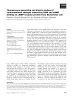
Fig. 2. Incubation of S512A C5a peptidase precursor with wild-type C5a
peptidase (panel A) and streptococcal cysteine protease (panel B) ana-
lyzed by SDS/PAGE using 10–20% gradient gel. Lanes 0 contain the
starting material. Lanes 15, 30, 45, 60, 120, 240, 360, and 1320 repre-
sent increasing times of incubation. The outer lanes in the gels contain
molecular mass standards as indicated. Arrows show relative mobility
of S512A C5a peptidase bands after incubation with wild-type C5a
peptidase (panel A) or streptococcal cysteine protease (panel B).
4844 E. T. Anderson et al. (Eur. J. Biochem. 269) Ó FEBS 2002
towards the 19-mer peptide. Maximum activity of C5a
peptidase was observed in the narrow range of temperatures
between 40 and 43 °C. A further increase in temperature
caused a sharp decline in C5a peptidase activity and
subsequently its complete inactivation at 60 °C. Heat-
induced unfolding of the C5a peptidase was studied
using fluorescence and circular dichroism spectroscopy
(Fig. 5B,C,D). Figure 5B presents a melting curve obtained
by heating C5a peptidase while monitoring the ratio of
fluorescence intensity at 350 nm to that at 320 nm as a
measure of the spectral shift that accompanies unfolding. At
neutral pH, in response to heating, the protein exhibited a
high magnitude sigmoidal denaturation transition with a
midpoint (T
m
)of50°C. The midpoint of the less pro-
nounced second transition was observed at 70 °C. The
biphasic nature of denaturation curve suggested that the
compact structure of C5a peptidase is formed by at least two
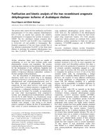
Fig. 3. Treatment of casein-resorufin (panel A) and 19-mer synthetic
peptide VVASQLRANISHKDMQLGR (panel B) with recombinant
C5a peptidase species. Casein-resorufin or 19-mer peptide substrate
were incubated in the presence of the wild-type C5a peptidase (empty
squares) and S512A C5a peptidase mutant (filled diamonds). Subtilisin
from B. subtilis (filled squares) was used as a positive control in
caseinolytic experiments.
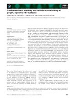
Fig. 4. HPLC analysis of C5a peptidase-catalyzed cleavage of 19-mer
synthetic peptide VVASQLRANISHKDMQLGR. The19-mer peptide
(345 l
M
) was incubated in the absence (panel A) or presence (panel B)
of C5a peptidase (0.28 l
M
) in NaCl/Tris, pH 7.4, 5 m
M
CaCl
2
for
120 min at 25 °C. The products of enzymatic hydrolysis were identified
by NH
2
-terminal sequence and mass spectral analysis. Peaks corres-
ponding to each peptide are labeled as peak 1 – (filled diamonds)
VVASQLRANISHKDMQLGR, peak 2 – (empty triangles)
VVASQLRANISH, peak 3 – (empty diamonds) SQLRANISH, and
peak 4 – (empty squares) KDMQLGR. The VVA product of hydro-
lysis was not recovered from the reaction mixture. Accumulation of
peptide products in reaction mixture was monitored and plotted as a
function of incubation time (panel C).
Ó FEBS 2002 Enzymology of C5a peptidase (Eur. J. Biochem. 269) 4845
domains with different stability. Melting of C5a peptidase in
the presence of 5 m
M
CaCl
2
did not affect denaturation
profile and transition midpoints (Fig. 5C, curve a). The
addition of 2 m
M
EDTA and heating under these condi-
tions again produced a biphasic denaturation curve with a
high amplitude first transition and low amplitude second
transition. The T
m
of the major transition, however, was
shifted about 6 °C to lower temperature (Fig. 5C, curve b),
suggesting that the compact structure of C5a peptidase in
the presence of EDTA was destabilized. Heat induced
denaturation data obtained in the presence or absence of
EDTA demonstrated that C5a peptidase contains high
affinity metal-binding site(s) that is (are) presumably
occupied. Circular dichroism spectroscopy measurements
revealed that C5a peptidase has a spectrum in the far UV
region that exhibits characteristic positive band at 194 nm
and negative bands at 210 nm and 215 nm. Heating of C5a
peptidase up to 98 °C abolished these features (Fig. 5D
inset). Monitoring of the ellipticity at 205 nm during heating
produced a sigmoidal biphasic transition curve indicative of
cooperative unfolding for a multidomain protein (Fig. 5D).
Again, the midpoints for low and high temperature
transitions were observed near 50 and 70 °C, respectively.
Results of denaturation experiments are consistent with
peptidase activity data and suggest that the decrease of
activity of C5a peptidase at temperatures above 43 °Cis
associated with the beginning of thermal unfolding.
Kinetic parameters of C5a peptidase: hydrolysis
of peptide and
p
NA substrates
In order to investigate the kinetic parameters for peptide
cleavage by C5a peptidase, we synthesized two derivatives of
the 19-mer parent peptide. These include the 16-mer
SQLRANISHKDMQLGR and the 12-mer VVASQLR
ANISH peptides. Based on the data obtained with the 19-
mer peptide, each of these peptides contains a single potential
cleavage site, and therefore can be easily utilized for kinetic
studies. This was confirmed by HPLC assay with subse-
quent mass-spectroscopic evaluation of generated products
(Figs 6A,B and 7A,B). To determine kinetic constants for
the hydrolysis of peptides by C5a peptidase, time course
experiments using different substrate concentrations were
performed in NaCl/Tris, pH 7.4, 5 m
M
CaCl
2
at 25 °C. In
each case, the initial velocity of hydrolysis of the peptide
bond was obtained. Using nonlinear regression analysis,
these data were fitted to the Michaelis–Menten equation to
yield V
max
and apparent K
m
values (Figs 6C and 7C). The
kinetic parameters for C5a peptidase-catalyzed cleavage of
16- and 12-mer synthetic peptides corresponding to the
COOH-terminus of human complement fragment C5a are
summarized in Table 1. As can be seen from Table 1, C5a
peptidase hydrolyzes the 16-mer peptide with catalytic
efficiency (k
cat
/K
m
) about 14-fold higher than the 12-mer.
Fig. 5. Effect of temperature on proteolytic activity of C5a peptidase
(panel A) and heat-induced unfolding of C5a peptidase detected by
fluorescence (panels B, C) and circular dichroism (panel D) spectroscopy.
Panel A shows relative proteolytic activity of C5a peptidase at various
temperatures in NaCl/Tris, pH 7.4. Reactions were performed using
19-mer synthetic peptide VVASQLRANISHKDMQLGR as a sub-
strate. The pH dependence of C5a peptidase relative activity is shown
in the inset. Reactions were performed at 25 °C in 100 m
M
NaAc
(pH 4.5–5.0), 100 m
M
Mes (pH 5.5–6.5), 100 m
M
Hepes (pH 7.0–8.0),
100 m
M
Ampso (pH 8.5–9.5), 100 m
M
Caps (pH 10.0–11.0) (filled
squares) and 100 m
M
Tris (pH 7.0–9.0) (empty squares). Panel B
illustrates fluorescence-detected thermal denaturation of C5a peptidase
in NaCl/Tris, pH 7.4. Fluorescence-detected melting curves of C5a
peptidase in NaCl/Tris, pH 7.4, 5 m
M
CaCl
2
(a) and NaCl/Tris,
pH 7.4, 2 m
M
EDTA (b) are presented in panel C. Protein solutions
were heated while monitoring the ratio of fluorescence at 350 nm to
that at 320 nm with excitation at 280 nm. Panel D shows changes in
ellipticity at 205 nm upon heating of C5a peptidase sample; the CD
spectra of C5a peptidase at 25 and 98 °Carepresentedintheinset.All
CD measurements were performed in NaCl/P
i
,pH7.4.
4846 E. T. Anderson et al. (Eur. J. Biochem. 269) Ó FEBS 2002
This is consistent with our data generated using 19-mer
parent peptide that contains both cleavage sites (Fig. 4C).
The low efficiency of hydrolysis of the 12-mer peptide
compared to its 16-mer counterpart was a result of a sixfold
reduction in k
cat
value and threefold increase in K
m
value.
When 5 m
M
EDTA was included into reaction buffer,
hydrolysis of both 16-mer and 12-mer peptides was
significantly inhibited (Figs 6C and 7C), again suggesting
existence of metal-binding site(s) within C5a peptidase.
Based on the sequence of 16-mer SQLRANISHKDMQ
LGR peptide, we designed a water-soluble chromogenic
pNA substrate, Ac-SQLRANISH-pNA. Incubation of
Fig. 6. HPLC analysis of C5a peptidase-catalyzed cleavage of 16-mer
synthetic peptide SQLRANISHKDMQLGR. The 16-mer peptide
(410 l
M
) was incubated in the absence (panel A) or presence (panel B)
of C5a peptidase (0.28 l
M
) for 120 min. The products of enzymatic
hydrolysis were identified by NH
2
-terminal sequence and mass spectral
analysis. Peaks corresponding to each peptide are labeled as peak
1 – SQLRANISHKDMQLGR, peak 2 – SQLRANISH, and peak 3 –
KDMQLGR. Initial rate of hydrolysis V was plotted vs. the concen-
tration of the substrate SQLRANISHKDMQLGR [S] (panel C).
Experiments were performed in NaCl/Tris, pH 7.4, containing either
5m
M
CaCl
2
(filled squares) or 5 m
M
EDTA (empty squares) at 25 °C.
Fig. 7. HPLC analysis of C5a peptidase-catalyzed cleavage of 12-mer
synthetic peptide VVASQLRANISH. The12-mer peptide (550 l
M
)was
incubated in the absence (panel A) or presence (panel B) of C5a
peptidase (0.28 l
M
) for 360 min. The products of enzymatic hydrolysis
were identified by NH
2
-terminal sequence and mass spectral analysis.
Peaks corresponding to each peptide are labeled as peak 1 –
VVASQLRANISH, and peak 2 – SQLRANISH. The VVA product
of hydrolysis was not recovered from the reaction mixture. Initial rate
of hydrolysis V was plotted vs. the concentration of the substrate
VVASQLRANISH [S] (panel C). Experiments were performed in
NaCl/Tris, pH 7.4, containing either 5 m
M
CaCl
2
(filled squares) or
5m
M
EDTA (empty squares) at 25 °C.
Ó FEBS 2002 Enzymology of C5a peptidase (Eur. J. Biochem. 269) 4847
Ac-SQLRANISH-pNA in the presence of C5a peptidase
was accompanied by increase of absorbance at 405 nm,
suggesting enzymatic release of p-nitroaniline. The linear
dependence of Ac-SQLRANISH-pNA cleavage with
enzyme concentration is demonstrated in Fig. 8. In contrast,
upon incubation of C5a peptidase with Suc-AAPF-pNA, a
substrate commonly used for kinetic analysis of subtilisins
[21,22], hydrolysis was not detected. This observation is
consistent with limited substrate specificity of C5a peptidase
and further illustrates significant differences in the organ-
ization of its substrate-binding site compared to that of the
classical subtilisins. The pH-dependence of the hydrolysis of
Ac-SQLRANISH-pNA reveals the optimum activity of
C5a peptidase in the alkaline region (pH 8.5–9.5) (Fig. 8,
inset). At pH 8.6 activity of C5a peptidase towards
Ac-SQLRANISH-pNA was about 60% higher than that
observed at pH 7.4. Analysis of Ac-SQLRANISH-pNA
cleavage by C5a peptidase revealed that estimated Michaelis
constant value is too high and exceeds maximum substrate
concentration (2 m
M
) used in the experiments and therefore
prevents accurate determination of individual kinetic
parameters. Instead, the specificity constant k
cat
/K
m
was
determined directly. Specificity of C5a peptidase towards
Ac-SQLRANISH-pNA was only 13
M
)1
Æs
)1
(Table 1). This
value is about 230-fold lower than that obtained for the
parent SQLRANISHKDMQLGR extended peptide sub-
strate. Thus, substitution of the lysine residue at P1¢ position
to pNA moiety and the lack of residues at the P2¢ through
P7¢ positions, resulting in a drastic reduction of catalytic
efficiency, indicate the importance of C5a peptidase inter-
actions with the P¢ side of the substrate.
DISCUSSION
In Gram-positive bacteria, extracellular proteases are syn-
thesized initially as inactive precursors containing an amino-
terminal extension that is composed of the signal peptide
and propeptide. The signal sequence, or prepeptide, is
involved in translocation of precursor through the cyto-
plasmic membrane. One of the major functions of the pro-
sequence region is to prevent unwanted protein degradation
and to enable spatial and temporal regulation of proteolytic
activity. The pro-sequence region associates to the protease
module, thus preventing access of substrate(s) to the active
site [23]. Zymogen conversion to the active enzyme occurs
by limited proteolysis of the inhibitory pro-sequence
segment and may be either autocatalytic or involve acces-
sory molecules. The length of propeptide may vary and
range from short polypeptide segments to independently
folded domains comprising more than 100 residues [24,25].
Often the precise length of the mature, active enzyme is not
known due to the fact that the NH
2
-terminal processing
site(s) has not been mapped. Such information was not
available for C5a peptidase from pathogenic Streptococcus
pyogenes. One of the aims of this study was to map the pro-
sequence region of C5a peptidase and to investigate the
mechanism(s) of its maturation. Recombinant wild-type
C5a peptidase and its S512A mutant, both lacking NH
2
-
terminal signal sequence and COOH-terminal membrane
anchor sequence (Asn32 – Gln1038), were overexpressed in
E. coli and isolated from the soluble fraction of cell lysate.
Mobility of purified S512A C5a peptidase mutant was
slightly decreased on SDS/PAGE compared to that of the
wild-type enzyme, suggesting partial proteolytic degrada-
tion of the latter. Sequence analysis of wild-type C5a
peptidase confirmed the loss of its NH
2
-terminal 50 amino
residues, while the enzymatically inactive S512A mutant
Table 1. Kinetic parameters for the hydrolysis of peptide and pNA substrates by C5a peptidase. The arrow (fl) represents location of scissile bond.
ND, not determined. The standard errors of the given k
cat
and K
m
values did not exceed 20%. Kinetic constants for hydrolysis of peptide substrates
were obtained in NaCl/Tris, pH 7.4, 5 m
M
CaCl
2
. Specificity constant for hydrolysis of pNA substrate was obtained in 100 m
M
Tris/HCl, pH 8.6,
5m
M
CaCl
2
.
Substrate
(Pn…, P2, P1 fl P1¢,P2¢,… Pn¢)
k
cat
(s
)1
)
K
m
(l
M
)
k
cat
/K
m
(
M
)1
Æs
)1
)
SQLRANISH fl KDMQLGR 1.1 360 3050
VVA fl SQLRANISH 0.2 936 216
Ac-SQLRANISH fl pNA ND >2000 13
Fig. 8. Incubation of Ac-SQLRANISH-pNA and Suc-AAPF-pNA with
different amounts of C5a peptidase. Reactions were carried out at 25 °C
for 180 min in 100 m
M
Tris, pH 8.6, 5 m
M
CaCl
2
in the presence of
220 l
M
Ac-SQLRANISH-pNA and in 100 m
M
Tris, pH 8.6, 5 m
M
CaCl
2
(containing 2% dimethylsulfoxide) in the presence of 400 l
M
Suc-AAPF-pNA. The pH-activity profile of C5a peptidase for
Ac-SQLRANISH-pNA is shown in the inset. Reactions were per-
formedin100m
M
Mes (pH 6.0–6.5), 100 m
M
Hepes (pH 7.0–8.0),
100 m
M
Ampso (pH 8.5–9.5), 100 m
M
Caps (pH 10.0–11.0) in the
presence (filled squares) or absence (circles) of the enzyme. Reactions
were also carried out in the presence (empty squares) or absence (tri-
angles) of C5a peptidase in 100 m
M
Tris (pH 7.0–9.0).
4848 E. T. Anderson et al. (Eur. J. Biochem. 269) Ó FEBS 2002
demonstrated an intact NH
2
-terminus. Several preparations
of wild-type C5a peptidase and S512A mutant consistently
displayed truncated and intact NH
2
-termini, respectively.
The observed truncation of wild-type C5a peptidase
suggested the existence of an autocatalytic processing
reaction since substitution of the reactive serine at position
512 to alanine abolished not only proteolytic activity of C5a
peptidase, but also prevented the loss of its NH
2
-terminal
segment. To determine whether maturation of the C5a
peptidase precursor can proceed through either intermo-
lecular or intramolecular autolysis, the S512A mutant was
incubated in the presence of different concentrations of
wild-type peptidase. Even at enzyme/substrate ratio of
1:10(
M
/
M
) and extended incubation time (up to 22 h),
processing was not detected. In addition, cleavage was not
observed upon incubation of wild-type C5a peptidase with
synthetic peptides (QTPDEAEETI and AEETIADDA
NDL) corresponding to the NH
2
-terminal segment of the
precursor and containing autocatalytic cleavage sites. These
results demonstrate an intramolecular nature of processing
of the C5a peptidase precursor and indicate that autocata-
lytic cleavage of the propeptide requires its linkage with the
catalytic domain of the enzyme. Such a conclusion is
consistent with the extracellular surface-associated status of
C5a peptidase. Covalent attachment of the carboxyl group
of threonine within the LPXTN motif to the peptidoglycan
[7] may limit intermolecular contact between C5a peptidase
molecules. The surface-associated location of C5a pepti-
dase, however, can be affected by secreted cysteine protease
streptopain. Berge and Bjorck [20] demonstrated that
functionally active 116 kDa fragment of C5a peptidase
can be released from the surface of S. pyogenes by
streptopain. In this study we established that streptococcal
cysteine protease is also involved in the processing of C5a
peptidase precursor. Treatment of S512A C5a peptidase
with streptococcal cysteine protease resulted in NH
2
-
terminal truncation of the peptidase. This was evident both
from the shift in mobility on SDS/PAGE and from NH
2
-
terminal sequence of the truncated protein. Truncated C5a
peptidase mutant displayed a single NH
2
-terminal sequence
starting at Lys90 (KTADTPATSK). This result is consis-
tent with an earlier finding where, released from the surface
of S. pyogenes, C5a peptidase exhibited the same NH
2
-
terminal sequence starting at Lys90 [20]. The data presented
above suggest that secreted streptococcal cysteine protease is
involved in the maturation of C5a peptidase precursor. Such
activity also indicates that streptococcal cysteine protease
potentially may act as an activator for C5a peptidase. Thus,
C5a peptidase is synthesized as a precursor, comprised of a
31 amino acid residues signal sequence, followed by a 47–58
residues long pro-sequence region (Fig. 9) and a 1078
residues mature sequence. Processing of C5a peptidase
precursor can be achieved via two alternative mechanisms.
These include intramolecular autocatalytic processing and
heterologous intermolecular processing achieved through
the action of secreted cysteine protease. It seems likely that a
combination of both mechanisms might function in vivo
with the intramolecular route dominating during periods of
minimum synthesis and secretion of the cysteine protease.
The role of a heterologous intermolecular mechanism in the
processing of C5a peptidase precursor may be more
prominent during stages of maximal synthesis of strepto-
coccal cysteine protease.
Spectroscopic analysis of C5a peptidase revealed that it is
folded into a compact structure that undergoes unfolding
transition when heated. The thermal denaturation process,
detected by changes in tryptophan fluorescence intensity
ratio and by changes of the ellipticity, was biphasic and two
cooperative transitions were observed at T
m1
¼ 50 °Cand
T
m2
¼ 70 °C (Fig. 5B,D). The complex biphasic melting
behavior of C5a peptidase is interpreted in terms of
sequential denaturation of different compact structures with
different stability, thus suggesting that the protein is
composed of several structural domains. This is consistent
with data obtained for C5a peptidase using multiple
sequence alignment and database homology searching
methods [5,26]. It was shown that C5a peptidase is composed
of NH
2
-terminal serine protease domain (360 residues SP
domain, Ala89/Ile102–Pro333, Gly468–Thr582), protease-
associated or insert domain (134 residues PA or I domain,
Asp334–Ser467), and COOH-terminal domain (450 residues
A domain, Met583–Ser1032) [5,26,27]. The size of each of
these segments is sufficient to form at least one independently
folded structural domain. Thus, our thermal denaturation
data provide experimental evidence supporting multidomain
organization of C5a peptidase. Upon heating in neutral pH,
the beginning of denaturation process of C5a peptidase
coincides with the loss of its enzymatic activity. Both
processes were observed at temperatures above 43 °C,
suggesting that inactivation of C5a peptidase is a result of
thermal unfolding of its compact structure. The temperature
range (40–43 °C) of C5a peptidase maximum activity
correlates with the maximum temperature of the human
body.
When melting experiments were performed in the pre-
sence of 5 m
M
CaCl
2
, thermal stability of C5a peptidase was
not affected. Unfolding curves produced in NaCl/Tris,
pH 7.4 with or without Ca
2+
were essentially undistin-
guishable (Fig. 5B,C). In the presence of 2 m
M
EDTA, the
major high amplitude transition of C5a peptidase shifted to
a lower temperature by approximately 6 °C, having a
midpoint at 44 °C, while the low amplitude second transi-
tion remained unaffected (Fig. 5C). Reduction of the
intrinsic stability of the protein in the presence of EDTA
and the absence of detectable stabilization effect in the
presence of 5 m
M
Ca
2+
indicate that C5a peptidase
contains occupied high affinity metal ion binding site(s).
Based on the known crystal structures of subtilisins
Fig. 9. Propeptide region of recombinant C5a peptidase precursor. The
positions of identified autocatalytic cleavage sites are indicated as (+).
The late intermediate of autocatalytic processing starts at Ala72, while
the terminal product of maturation starts at Asp79. The position of
identified cleavage site catalyzed by streptococcal cysteine protease is
shown as (fl). Streptococcal cysteine protease generates the mature
sequence that starts at Lys90. The NH
2
-termini of the C5a peptidase
mature sequence produced either via intramolecular autocatalytic
processing or via the action of streptococcal cysteine protease are
depicted as (fi). The three NH
2
-terminal extra residues MEF that are
not part of the natural protein are shown in italic.
Ó FEBS 2002 Enzymology of C5a peptidase (Eur. J. Biochem. 269) 4849
Carlsberg, subtilisin Novo [28], thermitase [29], and protei-
nase K [30] three major types of calcium-ion binding sites
have been identified for the subtilisin family of serine
proteases. These include strong Ca1, Ca2 (K
d
£ 10
)10
M
)
and weak Ca3 (K
d
¼ 10
)4
)10
)7
M
)Ca
2+
binding sites. The
occupancy of these sites depends on the calcium ion
concentration in solution. Sequence alignments and mode-
ling studies suggested that the Ca1 and Ca3 sites are most
common in subtilisins, whereas the Ca2 site is less common
[5]. Our thermal denaturation data demonstrated that C5a
peptidase forms strong metal ion binding site(s) that may
belong to either Ca1 or Ca2. Influence of Ca
2+
and EDTA
on the C5a peptidase thermal stability was also consistent
with their effect on its enzymatic activity. Incorporation of
5m
M
CaCl
2
into the reaction buffer did not cause any
detectable changes in C5a peptidase activity toward tested
peptide substrates, while 5 m
M
EDTA drastically reduced it
(Figs 6C and 7C).
Earlier studies demonstrated that incubation of C5a
peptidase with complement C5, C3, albumin, myosin,
ovalbumin and cytochrome c and subsequent SDS/PAGE
analysis of reaction mixtures resulted in the absence of
detectable cleavage of these protein substrates [3,4]. Highly
restricted macromolecular specificity of C5a peptidase was
further confirmed in this study using chromogenic substrate
resorufin-casein. Careful spectroscopic evaluation of reac-
tion mixtures containing increasing amounts of C5a pepti-
dase and resorufin-labeled casein revealed absence of
detectable casein cleavage (Fig. 3A). When the 19-mer
synthetic peptide corresponding to COOH-terminus of
human C5a was tested as a substrate, it was effectively
hydrolyzed by C5a peptidase (Fig. 3B). In addition to the
already known cleavage site between His67-Lys68, we
identified a novel secondary cleavage site between Ala58-
Ser59 (Figs 4 and 7). Since identification of secondary
cleavage site was performed using synthetic peptides it
represents an in vitro observation. Accessibility of the Ala58-
Ser59 peptide bond within human C5a fragment for
streptococcal peptidase has to be further investigated.
Catalytic efficiency of cleavage of the Ala58-Ser59 peptide
bond by C5a peptidase was lower compared to His67-Lys68
bond (Fig. 4C). This was further confirmed using the 16-
mer SQLRANISHKDMQLGR and 12-mer VVASQLR
ANISH peptide substrates (Figs 6 and 7). The reduced
catalytic efficiency of hydrolysis of the 12-mer peptide
compared with its 16-mer counterpart was due to a sixfold
reduction in k
cat
value and a threefold increase in K
m
value
(Table 1). This could be caused by the lack of P9–P4 within
12-mer peptide and/or poor acceptance of amino acid
residue(s) at specific P–P¢ position(s). The importance of
C5a peptidase interactions with the P¢ side of the substrate is
suggested by at least a sixfold higher K
m
and a 230-fold
lower k
cat
/K
m
value obtained for the Ac-SQLRANISH-
pNA substrate compared to its parent extended SQLRA
NISHKDMQLGR peptide (Table 1). The binding energy
gained through interactions between substrates and pro-
teases at non-S1 sites can contribute to stabilization of the
transitional state for peptide bond hydrolysis [31–33]. As a
result, hydrolysis of substrates that lack specific P or P¢ can
be less efficient than hydrolysis of longer substrates that
contain these residues. Interestingly, substrates containing
pNA aromatic leaving group are considered more chemic-
ally labile than the extended peptide substrate [33,34] and
yet a poor hydrolysis of Ac-SQLRANISH-pNA was
observed, suggesting contribution of S¢ subsites to catalysis.
Based on data presented in this study, one can conclude that
the substrate-binding site of C5a peptidase is rather stringent
and requires extended interactions with P and P¢ positions of
the polypeptide substrate. In addition to the restricted
recognition of the primary structure at the cleavage site,
other mechanism(s) may also contribute to macromolecular
specificity of C5a peptidase toward human C5a fragment.
An additional site within C5a peptidase, distal from the
binding cleft surrounding P1-P1¢, may be involved in
recognition and binding of human C5a fragment. It was
suggestedthatsuchanexositemightbeformedeitherby
protease-associated domain (PA domain) [27] or by the
COOH-terminal region (A domain) [26] of the C5a pepti-
dase. Further work is required to determine the interactions
between C5a peptidase and its substrate(s) as well as the
effects of such interactions on the efficiency of hydrolysis.
ACKNOWLEDGEMENTS
We would like to thank Melissa Naschke for assistance with protein
sequence analysis and Christopher Colocillo for synthesis of peptides
used in this study. This work was supported in part by NIAID grant
AI2006 (to P. C).
REFERENCES
1. Schechter, I. & Berger, A. (1967) On the size of the active site of
proteases. Biochem. Biophys. Res. Commun. 27, 157–162.
2. Chen, C.C. & Cleary, P.P. (1990) Complete nucleotide sequence of
the streptococcal C5a peptidase gene of Streptococcus pyogenes.
J. Biol. Chem. 265, 3161–3167.
3. Wexler, D.E., Chenoweth, D.E. & Cleary, P.P. (1985) Mechanism
of action of the group A streptococcal C5a inactivator. Proc. Natl
Acad. Sci. USA 82, 8144–8148.
4. Cleary, P.P., Prahbu, U., Dale, J.B., Wexler, D.E. & Handley, J.
(1992) Streptococcal C5a peptidase is a highly specific endo-
peptidase. Infect. Immun. 60, 5219–5223.
5. Siezen, R.J., de Vos, W.M., Leunissen, A.M. & Dijkstra, B.W.
(1991) Homology modeling and protein engineering strategy of
subtilases, the family of subtilisin-like serine proteinases. Protein
Eng. 4, 719–737.
6. Navarre, W.W. & Schneewind, O. (1994) Proteolytic cleavage and
cell wall anchoring at the LPXTG motif of surface proteins in
Gram-positive bacteria. Mol. Microbiol. 14, 115–121.
7. Schneewind, O., Fowler, A. & Faull, K.F. (1995) Structure of the
cell wall anchor of surface proteins in Staphylococcus aureus.
Science 268, 103–106.
8. Cleary, P.P. (1998) C5a peptidase. In Handbook of Proteolytic
Enzymes (Barrett, A.J., Rawlings, N.D. & Woessner, J.F., eds),
pp. 308–310. Academic Press, London, UK.
9. Cunningham, M.W. (2000) Pathogenesis of group A streptococcal
infections. Clin. Microbiol. Rev. 13, 470–511.
10. Fisher, C.L. & Pei, G.K. (1997) Modification of a PCR-based site-
directed mutagenesis method. Biotechniques 23, 570–574.
11. Edelhoch, H. (1967) Spectroscopic determination of tryptophan
andtyrosineinproteins.Biochemistry 6, 1948–1954.
12. Gill, S.C. & von Hippel, P.H. (1989) Calculation of protein
extinction coefficients from amino acid sequence data. Anal. Bio-
chem. 182, 319–326.
13. Smith, P.K., Krohn, R.I., Hermanson, G.T., Mallia, A.K.,
Gartner, F.H., Provenzano, M.D., Fujimoto, E.K., Goeke, N.M.,
Olson, B.J. & Klenk, D.C. (1985) Measurement of protein using
bicinchoninic acid. Anal. Biochem. 150, 76–85.
4850 E. T. Anderson et al. (Eur. J. Biochem. 269) Ó FEBS 2002
14. Barrett, A.J., Kembhavi, A.A., Brown, M.A., Kirschke, H.,
Knight, C.G., Tamai, M. & Hanada, K. (1982) 1-trans-Epoxy-
succinyl-leucylamido (4-guanidino) butane (E-64) and its analo-
gues as inhibitors of cysteine proteinases including cathepsins B, H
and L. Biochem. J. 201, 189–198.
15. Matsuka, Y.V., Pillai, S., Gubba, S., Musser, J.M. & Olmsted,
S.B. (1999) Fibrinogen cleavage by the Streptococcus pyogenes
extracellular cysteine protease and generation of antibodies that
inhibit enzyme proteolytic activity. Infect. Immun. 67, 4326–4333.
16. Matsuka, Y.V., Medved, L.V., Brew, S.A. & Ingham, K.C. (1994)
The N-terminal fibrin-binding site of fibronectin is formed by
interacting fourth and fifth finger domains. J. Biol. Chem. 269,
9539–9546.
17. Liu, T Y. & Elliott, S.D. (1965) Streptococcal proteinase: the
zymogen to enzyme transformation. J. Biol. Chem. 240, 1138–
1142.
18. Elliott, S.D. & Liu, T Y. (1970) Streptococcal proteinase. Meth.
Enzymol. 19, 252–261.
19. Elliott, S.D. (1945) A proteolytic enzyme produced by group A
treptococci with special reference to its effect on the type-specific
Mantigen.J. Exp. Med. 81, 573–592.
20. Berge, A. & Bjorck, L. (1995) Streptococcal cysteine proteinase
releases biologically active fragments of streptococcal surface
proteins. J. Biol. Chem. 270, 9862–9867.
21. Del Mar, E.G., Largman, C., Brodrick, J.W. & Geokas, M.C.
(1979) A Sensitive New Substrate For Chymotrypsin. Anal. Bio-
chem. 99, 316–320.
22. Carter, P. & Wells, J.A. (1988) Dissecting the catalytic triad of
serine protease. Nature 332, 564–568.
23. Hu, Z., Haghjoo, K. & Jordan, F. (1996) Further evidence for the
structure of the subtilisin propeptide and for its interactions with
mature subtilisin. J. Biol. Chem. 271, 3375–3384.
24. Khan, A.R. & James, M.N.G. (1998) Molecular mechanisms for
the conversion of zymogens to active proteolytic enzymes. Protein
Sci. 7, 815–836.
25. Ikemura, H., Takagi, H. & Inouye, M. (1987) Requirement of pro-
sequence for the production of active subtilisin E in Escherichia
coli. J. Biol. Chem. 262, 7859–7864.
26. Siezen, R.J. (1999) Multi-domain, cell-envelope proteinases of
lactic acid bacteria. Antonie Van Leeuwenhoek 76, 135–155.
27. Mahon, P. & Bateman, A. (2000) The PA domain: a protease-
associated domain. Protein Sci. 9, 1930–1934.
28. McPhalen, C.A. & James, M.N.G. (1988) Structural comparison
of two serine proteinase-protein inhibitor complexes: eglin-c-sub-
tilisin Carlsberg and CI-2-subtilisin Novo. Biochemistry 27, 6582–
6598.
29. Gros, P., Kalk, K.H. & Hol, W.G.R. (1991) Calcium binding to
Thermitase. J. Biol. Chem. 266, 2953–2961.
30. Bajorath, J., Raghunathan, S., Hinrichs, W. & Saenger, W. (1989)
Long-range structural changes in proteinase K triggered by cal-
cium ion removal. Nature 337, 481–484.
31. Fiedler, F. (1987) Effects of secondary interactions on the kinetics
of peptide and peptide ester hydrolysis by tissue kallikrein and
trypsin. Eur. J. Biochem. 163, 303–312.
32. Bauer, C A., Brayer, G.D., Sielecki, A.R. & James, M.N.G.
(1981) Active site of alpha-lytic protease. Enzyme–substrate
interactions. Eur. J. Biochem. 120, 289–294.
33. Coombs, G.S., Dang, A.T., Madison, E.L. & Corey, D.R. (1996)
Distinct mechanisms contribute to stringent substrate specificity of
tissue-type plasminogen activator. J. Biol. Chem. 271, 4461–4467.
34. Madison, E.L., Coombs, G.S. & Corey, D.R. (1995) Substrate
specificity of tissue type plasminogen activator. J. Biol. Chem. 270,
7558–7562.
Ó FEBS 2002 Enzymology of C5a peptidase (Eur. J. Biochem. 269) 4851