Báo cáo khoa học: Genomic structure, promoter analysis and functional mutation pptx
Bạn đang xem bản rút gọn của tài liệu. Xem và tải ngay bản đầy đủ của tài liệu tại đây (725.87 KB, 13 trang )
Human aquaporin adipose (AQPap) gene
Genomic structure, promoter analysis and functional mutation
Hidehiko Kondo
1
, Iichiro Shimomura
1
, Ken Kishida
1
, Hiroshi Kuriyama
1
, Yasunaka Makino
2
,
Hitoshi Nishizawa
1
, Morihiro Matsuda
1
, Norikazu Maeda
1
, Hiroyuki Nagaretani
1
, Shinji Kihara
1
,
Yoshihisa Kurachi
2
, Tadashi Nakamura
1
, Tohru Funahashi
1
and Yuji Matsuzawa
1
1
Department of Internal Medicine and Molecular Science, Graduate School of Medicine, Department of Pharmacology II,
and
2
Graduate School of Medicine, Osaka University, Yamadaoka, Suita, Japan
Aquaporin adipose ( AQPap), which we identified from
human adipose t issue, is a glycerol channel in adipocyte
[Kishida et al. (2000) J. Biol. Chem. 275, 20896–20902]. In
the c urrent study, we determined the genomic structure of
the human AQPap gene, and identified three AQPap-like
genes that resembled ( 95%) AQPap, with little expression
in human tissues. The AQPap promoter contained a puta-
tive peroxisome proliferator response element (PPRE) at
)46 to )62, and a putative insulin response element (IRE) a t
)542/)536. Deletion of the PPRE abolished the pioglita-
zone-mediated induction of AQPap promoter activity in
3T3-L1 adipocytes. Deletion and single base pair substitu-
tion analysis of the I RE abolished the insulin-m ediated
suppression of the human AQPap gene. Analysis of AQPap
sequence i n human subjects revealed three missense muta-
tions (R12C, V59L and G264V), and two silent mutations
(A103A and G250G). The cRNA injection of the missense
mutants into Xenopus oocytes revealed the a bsence of the
activity to transport glycerol and water in the AQPap-
G264V protein. In the subject homozygous for AQPap-
G264V, exercise-induced increase in plasma glycerol was not
observed in spite of the increased plasma noradrenaline. We
suggest that A QPap is responsible for the increase of plasma
glycerol during exercise in humans.
Keywords: mutation; aquaporin adipose; genome; glycerol
channel; promoter.
In response to energy demand in fasting and exercise,
triglyceride stored in adipocytes is hydrolyzed to glycer ol and
free fatty acid (FFA) by hormone-sensitive lipase, and both
products are promptly released into the blood stream. M any
studies have demonstrated that the transport of FFA is
facilitated by several membrane proteins, such as fatty a cid
transport p rotein (FATP) [1,2], plasma membrane fatty acid-
binding proteins [3], and fatty acid translocase [1,4]. On the
other hand, the molecular mechanism underlying glycerol
transport across the cell membrane has not been well
characterized. R ecently, from human adipose tissue, we
cloned and identified a dipose-specific glycerol channel,which
belonged to the aquaporin (AQP) family [5]. Therefore, we
designated this molecu le as aquaporin adipose (AQPap).
To date, 11 kinds of AQP have been identified and cloned
from various mammalian tissues [5–17]. The members of t he
AQP family can be classified i nto two subgroups: aquapo-
rins that are selective water channels, and aquaglyceropo-
rins that transport glycerol as well as water. Functional
studies demonstrated that AQPap facilitated glycerol
transport in Xenopus oocytes injected with its c RNA [5].
Thus, AQPap belongs to aquaglyceroporin together with
AQP3andAQP9,whichareexpressedinthekidneyand
liver, respectively [9,16].
It has b een shown that plasma glycerol accounts for
around 90% substrates for hepatic gluconeogenesis at
fasted condition in rodents [18]. Adipose t issue is the major
source of plasma glycerol. We showed that AQPap mRNA
levels increased after fasting and d ecreased with refeeding, in
the white adipose tissue o f mice [19]. Insulin deficiency
generated by streptozotocin enhanced the mRNA levels in
adipose tissue. These changes of AQPap mRNA level were
mediated through the heptanucleotide designated negative
insulin response element (IRE) in the promoter of mouse
AQPap gene [20]. The concentrations of plasma glycerol
increased with the augmented function of AQPap in f asting
and insulin deficient condition [19]. On the other hand, in
the severe insulin resistant states of db/db mice, the mRNA
expression levels of adipose AQPap were increased in spite
of hyperinsu linemia, resulting in the higher concentrations
of plasma glycerol and hepatic glucose production [19].
However, physi ological regulation and significance of
AQPap in human has not been characterized.
Correspondence to I. Shimomura, Department of Internal Medicine
and Molecular Science, Graduate School of Medicine,
Osaka University, 2-2 Yamadaoka, Suita, 565-0871, Japan.
Fax: + 81 6 6879 3739, Tel.: + 81 6 6879 3732,
E-mail:
Abbreviations: AQP, aquaporin; AQPap, aquaporin adipose;
DMEM, Dulbecco’s modified Eagle’s medium; PPRE, peroxisome
proliferator-activated receptor response element ; IRE, insulin
response element; FFA, f ree fatty acid; FATP, fatty acid transport
protein; PGZ, pioglitazone; BAC, bacterial artificial chr omosome;
RH mapping, radiation-hybrid mapping; BMI, body mass index;
PPAR, peroxisome proliferator-activated receptor; RXR, retinoid X
receptor; PEPCK, phosphoenolpyruvate carboxykinase; IGFBP-1,
insulin-like growth factor-binding protein-1; G6Pase, glucose-6-
phosphatase; IRS-2, insulin receptor substrate-2.
(Received 19 November 2001, revised 29 January 2002, accepted 4
February 2002)
Eur. J. Biochem. 269, 1814–1826 (2002) Ó FEBS 2002 doi:10.1046/j.1432-1033.2002.02821.x
In the current study, we determined the genomic structure
of the human AQPap gene, analyzed the promoter region,
searched the genetic mutation in human subjects and
identified the nonfunctional genetic mutation of AQPap
gene which caused the lack of increase in plasma glycerol by
endurance exercise.
MATERIALS AND METHODS
Materials
Total RNA prepared from hu man testis, heart, brain, lung,
liver, kidney, spleen, and skeletal muscle RNAs were
obtained from Clontech (Palo Alto, CA, USA). Bovine
pancreatic insulin was purchased from Sigma (St Louis,
MO, USA). Pioglitazone (PGZ) was generously given by
Takeda Chemicals (Osaka, Japan). Human abdominal
subcutaneous and mesenteric fat tissues were obtained from
the subjects (age 35–53 years) after an overnight fast.
Written informed consent was obtained from all subjects
before their enrollment in the study.
Isolation of the human AQPap gene
Two bacterial artificial chromosome (BAC) clones (BAC-
33-J3 and BAC-6-J7) were isolated by screening the human
BAC DNA library (Genomic Systems, Inc. St Louis, MO,
USA) using a human AQPap cDNA fragment including
(position 172–1120 in [5]) as a probe.
Double-stranded sequencing of the BAC clones was
performed using the DYEnamic ET termination cycle
sequencing kit (Amersham, Piscataway, NJ, USA) and
sequence primers synthesized on the basis o f the published
cDNA sequen ce of human AQPap [5]. Genomic structure
of the clones was determined by primer walking. All exons
and exon–intron boundaries were sequenced. Intron
sequences, except f or introns 2 a nd 3, were also determined.
Nucleotide sequences were analyzed and assembled using
MACDNASIS PRO
(Hitachi Software Engineering Co., Kanag-
awa, Japan). The nucleotide sequence o f the AQPap cloned
in BAC-33-J3 has been deposited in DDBJ under accession
numbers AB052624, AB052625, and AB052626. T he
sequence of the wAQPap-1 cloned in BAC-6-J7 has been
deposited under accession numbers AB052627, AB052628,
AB052629, and AB052630. The sizes of introns 2 and 3 w ere
determined by PCR amplification.
Southern blot analysis
Southern blot analysis on human genomic DNA was
performed by standard procedures [21]. The blot was
hybridized to 784 bp of AQPap genomic probe
(1 · 10
6
c.p.m. Æ mL
)1
) containing the region from exon 4
to the proximal part of exon 7. This probe was prepared by
PCR amplification of the AQPap gene cloned in BAC-6-J7
using the following primers; 5¢-ATCTCTGGAGCCCA
CATGAA-3¢ and 5¢-GACCACGAGGATGCCTATCA-3¢.
RT-PCR analysis of AQPap and AQPap-like genes
The first strand cDNA was synthesized by reverse tran-
scriptase from the equal amount of total RNA (200 ng)
prepared from various human tissues using oligo d(T)
12)16
primer. U sing this cDNA as a template, RT-PCR was
carried out using the following primers. AQPap: 5¢-CAAA
GATCCAGGAAATACTGC-3¢,and5¢-CCCAGCGCAC
AGTTAGCA-3¢; AQPap-like; 5¢-AAATATGGTGCGAG
GAAGATG-3¢,and5¢-CCCAGCGCACAGTTAGTG-3.
PCR condition was a s fo llows: denaturation 94 °Cfor
1 min; annealing a t 60 °C for 2 min; and extension at 72 °C
for 1.5 min. After 20–30 cycles of PCR, amplified DNAs
were separated by a garose gel e lectrophoresis, and analyzed
with a digital fluorodensitometer (FM-BIO100, Hitachi
Software Engineering Co., Kanagawa, Japan) after ethidi-
um bromide staining. RNA samples were tested for integrity
by RT-PCR u sing b-actin primers ( 5¢-TGACAGGATG
CAGAAGGAGAT-3¢ and 5¢-CTCCTGCTTGCTGATC
CACAT-3¢).
Radiation-hybrid mapping (RH Mapping)
The chromosomal mapping of the AQPap gene was
performed using the Gene Bridge 4 Radiation Hybrid p anel
(Research Gen etics) according to the manufacturer’s
instructions, using specific primers designed to amplify the
359-nucleotide sequence containing exon 3 and intron 3 o f
AQPap gene. Primers used were: 5 ¢-CAAAGATCCA
GGAAATACTGC-3¢ and 5¢-GCCTCTTCAATCTCTT
TATC-3¢. Results were analyzed on the w eb site at http://
www-genome.wi.mit.edu/cgi-bin/contig/rhmapper.
5¢ RACE
5¢ RACE was performed using the 5¢ RACE System for
Rapid Amplification of cDNA Ends, Version 2.0 (Life
Technologies, Gaithersburg, MD, USA) according to the
manufacturer’s instructions. Total RNA was prepared from
human mesenteric fat by the standard acid guanidium
phenol/chloroform method [22]. First strand cDNA was
synthesized from the total RNA using AQPap-specific
primer, 5¢-CCCAGCGCACAGTTAGCA-3¢. T he cDNA
was tailed with terminal deoxynucleotidyl transferase and
dCTP, and amplified by PCR using the 5¢ RACE Abridged
Anchor Primer and nested primer, 5¢-CCCAAGTTGA
CACCAAGGTA-3¢. P CR products were cloned into
pGEM-T easy (Promega) and the nucleotide s equences
were analyzed.
Luciferase assay
The human AQPap promoter regions ()681/+11 or )681/
+147) were amplified from the AQPap genomic clone using
a MluI site-added 5 ¢ primer and XhoI site-added 3¢ primers.
The human AQPap promoter–luciferase reporter plasmids
were constructed by excising the amplified promoter
fragment of AQPap and inserting it into the MluIand
XhoI site of the control pGL3 basic luciferase expression
vector (Promega). Partial deletion mutants o f p GL3-
AQPap luciferase p lasmid were constructed u sing the
QuickChange Site-Directed Mutagenesis kit. The peroxi-
some proliferator response e lement (PPRE)-deleted con-
struct was designed to lack the PPRE consensus region
()46/)62) from the w ild-type construct ()681/+11). IRE-
deleted constructs were designed to lack the e ach IRE region
()629/)623, )542/)536, or )121/)115) from the wild-type
construct ()681/+147). The plasmids for transfection were
Ó FEBS 2002 Genetic analysis of human AQPap gene (Eur. J. Biochem. 269) 1815
purified using t he Endofree
TM
Plasmid kit (Qiagen, Valen-
cia, CA, U SA). PCR-generated f ragments of full-length
PPARc2 and AF-2-deleted mutant DPPARc were sub-
cloned into t he XhoI site of the pcDNA3.1 expression vector
(Invitrogen, Groningen, the Netherlands). The pcDNA3.1-
PPARc expression vector was a generous gift from
D. Mangelsdorf (University of Texas South-western Med-
ical Center, Dallas, Texas, USA). The DPPARc mutant
construct lacks 11 amino acids (PLLQEIYKDLY) in the
activation function-2 (AF-2) d omain, at its C-terminus.
3T3-L1 preadipocytes were grown to confluence and then
induced to differentiate i nto adipocytes according to the
modified method of Rubin et al. [23]. Briefly, 3T3-L1 cells
were grown o n a 12-well plate in Dulb ecco’s modified
Eagle’s medium (DMEM) supplemented with 10% fetal
bovine serum. The cells were grown to confluence a nd were
differentiated by incubation in DMEM with 1 0% fetal
bovine serum containing 0.5 m
M
1-methyl-3-isobutyl-
xanthine, 1 l
M
dexamethazone, a nd 5 lgÆmL
)1
insulin for
48 h. The differentiated cells were maintained in DMEM
with 10% f etal bovine serum until their use in the
transfection experiments. For each 12-well culture plate,
1 lgoffirefly(Photinus pyralis) l uciferase plasmid co n-
structed from pGL3-basic luciferase expression vector and
10 ng of a sea pansy (Re nilla reniformis)luciferasepRL-
SV40 plasmid (Promega, Wisconsin, USA) were complexed
with LipofectAMINE
TM
2000 (Life Technologies, Tokyo,
Japan) following the manufacturer’s protocol and then used
for transfection. For analysis of the regulation by pioglit-
azone (PGZ), an equal volume of DMEM containing 20%
fetal bovine s erum and 20 l
M
PGZwasadded4hafter
transfection, and the cells were maintained for an additional
44-h period. For analysis of the regulation by insulin, an
equal volume of DMEM containing 20% fetal bovine
serum was added 4 h a fter transfection. The transfection
mixture was removed 24 h after t ransfection and the cells
were maintained in DMEM containing 0.5% fatty acid f ree
BSA and 1 l
M
insulin. The cells were harvested with passive
lysis buffer (Promega). Luciferase activities were measured
with the Dual L uciferase Reporter Assay System (Promega)
according to the manufacturer’s protocol.
Gel electromobility shift assays
PCR-generated fragments of full-length RXRa was sub-
cloned into t he XhoI site of the pcDNA3.1 expression vector
(Invitrogen). cDNAs for PPARc2, RXRa and DPPARc
were transcribed and translated in vitro from the plasmids
pPPARc2, pRXRa and pDPPARc,usingtheTNTÒ Quick
Coupled Transcription/Translation S ystems (Promega). The
translation products were verified by SDS/PAGE.
A double-stranded o ligonucleotide, PPREwt, spanning
nucleotides )67 t o )33 of the human AQPap upstream
sequence w ere
32
P-radiolabeled with polynucleotide kinase
(Promega). A 15-lL reaction solution containing endlabeled
PPRE oligonucleotide probe (2 · 10
5
c.p.m.) and 1 lLof
in vitro translation reaction was incubated for 20 min at
25 °Cand15 minat4 °C in a buffer containing 20 m
M
N-2-
hydroxyethylpiperazine-N¢-2-ethanesulfonic acid (pH 8.0),
60 m
M
KCl, 1 m
M
dithiothreitol, 10% glycerol, and 1 lg
poly (dI-dC). The DNA–protein complexes were resolved
from the free probe by electrophoresis on a 4% polyacryla-
mide gel in 0.5 · Tris/borate/EDTA buffer (1 · Tris/
borate/EDTA contains 9 m
M
Tris, 90 m
M
boric acid,
20 m
M
EDTA). The gels were dried and autoradiographed
at )80 °C. Double-stranded oligonucleotides composed of
the following sequences were used for binding and compe-
tition analysis. PPREwt, 5¢-GCTGCTCCTGCTC
CTC
CAGGGGAGAGGTCAGTAAG-3¢;PPREmut,5¢-GC
TGCTCCTGCTC
CTCCAGGGGtGtcGTCAGTAAG-3¢.
PPRE sequence is underlined. The mutated b ases are shown
in lowercase letters.
Mutation analysis of the gene for AQPap
We searched for the mutation of the AQPap gene in 160
unrelated adult Japanese subjects (84 men, average age
(± SD) 57 ± 13 years old, and 76 women, average age
(± SD) 60 ± 15 years old; BMI 25.1 ± 6.1 kg Æm
)2
).
Sixty-four of the subjects (34 men and 30 women ) were
patients of noninsulin-dependent diabetes mellitus with a
BMI of less than 30 kgÆm
)2
. Sixteen (seven men a nd nine
women) were no ndiabetic obese subjects with a BMI g reater
than 30 kg Æm
)2
. Nine ( five male a nd four female) were
diabetic obese subjects. The remaining 71 (38 male and 33
female) were nondiabetic and nonobese subjects. W e
isolated the genomic DNA of the subjects from peripheral
blood leukocytes. Written informed consent was obtained
from all subjects before their enrolment in the study. The
entire open reading frame of the AQPap gene (exons 2–8)
was amplified as three fragments by PCR using specific
primer sets. Amplification of exon 2 was performed using
primers designed on t he basis of the flanking intron
sequences (5¢-CAAGGTCTGATGGAAGTGTG-3¢ and
5¢-GCCAGAAAGCTAACAAGGCT-3¢). Exon 3 was
amplified using primers consisting of the flanking intron
sequences (5¢-C TCTCAAGTGTCTCCAATTCCA-3¢ and
5¢-GCCTCTTCA ATCTCTTTATC-3¢). Exons 4–8 were
amplified as a single DNA fragment using the following
primers: 5¢-CTCAGGTCTGAGAGGCCTCAGCA-3¢
derived from intron 3, and 5¢-TCGGACAAGCCTTGCT
TTATTG-3¢ derived from the 3¢ untranslat ed region. The
amplification conditions consisted of an initial denaturation
step of 94 °C for 2 min, followed by 30–35 cycles of 94 °C
for 1 min, 60 °Cfor2min,72°C for 1.5 m in. The PCR
products were directly sequenced on an ABI377 automatic
sequencer. Oligonucleotides used for sequencing are sum-
marized in Table 1.
Functional analysis of human AQPap
A plasmid p SP/AQPap, in which human AQPap cDNA
was inserted into t he BamHI and HindIII sites of the pSP
poly(A) vector (Promega) [5], was used as a template for
site-directed mutagenesis. Mutagenesis was performed using
QuickChange Site-Directed Mutagenesis Kit (Promega)
and mutagenic oligonucleotides.
In vitro transcription of cRNA from the plasmids
encoding AQPap and AQPap mutants, and injection of
the resulting cRNA into Xenopus oocytes were performed a s
previously described [5]. Oocytes were injected with 10 ng of
AQPap cRNA (0.5 lgÆlL
)1
) and incubated in Barth’s
buffer at 18 °C. After 48 h of incubation, osmotic water
permeability and uptake of glycerol was measured.
For measurement of the uptake of glycerol, groups of five
to eight o ocytes were incubated in modified Barth’s buffer
1816 H. Kondo et al. (Eur. J. Biochem. 269) Ó FEBS 2002
(96 m
M
NaCl, 2 m
M
KCl, 1.8 m
M
CaCl
2
,1m
M
MgCl
2
,
25 lgÆmL
)1
gentamycin, 5 m
M
Hepes, pH 7.4) containing
2 lCiÆmL
)1
of [U-
14
C]glycerol (Amersham; nonradioactive
glycerol was a dded to give a 1 m
M
final concentration) at
room temperature. After 20 min of incubation, oocytes
were rapidly rinsed five times in i ce-cold Barth’s buffer. Th e
oocytes were lysed in 400 lL of 5% SDS overnight, and the
radioactivity was measured by liquid scintillation counting.
For measurement of osmotic water permeability, th e
oocytes were transferred from 200 to 20 mOsm modified
Barth’s buffer, and the swelling was monitored w ith a
Nikon phase-contrast microscope equipped for videore-
cording. The oocyte volume was calculated from the
recorded images with a microcomputer-imaging device
(MCID-M2, Imaging Research Inc., Ontario, Canada).
Osmotic water permeability (P
f
,cmÆs
)1
) was calculated from
the initial rates o f s welling, d( V/V
0
)/dt, oocyte surface-
to-volume ratio (S/V
0
¼ 50 cm
)1
) and partial molar vol-
ume of water (V
w
¼ 18 cm
3
Æmol
)1
) from the relation,
P
f
¼ (d(V/V
0
)/dt)/((S/V
0
)V
w
/(osm
in
) osm
out
)) [24], where
osm
in
) osm
out
¼ 180 mO sm.
Immunoblotting
For the isolation of total membranes, eight oocytes were
homogenized in 160 lL of a homogenization buffer ( 20 m
M
Tris/HCl (pH 7 .4), 5 m
M
MgCl
2
,5m
M
NaH
2
PO
4
,80m
M
sucrose, 1 m
M
EDTA, 1 m
M
dithiothreitol, 1 m
M
phenyl-
methanesulfonyl fluoride, and 5 lgÆmL
)1
leupeptin and
pepstatin), and centrifuged twice for 5 m in at 200 g at 4 °C.
Next, the membranes were isolated by 20 min centrifuga-
tion at 4 °C for 14 000 g, and resuspended in 15 lLof
Laemmli buffer. The membrane proteins were denatured at
37 °C f or 30 min, electrophoresed through a 12.5% SDS/
polyacrylamide gel, and transferred to a nitrocellulose
membrane (Schleicher & Shuell, Dassel, Germany). For
immunodetection, the membrane was incubated with a
1 : 500 d ilution of rabbit polyclonal anti-(human AQPap)
Ig (Chemicon International, Inc., Temecula, CA, USA). As
a secondary antibody, a 1 : 1000 dilution of affinity-purified
anti-(rabbit IgG) Ig conjugated to horseradish peroxidase
(Amersham) was used. Proteins were visualized using
enhanced chemiluminescence (Ame rsham).
Exercise experiment
One AQPap-G264V homozygous subject and two AQPap
wild-type subjects gave their informed consent in accord-
ance with the procedures approved by the Ethics Commit-
tees of Osaka University.
To determine the maximum oxygen consumption (
_
VV
O2
max), each subject first underwent a test on an electrical
braked cycle e rgometer, using a c ontinuous incremental
workload test to the stage of volitional e xhaustion. Resist-
ance was inc reased by 35 WÆmin
)1
until exhaustion. Oxygen
consumption (
_
VV
O2
) was acquired and recorded at 10-s
intervals. The average
_
VV
O2
max (±SE) was 33.9 ± 1.7
(mL O
2
Ækg
)1
Æmin
)1
).
The exercise experiments were performed using a cycle
ergometer after a 20-h fast. When the subjects had rested for
15 min on the cycle ergometer, they exercised for 30 m in at
50% of their
_
VV
O2
max. Venus blood was drawn at various
times for the determination of plasma glycerol and
noradrenaline.
Plasma glycerol was measured by a fluorometric/colori-
metric enzyme method [25]. C oncentration of plasma
noradrenaline was determined by HPLC.
RESULTS
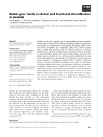
Genomic structure of human AQPap gene
Two positive BAC clones (BAC-33-J3 and BAC-6-J7) were
isolated by screening of the human BAC DNA library using
full-length human AQPap cDNA as a probe. Primer
walking and direct sequencing of the clone B AC-33-J3
revealed that it contained the entire coding sequence of
AQPap gene. The nucleotide sequences of the exon/intron
boundaries and the size of the exons and introns are shown
in Fig. 1B. The human AQPap gene contained eight exons
within 18 kb of genomic DNA with large second and third
introns (Fig. 1A). All of the exon–intron boundaries were
consistent with the GT/AG rule (Fig. 1B). The putative
translation initiation codon was located in exon 2.
Presence of multiple AQPap-like genes in human genome
DNA sequencing of another BAC clone, BAC-6-J7,
revealed that it contained the pseudogene designated
wAQPap-1 which had high ( 95%) homology of its
nucleotide sequence with AQPap (Fig. 2A). The genomic
organization o f t he wAQPap-1 gene was very similar t o that
of the genuine AQPap (Fig. 2A), and the exon–intron
boundaries were completely consistent with the GT/AG
rule. T he wAQPap-1 gene contained a termination codon in
exon 3, and an insertion of single nucleotide in exon 7
resulting in a frame shift of the coding region.
BLAST
search analysis detected two BAC clones (RP11-
251017 and RP11–15E1) containing DNA sequences
resembling the AQPap gene, which we designated
wAQPap-2 and wAQPap-3, respectively (Fig. 2A). Both
wAQPap-2 and -3 genes had 95% nucleotide sequence
similarity with AQPap. The wAQ Pap-3 gene had 99%
Table 1. Nucleotide sequences of th e sequencing primers used for
mutation analysis. The entire open reading frame (exons 2 to 8) was
amplified as three fragments by P CR as described in Mate rials and
methods section. Both strands of the PCR products containing each
exon were sequ en ced using the forward or reverse sequ encing p rimer.
Exon Direction Sequence (5¢ to 3¢)
2 Forward
CCCAAGTTCTGTGTCCTCCA
Reverse CTGAGTGCAGTTGAGTTGAAG
3 Forward ACTCAGCTGGGAGTTGAAGAG
Reverse CCAGTGCATGGTTTCATTTGAC
4 Forward GAGGAGCTAGAACTGAGCTCTGA
Reverse TTGGGGACACCTGGTCTTG
5 Forward TTGTTTGTTCTGCTCTCACTC
Reverse ACACTGAGGTCCAATCTGCCCAT
6 Forward TAACCTCATTTCTGGGACCCCGGT
Reverse TGCTGGCTCCGTCCTGAGGG
7 Forward CCGAGGTCCTGTGGCTTGGG
Reverse TGTGCTGCCCCTCACATCACC
8 Forward GGATGACTCCTCTGCTCAAC
Reverse GATGGGATCACAAATAATCTCTG
Ó FEBS 2002 Genetic analysis of human AQPap gene (Eur. J. Biochem. 269) 1817
homology with the wAQPap-1 gene, but had no frame shift
mutation, unlike wAQPap-1. wAQPap-2 gene s howed high
homology ( 98%) with the wAQPap-1 and -3 genes.
We grouped together the sets of genes similar to AQPap,
including wAQPap-1, -2 and -3, as AQPap-like genes.
Figure 2B shows genomic Southern blotting using BamHI
digested DNA and radiolabeled probe. The 0.8-kb probe
fragment of genomic DNA was obtained from AQPap
gene, and the probe region had 96% seq uence identity to
wAQPap-1, -2 and -3. From t h e b lot, AQPapand wAQ Pap-2
genes appeared to exist a s a single copy gene. BamHI
digestion was expected to produce a 7.7-kb band for
wAQPap-1 and wAQPap-3, 5.6-kb signal for wAQPap-2,
and 3.5-kb signal for AQPap, respectively. The signal
intensity of the 7.7-kb band was around threefold greater
than that of the 3.5-kb band for the AQPap gene in spite of
the lower affinity of the probe, suggesting that the 7.7-kb
band represents two or more AQPap-like genes including
wAQPap-1 and -3 (Fig. 2 B). Indeed, the amplification of
exon 7 using the specific primers for wAQPap-1 and -3
and the following direct sequencing revealed that the
human genome contained gene(s) with frame-shift muta-
tion (i.e. wAQPap-1) a nd gene(s) without the mutation
(i.e. wAQPap-3) (data not shown).
Figure 2C estimated by RT-PCR using specific primers
the mRNA amounts of the AQPap and AQPap-like genes
in various tissues. The signal for the AQPap transcript was
detected most abundantly in white fat, in w hich the
transcript signal emerged after 20 cycles of RT-PCR
(Fig. 2 C, upper panel). Twenty-five cycles of PCR also
detected trace amounts of the transcript in the testis, heart,
and kidney. On the other hand, when the primers
completely conserved in the wAQPap-1, -2 and -3 g enes
were used, no PCR products were detected in any of the
examined tissues by 25 cycles of PCR. Taken togeth er, the
expression of AQPap-like genes was, if any, extremely low,
strongly suggesting these three AQPap-like genes to be
nonfunctional pseudogenes of the genuine AQPap.
Chromosome localization of the AQPap gene
Because of the presence of the multiple homologous genes
of AQPap in the human genome, it was suspected that the
genuine AQPap gene might be localized to the other
chromosome region different from the region determined
using fluorescent in situ hybridization (FISH) method [26].
Therefore, RH mapping using the AQPap-specific primer
set was performed. The mapping revealed that AQPap gene
was localized to the marker D 9S165 with 0.0 cR
(LOD > 15) RH distance (Table 2). D9S165 has been
mapped between the markers D9S1788 and WI-5340,
both of which reside in chromosome 9p13.3-p21.1 in the
Fig. 1. Genomic structure of the human
AQPap gene. (A) G enomic structure of the
human AQPap gene was organized. S izes of
intron-2 and -3 were d etermined by P CR
amplification using primers to the flanking
region. Eight exons are represented by boxes
and numbered; solid area s indicate coding
regions. (B) Intron numbers and sizes in base
pairs are sho wn. Bou nd ary exon sequences are
capitalized. Intron sequences are shown in
lowercase l etters. The dotted line indicates the
intervening intron sequences.
Fig. 2. Multiple AQPap-like genes. (A) Restriction map of AQPap
(AL353675), wAQPap-1 (AB052627), wAQPap-2 (AL137070), a nd
wAQPap-3 genes (AL136317). Exons are represented by solid boxes
and are numbered. The BamHI restriction enzyme sites are indicated
by a capital B . The closed box repre sents the region of the probe used
for the So uthern blot analysis. (B ) Human genomic D NA (10 lg) was
completely digested with BamHI and t hen subjected to Southern blot
analysis using
32
P-radiolabeled genomic probe as d escribed in the
Materials and methods. (C) Tissue distribution o f mRNAs fo r AQPap
and AQPap-like genes. Total RNAs from indicated human tissues
were subjected to RT-PCR analysis using primers specific for AQPap
and AQPap-like, respectively, a s described in the Materials and
methods.
1818 H. Kondo et al. (Eur. J. Biochem. 269) Ó FEBS 2002
Whitehead Y AC map. Thus the AQPap gene was assigned
to chromosome 9p13.3-p21.1, close, but not identical, to the
region described previously [26]. Computer analysis re-
vealed both t he AQPap and A QP3 genes were colocalized in
a B AC clone (RP11-115015) found by a
BLAST
search,
indicating their close localization t o Chr9p13.3-p21.1.
A mapping search revealed that both RP11-251017
containing wAQPap-2 gene and RP11-15E1 containing
wAQP ap-3 gene were localizeded to Chr9p13.1, which i s a
region that is more than 50 Mb closer to the centromere
than AQPap and AQP3 gene locus.
Promoter of the AQPap gene
The 5¢ flanking region of the AQPap gene was sequenced,
and transcriptional initiation sites of the AQPap gene were
determined by 5¢ RACE using RNA isolated from human
fat tissues (Fig. 3). Several different 5¢ ends were obtained
by 5¢ RACE. RNase protection assay also revealed the
presence of many transcription start sites using the RNA
obtained from t he adipose tissues of five i ndividuals (data
not shown).
A search of the promoter region of the AQPap gene for
canonical consensus sequences revealed the p resence of
several putative binding sites f or transcription f actors
(Fig. 3). Several binding sites f or CCAAT enhancer binding
protein (C/EBP), and cAMP-regulatory element binding
protein (CRE-BP), were identified in the promoter.
An Alu repetitive sequence was detected in position
)1276 to )1509 of the promoter o f the AQPap gene. Alu
sequences were found also at the corresponding sites of
wAQPap-1, -2 and -3, respectively (data not shown). The
proximal promoter regions of the AQPap-like genes showed
high similarity ( 98% homology) with that of AQPap
downstream of the Alu sequences, whereas they quite
differed from the AQPap promoter upstream of the Alu
Table 2. Gene Bridge 4 Panel Radiation Hybrid mapping data. The
chromosomal mapping of the AQPap gene was performed using the
Gene Bridge 4 R adiation Hybrid panel and AQPap-specific primers as
described in Materials and metho ds. Results were analyz ed on the w eb
site. The results of PCR was expressed as a vector of 0’s and 1’s;
0 ¼ negative, 1 ¼ positive. The quoted LOD score is the highest,
for which the linkage between AQPap and the flanking marker is
supported.
Gene/Locus Data vector
LOD
score
Flanking
marker
AQPap 010000101100010101100010 15 D9S165
000000100101001100100100
110000011001010010010000
010010000000101000010
Fig. 3. Promoter sequence of AQPap gene.
The sequence of the human AQPap promoter
and its 5¢ flanking sequence are shown. The
nucleotide corresponding to the 5¢ end of
AQPap cDNA (5) is designated +1. Tran-
scription start sites predicted by 5 ¢ RACE are
marked by overscored filled circles. Putative
transcription factor binding sites are predicted
by the sequence m otif search pro gram,
MATINSPECTOR PROFESSIONAL
(http://www.
genomatix.de/cgi-bin/matinspector/matin
spector.pl). Three putative IREs and one
putative PPRE are boxed with solid and
broken lines, r espe ctively. T he i ntron s eque nce
is shown in lowercase letters.
Ó FEBS 2002 Genetic analysis of human AQPap gene (Eur. J. Biochem. 269) 1819
sequences, s uggesting t he evolutionar y link between the
AQPap and wAQPap genes after the Alu sequence. The
sequence dissimilarity upstream regions of the Alu sequence
might account for the differential expression levels between
the AQPap and AQPap-like genes.
PPARc-mediated induction of AQPap transcription
Inspection of the human AQPap promoter revealed a
putative PPRE of the direct repeat 1 type at )46 to )62,
which is similar to the consensus PPRE sequence (Fig. 4A)
[27–31]. To determine the function of the AQPap promoter
as well as its putative PPRE, transient reporter assays were
performed using the wild-type ()681/+11) and PPRE-
deleted constructs (Fig. 4B). These reporter plasmids were
transfected to 3 T3-L1 p readipocytes or adipocytes, and
treated with or without PGZ. The basal luciferase activity of
the wild-type construct was increased significantly when
transfected t o adipocytes, in comparison to preadipocytes.
This differentiation mediated-modulation of AQPap pro-
moter activity was totally abolished when the construct was
deprived of PPRE in the DPPRE construct. The wild-type
construct containing native )681/+11 regions showed a
sixfold increase of luciferase activity when the cells were
treated with PGZ. However, the construct lacking the
PPRE region ()62/)46) specifically from the construct
()681/+11) showed no responses to treatment with PGZ
(Fig. 4B). These results indicate tha t the P PRE site i n the
human AQPap promoter is important for a high AQPap
mRNA expression in differentiated adipose cells, and that
Fig. 4. PPAR c-mediated induction of human AQPap gene transcription through PPRE. (A) The putative PPRE sequence in the promoter region of
the human AQPap gene, compared with the classical PPRE consensus sequence. The bold (upp er case) letters de note conserved base(s) . (B) Firefly
luciferase constructs of pAQPap-P PRE wild-type, pAQPap-PPRE deleted construct, or control pGL3-basic were cotransfected with pRL-SV40
into 3T3-L1 preadipo cytes (left) o r differentiated 3T3-L1 ad ipocytes (right), and incu bated in th e medium containing P GZ (final 10 l
M
;solidbar)
or co ntrol dimethylsulfoxide (DMSO) (open bar) as d escribed in the Materials and methods. The cells were harvested for the measurement of
luciferase activity. The value of pAQPap-luciferase activity in the v ehicle (DMSO)-treated g roup was arbitrarily set as 1 .0. The n ormalized luci ferase
activities are shown as mean ± SE (n ¼ 3). An a sterisk denotes a significant difference (P < 0.01, Student’s t-test) b etween the control (DMSO)
group and the PGZ-treated group. (C) Direct an d specific binding o f PPARc/RXRa complex to the human AQPap PPRE. Electrophoretic
mobility shift assays were performed as described in the Materials and methods. The
32
P-radiolabeled PP REwt o ligonucleot ide was in cu bated with
in vitro synthesized PPARc and/or RXRa proteins. The competitive gel mobility sh ift assay was performed using
32
P-radiolabeled PPREwt as the
input probe and unlabeled oligonucleotides (PPREwt or PPREmut) as competitors at 10-, and 50-fold molar excess. ( D) Schematic illustrations of
PPARc and DPPAR c expression vecto rs, and t he effect o f DPPARc on human AQPap transcription. DPPARc construct was deprived of th e last 11
amino acids in the carboxyl terminus in the AF-2 d omain of PPARc. 3T3-L1 preadipocytes were transiently transfected with 10 ng pRL-SV40
plasmids, 1 lg pAQPap-Luciferase ()681/+11), 200 ng PPARc-, and increasing amoun ts of DPPARc- expression vectors for 4 h, and then the
medium was supplemented with or without 10 l
M
PGZ for 24 h before harvest. The total amount of DNA added was adjusted to 2.21 lgusing
empty pcDNA3.1. The value of pAQPap-luciferase activity in lane 1 was arbitrarily set as 1.0. The normalized luciferase activities are shown as
mean ± SE ( n ¼ 3).
1820 H. Kondo et al. (Eur. J. Biochem. 269) Ó FEBS 2002
the site is responsible for t he induction of AQPap
transcription by thiazolidinedione.
PPARc’s partner f or transcriptional activation is RXR a
[32]. To determine whether PPARc binds to the AQPap
PPRE as complexes with RXRa, gel mobility shift assays
were performed with double-stranded oligonucleotides
containing AQPap PPRE (Fig. 4C). The
32
P-radiolabeled
double-stranded PPREwt oligonucleotides were incubated
with in vitro translated PPARc protein (Fig. 4C). Neither
PPARc nor RXRa alone bound to AQPap PPRE (lanes 2
and 3). When PPARc and RXRa were produced together,
the mobility of
32
P-radiolabeled PPRE oligonucleotides
were shifted to a higher range, which indicated binding of
the PPARc/RXRa complex to AQPap PPRE (lane 4). The
addition of excessive unlabelled PPREwt oligonucleotides
distinguished the signal of the
32
P-radiolabeled PPREwt
oligonucleotides’ binding to PPAR c/RXRa (lanes 5 and 6).
On the other hand, addition of PPREmut oligonucleotides
containing three-base substitutions, previously reported t o
diminish the binding of PPARc to P PRE in t he promoter o f
FATP1 gene [ 29], did not affect the specific signal (lanes 7
and 8). These results indicate the specific binding of PPARc/
RXRa complex to the human AQPap PPRE.
To further confirm the PPARc-dependent enhancement
of AQPap gene expression, we generated dominant negative
PPARc expression constructs. In t he other nuclear receptor-
type transcriptional factors, the mutant construct lacking
the c arboxyl activation function-2 (AF-2) domain possessed
a dominant n egative effect on transcription of the target
genes [33,34]. We generated a mutant PPARc expression
construct, designated DPPAR c, which lacked the last 1 1
amino a cids in the AF-2 domain (Fig. 4D). The mutant
protein derived from the DPPARc construct had the ability
to bind to the AQPap PPRE oligonucleotides as a complex
with RXRa, at a similar strength to the wild-type P PARc
iprotein (data not shown). Figure 4 D demonstrates that the
expression of PPARc induced the basal luciferase activity of
the AQPap promoter in 3T3-L1 preadipocytes (lane 6 vs.
lane 1), a nd a further increase was observed following
incubation with PGZ (lane 16). These increases in promoter
activities were re duced by transfection of the DPPARc
construct, in a dose-dependent manner (lanes 6–10, and
lanes 16–20). These data a lso confirmed the specific
activation of AQPap gene transcription by PPARc.We
identified the PPRE site ()93/)77) in the promoter of the
mouse AQPap gene and observed similar findings in the
characterization of the promoter [35].
Negative IRE in the human AQPap gene promoter
Recently, we reported that mRNA expression and promoter
activities of the m ouse AQPap gene were negatively
regulated by insulin through an IRE in its promoter. In
the p romoter of the human AQPap gene, we identified three
regions identical or similar to the core negative IRE [T(G/
A)TTTT(G/T)], which were found previously in the
promoters of genes such as PEPCK [36], IGFBP-1 [36],
G6Pase [37], and IRS-2 [38] (Fig. 5A). These three core
regions were designated as IRE1, IRE2 and IRE3, respec-
tively (Fig. 5A). To determine whether there is a specific
region required for insulin-mediated repression of AQPap
transcription, deletion mutants of each IRE in the human
AQPap promoter were subcloned into luciferase vectors
(Fig. 5B). The wild-type construct contained native )681/
+147 regions having all t hree IREs, a nd showed 50%
inhibition of luciferase activity after treatment with insulin
(Fig. 5B). Constructs lacking IRE1 and IRE3 also showed
insulin-mediated suppression of luciferase activity, to a
similar degree to that of the wild-type construct ( )681/
+147). In contrast, constructs lacking IRE2 were t otally
resistant to the inhibitory effect of insulin on promoter
activities by reducing the basal promoter activity. These
results demonstrate that the IRE2 sequence ()542/)536) is
required for mediation o f the suppressing effect of insulin on
the transcription of the human AQPap gene.
We co nducted a detailed analysis of the promoter activity
between t he wild-type ()681/+147) and IRE2-deleted
mutant ()681/+147, DIRE1) (Fig. 5C). Insulin suppressed
the wild-type luciferase activity in 3 T3-L1 adipocytes, in
dose- and time-dependent fashions. In the absence of
insulin, the wild-type AQPap promoter produced a higher
luciferase activity than the deletion mutant ()681/+147,
DIRE2) promoter. In the presence of insulin, the activity of
the wild-type AQPap p romoter was reduced to the level of
the mutant promoter, which was not affected by insulin. To
further elucidate the significance of IRE2 for the insulin-
mediated repression of the human AQPap gene, we
prepared the luciferase plasmids with a single transversion
mutation in IRE2 of the human AQPap promoter ( )681/
+147). The activity of the wild-type AQPap promoter was
reduced by 51% in the presence of insulin, similar to
Fig. 5B,C. Each mutation in base pairs 2 and 3 of the
heptanucleotide sequence completely blocked the insulin-
sensitive repression of human AQPap transcription
(Fig. 5D), indicating that IRE2 was responsible for the
insulin-mediated suppression of the human AQPap tran-
scription, similar to mouse AQPap [20].
Genetic mutations of the AQPap gene in human subjects
and functional analysis of the mutant proteins
The entire coding regions of the AQPap genes were
amplified from the genomic DNA derived f rom 160
Japanese subjects, and then d irectly sequ enced. Primers
used for this analysis are shown in Table 1. Direct
sequencing revealed that the genuine AQPap gene could
be amplified without contamination of the AQPap-like
genes. We found three missense mutations (Fig. 6A): a
C fi T substitution at nucleotide 206 in exon 3 led to the
amino-acid substitution from arginine to cysteine at posi-
tion 12, which resides in the N-terminal cytoplasmic domain
(R12C); a G fi C substitution at nucleotide 347 in exon 4
caused the amino-acid substitution from valine to l eucine at
position 59, which resides in first transmembrane domain
(V59L); and a G fi T substitution at nucleotide 9 63 in exon
8 led to the amino-acid substitution from glycine to valine at
position 2 64, which resides in the sixth transmembrane
domain. The other two were G fi A substitutions at
nucleotide 480 in exon 5 and nucleotide 922 in exon 8,
neither of which caused an amino-acid conversion (A103A
and G250G, respectively). Among the 160 subjects exam-
ined, these mutations were found in one subject for R12C,
13 for V59L, eight for A103A, one for G250G, a nd six for
G264V. One subject was homozygous for G264V. The
frequency of e ach mutation were not significantly associated
with the phenotype of diabetes or obesity (Table 3).
Ó FEBS 2002 Genetic analysis of human AQPap gene (Eur. J. Biochem. 269) 1821
Next we examined the functions of these mutant AQPap
proteins. Glycerol and water permeabilities of oocytes
microinjected w ith cRNA for mutant or wild-type AQPap
were compared (Fig. 6). Two days after the injection of
50 ng of cRNA, the protein expressions of the wild-type and
each mutant were confirmed by immunoblotting (Fig. 6 B).
Under this condition, the g lycerol permeability of oocytes
expressing AQPap-R12C, and AQPap-V59L was similar to
that of wild-type AQPap (Fig. 6C). However, t he glycerol
permeability of oocytes expressing the G264V mutant was
lower, and comparable to that of c ontrol oocytes injected
with H
2
O (Fig. 6C). Furthermore, the oocytes expressing
AQPap-G264V showed much lower w ater permeability,
comparable to that of the control oocytes, whereas the water
permeabilities of oocytes expressing wild-type AQPap,
AQPap-R12C, and AQPap-V59L were similar (Fig. 6D).
As mentioned a bove, one subject (48-year-old-man)
was homozygous for the nonfunctional G264V mutation.
In the subject, BMI (23.7 kgÆm
)2
) and plasma concentra-
tions of glycerol (104 lmolÆL
)1
), glucose (4.74 mmolÆL
)1
),
total c holesterol (4.32 mmolÆL
)1
), HDL-cholesterol (1.23
mmolÆL
)1
), triglyceride (1.06 mmolÆL
)1
)werewithina
normal range. His fertility has not been disturbed because
he has three children. Figure 7 shows the changes of
plasma noradrenaline and glycerol levels after cycle
ergometer exercise in the G264V homozygous subject and
two control s ubjects. Endurance exercise on a cycle
ergometer c aused remarkable i ncrease i n t he plasma
noradrenaline level in the control and mutant subjects
(Fig. 7 B). The plasma glycerol level increased markedly in
the control subjects during t he exercise as has been
described p reviously [39] (Fig. 7 B). However, t he plasma
Fig. 5. Insulin-mediated suppression of human AQPap gene transcription through the IRE. (A) Consensus sequence of IRE and the p utative IREs of
the AQPap gene (IR E 1: )629/)623, IRE2: )542/)536, IRE3: )121/)115). (B) S chemat ic p resentation o f the plasm id c onstru cts u sed t o i de ntify t he
insulin response sequence in the promoters of the human AQPap gene. 3T3-L1 adipocytes were cotransfected with pRL-SV40 plasmid and the
indicated constructs, and in cubated in the presence (solid bar) or absence (open bar) o f insulin as described in t he Materials and methods. T welve
hours later, cells were harvested for the measurement of luciferase activity. T he value for noninsulin treated pGL 3-AQPap luciferase activity was
arbitrarily set as 1.0. The normalized luciferase activities are shown as me an ± SE (n ¼ 5). An asterisk d enotes a significant difference (P < 0.05,
Student’s t-test) between the control group and the insulin-treated g roup. (C) Dose- and time-curve of insulin-mediated inhibition of AQPap
promoter activity in the 3T3-L1 adipocytes. 3T 3-L1 ad ipocytes were cotran sfected with p RL-SV40 plasmids, and e ither pAQPap (WILD )-
Luciferase (closed circle) or pAQPap (DIRE2)-Luciferase (open circle) for 18 h, and then incubated for 12 h with the indicated concentration o f
insulin (left) or incubated with 1 l
M
insulin for t he indicated time (right). T he cells were harvested for meas urement of luciferase a ct ivities. The value
for pAQPap (WILD)-luciferase activity, in the absence of insulin (left) or 0 time of insulin incubation (right), was arbitrarily set as 1.0, respectively.
Data are expressed as mean ± SE (n ¼ 4). (D) Cells were cotransfected with 10 ng pRL-SV40, and 1.0 lgofpAQPap-Luciferase(WILD)orthe
indicated m utant plasmids i n which the indicated single base p air was substituted in t he IRE2 sequence. After incubation for 1 8 h, t he serum-free
DMEM containin g 0.5% BSA was sup plemen ted in the absence (open bar) o r p resen ce (c losed b ar) of 1 l
M
insulin for 1 2 h. The percent inhibition
of AQPap-mediated luciferase activity by insulin is shown (mean ± SE, n ¼ 5). An asterisk denotes a significant d ifferen ce (P <0.05,Student’s
t-test) between the insu lin-treated and th e nontreated group s.
1822 H. Kondo et al. (Eur. J. Biochem. 269) Ó FEBS 2002
glycerol level was not increased in a subject homozygous
for G264V mutation during exercise.
DISCUSSION
In the current study, w e demonstrated that the human
AQPap gene is composed of eight exons, which span
18 kb, and is mapped to chromosome 9p13.3–21.1.
Ishibashi et al. cloned AQP7 from human testis independ-
ently [26]. Human AQP7 had an identical sequence in the
coding region to that of human AQPap. A previous study
claimed that the human AQPap/AQP7 gene was composed
of six exons distributed over 6.5 kb [26]. In the current
analysis, we identified two additional exon–intron bound-
aries in the part of exon 1 that was originally described [26].
This discrepancy w as unlikely t o have arisen from the
existence of another AQPap-related gene, because such an
exon including exon-1, -2, and -3 was not amplified by PCR
(data not shown).
Furthermore, we identified t hree AQPap-like genes
through the course of screening the human genomic library
and the database search. T he wAQPap-1 g ene isolated from
BAC genomic library was a pseudogene, because it had an
inframe termination codon in exon 3, and a frame shift
mutation in exon 7. The two AQPap-like genes were also
identified by
BLAST
search analysis. RT-PCR a nalysis
showed that the AQPap-like genes were little, if any,
expressed in human tissues, which strongly suggested that
these three AQPap-like genes were pseudogenes.
RH mapping revealed that the AQPap gene was localized
to chromosome 9p13.3-p21.1. AQPap-like genes were found
Fig. 6. Protein expression and functional analysis of the mutant human
AQPap genes. (A) Three identified m issense mutations in the topology
of AQPap. NAA and NPS are amino-acid residues composed of NPA
motifs highly conserved a mong the AQP family proteins. (B) On day 2
after the injection of water (H
2
O) or 10 ng of cRNA encoding wild
type-AQPap (WT), AQPap-R12C ( R12C), AQPap-V59L ( V59L), an d
AQPap-G264V (G264V), total m embrane proteins were purified from
the oocytes. Immunoblotting for AQPap proteins was performed as
described in the Materials and methods section. (C) Glycerol per-
meabilities of the oocytes injected with water o r c RNAs wer e measur ed
on da y 2 after injection. Data are represen ted as mean ± SE ( n ¼ 4).
(D) Water permeability (P
f
) of the oocytes injected with water or
cRNA was measured in a standard swelling assay on day 2 after
injection. Data are expressed as mean ± SE (n ¼ 5).
Table 3. Frequency of mutation in AQPap gene. Mutation analysis AQPap gen e was performed for a total of 160 Japan ese subjects as described in
Materials and methods. Frequencies of the subjects carrying AQPap mutations were indicated.
n R12C V59L A103A G250G G264V
Non-DM, non-obese 71 0 5 2 0 3
a
DM 64 1 6 3 0 3
Obese 16 0 1 2 1 0
DM + obese 9 0 1 1 0 0
Total 160 1 13 8 1 6
a
One of the three subjects was homozygous for G264V mutation. The other mutations were identified in heterozygous form.
Fig. 7. Exercise-induced changes in plasma noradrenaline and glycerol
in the control and the AQPap-G264V homozygous subjects. (A) Sche-
matic illustration of the protocol of exercise experiment. (B) T he
plasma noradrenaline (left) and glycerol (right) levels of a subject
homozygous for G264V mutation (d)werecomparedwiththoseof
two control subjects with wild type-AQPap gene (s,n). T ime 0 rep-
resents the base-line (i.e. resting). Concentration of plasma noradren-
alineattime0was105pgÆmL
)1
in the G264V homozygote, and 195
and 227 pgÆmL
)1
in the control subjects. Concentration of plasma
glycerol at time 0 w as 104.4 lmol ÆL
)1
in the G264V hom ozygote, and
55.7 and 97.2 lmolÆL
)1
in the control subjects.
Ó FEBS 2002 Genetic analysis of human AQPap gene (Eur. J. Biochem. 269) 1823
to exist very close to the site of the genuine AQPap at
Chr9p13. Previously, Ishibashi et al. showed that the
human AQPap/AQP7 was localized at Chr9p13 using the
FISH method [26], by which it is difficult to pin point the site
ofthegeneinthechromosome.
The current study demonstrated that the promoter
activity of the human AQPap gene was increased by
PGZ, a synthetic PP ARc agonist, through the PPRE site at
)62 /)46 in the p romoter region. An identical PPRE site was
found in the promoter of the mouse A QPap [35]. The
AQPap mRNAs were increased in 3T3-L1 cells following
PPARc induction during the differentiation into adipocytes
[35]. Promoter analysis of h uman AQPap in the current
study confirmed that the abundant expression of AQPap
mRNA in the adipose tissue of humans [5] was also
attributed to the specific and abundant expression of
PPARc in human adipose tissue.
The promoter of the human AQPap gene had the site that
was negatively regulated by insulin. In our recent study, the
mRNA and the promoter activity of mouse AQPap were
suppressed by the action of insulin [20]. IRE in the human
AQPap gene promoter which was accountable for insulin-
mediated suppression of human AQPap transcription had a
heptanucleotide consensus sequence similar to those in the
promoters o f the m ouse AQPap [20], rat PEPCK [36],
mouse FATP1 [40] and human IRS-2 [38] genes. The
deletion mutant of this specific IRE decreased b asal
transcription a ctivity in the absence of i nsulin and abolished
insulin-mediated repression. Previous in vivo studies using
mice have demonstrated that AQPap mRNA increased and
decreased during fasting and refeeding, respectively, in
coordination with adipose FATP1 mRNA expression.
Physiologically coordinated regulation of the AQPap and
FATP1 genes by insulin should be efficient for supplying
glycerol and FFA in accordance with nutritional conditions.
However, in the adipose tissue of insulin-resistant animals,
AQPap and FATP1 mRNA levels were increased, despite
high concentrations of plasma insulin, leading to higher
plasma glycerol and FFA levels [19,41]. Increased influx of
glycerol and FFA into the liver enhances hepatic glucose
production and output [18,42]. Insulin-mediated suppres-
sion of th e human AQPap g ene through functional IRE
similar to mouse AQPap, suggests that human AQPap g ene
transcription is also regulated in response to nutritional
conditions through the IRE, and is augmented in insulin-
resistant states, resulting in i ncreased plasma glycerol
and hepatic glucose production in obese, i nsulin-resistant
subjects .
Among three missense mutations (R12C, V 59L, and
G264V) identified in human subjects, G264 V m utant
protein was incapable of transporting glycerol as well as
water. Murata et al. [43] documented that the conserved
GxxxGxxxG motif [44] in the third and sixth transmem-
brane domains was important for functional conformation
of the AQP family protein; glycine can be sometimes
replaced by alanine in the motif. In the sixth transmembrane
domain of human AQPap, A260, G264, and G268 formed
the motif. Functional defect in the G264V mutant might be
caused by th e disturbance of this motif.
Intense endurance exercise increases the level of p lasma
catecholamines [45]. The increase in catecholamines pro-
motes the release of glycerol and FFA, hydrolyzed from
stored triglyceride in adipose tissue [39]. Control subjects
with th e w ild-type AQPap gene show ed an increase in
plasma glycerol in parallel with the change in noradrenaline
as has been described previously [39]. However, a G264V
homozygous subject lacked the response of plasma glycerol
to exercise. These findings indicate that AQPap is respon-
sible for the exercise-induced increase in plasma glycerol in
humans. Normal adiposity and a normal plasma glycerol i n
a G264V homozygous subject, in spite of lacking functional
AQPap, suggests the existence of another pathway to
maintain plasma glycerol in the sedative condition.
From the observations of insulin resistant mice, it is
conceivable that the excessive function of AQPap m ay result
in disturbed g lucose homeostasis. Based on the g enetic
information described in this paper, a trial to identify the
mutations causing increased function or increased expres-
sion of AQPap is now under way.
ACKNOWLEDGEMENTS
We are indebted to Dr David Mangelsdorf (University of Texas S outh-
western Medical Center) for providing the mouse P PARc construct,
and Dr Makoto M akishim a for a valuable suggestion to generate t he
dominant negative PPARc construct. We thank Drs Yasuaki Fuku-
moto (Gracia Ho spital), Kazuya Yamagata, and K ikuko Hotta for
providing th e samples. We are grateful to Y uko Mats ukawa and
Sachiyo Tanaka for excellent tec hnical assistance. This work was
supported in part by the fund from the ÔResearch for the FutureÕ
Program from the Japan Society for the Promotion of Science: JSPS-
RFTF97L00801 and Grants-in-Aid from the Ministry of Education,
Science, Sports and Culture of Japan (09307019, 10557100, 10557101,
10671035, 13 671189 ).
REFERENCES
1. Motojima, K., Passilly, P., Peters, J. M., Gonzalez, F.J. & Latruffe,
N. (1998) Expression of putative fatty acid transporter genes are
regulated by p eroxisome proliferator-activated receptor a and c
activators in a tissue- and inducer-specific manner. J. Biol. Chem.
273, 16710–16714.
2. Schaffer, J.E. & Lodish, H.F. (1994) Expression cloning a nd
characterization of a novel adipocyte long chain fatty acid trans-
port protein. Cell 79, 427–436.
3. Stremmel,W.,Strohmeyer,G.,Borchard,F.,Kochwa,S.&Berk,
P.D. (1985) Isolation and partial characterization of a fatty acid
binding protein in rat liver plasma membranes. Proc. Natl Acad.
Sci. USA 82, 4–8.
4. Ibrahimi, A., Sfeir, Z., Magharaie, H., Amri, E.Z., Grimaldi, P. &
Abumrad, N.A. (1996) Expre ssion of the CD36 homolo g (FAT)
in fi broblast cells: effects on fatty acid transport. Proc. Natl Acad.
Sci. USA 93, 2646–2651.
5. Kuriyama, H., Kawamoto, S., Ishida, N., Ohno, I., Mita, S.,
Matsuzawa, Y., Matsubara, K. & Okubo, K. (1997) Molecular
cloning and expression of a novel human aquaporin from adipose
tissue with glycerol permeability. Biochem. Biophys. Res. Commun.
241, 53–58.
6. Gorin, M.B., Yancey, S.B., Cline, J., Revel, J.P. & Horwitz, J.
(1984) The major intrinsic protein (MIP) of the bovine lens fiber
membrane: characterization and structure based on cDNA clon-
ing. Cell 39, 49–59.
7. van Hoek, A.N. & Verkman, A.S. (1992) Functional reconstitu-
tion o f the i solated erythrocyte water channel CHIP28. J. Biol.
Chem. 267, 18267–18269.
8. Fushimi, K., Uchida, S., H ara, Y., H irata, Y., M arumo, F. &
Sasaki, S. (1993) Cloning and expression of apical mem-
brane wa ter channel of rat kidney collecting tub ule. Nature 361,
549–552.
1824 H. Kondo et al. (Eur. J. Biochem. 269) Ó FEBS 2002
9. Ishibashi, K., Sasaki, S ., Fushimi, K., Uchida, S., Kuwahara, M.,
Saito, H., Furukawa, T., Nak ajima, K., Yamaguchi, Y.,
Gojobori, T . & M arumo, F. (1994) Molecular c loning and
expression of a member of the aquaporin family with permeability
toglycerolandureainadditiontowaterexpressedatthebaso-
lateral membrane of kidney collecting d uct cells. Proc. Natl A cad.
Sci. USA 91, 6269–6273.
10. Jung, J.S., Bhat, R.V., Preston, G.M., Guggino, W.B., Baraban,
J.M. & Agre, P. (1994) Molecular characterization of an aqua-
porin cDNA f ro m brain: c and idate osm oreceptor and regulator of
water balance. Proc.NatlAcad.Sci.USA91, 13052–13056.
11. Raina, S., Preston, G.M., Guggino, W.B. & Agre, P. (1995)
Molecular cloning and characterization of a n aquaporin cDNA
from salivary, lacrimal, and respiratory tissues. J. Biol. Chem. 270,
1908–1912.
12. Ma, T., Frigeri, A., Skach, W. & Verkman, A.S. (1993) Cloning
of a novel rat kidney c DNA homologous to CHIP28 and
WCH-CD water channels. Biochem. Biophys. Res. C ommun. 197,
654–659.
13. Ishibashi, K., Kuwahara, M., Gu, Y., Kageyama, Y., Tohsaka,
A., Suzuki, F., Marumo, F. & Sasaki, S. (1997) Cloning and
functional expression of a new water channel abundan tly
expressed in the testis permeable to water, glycerol, and urea.
J. Biol. Chem. 272, 20782–20786.
14. Koyama, Y., Y amamoto, T., Kondo, D., Funaki, H., Yaoita, E.,
Kawasaki, K., Sato, N., Hatakeyama, K. & Kihara, I. (1997)
Molecular cloning of a new aquaporin from rat pancreas and liver.
J. Biol. Chem. 272, 30329–30333.
15. Ishibashi, K., Kuwahara, M ., Kageyama, Y., T ohsaka, A.,
Marumo, F. & Sasaki, S. (1997) Cloning and functional expres-
sion of a second new aquaporin abun dantly expressed in t estis.
Biochem. Biophys. Res. Commun. 237, 714–718.
16. Tsukaguchi, H., Shayakul, C., Berger, U.V., Mackenzie, B.,
Devidas, S., Guggino, W.B., van H oek, A.N. & Hediger, M.A.
(1998) Mole cular characterization of a broad selectivity neutral
solute channel. J. Biol. Chem. 273, 24737–24743.
17. Hatakeyama, S., Yoshida, Y., Tani, T., Koyama, Y., Nihei, K.,
Ohshiro,K.,Kamiie,J.I.,Yaoita,E.,Suda,T.,Hatakeyama,K.&
Yamamoto, T. (2001) Cloning of a new a quaporin (aqp10)
abundantly expressed in duodenum and jejunum. Biochem. Bio-
phys. Res. Commun. 287, 814–819.
18. Peroni, O., Large, V. & Beylot, M. (1995) Measuring gluconeo-
genesis with [2-
13
C]glycerol and mass isotopomer distribution
analysis of glucose. Am. J. Physiol. 269, E516–E523.
19. Kishida, K., Kuriyama, H., Funahashi, T., Shimomura, I.,
Kihara, S., Ouchi, N., Nishida, M., Nishizawa, H., Matsuda, M.,
Takahashi, M. et al. (2000) Aquaporin a dipose: a p utative g lycerol
channel in adipocytes. J. Biol. Chem. 275, 20896–20902.
20. Kishida, K., Shim omura, I., Kondo, H., Kuriyama, H., Makino,
Y.,Nishizawa,H.,Maeda,N.,Matsuda,M.,Ouchi,N.,Kihara,
S. et al. (2001) Ge nomic structure and insulin-mediated repression
of the a quaporin adipose ( AQPap), a dipose-specific glycerol
channel. J. Biol. Chem. 276, 36251–36260.
21. Sambrook, J ., Fritsch, E.F. & Maniatis, T . (1989) Molecular
Cloning: a Laboratory Manual, 2nd edn. Cold Spring Harbor
Laboratory Press, Cold Spring Harbor, New York.
22. Chomczynski, P. & Sacci, N. (1987) Single-step method of RNA
isolation by ac id g uanidinium thioc yan ate-phenol-chloro form
extraction. An al. Biochem . 162, 156–159.
23. Rubin, C.S., Hirsch, A., Fung, C. & Rosen, O.M. (1978) Devel-
opment of hormone receptors and hormo nal r esponsive ness
in vitro. Insulin receptors and insulin sensitivity in the pre-
adipocyte and adipocyte forms of 3T3-L1 cells. J. Biol. Chem. 253,
7570–7578.
24. Zhang, R. & Verkman, A.S. (1991) Water and urea permeability
properties of Xenopus oocytes: expression of mRNA from toad
urinary bladder. Am. J. Physiol. 260, C26–C34.
25. Winartasaputra, H., Mallet, V.N., Kuan, S.S. & Guilbault, G.G.
(1980) Fluoro metric and colorimetric e nzymic determination of
triglycerides (triacylglycerols) in serum. Clin. Chem. 26, 613–617.
26. Ishibashi, K., Yamauchi, K., Kageyama, Y., Saito-Ohara, F.,
Ikeuchi, T., Marumo, F. & Sasaki, S. (1998) Molecular char-
acterization of human aquaporin-7 gene and its chromosomal
mapping. Biochim. Biophys. Acta. 1399, 62–66.
27. Barak, Y., Nelson, M.C., Ong, E.S., Jones, Y.Z., Ruiz-Lozano, P.,
Chien, K.R., Koder, A. & Evans, R.M. (1999) PPARPPc is
required f or placental, c ardiac, and adipose tissue development.
Mol. Cell 4, 585–595.
28. Kubota,N.,Terauchi,Y.,Miki,H.,Tamemoto,H.,Yamauchi,
T., Komeda, K., Satoh, S., Nakano, R., Ishii, C., Sugiyama, T.
et al. (1999) PPARc mediates high-fat d iet-induced adipocyte
hypertrophy and insulin resistance. Mol. Cell 4, 597–609.
29. Tontonoz, P., H u, E., Graves, R.A., B ud avari, A.I. & Spiegelman,
B.M. (1994) mPPARc 2: tissu e-specific regulator of an adipo cyte
enhancer. Genes Dev. 8, 1224–1234.
30. Frohnert, B.I., Hui, T.Y. & Bernlohr, D.A. (1999) Identification
of a functional peroxisome proliferator-responsive element in the
murine fatty acid transp ort p ro tein gene. J. Biol . C hem. 27 4, 3970–
3977.
31. Ranganathan, S. & Kern, P.A. (1998) Thiazolidinediones inhibit
lipoprotein lipase activity in adipocytes. J. Biol. Chem. 273, 26117–
26122.
32. Mangelsdorf, D.J. & Evans, R.M. (1995) The RXR heterodimers
andorphanreceptors.Cell 83, 841–850.
33. Schulman, I.G., Shao, G . & Heyman, R.A. (1998 ) T ransactivation
by retinoid X receptor-peroxisome proliferator-activated receptor
c (PPARc) heterodimers: intermolecular synergy requires only the
PPARc hormone-dependent activation function. Mol. Cell. Biol.
18, 3483–3494.
34. Ananthanarayanan, M., Balasubramanian, N., Makishima, M.,
Magelsdorf, D.J. & Suchy, F.J. (2001) Human bile salt export
pump promoter is transactivated by the farnesoid X receptor/bile
acid receptor. J. Biol. Chem. 276, 28857–28865.
35. Kishida, K., Shimomura, I., Nishizawa, H., Maeda, N., Kuri-
yama, H., Kondo, H., Matsuda, M., Nagaretani, H., Ouchi, N.,
Hotta, K. et al. (2002) Enhancement of t he aquaporin adipose
(AQPap) gene expression by a peroxisome proliferator-activated
receptor c. J. Biol. Chem., in press.
36. Hall, R.K., Yamasaki, T., Kucera, T., Waltner-Law, M., O’Brien,
R. & Granner, D.K. (2000) Regulation of phosphoenolpyruvate
carboxykinase and insuline-like growth factor-binding protein-1
gene expression by insuline: The role of winged helix/forkhead
proteins. J. Biol. Chem. 275, 30169–30175.
37. Schmoll,D.,Walker,K.S.,Alessi,D.R.,Grempler,R.,Burchell,
A., Gu o, S., Walther, R. & U nterman, T.G. (2000) Regulation of
glucose-6-phosphatase gene expression by protein kinase Ba and
the forkhead transcription factor FKHR. Evidence for insulin
response unit-dependen t and -independent effects of insulin on
promoter activity. J. Biol. Chem. 275, 36324–36333.
38. Zhang, J., Ou, J., Bashmakov, Y., Horton, J.D., Brown, M.S. &
Goldstein, J.L. (2001) Insulin inhibits transcription of IRS-2 gene
in rat liver through an insulin response element (IRE) that
resembles IREs of other insulin-repressed genes. Proc. Natl Acad.
Sci. USA 98, 3756–3761.
39. Arner, P., Kriegholm, E., Engfeldt, P. & Bolinder, J. (1990)
Adrenergic regulation of lipolysis in situ at rest and during
exercise. J. Clin. Invest. 85, 893–898.
40. Hui, T.Y., Frohnert, B.I., Smith, A.J., Schaffer, J.E. & Bernlohr,
D.A. (1998) Characterization of the murine fatty acid transport
protein gene and its insu lin response sequence. J. Biol. Chem. 273,
27420–27429.
41. Berk, P.D., Zhou, S.L., Kiang, C.L., Stump, D., Bradbury, M. &
Isola, L.M. (1997) Uptake of long chain free fatty acids is selec-
tively up-regulated in adipocytes of Zucker rats with genetic
Ó FEBS 2002 Genetic analysis of human AQPap gene (Eur. J. Biochem. 269) 1825
obesity and n on-insulin-dependent diabetes mellitus. J. Biol.
Chem. 272, 8830–8835.
42. Rebrin, K., Ste il, G.M., Mittelman, S.D. & Bergman, R.N. (1996)
Causal lin kage betwee n insulin suppressio n of lipolysis and sup-
pression of liver glucose output in dogs. J. Clin. Invest. 98,741–
749.
43. Murata, K., Mitsuoka, K., Hirai, T., Walz, T., Agre, P.,
Heymann, J.B., Engel, A. & Fujiyoshi, Y. (2000) Structural
determinants of water permeat ion through aquaporin-1. Nature
407, 599–605.
44. Heymann, J.B. & Engel, A. (2000) Structural clues in the
sequences of t he aquaporins. J. Mol. Biol. 295, 1039–1053.
45. MacLaren, D.P.M., Reilly, T., Campbell, I.T. & Frayn, K.N.
(1994) Hormonal and metabolite responses to glucose and mal-
todextrin i ngestion with or without the addition of guar gum. Int.
J. Sports Me d. 15, 466–470.
1826 H. Kondo et al. (Eur. J. Biochem. 269) Ó FEBS 2002