Báo cáo khoa học: In vitro and in vivo self-cleavage of Streptococcus pneumoniae signal peptidase I pot
Bạn đang xem bản rút gọn của tài liệu. Xem và tải ngay bản đầy đủ của tài liệu tại đây (269.33 KB, 9 trang )
In vitro
and
in vivo
self-cleavage of
Streptococcus pneumoniae
signal peptidase I
Feng Zheng, Eddie L. Angleton, Jin Lu and Sheng-Bin Peng
Infectious Diseases Research, Lilly Research Laboratories, Indianapolis, IN, USA
We have previously demonstrated that Streptococcus
pneumoniae signal peptidase (SPase) I catalyzes a self-
cleavage to result in a truncated product, SPase37–204
[Peng, S.B., Wang, L., Moomaw, J., Peery, R.B., Sun,
P.M., Johnson, R.B., Lu, J., Treadway, P., Skatrud, P.L.
& Wang, Q.M. (2001) J. Bacteriol. 183, 621–627]. In this
study, we investigated the effect of phospholipid on in vitro
self-cleavage of S. pneumoniae SPase I. In the presence of
phospholipid, the self-cleavage predominantly occurred at
one cleavage site between Gly36–His37, whereas the self-
cleavage occurred at multiple sites in the absence of
phospholipid, and two additional self-cleavage sites,
Ala65–His66 and Ala143–Phe144, were identified. All
three self-cleavage sites strongly resemble the signal pep-
tide cleavage site and follow the ()1, )3) rule for SPase I
recognition. Kinetic analysis demonstrated that self-cleav-
age is a concentration dependent and intermolecular
event, and the activity in the presence of phospholipid is
25-fold higher than that in the absence of phospholipid.
Biochemical analysis demonstrated that SPase37–204, the
major product of the self-cleavage totally lost activity to
cleave its substrates, indicating that the self-cleavage
resulted in the inactivation of the enzyme. More impor-
tantly, the self-cleavage was demonstrated to be happening
in vivo in all the growth phases of S. pneumoniae cells. The
bacterial cells keep the active SPase I at the highest level
in exponential growth phase, suggesting that the self-
cleavage may play an important role in regulating the
activity of the enzyme under different conditions.
Keywords: Streptococcus pneumoniae; signal peptidase I; self-
cleavage; inactivation; regulation.
Many secreted and membrane proteins of both prokaryotic
and eukaryotic cells are initially synthesized as a precursor
(or preprotein) with an N-terminal extension known as a
signal (or leader) peptide. This signal sequence is involved in
guiding the protein into the targeting and translocating
pathway by interacting with the membrane and other
components of the cellular secretory machinery [1]. The
signal peptides of secreted proteins are normally removed by
signal peptidase (SPase) that spans in the cytoplasmic
membrane in bacteria after the proteins have been translo-
cated across the membrane. Two major bacterial SPases,
SPase I and SPase II, with different cleavage specificity,
have been identified. SPase I is responsible for processing
majority of the secreted proteins [2,3], whereas SPase II
exclusively processes glyceride-modified lipoproteins [4].
There is no sequence similarity and substrate overlap
between these two types of SPases.
In bacteria, the majority of protein translocation
occurs post-translationally via the Sec system [5,6]. The
Sec system is composed of multiple proteins SecA, SecB,
SecD, SecE, SecF, SecG and SecY. In Escherichia coli,
the homotetramer SecB, a chaperone protein, interacts
with the newly synthesized precursor and targets the
protein to the SecAYEG translocase at the cytoplasmic
membrane surface. The secretory precursor then interacts
with the membrane-associated homodimer SecA, which
contains an ATP binding domain and utilizes the energy
from ATP hydrolysis to translocate the precursor through
the membrane protein channel thought to be formed
from components SecYEG. SPase I is a membrane-
bound endopeptidase that presumably is localized in close
proximity to Sec YEG. it is typically anchored to the
cytoplasmic membrane by one transmembrane segment in
most of the gram-positive enzymes, or two transmem-
brane segments in most of the gram-negative enzymes in
the N-terminus. Topological analysis demonstrated that
the C-terminal catalytic domain of E. coli SPase I resides
on the outer surface of the cytoplasmic membrane, and is
thus localized in the periplasm of the cells [7–9]. SPase I
functions to cleave away the signal peptides from the
translocated precursors, thereby releasing the mature
proteins from the membrane and allowing them to their
final destinations in the periplasm, outer membrane, or
extracellular milieu. Inhibition of SPase I leads to the
accumulation of secretory precursors in the cell mem-
brane and eventual cell death [10–13]. Therefore, SPase I
is an essential component for bacterial growth, and a
potential target for development of novel antibacterial
agents.
Proteases in general are divided into four classes accord-
ing to their mechanism of action, they are serine, cysteine,
metallo- and aspartyl proteases. However, recent investiga-
tions have unambiguously demonstrated that SPase I is not
a member of any of these four traditional classes, it is not
Correspondence to S. B. Peng, Lilly Research Laboratories, Lilly
Corporate Center, Indianapolis, IN 46285, USA.
Fax: + 1317 2769086, Tel.: + 1317 4334549,
E-mail:
Abbreviations:SPase,signalpeptidase;IPTG,isopropyl-
b-thiogalactopyranoside; BHI, brain heart infusion.
(Received 8 April 2002, revised 20 June 2002, accepted 28 June 2002)
Eur. J. Biochem. 269, 3969–3977 (2002) Ó FEBS 2002 doi:10.1046/j.1432-1033.2002.03083.x
sensitive to any of the standard protease inhibitors [2,14].
The catalytic mechanism of the bacterial SPase I has been
studied by site-directed mutagenesis using E. coli enzyme
[15,16], Bacillus subtilis SipS [17] and S. pneumoniae enzyme
[18]. In these cases, a conserved serine and a conserved lysine
were identified to be critical for enzymatic activity. These
results suggest that these enzymes belong to a novel class of
serine proteases that utilize a serine and a lysine to form a
catalytic dyad. Therefore, this class of protease is a unique
serine protease that does not utilize a histidine as a catalytic
base, but may instead employ a lysine side chain to fulfill
this role [15,16,19]. This serine–lysine catalytic dyad struc-
ture has been recently confirmed by structural analysis in
E. coli SPase I [20].
A precedent for a mechanism involving a serine–lysine
dyad for a peptidase has been previously reported [21–23].
The LexA protein, which is involved in the SOS response in
E. coli, undergoes a specific self-cleavage reaction that
inactivates the protein. The self-cleavage of LexA protein is
important in the SOS response in bacteria [21–23]. In vitro
self-cleavage was also observed in all investigated bacterial
SPase I including enzymes from E. coli [24], B. subtilis [25]
and S. pneumoniae [18]. In E. coli, the self-cleavage of
SPase I occurred in a hydrophilic domain connecting the
two transmembrane segments at the N-terminus [24]. In
B. subtilis, the self-cleavage of SPase I (SipS) occurred
immediately after the active residue serine [25]. In S. pneu-
moniae, the self-cleavage site was identified between Gly36
and His37, one residue away from the active residue Ser38
[18]. Although in vitro self-cleavage is common in all
investigated bacterial SPase I, the further studies on this
basic biochemical property of the enzyme are very limited so
far.
We have previously demonstrated that S. pneumoniae
SPase I catalyzes a self-cleavage reaction in the presence
of phospholipid. After self-cleavage, the N-terminal 36
residues are removed from the protein, the major
product (SPase37–204) consisting of amino acids
37–204, contains the two critical residues, Ser38 and
Lys76, required for the formation of the catalytic dyad.
However, we do not know if the self-cleavage is
activating or inactivating the enzyme, and if the self-
cleavage is happening in vivo within the bacterial cells.
In the current study, we have shown that phospholipid
enhances the activity of self-cleavage, and self-cleavage
results in the inactivation of the enzyme. More impor-
tantly, we have demonstrated for the first time that
the self-cleavage of SPase I is happening within the
S. pneumoniae cells in all the growth phases, and the
cells maintain the active SPase I at the highest level in
the exponential growth phase.
MATERIALS AND METHODS
Materials and bacterial strains
Restriction enzymes, T4 DNA ligase and Taq DNA
polymerase were purchased from Life Technologies, BRL
Inc. The peptide substrate (KLTFGTVKPVQAIA
GYEWL) was developed and synthesized based upon the
signal peptide of prestreptokinase of S. pyogenes,as
described previously [26,27]. CM- and DEAE-Sepharose
and an ECL kit for Western blot analysis were obtained
from Amershan-Pharmacia. Ni-nitrilotriacetic acid agarose
was from Qiagen. E. coli lipid extract and pure phospho-
lipids were purchased from Avanti Polar Lipid, Inc.
Trifluoroacetic acid, EDTA, and acetonitrile were pur-
chased from Fisher Scientific. Chemicals for SDS/PAGE
were from Nova. All HPLC measurements were performed
on a HP 1100 system using C18 reversed-phase column.
E. coli strain Bl21(DE3)pLysS was from Novagen, and
S. pneumoniae R6 strain was from American Type Culture
Collection.
Purification of
S. pneumoniae
SPase I
The expression vector, pET16b-spi, that directs the
synthesis of the full length S. pneumoniae SPase I, was
constructed and transformed into E. coli strain
BL21(DE3) pLysS. The transformed E. coli cells were
grown and induced with IPTG for protein expression at
30 °C. The overexpressed protein was purified as
described previously [18]. Typically, 1 L of IPTG-induced
E. coli cells were lysed by sonication in 20 mL of lysis
buffer containing 300 m
M
NaCl and 50 m
M
Na
2
HPO
4
(pH 8.0). The lysate was then centrifuged at 50 000 g for
1hat4°C. The resultant supernatant was discarded, and
the pellet was resuspended and sonicated in 20 mL of lysis
buffer with 1% Triton X-100. After centrifugation at
50 000 g for 1 h, the supernatant was diluted with 80 mL
of lysis buffer, and loaded to a 2 mL of Ni-nitrilotriacetic
acid column, that was then washed with 50 mL of lysis
buffer with 0.1% Triton X-100 and 15 m
M
imidazole.
Finally, the protein was eluted with 10 mL of elution
buffer containing 20 m
M
Tris/HCl (pH 8.0), 20% glycerol,
0.1% Triton X-100 and 100 m
M
imidazole. The purity of
the protein was analyzed by SDS/PAGE, and selected
fractions were utilized for enzyme assays.
Purification of truncated SPase37–204 from
self-cleaved SPase I
Purified SPase I (5 mg) was incubated for 2 h at 37 °Cin
5 mL of reaction buffer containing 20 m
M
Tris/HCl
(pH 8.0), 100 m
M
imidazole, 20% glycerol, 0.1% Triton
X-100 and 1 lgÆlL
)1
of E. coli lipid extract. The reaction
mixture was diluted to 50 mL with 20 m
M
Tris/HCl
(pH 8.0), and loaded into a 2 mL of Ni-nitrilotriacetic
acid agarose column. The pass flow from Ni-nitrilotriace-
tic acid agarose column was loaded to a 1 mL of
CM-Sepharose column, and subsequently to a 1 mL of
DEAE-Sepharose column that was preequilibrated with
buffer A consisting of 20 m
M
Tris/HCl (pH 8.0), 20%
glycerol. After washing the DEAE-Sepharose column with
5 mL of buffer A, the protein was eluted with 10 mL of
0–400 m
M
NaCl gradient prepared in buffer A, 1 mL of
fractions were collected, and selected fractions were
utilized for functional assay.
Overexpression and purification of truncated
S. pneumoniae
SPase37–204
To develop a simpler method to purify S. pneumoniae
SPase37–204, we constructed an expression vector,
pET23b-spiD1-36, that directs the expression of SPase I
lacking the N-terminal 36 amino acids with a C-terminal
3970 F. Zheng et al. (Eur. J. Biochem. 269) Ó FEBS 2002
histidine tag. Briefly, the encoding region for residues
37–204 of S. pneumoniae SPase I was amplified by PCR
using genomic DNA as the template and two oligonucleo-
tides as primers (5¢-GCAATGTTCGCGTACATATGCA
TTCCATGGATCCGACC-3¢ and 5¢-TGGTGGTGCTC
GAGAAATGTTCCGATACGGGTGATTGGCCAGA
AGCG-3¢), which were designed to contain NdeIandXhoI
restriction sites at the 5¢ ends, respectively. The primers were
synthesized in accordance with the published sequence of
S. pneumoniae SPase I [28]. The PCR product was puri-
fied and cloned into the NdeIandXhoI sites of vector
pET23b, resulting in pET23b-spiD1-36. The identity of the
cloned gene was confirmed by DNA sequencing. For
expression of SPase37–204, E. coli strain BL21(DE3)-
pLysS was transformed with pET23b-spiD1-36, grown
and induced with 0.4 m
M
IPTG at 30 °C, as described
previously [29]. For purification, 1 L of the IPTG-induced
E. coli cells were harvested by centrifugation, resuspended
in 40 mL of lysis buffer, and sonicated for 5 min on ice.
The lysate was then centrifuged at 50 000 g for 1 h. The
resultant supernatant was loaded onto a 2 mL preequil-
ibrated Ni-nitrilotriacetic acid column, which was then
washed with 50 mL of lysis buffer with 15 m
M
imidazole.
The protein was finally eluted out with 10 mL of elution
buffer, and 1 mL of fractions were collected and analyzed
by SDS/PAGE.
In vitro
self-cleavage of
S. pneumoniae
SPase I
For in vitro self-cleavage of S. pneumoniae SPase I in the
presence of phospholipid, 20 lL of reaction containing
5 lg of purified SPase I was incubated in 37 °Cfor2hin
20m
M
Tris/HCl (pH 8.0), 0.05% Triton X-100, 10%
glycerol, and 50 lgofE. coli lipid extract. For self-
cleavage in the absence of phospholipid, 20 lL of reaction
containing 5 lg of purified SPase I was incubated at
37 °C in the same buffer without phospholipid. Typically,
the reactions were terminated by the addition of SDS
sample buffer, and separated on a 4–20% SDS/poly-
acrylamide gel; the gel was then stained with Coomassie
Brilliant Blue. For kinetic analysis of self-cleavage, reac-
tions (20 lL) containing different concentrations of
SPase I were incubated at 37 °Cfor30mininthe
presence of 50 lg phospholipid or 4 h in the absence of
phospholipid. Densitometer analysis was performed using
a Personal Densitometer SI and
IMAGE QUANT
5.0 soft-
ware from Molecular Dynamics. Specific activities of self-
cleavage in the presence or absence of phospholipid were
calculated according to the cleavage of SPase I at
concentration of 1 mgÆmL
)1
.
N-Terminal peptide sequencing
To determine the self-cleavage sites of S. pneumoniae
SPase I, the proteolytic products of the self-cleavage were
fractionated on a 4–20% SDS/polyacrylamide gel, and
transferred to a poly(vinylidene difluoride) (PVDF) mem-
brane by electroblotting. The membrane was then briefly
stained by Commassie bright blue and destained by 50%
methanol. The visualized protein bands on the membrane
were excised and the N-terminal amino acid sequence
of each protein was determined by automated Edman
degradation.
Cleavage of prestreptokinase by
S. pneumoniae
SPase I and SPase37–204
We have previously demonstrated that prestreptokinase is a
native substrate of S. pneumoniae SPase I. The gene
encoding S. pyogenes prestreptokinase was amplified by
PCR based upon the published sequence [26], and cloned
into the expression vector pET23b to result in pET23b-ska.
For the expression of the prestreptokinase, E. coli strain
BL21(DE3)pLysS was transformed with pET23b-ska,
grown and induced by IPTG. The overexpressed prestrep-
tokinase was then solubilized with 1% Zwittergent 3–16,
and purified with Ni-nitrilotriacetic acid column, as
described previously [18]. For the cleavage of prestrepto-
kinase, typically, reactions (20 lL) containing 0.1 lgSPase I
or SPase37–204 were incubated with 5 lg of purified
prestreptokinase at 37 °C for 1 h in the buffer containing
20m
M
Tris/HCl (pH 8.0), 0.02% Triton X-100, 5%
glycerol and 50 lgofE. coli total lipid extract. The reactions
were then terminated by the addition of SDS sample buffer,
and the proteins were separated on a 4–20% SDS/poly-
acrylamide gel, and stained by Coomassie Brilliant Blue.
Cleavage of a peptide substrate by
S. pneumoniae
SPase I and SPase37–204
A peptide substrate, KLTFGTVKPVQAIAGYEWL was
developed and synthesized based upon the signal peptide
of the prestreptokinase of S. pyogene [26,27]. Typically,
cleavage reactions were performed in 50 lL reaction
mixtures containing 20 m
M
Tris/HCl (pH 8.0), 50 lgof
E.coli lipid extract, 0.1 l
M
SPase I and 100 l
M
of the
peptide substrates. Reactions were incubated at 37 °Cfor
2 h and terminated by the addition of an equal volume of
8
M
urea. Cleavage of the peptide substrate was deter-
minedbyHPLCusingaHewlettPackardSeries1100
system equipped with an autosampler. The reaction
mixtures were injected into a reversed-phase column
(Vydac C18) and the fragments were separated using a
0–67% linear gradient of buffer B in buffer A (buffer
A ¼ 0.1% trifluoroacetic acid in water, buffer B ¼ 90%
acetonitrile and 0.1% trifluoroacetic acid) with a flow rate
of 1 mLÆmin
)1
. Peak detection was accomplished by
monitoring the absorbance at 214 nm.
Preparation of a polyclonal antibody against
S. pneumoniae
SPase I
Two S. pneumoniae SPase I-specific peptides, SP-Ab1
(CHEEDGNKDIVKRVIG) and SP-Ab2 (CLADYIK
RFKDDKLQS), were synthesized based upon the deduced
amino acid sequence [28]. A cysteine was artificially added
to the N-terminus of each peptide to increase the coupling
efficiency. After purification, the synthetic peptides were
coupled to keyhole limpet hemocyanin, and utilized for
immunization of New Zealand white rabbits to generate
polyclonal antibodies, as described previously [30].
Detection of
in vivo
self-cleavage of
S. pneumoniae
SPase I by Western blot analysis
Fresh S. pneumoniae cells were prepared by growing the
cells in 5 mL of brain–heart infusion (BHI) broth in a
Ó FEBS 2002 Self-cleavage of S. pneumoniae SPase I (Eur. J. Biochem. 269) 3971
series of dilution at 37 °Cwith5%CO
2
overnight. The
culture with D
620
< 0.3 was carefully centrifuged, washed
once with fresh BHI broth. The cells were then
resuspended in fresh BHI broth with an initial
D
620
¼ 0.075, and grown at 37 °Cand5%CO
2
for a
period up to 5 h. 2 mL of culture was harvested at
different time points by centrifugation and immediately
lysed with a solution containing 1· SDS sample buffer,
B-PER II bacterial extraction reagent from Pierce and a
protease inhibitor cocktail from Roche to protect proteins
from nonspecific proteolysis. After boiling for 10 min, the
samples (20 lg of total protein each) were separated on a
4–20% SDS/polyacrylamide gel and transferred electroph-
oretically to a PVDF membrane. Immunodetection was
performed using immune serum against S. pneumoniae
SPase I at 1 : 2000 dilution or a polyclonal antibody
against S. pneumoniae Eraat1:1000dilution.Densitom-
eter analysis was performed using a Personal Densitometer
S1 and
IMAGE QUANT
5.0 software. The ratio of the
full length SPase/cleaved SPase was calculated based upon
the relative intensity of the two protein bands from
Western blot analysis.
RESULTS
In vitro
self-cleavage of
S. pneumoniae
SPase I
We have previously reported that S. pneumoniae SPase I
catalyzes a specific self-cleavage reaction in vitro.A
similar self-cleavage reaction was also observed in
purified E. coli SPase I [24]. The self-cleavage of E. coli
SPase I was initially speculated to be protected within the
bacterial cells by the interaction between the enzyme and
the cytoplasmic membrane. In this study, we were
interested in investigating the effect of phospholipid on
the self-cleavage of S. pneumoniae SPase I. In the pres-
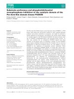
ence of phospholipid, the self-cleavage occurred predom-
inantly at one cleavage site to produce two protein
bands, b1 and b4, as demonstrated in Fig. 1A; no other
cleavage site was observed. In the absence of phosphol-
ipid, the highly purified S. pneumoniae SPase I, when
incubated at 37 °C, also resulted in the self-cleavage of
the enzyme. Interestingly, the self-cleavage was somewhat
different, it occurred at multiple sites and resulted in at
least five identifiable protein bands, b1, b2, b3, b4, and
b5 with molecular masses ranging from 8 to 19 kDa, as
shown in Fig. 1B. To confirm the specificity of the self-
cleavage, we also tested the self-cleavage of two SPase I
mutants, S38A and K76A that lost their activity to
catalyze substrate cleavage as described previously [18].
Results demonstrated that the purified S38A and K76A
were unable to catalyze self-cleavage in the presence or
absence of phospholipid, confirming that the self-cleavage
in both conditions was specific and not due to the
possibly contaminating proteases (data not shown).
Additionally, all the major protein bands from the self-
cleavage of SPase I were separated, excised and subjected
for N-terminal peptide sequencing. The sequences
obtained from five cleaved protein bands were summa-
rized in Table 1. In the absence of phospholipid, three
self-cleavage sites, Gly36–His37, Ala65–His66, and
Ala143–Phe144 were identified as indicated in Fig. 1C.
In the presence of phospholipid, only one self-cleavage
site (Gly36–His37) was identified. The peptide sequence
GHHHHHHHHHHSSG from products b2 and b4
was the histidine tag fused to the N-terminus of the
enzyme.
Kinetics of SPase I self-cleavage
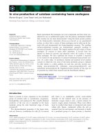
Kinetic analysis demonstrated that self-cleavage was a
protein concentration dependent event. Titration experi-
ments revealed that the specific activities of self-cleavage in
the presence of phospholipid were increasing when SPase I
concentrations were increased (Fig. 2A). A similar protein
concentration-dependent self-cleavage of SPase I was also
observed in the absence of phospholipid (Fig. 2B). It
suggests that self-cleavage of SPase I is catalyzed through
an intermolecular mechanism. The activities of self-cleav-
age in the presence or absence of phospholipid were
calculated to be 0.025 or 0.001 min
)1
, respectively, at
SPase I concentration of 1 mgÆmL
)1
,anda25-fold
Fig. 1. Self-cleavage of S. pneumoniae
SPase I. Reactions (20 lL) containing 5 lgof
wild type SPase I were incubated at 37 °Cin
the presence (A) or absence (B) of phospholi-
pid. The samples were separated on 4–20%
SDS/polyacrylamide gels, and stained with
Coomassie Brilliant Blue. Lane 1, purified full
length SPase I before incubation; lane 2, full
length SPase I after incubation. Protein bands
corresponding to degradation products of
S. pneumoniae SPase I were indicated as b1,
b2, b3, b4 and b5. (C) Amino acid sequence of
S. pneumoniae SPase I. The self-cleavage sites,
identified by automated Edman degradation,
are marked with arrows. The peptides utilized
for antibody preparation are underlined.
3972 F. Zheng et al. (Eur. J. Biochem. 269) Ó FEBS 2002
stimulation by phospholipid was observed. Therefore,
phospholipid greatly stimulates the self-cleavage of
SPase I.
Self-cleavage sites of
S. pneumoniae
SPase I resemble
signal peptide cleavage sites
Although signal peptides of secreted proteins do not
show a great deal of sequence identity, they do share
some common structural properties. Statistical analysis of
the amino acid sequences surrounding signal peptide
cleavage sites has led to the so called ()1, )3) rule that
states that the residues at the )1and)3 positions relative
to the SPase I cleavage site must be small and neutral
residues [31–33]. The residues at )1 position are usually
Ala, Gly, and Ser, and at )3 position are usually Ala,
Val, Gly, Ser, and Thr. Sequence analysis around the
three self-cleavage sites identified from S. pneumoniae
SPase I revealed that all these self-cleavage sites strongly
resemble the signal peptide cleavage sites, and follow the
()1, )3) rule well with the most common alanine or
glycine in the )1 position, and an alanine or a valine at
)3 position as demonstrated in Fig. 3. This result further
supports that the self-cleavage of SPase I with or without
phospholipid is specific and not caused by possibly
contaminating proteases from the purification or by
careless handling of the protein. Similarly, the self-
cleavage sites identified from E. coli and B. subtilis SPases
also follow the ()1, )3) rule for signal peptidase
recognition as aligned in Fig. 3.
Purification of truncated
S. pneumoniae
SPase37–204
from self-cleaved products and overexpressed
E. coli
cells
We have demonstrated that S. pneumoniae SPase I pre-
dominantly catalyzes self-cleavage at one cleavage site
between Gly36 and His37 in the presence of phospholipid.
The major product of this cleavage, SPase37–204 still
contains residues Ser38 and Lys76, which are two active
residues to form a catalytic dyad [18]. Therefore, we are
interested in comparing the enzymatic activity of the full
length enzyme with this truncated product. For this
purpose, we developed a procedure to purify SPase37–204
from the reaction mixture of the self-cleavage. When
Fig. 2. Kinetic analysis of Self-cleavage of S. pneumoniae SPase I. Reactions (20 lL) containing different concentrations of SPase I as indicated
were incubated at 37 °C for 30 min or 4 h in the presence (A) or absence (B) of phospholipid. The samples were separated on 4–20% SDS/
polyacrylamide gels and stained with Coomassie Brilliant Blue. Densitometer analysis was performed with a Personal Densitometer SI and
IMAGE
QUANT
5.0 software from Molecular Dynamics. Percentage of the self-cleavage was calculated according to the decrease of the full length SPase I in
each reaction.
Table 1. N-terminal sequences of self-cleaved products of S. pneumoniae SPase I. N-terminal sequences of two cleaved products, b1 and b4 in the
presence of phospholipid, and five cleaved products, b1, b2, b3, b4 and b5 in the absence of phospholipid were determined by automated Edman
degradation.
Peptide sequences
Product M
r
(kDa) Phospholipid No phospholipid
b1
b2
19
18
HSMDPTLADGE HSMDPTLADG
GHHHHHHHHHHSS
b3 15 HEEDGNKDIV
b4 9 GHHHHHHHHHHSSGF GHHHHHHHHHHSSG
b5 8 FTVDVNYNTNFSFT
Fig. 3. Self-cleavage sites of S. pneumoniae SPase I resemble signal
peptide cleavage sites. The three self-cleavage sites of S. pneumoniae
SPase I identified by automated Edman degradation were aligned
along with the self-cleavage sites identified from E. coli and B. subtilis
enzymes. Self-cleavage sites are marked with arrow. The )1and)3
positions relative to the cleavage sites are highlighted.
Ó FEBS 2002 Self-cleavage of S. pneumoniae SPase I (Eur. J. Biochem. 269) 3973
reaction mixture was passed through a Ni-nitrilotriacetic
acid agarose column, the uncleaved SPase I and the
N-terminal product containing a histidine tag bound to
the Ni-nitrilotriacetic acid column. The C-terminal product,
SPase37–204, passed through the Ni-nitrilotriacetic acid
column, was further purified by chromatography utilizing a
CM- and a DEAE-Sepharose column as described under
the Materials and methods. The purified SPase37–204, we
called it the self-cleavage-generated SPase37–204, was
utilized for activity analysis. As the self-cleavage-generated
SPase37–204 may have lost its activity due to a relatively
complicated and lengthy purification protocol, we also
constructed an expression vector, pET23b-spiD1-36 to
direct the overexpression of SPase37–204 with a C-terminal
His tag. The overexpressed SPase37–204 was easily solubi-
lized by a simple salt extraction and purified to near
homogeneity by one step Ni-nitrilotriacetic acid agarose
chromatography as described under the Materials and
methods. Approximately 5 mg of purified protein was
obtained from 1 L of IPTG-induced E. coli cells.
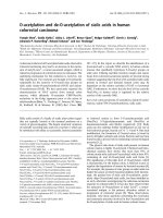
SPase37–204 loses its ability to cleave its native
substrate, prestreptokinase
We have previously identified prestreptokinase, an extra-
cellular protein in pathogenic streptococci to be cleaved
between Ala26 and Ile27 by S. pneumoniae SPase I [18]. To
evaluate the activity of SPaseD37–204, we incubated the
substrate with the self-cleavage-generated SPase37–204 in
the presence of phospholipid. As demonstrated in Fig. 4,
lane 2, the purified SPase37–204 was unable to cleave
prestreptokinase. It indicated that the major product of the
SPase I from self-cleavage was not active. Similarly, the
overexpressed SPase37–204, that was purified simply by one
step Ni column was unable to cleave prestreptokinase either
as shown in Fig. 4, lane 3, whereas the full length SPase I
cleaved the substrate effectively (Fig. 4, lane 4). These
results indicate that SPase I lacking the N-terminal 36
amino acids has lost its activity to cleave its native substrate,
prestreptokinase.
SPase37–204 loses its ability to cleave a peptide
substrate
Based upon the signal peptide sequence of prestreptokinase,
we developed a peptide substrate, KLTFGTVKPVQAIA
GYEWL that was effectively and specifically cleaved
between Ala and Ile by the full length S. pneumoniae
SPase I [27]. As demonstrated by HPLC analysis in Fig. 5,
this 19 amino acid peptide substrate had a retention time of
4.25 min. When it was incubated with the full length
SPase I in the presence of phospholipid, two products were
generated with retention times of 3.32 and 3.88 min,
respectively (Fig. 5A). Mass spectrum analysis of the two
products confirmed that the cleavage specifically occurred
between residues Ala–Ile as expected (data not shown).
However, when the peptide substrate was incubated with
Fig. 4. SDS/PAGE analysis of purified prestreptokinase and its cleav-
age by full length SPase I and SPase37–204. Reactions containing
0.1 lg SPase I or SPase37–204 were incubated with 5 lgofpurified
prestreptokinase at 37 °C for 1 h in the buffer containing 20 m
M
Tris/
HCl (pH 8.0), 0.02% Triton X-100, 5% glycerol and 50 lg phos-
pholipid. The reactions were terminated by the addition of SDS sample
buffer, and the proteins were separated on a 4–20% SDS-poly-
acrylamide gel, and stained by Coomassie Brilliant Blue. Lane 1,
prestreptokinase (pre-Ska); lane 2, prestreptokinase plus self-cleavage-
generated SPase37–204; lane 3, prestreptokinase plus overexpressed
SPase37–204; and lane 4, prestreptokinase plus full length SPase I.
Prestreptokinase was processed to mature streptokinase (mSka) upon
incubation with full length SPase I, as demonstrated in lane 4.
Fig. 5. HPLC analysis of the peptide substrate cleavage by full length
SPase I and SPase37–204. The peptide substrate, KLTFGTVK
PVQAIAGYEWL was incubated at 37 °Cfor2hwithfulllength
SPase I (A), self-cleavage-generated SPase37–204 (B), or overex-
pressed SPase37–204 (C). The cleavage of the peptide substrate was
determined by HPLC using a Hewlett Packard Series 1100 system with
a reversed-phase column (Vydac C18) as described under the Materials
and methods. The peaks labeled 1, 2 and 3 correspond to the substrate,
the C-terminal cleavage product and the N-terminal cleavage product,
respectively.
3974 F. Zheng et al. (Eur. J. Biochem. 269) Ó FEBS 2002
the self-cleavage-generated and the overexpressed SPase37–
204, there was no product peak formed at the expected
retention time in the HPLC profiles (Fig. 5B,C). The result
was in accordance with that observed with the native
substrate, prestreptokinase. Taken together, these results
confirmed that S. pneumoniae SPase37–204, the major
product of the self-cleavage lost its ability to cleave its
substrates. Therefore, the self-cleavage of SPase I is believed
to inactivate the activity of the enzyme.
In vivo
self-cleavage of
S. pneumoniae
SPase I
As the in vitro self-cleavage inactivates the protease activity
of S. pneumoniae SPase I, and this self-cleavage is actually
stimulated, not protected by phospholipid, we are interested
in exploring the reality of the self-cleavage within the
bacterial cells. Polyclonal antibody against S. pneumoniae
SPase I was obtained after immunization of rabbits with
synthetic peptides. To detect SPase I and its cleavage within
the bacterial cells, we performed a Western blot analysis on
S. pneumoniae cells. The whole cell lysates were prepared
from submerged cultures grown to different points through-
out their exponential and stationary growth phases as
indicated in Fig. 6A, separated on a SDS/polyacrylamide
gel, and transferred to a PVDF membrane for immunode-
tection with antidodies against S. pneumoniae SPase I and
Era, an essential membrane associated GTP binding protein
from S. pneumoniae [34]. As demonstrated in Fig. 6C, the
full length S. pneumoniae SPase I and its cleaved product
were detected in all the growth phases. This result confirmed
for the first time that the self-cleavage of SPase I is indeed
happening within the bacterial cells throughout all the
growth phases. The cleaved product reacting with the
peptide antibody against SPase I has a molecular mass of
11 kDa, equivalent to the molecular mass of peptides from
residues 36–143, a possible self-cleaved product based upon
self-cleavage sites identified. As expected, this protein band
reacted specifically with both peptide antibodies against
SPase I. It should be noted that the cell lysates were
prepared immediately after harvest by adding SDS sample
buffer and protease inhibitor cocktail, and boiling for
10 min to protect proteins from nonspecific proteolysis.
S. pneumoniae
cells maintain the full length SPase I
in the highest level in exponential growth phase
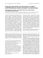
As demonstrated by Western blot analysis in Fig. 6C,
S. pneumoniae cells appeared to produce the overall SPase I
in the same level in all growth phases. However, differences
in full length and cleaved SPase I were observed in different
growth phases. In lag and stationary phases, the cells
showed lower levels of full length SPase I and higher levels
of cleaved product. In contrast, the cells had a higher level
of full length protein and a lower level of cleaved product
in exponential growth phase. In general, the bacterial cells
maintained the active SPase I in the highest level in
Fig. 6. Cell growth of S. pneumoniae and Western blot analysis of in vivo self-cleavage of S. pneumoniae SPase I. (A) Growth curve of
S. pneumoniae cells. The cells were grown at 37 °C in BHI broth, and harvested at different time points as indicated. The cell growth was
monitored by the measurement of absorbence at 620 nm. (B) Densitometer analysis of full length SPase I and the cleaved product in different
growth phases. The analysis was performed based upon the results of Western blot analysis using a Personal Densitometer SI and
IMAGE QUANT
5.0 software from Molecular Dynamics. The ratio of full length/cleaved SPase I was calculated based upon the relative intensity of the two
protein bands reacting with antibody. (C) Western blot analysis of in vivo self-cleavage of SPase I. 20 lg of whole cell lysate from different
growth phases was separated on a 4–20% SDS/polyacrylamide gel, and transferred to a PVDF membrane. Immunodetection was performed
using immune serum against S. pneumoniae SPase I at 1 : 2000 dilution. Lanes 1–9 were the whole cell lysate prepared from S. pneumoniae R6
cells grown in BHI broth for different time as indicated. (D) Western blot analysis with Era antibody.
Ó FEBS 2002 Self-cleavage of S. pneumoniae SPase I (Eur. J. Biochem. 269) 3975
exponential growth phase compared to lag and stationary
growth phases. Densitometer analysis demonstrated that
theratioofthefulllengthSPaseI/thecleavedSPaseIwas
0.81–1.0 in exponential phase, whereas the ratio was 0.36–
0.51 in lag phase and 0.39–0.55 in stationary phase
(Fig. 6B). This result is very reproducible, Fig. 6C shows
one example of several experiments. Clearly, a more
quantitative methodology, other than Western blot analysis,
needs to be developed to quantify the SPase I and its
cleaved products more accurately. In addition, Western blot
analysis with an antibody against S. pneumoniae Era
demonstrated that approximately an equal amount of
protein was loaded in each lane (Fig. 6D). The Era antibody
was selected because of its equally expression in all the
growth phases of S. pneumoniae.
DISCUSSION
A previous study demonstrated that S. pneumoniae SPase I
catalyzes a self-cleavage. In the presence of phospholipid,
the enzyme predominantly cleaves itself at one cleavage site
between Gly36 and His37 [18]. In this study, we found that
the self-cleavage occurred at multiple sites in the absence of
phospholipid, and two additional self-cleavage sites, Ala65–
His66 and Ala143–Phe144, were identified. All three self-
cleavage sites strongly resemble the signal peptide cleavage
site and follow the ()1, )3) rule for signal peptidase
recognition. Phospholipid was demonstrated to stimulate
the self-cleavage of S. pneumoniae SPase I. We also dem-
onstrated that the major product of the self-cleavage,
SPase37–204, totally lost its activity to cleave a native
substrate prestreptokinase and a peptide substrate, indicat-
ing that the self-cleavage inactivates the enzyme. More
importantly, we found that the self-cleavage of S. pneumo-
niae SPase I is also happening in vivo in all the growth
phases, and that the bacterial cells maintain the active
SPase I at the highest level in the exponential growth phase.
These results suggest that the self-cleavage of SPase I may
play an important role in regulating the activity of the
enzyme within the cells.
A number of genes encoding SPase I have been cloned
and sequenced from both Gram-negative and Gram-
positive bacteria including E. coli [9], Salmonella enterica
serovar Typhimurium [35], Haemophilus influenzae [36],
Staphylococcus aureus [37], Bacillus subtilis [38–40], S. pneu-
moniae [18,28], and Streptomyces lividans [41]. To date, the
in vitro biochemical studies on SPase I were performed
using enzymes from three species, E. coli, S. pneumoniae,
and Bacillus subtilis. Although significant differences in
primary sequences exist, these three enzymes share a
common biochemical property, i.e. in vitro self-cleavage.
In E. coli, the self-cleavage of SPase I occurs between the
residues Ala40 and Ala41, which are located in a hydro-
philic domain connecting the two transmembrane segments
at the N-terminus of the enzyme. Although the major
product of this self-cleavage is still active, its specific activity
is 100-fold less than the native enzyme [24]. In S. pneumo-
niae, the purified full length SPase I catalyzes an intermo-
lecular self-cleavage. The major product of this self-cleavage
totally lost its activity as demonstrated in this study. In
B. subtilis, the self-cleavage of SPase I (SipS) was observed
recently, the soluble form of SipS that lacks the N-terminal
membrane anchor is prone to self-cleavage, and the
self-cleavage also results in complete inactivation of the
enzyme [25]. Taken together, the self-cleavage was observed
in all bacterial SPases investigated so far, suggesting that it is
most likely a common biochemical property shared by
bacterial SPases. Another common biochemical property is
that the self-cleavage of the SPase I resulted in the complete
lose or dramatic decrease of the enzymatic activity, implying
that the self-cleavage, if occurring in vivo, may play an
important role in the regulation of the enzymatic activity
within the cells.
Interestingly, phospholipid was demonstrated to affect
self-cleavage of SPase I dramatically. In the absence of
phospholipid, SPase I cleaves itself at multiple sites, whereas
the self-cleavage predominantly occurs at one cleavage site
in the presence of phospholipid. We believe that the
interaction between SPase I and phospholipid somehow
changes the conformation of the enzyme, and makes
SPase I preferentially cleave itself at one specific site. More
importantly, phospholipid was shown to stimulate the self-
cleavage about 25-fold. This phospholipid stimulation was
also observed for substrate cleavage of the SPase I [18].
Therefore, we believe that the interaction of SPase I and
phospholipid may play an important role in the catalytic
mechanism of the enzyme.
Self-cleavage of the E. coli SPase I was previously
described [24]. Scientists working on this enzyme specu-
lated that the self-cleavage might be protected in vivo by
the interaction of the enzyme with cytoplasmic membrane.
Therefore, the possible physiological role of self-cleavage
was basically ignored. However, our investigation revealed
that the self-cleavage of S. pneumoniae SPase I was not
protected by the phospholipid mixture from E. coli lipid
extract. In contrast, the phospholipid mixture, which
composed mainly of phosphatidylethanolamine, phosphat-
idylglycerol, and cardiolipin, actually stimulated the self-
cleavage of the S. pneumoniae SPase I. These results
intrigued us to investigate the cleavage of S. pneumoniae
SPase I in vivo.AsshowninFig.6C,Westernblot
analysis demostrated that the self-cleavage of SPase I is
indeed occuring in vivo in S. pneumoniae throughout all
the growth phases. Although, at this moment, we can not
conclusively explain why self-cleavage is happening in vivo,
one speculation is that it may be involved in regulating the
activity of the enzyme. It is not difficult to imagine that
bacteria may secrete proteins at different levels at different
growth phases and various conditions, and thus may
require differential SPase I activity. Indeed, as we have
shown in this study, S. pneumoniae cells maintain the full
length SPase I in the highest level in exponential
phase compared to lag and stationary growth phases.
Bacterial cells in exponential phase secrete more proteins,
therefore a higher level of SPase activity may be required
for increasing the secretion capacity. It appears that
the self-cleavage of SPase I may play a role in bacterial
cells to control the overall activity of SPase I at a certain
level. Further investigation to establish this hypothesis is
needed.
REFERENCES
1. Wickner, W., Driessen, A.J.M. & Hartl, F U. (1991) The
enzymology of protein translocation across the Escherichia coli
plasma membrane. Annu.Rev.Biochem.60, 101–124.
3976 F. Zheng et al. (Eur. J. Biochem. 269) Ó FEBS 2002
2. Dalbey, R.E., Lively, M.O., Bron, S. & van Dijl, J.M. (1997) The
chemistry and enzymology of type I signal peptidase. Protein Sci.
6, 1129–1138.
3. Paetzel, M., Dalbey, R.E. & Strynadka, N.C.J. (2000) The struc-
ture and mechanism of bacterial type I signal peptidases, a novel
antibiotic target. Pharmacol. Therapeutics 87, 27–49.
4. Innis, M.A., Tokunaga, M., Williams, M.F., Loranger, J.M.,
Chang, S.Y. & Wu, H.C. (1984) Nucleotide sequence of the
Escherichia coli prolipoprotein signal peptidase (lsp)gene.Proc.
Natl Acad. Sci. U.S.A. 81, 3708–3712.
5. Driessen, J.A., Fekkes, P. & van der Wolk, J.P. (1998) The Sec
system. Curr. Opin. Microbiol. 1, 216–222.
6. Economou, A. (1999) Following the leader: bacterial protein
export through the Sec pathway. Trends Microbiol. 7, 315–320.
7. Moore, K.E. & Miura, S. (1987) A small hydrophobic domain
anchors leader peptidase to the cytoplasmic membrane of
Escherichia coli. J. Biol. Chem. 262, 8806–8813.
8. San Millan, J.L., Boyd, D., Dalbey, R.E., Wickner, W. & Beck-
with, J. (1989) Use of phoA fusions to study the topology
of Escherichia coli inner membrane protein leader peptidase.
J. Bacteriol. 171, 5536–5541.
9. Wolfe, P.B., Wickner, W. & Goodman, J.M. (1983) Sequence of
the leader peptidase gene of Escherichia coli and the orientation of
leader peptidase in bacterial envelope. J. Biol. Chem. 258, 12073–
12080.
10. Dalbey, R.E. & Wickner, W. (1985) Leader peptidase catalyzes the
release of exported proteins from the outer surface of the
Escherichia coli plasma membrane. J. Biol. Chem. 260, 15925–
15931.
11. Fikes, J.D. & Bassford, P.J. (1987) Export of unprocessed pre-
cursor maltose-binding protein to the periplasma of Escherichia
coli cells. J. Bacteriol. 169, 2352–2359.
12. Koshland,D.,Sauer,T.T.&Botstein,D.(1982)Diverseeffectsof
mutations in the signal sequence on the secretion of beta-lactamase
in Salmonella typhimurium. Cell 30, 903–914.
13. Kuhn, A. & Wickner, B. (1985) Conserved residues of the leader
peptide are essential for cleavage by leader peptidase. J. Biol.
Chem. 260, 15914–15918.
14. Tschantz, W.R. & Dalbey, R.E. (1994) Bacterial leader peptidase
I. Methods Enzymol. 244, 285–301.
15. Black, M.T. (1993) Evidence that the catalytic activity of pro-
karyote leader peptidase depends upon the operation of a serine-
lysine catalytic dyad. J. Bacteriol. 175, 4957–4961.
16. Tschantz, W.R., Sung, M., Delgado-Partin, V.M. & Dalbey, R.E.
(1993) A serine and a lysine residue implicated in the catalytic
mechanism of the Escherichia coli leader peptidase. J. Biol. Chem.
268, 27349–27354.
17. van Dijl, J.M., de Long, A., Venema, G. & Bron, S. (1995)
Identification of the potential active site of the signal peptidase
SipS of Bacillus subtilis. Structural and functional similarities with
LexA-like proteases. J. Biol. Chem. 270, 3611–3618.
18.Peng,S.B.,Wang,L.,Moomaw,J.,Peery,R.B.,Sun,P.M.,
Johnson, R.B., Lu, J., Treadway, P., Skatrud, P.L. & Wang, Q.M.
(2001) Biochemical characterization of signal peptidase I from
Gram-positive Streptococcus pneumoniae. J. Bacterol. 183,
621–627.
19. Black, M.T., Munn, J.G.R. & Allsop, A. (1992) On the catalytic
mechanism of prokaryotic leader peptidase I. Biochem. J. 282,
539–543.
20. Paetzel, M., Dalbey, R.E. & Strynadka, N.C.J. (1998) Crystal
structure of a bacterial signal peptidase in complex with a beta-
lactam inhibitor. Nature 396, 186–190.
21. Little, J.W. (1993) LexA cleavage and other self-processing reac-
tions. J. Bacteriol. 175, 4943–4950.
22. Peat, T.S., Frank, E.G., McDonald, J.P., Levine, A.S., Woodgate,
R. & Hendrickson, W.A. (1996) Structure of UmuD¢ protein
and its regulation in response to DNA damage. Nature 380,
727–730.
23. Roland, K.L. & Little, J.W. (1990) Reactions of LexA repressor
with diisopropyl fluorophosphate. A test of the serine protease
model. J. Biol. Chem. 265, 12828–12835.
24. Talarico, T.L., Dev, I.K., Bassfort, P.J. & Ray, P.H. (1991) Inter-
molecular degradation of signal peptidase I in vitro. Biochem.
Biophys. Res. Commun. 181, 650–656.
25. van Roosmalen, M.L., Jongbloed, J.D.H., Kuipers, A., Venema,
G., Bron, S. & van Dijl, J.M. (2000) A truncated soluble Bacillus
signal peptidase produced in Escherichia coli is subject to self-
cleavage at its active site. J. Bacteriol. 182, 5765–5770.
26. Huang, T.T., Malke, H. & Ferretti, J.J. (1989) The streptokinase
gene of group A streptococci: cloning, expression in Escherichia
coli, and sequence analysis. Mol. Microbiol. 3, 197–205.
27. Peng, S.B., Zheng, F., Angleton, E.L., Smiley, D., Carpenter, J.
& Scott, J.E. (2001) Development of an internally quenched
fluorescent substrate and a continuous fluorimetric assay for
Streptococcus pneumoniae signal peptidase I. Anal. Biochem. 293,
88–95.
28. Zhang, Y., Greenberg, B. & Lacks, S.A. (1997) Analysis of a
Streptococcus pneumoniae gene encoding signal peptidase I and
overproduction of the enzyme. Gene 194, 249–255.
29. Studier, F.W., Rosenburg, A.H., Dunn, J.J. & Dubendorff, J.W.
(1990) Use of T7 RNA polymerase to direct expression of cloned
genes. Methods Enzymol. 185, 60–89.
30. Peng, S.B., Crider, B.P., Tsai, S.J., Xie, X.S. & Stone, D.K. (1996)
Identification of a 14-Kda subunit associated with the catalytic
sector of Clathrin-coated vesicle H+-ATPase. J. Biol. Chem. 271,
3324–3327.
31. von Heijne, G. (1983) Patterns of amino acids near signal-
sequence cleavage sites. Eur. J. Biochem. 116, 17–21.
32. von Heijne, G. (1984) How signal sequences maintain cleavage
specificity. J. Mol. Biol. 173, 143–251.
33. von Heijne, G. (1986) A new method for predicting signal
sequence cleavage sites. Nucleic Acids Res. 14, 4683–4690.
34. Meier, T.I., Peery, R.B., Jaskunas, S.R. & Zhao, G. (1999) 16S
rRNA is bound to Era of the Streptococcus pneumoniae. J. Bac-
teriol. 181, 5242–5249.
35. van Dijl, J.M., van den Bergh, R., Reversma, T., Smith, H., Bron,
S. & Venema, G. (1990) Molecular cloning of the Salmonella
typhimurium lep gene in Escherichia coli. Mol. General Genet. 223,
233–240.
36. Fleischmann, R.D. et al. (1995) Whole genome random
sequencing and assembly of Haemophilus influenzae. Science 269,
496–512.
37. Cregg, K.M., Wilding, E.I. & Black, M.T. (1996) Molecular
cloning and expression of the spsB gene encoding an essential type
I signal peptidase from Staphylococcus aureus. J. Bacteriol. 178,
5712–5718.
38. Meijer,W.J.J.,deJong,A.,Bea,G.,Wiseman,A.,Tjalsma,H.,
Venema, G., Bron, S. & van Dijl, J.M. (1995) The endogenous
B. subtilis plasmids pTA1015 and pTA1040 contain signal pepti-
dase-encoding genes. Identification of a new structural module on
cryptic plasmids. Mol. Microbiol. 17, 621–631.
39. Tjalsma, H., Noback, M.A., Bron, S., Venema, G., Yamane, K. &
van Dijl, J.M. (1997) Bacillus subtilis contains four closely related
type I signal peptidase with overlapping substrate specificities.
J. Biol. Chem. 272, 25983–25992.
40. van Dijl, J.M., de Jong, A., Vehmaanpera, J., Venema, G. & Bron,
S. (1992) Signal peptidase I of B. subtilis: patterns of conserved
amino acids in prokaryotic and eukaryotic type I signal pepti-
dases. EMBO J. 11, 2819–2828.
41. Parro, V., Schacht, S., Anne, J. & Mellado, R.P. (1999) Four genes
encoding different type I signal peptidase are organized in a cluster
in Streptomyces lividans TK21. Microbiol. 145, 2255–2263.
Ó FEBS 2002 Self-cleavage of S. pneumoniae SPase I (Eur. J. Biochem. 269) 3977