Báo cáo khoa học: Temperature and salts effects on the peptidase activities of the recombinant metallooligopeptidases neurolysin and thimet oligopeptidase pdf
Bạn đang xem bản rút gọn của tài liệu. Xem và tải ngay bản đầy đủ của tài liệu tại đây (304 KB, 9 trang )
Temperature and salts effects on the peptidase activities
of the recombinant metallooligopeptidases neurolysin
and thimet oligopeptidase
Vitor Oliveira
1
, Reynaldo Gatti
2
, Vanessa Rioli
3
, Emer S. Ferro
3
, Alberto Spisni
2,4
, Antonio C. M. Camargo
5
,
Maria A. Juliano
1
and Luiz Juliano
1
1
Department of Biophysics, Escola Paulista de Medicina, Sa
˜
o Paulo, Brazil;
2
Centro de Biologia Molecular Estrutural, National
Laboratory of Synchrotron Light (CBME-LNLS), Campinas, Brazil;
3
Department of Histology, Institute of Biomedical Sciences,
Universidade de Sa
˜
o Paulo, Brazil;
4
Department of Experimental Medicine, University of Parma, Parma 43100, Italy;
5
Laboratory of
Biochemistry and Biophysics, Instituto Butantan, Sa
˜
o Paulo, Brazil
We report the recombinant neurolysin and thimet oligo-
peptidase (TOP) hydrolytic activities towards internally
quenched fluorescent peptides derived from the peptide
Abz-GGFLRRXQ-EDDnp (Abz, ortho-aminobenzoicacid;
EDDnp, N-(2,4-dinitrophenyl) ethylenediamine), in which
X was substituted by 11 different natural amino acids.
Neurolysin hydrolyzed these peptides at R–R or at R–X
bonds, and TOP hydrolyzed at R–R or L–R bonds,
showing a preference to cleave at three or four amino
acids from the C-terminal end. The kinetic parameters of
hydrolysis and the variations of the cleavage sites were
evaluated under different conditions of temperature and
salt concentration. The relative amount of cleavage varied
with the nature of the substitution at the X position as
well as with temperature and NaCl concentration. TOP
was activated by all assayed salts in the range 0.05–0.2
M
for NaCl, KCl, NH
4
Cl and NaI, and 0.025–0.1
M
for
Na
2
SO
4
. Concentration higher than 0.2 N NH
4
Cl and NaI
reduced TOP activity, while 0.5 N or higher concentration
of NaCl, KCl and Na
2
SO
4
increased TOP activity. Neu-
rolysin was strongly activated by NaCl, KCl and Na
2
SO
4
,
while NH
4
Cl and NaI have very modest effect. High
positive values of enthalpy (DH*) and entropy (DS*) of
activation were found together with an unusual tempera-
ture dependence upon the hydrolysis of the substrates.
The effects of low temperature and high NaCl concen-
tration on the hydrolytic activities of neurolysin and TOP
do not seem to be a consequence of large secondary
structure variation of the proteins, as indicated by the far-
UV CD spectra. However, the modulation of the activities
of the two oligopeptidases could be related to variations
of conformation, in limited regions of the peptidases,
enough to modify their activities.
Keywords: protease; peptide; metalloprotease; fluorescence;
enthalpy of activation in proteolysis; entropy of activation in
proteolysis.
Thimet oligopeptidase (TOP, EC 3.4.24.15) and neurolysin
(EC 3.4.24.16) are zinc-dependent peptidases, members of
the metallopeptidase M3 family and contain in their
primary sequence the HEXXH motif [1,2]. Rat neurolysin
has been the first member of the M3 family of which the 3D
structure has been determined [3]. As it has been shown that
this enzyme and thermolysin have common ancestors, the
M3 family was thus included in the clan MA [4]. It is
interesting to note that in the structure of neurolysin, the
catalytic center is located in a deep channel [3], which limits
the access only to short peptides [5,6]. This selectivity
toward hydrolysis of oligopeptides was also verified for
TOP [5–9]. The high primary sequence identity found for
these related peptidases, which is about 65% [2], allows
hypothesizing that they may share a similar folding
including the deep channel that constitutes the neurolysin
active site. This feature seems to prevent the unspecific
cleavage of other proteins and it is of particular relevance
for TOP and neurolysin as they are not expressed as inactive
precursors and they are present in high amount as soluble
enzymes in cytosol. On the other hand, membrane associ-
ated form of TOP [10] and neurolysin have been identified
[11,12]. The secretion of TOP has been reported in AtT20
[13,14] and MDCK cells [15] while neurolysin was showed
to be secreted by astrocytes [16].
Efficient oligopeptidases are required to metabolize
biologically active peptides before and after their interac-
tion with cell receptors, this is particularly relevant with
neuropeptides that lack classical reuptake mechanisms for
recycling components into the cell. TOP exhibits charac-
teristics of both metabolizing and processing enzymes,
and has multiple peptide substrates as GnRH [17],
neurotensin [18], bradykinin [19], somatostatin 1–14 [20],
and nociceptin [21]. TOP also processes Met- and
Correspondence to L. Juliano, Departamento de Biofisica, Escola
Paulista de Medicina, Rua Treˆ s de Maio, 100, 04044-020 Sa
˜
oPaulo,
SP, Brazil. Fax: + 55 11 5575 9040, Tel.: + 55 11 5575 9617,
E-mail:
Abbreviations:Abz,ortho-aminobenzoic acid; EDDnp,
N-(2,4-dinitrophenyl) ethylenediamine; IQF peptide, internally
quenched fluorescent peptide, TOP, thimet oligopeptidase.
Enzymes: thimet oligopeptidase (TOP, EC 3.4.24.15); neurolysin
(EC 3.4.24.16).
*Present address: Department of Experimental Medicine, University
of Parma, Parma, 43100, Italy.
(Received 10 April 2002, revised 3 July 2002, accepted 19 July 2002)
Eur. J. Biochem. 269, 4326–4334 (2002) Ó FEBS 2002 doi:10.1046/j.1432-1033.2002.03129.x
Leu-enkephalin from the enkephalin-containing peptides
[22], and the specific TOP inhibition increased Met-enke-
phalin antinociception in rodents [23]. TOP and neurolysin
are able to hydrolyze the biologically active peptide
neurotensin (NT) in vitro and they could be participating
in the catabolism of this biologically active peptide. In vivo
experiments have been showed that NT degradation is
blocked by TOP and neurolysin inhibitors [24] and a highly
specific neurolysin inhibitor potentiated the neurotensin-
induced antinociception of mice in the hot plate test when
administrated intracerebroventricularly [25]. In addition,
higher vertebrates produce large number of oligopeptides
generated by the proteasome system due to proteolysis of
intracellular and/or of foreign proteins. However, only some
of these peptides are presented to the immune system on cell
surface MHC class I molecules [26–28]. Recently it was
reported the involvement of TOP in sorting and hydrolysis
of the peptides generated by proteasomes [30–32].
A detailed analysis of the substrate specificity of neu-
rolysin and TOP was reported using internally quenched
fluorescent (IQF) peptides derived from bradykinin [5] and
neurotensin [6]. An outstanding feature of the hydrolytic
activities of neurolysin and TOP on these substrates is the
variability of the cleavage sites, which is a consequence of
modifications in size or in nature of the amino acids at
different positions of the substrates [5,6]. The 3D structure
determination of neurolysin supports this broad specificity
turning up the possibility of a reorganization of the flexible
loops of the enzyme binding site in order to accommodate
the substrates [3]. This very particular mechanism of
substrate interaction with a peptidase requires a detailed
study of the milieu composition and temperature influence
on neurolysin and TOP hydrolytic activities in order to
understand their unusual variations of specificity [5,6] and
broad spectrum of hydrolysis of biologically active peptides
as described above.
In the present work we report the neurolysin and TOP
hydrolytic activities towards IQF peptides derived from
Abz-GGFLRRVQ-EDDnp [Abz, ortho-aminobenzoic
acid; EDDnp, N-(2,4-dinitrophenyl) ethylenediamine], in
which Val was substituted by 11 different natural amino
acids. This sequence was chosen because we have previously
observed efficient hydrolysis by TOP of the peptide Abz-
GGFLRRV-EDDnp at L-R bond and the addition of Gln
at C-terminal site (Abz-GGFLRRVQ-EDDnp) resulted in
two cleavages, at L–R and or at R–R bond. We made
modifications at Val position in order to verify the influence
of the nature of the amino acid at this position on
determination of the cleavage sites and on the amount of
their hydrolysis. This series of peptides was chosen because
the P
2
¢ and P
3
¢ positions were demonstrated to be very
determinant on the specificity of neurolysin and TOP [5].
The kinetic parameters of hydrolysis and the variations of
the cleavage sites of this series of peptides by these two
oligopeptidases were evaluated in different conditions of
temperature and salts.
MATERIALS AND METHODS
Thimet oligopeptidase (TOP)
The purified recombinant rat testes TOP (rTOP) was
obtained as previously described [33]. Details about the
procedures applied for enzyme characterization and active
site titration were described elsewhere [5].
Neurolysin
The recombinant cDNA of porcine liver neurolysin (cyto-
solic form) was a kind gift from S. Hirose and A. Kato
(Department of Biological Sciences, Tokyo Institute of
Technology, Yokohama, Japan). Details concerning the
expression system including plasmid constructions and
vectors used were described elsewhere [34]. The procedures
for expression and purification of recombinant porcine liver
neurolysin were performed as previously reported for the
recombinant rat testes TOP [33]. The methods for the
determinations of enzyme purity and concentration were
also previously described [6].
Peptide synthesis
The IQF peptidescontaining N-[2,4-dinitrophenyl]-ethylene-
diamine (EDDnp) attached to glutamine were synthesized
by solid-phase strategy, which details are provided elsewhere
[35]. An automated bench-top simultaneous multiple solid-
phase peptide synthesizer (PSSM 8 system from Shimadzu)
was used for the synthesis of all the peptides by the Fmoc-
procedure. The final deprotected peptides were purified by
semipreparative HPLC using an Econosil C-18 column
(10 l, 22.5 · 250 mm) and a two-solvent system: (A)
trifluoroacetic acid/H
2
O (1 : 1000) and (B) trifluoroacetic
acid/acetonitrile/H
2
O (1 : 900 : 100). The column was eluted
at a flow rate of 5 mLÆmin
)1
with a 10 (or 30))50 (or 60)%
gradient of solvent B over 30 or 45 min. Analytical HPLC
was performed using a binary HPLC system from Shima-
dzu with a SPD-10AV Shimadzu uv-vis detector and a
Shimadzu RF-535 fluorescence detector, coupled to an
Ultrasphere C-18 column (5l,4.6· 150 mm) which was
eluted with solvent systems A and B at a flow rate of
1mLÆmin
)1
and a 10–80% gradient of B over 20 min. The
HPLC column eluates were monitored by their absorbance
at 220 nm and by fluorescence emission at 420 nm follow-
ing excitation at 320 nm. The molecular mass and purity of
synthesized peptides were checked by MALDI-TOF mass
spectrometry (TofSpec-E, Micromass) and/or peptide
sequencing using a protein sequencer PPSQ-23 (Shimadzu
Tokyo, Japan).
Kinetic assays
The Michaelis parameters were determined by initial rate
measurements. The hydrolysis of the fluorogenic peptidyl
substrates at 37 °Cin50m
M
Tris/HCl buffer pH 7.4
containing 100 m
M
NaCl was followed by measuring the
fluorescence at k
em
¼ 420 nm and k
ex
¼ 320 nm in a
Hitachi F-2000 spectrofluorometer. The 1-cm path-length
cuvette containing 2 mL of the substrate solution was
placed in a thermostatically controlled cell compartment for
5 min before the enzyme solution was added and the
increase in fluorescence with time was continuously recor-
ded for 5–10 min. For TOP an additional preincubation
time of 5 min with 0.5 m
M
of dithiothreitol were applied
before substrate addition. This amount of dithiothreitol was
chosen because it provided the maximum enzyme activation
in our conditions. The slope was converted into mols of
Ó FEBS 2002 Modulation of thimet oligopeptidase and neurolysin activities (Eur. J. Biochem. 269) 4327
hydrolyzed substrate per minute based on the fluorescence
curves of standard peptide solutions before and after total
enzymatic hydrolysis. The concentration of the peptide
solutions was obtained by colorimetric determination of the
2,4-dinitrophenyl group (17 300
M
)1
Æcm
)1
, extinction coef-
ficient at 365 nm). The enzyme concentrations for initial
rate determinations were chosen at a level intended to
hydrolyze less than 5% of the substrate present in the
reaction. The inner-filter effect was corrected using an
empirical equation as previously described [36]. The kinetic
parameters were calculated according Wilkinson [37] as well
as by using Eadie–Hofstee plots. All the obtained data were
fitted to nonlinear least square equations, using
GRAFIT
v3.0
from Erithacus Software [38].
The hydrolysis of the substrates cleaved at two peptide
bonds by TOP and neurolysin can be represented as shown
in Scheme 1, whose equation for velocity is Eqn (1). V
t
is
the sum of the velocities of formation of the products (P
a
and P
b
). V
a
max
is kp
a
· [E] and V
b
max
is kp
b
· [E], and [E] is
the total enzyme concentration in the assay. All the obtained
data with the peptides cleaved at two bonds fitted to
nonlinear least square plot of Eqn (1). The overall V
max
was
obtained from Eqn (1), whereas the separate values for
V
a
max
and V
b
max
were calculated using the ratio of the areas
taken from the integrated HPLC chromatogram analysis.
Additional data and discussion about this kinetic interpret-
ation can be found in more details in [5].
V
t
¼
½SÁðV
a
max
þ V
b
max
Þ
K
s
þ½S
ð1Þ
For the specificity rate constants (k
cat
/K
m
) which were
determined under first-order conditions, we used substrates
concentrations 10-fold less than K
m
. The obtained first-
order rate constants were divided by the total enzyme
concentration to provide k
cat
/K
m
. As the products Abz-
GGFL, Abz-GGFLR and their respective C-terminal
fragments were resistant to hydrolysis by TOP, and the
products Abz-GGFLR, Abz-GGFLRR and their respect-
ive C-terminal fragments were also resistant to hydrolysis by
neurolysin, we could determine the specificity rate constants
(k
cat
/K
m
) under first-order conditions, even for the peptides
hydrolyzed at two peptide bonds. This procedure was used
in the assays conduced at different temperatures and at
different salt concentrations [5].
Temperature dependence of the hydrolysis reaction
rates of the substrates by neurolysisn and TOP
The temperature dependence of the rate constants was
determined in thermostated cell holders. The reactions
were started after the thermal equilibrium had been
reached in the cell. Typically the reactions were carried
out in 1 mL of 50 m
M
Tris/HCl buffer pH 7.4 containing
100 m
M
NaCl. Activation parameters were calculated
from the linear plots of ln(k/T)vs.1/T (Eqn 2), where k is
the rate constant, R is the gas constant (8.314 JÆmol
)1
Æ
K
)1
), T is the absolute temperature, N
A
is Avogadro’s
number, h is Planck’s constant, the enthalpy of activa-
tion DH* ¼ –(slope)8.314 JÆmol
)1
, the entropy of activa-
tion DS* ¼ (intercept – 23.76)8.314 JÆmol
)1
ÆK
)1
.Thefree
energy of activation DG*, was calculated from Eqn (3)
(T ¼ 298.15 K).
ln
k
T
¼ ln
R
N
A
h
þ
DS
Ã
R
À
DH
Ã
RT
ð2Þ
DG
Ã
¼ DH
Ã
À TDS
Ã
ð3Þ
Dependence of the hydrolysis reaction rates
by neurolysisn and TOP on concentration and chemical
nature of salts
The dependence of the rate constants according to the
concentration and the chemical nature of salts were
determined in 1 mL of 50 m
M
Tris/HCl buffer pH 7.4
containing different concentrations of NaCl, KCl, NH
4
Cl
(0–2 N), NaI and Na
2
SO
4
(0–1 N). A strong fluorescence
quenching caused by the I
–
ion did not permitted the
experiments with 2 N NaI.
Determination of cleaved bonds
The cleaved bonds were identified by isolation of the
fragments by HPLC either comparing the retention times of
the products fragments with synthetic peptides encompas-
sing the expected hydrolysis products and/or by molecular
mass. The molecular masses were determined by MALDI-
TOF mass spectrometry and/or by sequencing, using a
protein sequencer PPSQ-23 (Shimadzu Tokyo, Japan).
Amino-acid analysis
The amino-acid compositions, the concentration of the
peptides and the purified rTOP were determined as follows:
the samples were digested for 22 h at 110 °Cin6NHCl
containing 1% phenol in vacuum sealed tubes and then
subjected to amino-acid analysis using a pico-Tag station
[39].
Circular dichroism
CD spectra were recorded at Jasco J-810 spectropolarimeter
with a Peltier system of cell temperature control. The
system was routinely calibrated with an aqueous
solution of recrystalized d-10 camphorsulphonic acid.
Ellipticity is reported as mean residue molar ellipticity, [h]
(degÆcm
2
Ædmol
)1
). The spectrometer conditions were typi-
cally: spectral range 195–260 nm, 100 mdeg sensibility;
0.2 nm resolution; 4 s response time; 20 nmÆmin
)1
scan rate,
7 accumulations at the appropriate temperature (10, 25 or
37 °C). The 100 mdeg sensibility is used in our routine that
leads to the lower noise-signal relationship. The control
baseline was obtained with solvent and all the components
without the proteins. All the data were obtained with three
Scheme 1.
4328 V. Oliveira et al. (Eur. J. Biochem. 269) Ó FEBS 2002
different solutions of the proteins. The quality of data was
certified by the correspondence of the amount of secondary
structures obtained by CD data deconvolution with those
from the 3D structure of neurolysin. The errors of
prediction on the range 195–260 nm and 200–260 nm were
5% using the
CDNN
program [40].
RESULTS
Kinetic parameters for the hydrolysis of IQF peptide
series Abz-GGFLRRXQ-EDDnp by TOP and neurolysin
Table 1 shows the kinetic parameters of the hydrolysis by
TOP and neurolysin of the substrates on the series Abz-
GGFLRRXQ-EDDnp and their peptide bonds cleaved in
the kinetic measurement conditions. TOP hydrolyzed the
peptides I to VI only at R–R bonds, which contain at
position X basic or aromatic amino acids, besides Pro and
Ala. On the other hand, the peptides VII to XII, which
contain hydrophobic or acidic amino acids at the X
position, besides Asn, were hydrolyzed either at R–R or
at L–R bonds, but preferentially at the R–R bond, except
the peptide XII that contains Asp. The higher specificity
constant (k
cat
/K
m
) values were obtained with the substrates
cleaved only at R–R bond, and the catalytic constant (k
cat
)
was the predominant component. Peptide XII and XIII (Qf
7 in Table 1) were exceptions in terms of preferential
cleavage site by TOP, which is directed to L–R bond by Asp
in peptide XII or by the absence of Gln in the peptide XIII,
however, their k
cat
/K
m
values were the lowest in the series.
On the other hand, the highest k
cat
/K
m
values obtained with
TOP were for substrates I and II containing Arg and His at
the X position, respectively.
Neurolysin, like TOP, hydrolyzed all the substrates at
R–R bond, but the alternative cleavage site was at R–X
bond in peptides II to IV, VII, IX and X, which contain at
the X position of the series Abz-GGFLRRXQ-EDDnp
essentially hydrophobic amino acids. The peptides hydro-
lyzed exclusively at R–R bonds contain at the X position
charged amino acids (Arg, Asp and Glu) or amino acids
with small hydrophobic side chain (Ala, Pro and Val). The
highest k
cat
/K
m
value for neurolysin was observed with
the hydrolysis of the substrate with Ala (peptide VI), and the
lowest k
cat
/K
m
values was the peptide with Asp (peptide
XII) at the X position.
Temperature dependence of the substrate hydrolysis
by TOP and neurolysin
The preference of cleavage at the L–R or R–R bond for
TOP and at the R–R or R–X bond for neurolysin in the
case of the substrates containing Val, Asp and Ile at the
X position in the studied series were determined at
temperature range 10–37 °C. These peptides were chosen
due to their different preferences for hydrolysis of the
susceptible bonds. Both peptidases were stable at 37 °C
for more than 20 min, which was longer than that used
in all enzyme assays. At 45 °C significant decrease of
activity was observed in the first 5 min, possibly due to
enzyme denaturation. A significant increase in the
percentage of hydrolysis by TOP at the R–R bond was
observed by decreasing temperature with the peptides
containing Val or Asp. No significant changes were
observed with the peptide having Ile or with any other
substrate assayed with neurolysin (Table 2). Both, the
dithiothreitol used for activation of TOP [41] and pH
variations from 6 to 9 did not affect the ratio of
hydrolysis between the two hydrolytic sites by the two
oligopeptidases.
The k
cat
/K
m
values were determined, at the temperature
range 10–37 °C in the presence of 0.1
M
NaCl, for
hydrolysis of Qf 7 and the substrates of the series
Table 1. Kinetics parameters for hydrolysis by TOP and neurolysin of the peptides derived from Abz-GGFLRRXQ-EDDnp. The parameters were
calculated as mean value ±S.D., which was never greater than 7%. The kinetic parameters for the hydrolysis of the substrates with two cleavage
sites were obtained using Eqn 2. k
cat
/K
m
¼ (k
a
cat
+ k
b
cat
)/K
m
. L–R, R–R and R–X indicate the cleavage sites. Qf 7 is the abbreviation used for the
peptide Abz-GGFLRRV-EDDnp. The kinetic experiments were conduced at 37 °Cin50m
M
Tris/HCl buffer containing 0.1
M
NaCl. For XIII,
cleavage site is L–R.
Number X
TOP (24.15) Neurolysin (24.16)
k
cat
(s
)1
)
K
m
(l
M
)
k
cat
/K
m
(l
M
)1
Æs
)1
)
k
cat
(s
)1
)
K
m
(l
M
)
k
cat
/K
m
(l
M
)1
Æs
)1
)
L–R R–R R–R R–X
I R – 11 1.5 7.3 3.3 – 2.3 1.4
II H – 10 1.4 7.1 1.6 0.6 1.7 1.3
III Y – 19 4.3 4.4 0.3 0.2 0.6 0.8
IV F – 15 3.6 4.2 0.2 0.3 0.7 0.7
V P – 7.4 2.2 3.4 1.8 – 1.2 1.5
VI A – 15 4.8 3.1 3.2 – 0.5 6.4
VII N 1.6 5.5 1.8 3.9 0.6 0.3 1.5 0.6
VIII V 1.7 2.8 1.7 2.6 1.4 – 2.1 0.7
IX I 0.8 4.3 2.0 2.5 0.7 0.1 1.8 0.4
X L 1.8 3.5 2.5 2.1 0.2 0.5 0.7 1.0
XI E 0.7 2.2 1.5 1.9 1.1 – 1.9 0.6
XII D 3.4 1.6 3.2 1.6 1.0 – 3.3 0.3
XIII Qf 7 0.7 – 1.7 0.4 2.0 – 2.2 0.9
Ó FEBS 2002 Modulation of thimet oligopeptidase and neurolysin activities (Eur. J. Biochem. 269) 4329
Abz-GGFLRRXQ-EDDnp containing Val, Ile and Ala at
the X position. The peptide containing Asp were only
assayed with TOP. Linear Eyring plots (ln[k/T ]vs.1/T )
were obtained for the hydrolysis of Qf 7 by both enzymes
and for the hydrolysis of the substrate containing Val by
neurolysin. The Eyring plots for the hydrolysis by TOP of
the peptides containing Ile, Val and Asp deviated from the
linearity above 25 °C. The plot for the reaction of the
peptide containing Ala with TOP was not linear in all
studied range of temperature (Fig. 1A). The Eyring plots
obtained for neurolysin reactions with the peptides
containing Ala and Ile at the X position gave two linear
fittings, above and below % 22 °C (Fig. 1B), indicating
different rate-limiting steps at each temperature range
(above and below 22 °C).
The DG* DH*andDS* values were taken from Eyring
plots and are shown in Table 3. In addition to the
temperature dependence of the catalytic constants accord-
ing to the substrates, the positive and high values of DH*
and DS* for hydrolysis are of note.
Influence of the NaCl on TOP and neurolysin activities
The influence of NaCl concentration on k
cat
/K
m
values of
TOP and neurolysin activities on the peptides containing
Ala, Val, Asp and Ile at the X position in the series Abz-
GGFLRRXQ-EDDnp was examined in the salt concen-
tration range 0–0.5
M
, and the data are presented in
Table 4. The k
cat
/K
m
values increased with the increasing
of NaCl concentration for all the assayed substrates,
except with the peptide containing Asp. The higher NaCl
effects were observed for the hydrolysis of the peptide
with Ile at the X position by neurolysin and TOP
(Table 4). NaCl was observed also to modulate the
preference of both enzymes for their cleavage sites,
namely, the increase of NaCl concentration further
enhanced the percentage hydrolysis by TOP and neuro-
lysin at R–R bond (Table 4).
The activation parameters of TOP and neurolysin
activities on Qf7 were also determined in the presence of
2
M
NaCl. In this condition, the Eyring plots obtained for
TOP and neurolysin gave two linear fittings, above and
below % 22 °C, which contrast with linear plots obtained in
the absence of salt. Similar to all others, substrates with
similar temperature behavior resulted in DH*andDS*
values at temperature range 25–37 °C significantly lower
than those at 10–20 °C(Table3).
Influence of the chemical nature of salts on the TOP
and neurolysin activities
Using Qf7 as a reference substrate, which was hydrolyzed
only at L–R bond, we studied the effects of different salts on
TOP and neurolysin activities. The results are shown in
Fig. 2A,B, respectively. TOP was activated by all the
assayed salts (NaCl, KCl, Na
2
SO
4
,NH
4
Cl and NaI) at
low concentrations. However, the increase of NH
4
Cl or NaI
concentrations reduced TOP activity, in contrast to NaCl,
KCl and Na
2
SO
4
that progressively increased the enzyme
activity from 0.5 till 2 N salt concentrations. In the case of
neurolysin, NaCl, KCl and Na
2
SO
4
exhibited the more
intense activation effect (one order of magnitude more than
that with TOP), and the effects of salts were proportional to
their concentration. NH
4
Cl and NaI exhibited small
activation without any inhibitory activity as observed with
TOP.
Fig. 1. Eyring plots for substrate hydrolysis reaction by TOP and neu-
rolysin. (A) Eyring plots for the hydrolysis carried out with TOP on
Qf 7 (black circles), Abz-GGFLRRAQ-EDDnp (open circles) and
Abz-GGFLRRVQ-EDDnp (black squares). (B) Eyring plots for the
hydrolysis carried out with neurolysin on Qf 7 (black circles), Abz-
GGFLRRAQ-EDDnp (open circles). The hydrolysis reactions were
carried out in Tris buffer 50 m
M
, pH 7.4 containing NaCl 100 m
M
.
Table 2. Influence of temperature on the preference cleavage sites of
TOP on the peptides derived from Abz-GGFLRRXQ-EDDnp. The
kinetic experiments were conduced in 50 m
M
Tris/HCl buffer con-
taining 0.1
M
NaCl.
T °C
Cleaved bond %, TOP
X ¼ V X ¼ D X ¼ I
L–R R–R L–R R–R L–R R–R
10 18 82 39 61 11 89
20 26 74 50 50 9 91
30 27 73 59 41 11 89
37 38 62 64 36 17 83
4330 V. Oliveira et al. (Eur. J. Biochem. 269) Ó FEBS 2002
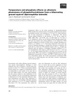
Circular dichroism spectra of TOP and neurolysin
The CD spectra of TOP and neurolysin show a predomi-
nance of a helical structures as shown in Fig. 3 (without
smoothing and curve fitting). For neurolysin spectra, the
results obtained in the deconvolution of the CD data, using
the
CDNN
program [40], are consistent with the helix content
found in the neurolysin crystal structure, as the helix content
from the crystal structure was 53% [3]; and from the
deconvolution of CD at 37 °C in the absence of NaCl (195–
260 nm) was 51%. For the spectrum of TOP in the same
conditions the deconvolution indicated 45% of a helix
content. This result is close to that of neurolysin and
consistent with consensus secondary structure prediction
obtained from different algorithms (
DPM
,
DSC
,
GOR
4,
HNNC
,
PHD
,
PREDATOR
,
SIMPA
96,
POPM
) performed at the internet
site . The variation of the NaCl
concentration from 0 to 1
M
did not affect the a helical
component in a detectable manner in the far-UV CD assay.
Changes in the secondary structure of TOP and neurolysin
with the temperature (10 and 37 °C) were also not detected
in the CD experiments in the presence and in the absence of
NaCl.
DISCUSSION
TOP and neurolysin hydrolyzed all the assayed substrates of
the series Abz-GGFLRRXQ-EDDnp at three or four
amino acids from their C-terminal ends. These results agree
with previously reported hydrolysis by both enzymes of IQF
peptides derived from neurotensin [6] and the hydrolysis of
repetitive sequences of tri-peptides [42] by TOP three or four
amino-acid residues from the C-terminal end of the
substrates. TOP and neurolysin also hydrolyze IQF peptides
derived from bradykinin by a similar way. In this case,
depending on the sequence and size of the substrates, the
hydrolysis were observed 6–10 amino acids from the
C-terminal end of the peptides but with very low efficien-
cies [5]. Comparatively, neurolysin hydrolyzed closer to
C-terminal end than TOP the series Abz-GGFLRRXQ-
EDDnp, as also observed for hydrolysis of neurotensin
derivatives [6]. In fact, despite the R–R bond being the
Table 3. Activation parameters for TOP and neurolysin reactions with the substrates of the series Abz-GGFLRRXQ-EDDnp and Abz-GGFLRRV-
EDDnp (Qf 7). The kinetic experiments were conduced in 50 m
M
Tris/HCl buffer containing 0.1
M
NaCl. The parameters were calculated as mean
value ± S.D.
Enzyme Substrate
Temperature
Range
a
°C
DG*
kJÆmol
)1b
DH*
kJÆmol
)1
DS*
JÆmol
)1
ÆK
)1
TOP Qf 7 10–37 43.4 134 ± 3 304 ± 6
Qf7
c
10–20 40.9 203 ± 5 544 ± 10
25–37 40.7 109 ± 3 229 ± 5
X=V 10–25 40.0 159 ± 2 399 ± 5
X=D 10–25 41.4 141 ± 14 334 ± 31
X=I 10–25 40.2 152 ± 1 375 ± 2
Neurolysin Qf7 10–37 44.2 139 ± 1 318 ± 3
Qf7
c
10–20 31.5 177 ± 4 488 ± 10
25–37 32.8 68 ± 2 118 ± 4
X=V 10–37 43.3 94 ± 4 170 ± 7
X=I 10–20 43.4 202 ± 7 532 ± 18
25–37 44.5 97 ± 4 176 ± 6
X=A 10–20 36.9 138 ± 1 339 ± 1
25–37 36.8 80 ± 3 145 ± 4
a
Temperature range where the Eyring plots are linear.
b
At 298.15 K (25 °C).
c
Parameters determined in the presence of 2
M
of NaCl.
Table 4. Influence of NaCl concentration on the specificity constant (k
cat
/K
m
) for hydrolysis of peptides derived from Abz-GGFLRRXQ-EDDnp by
TOP and neurolysin. The unit of k
cat
/K
m
is l
M
)1
Æs
)1
. The effect of NaCl on the variation of cleavage sites for neurolysin was perfomed only for the
substrate containing Ile. The kinetic experiments were conduced at 37 °Cin50m
M
Tris/HCl pH 7.4.
NaCl (
M
)
X=A
k
cat
/K
m
X=V
a
k
cat
/K
m
L–R R–R
X=D
a
k
cat
/K
m
L–R R–R
X=I
a
k
cat
/K
m
L–R R–R R–I
TOP
0 2.6 1.4 46 54 2.8 76 24 2.1 22 78
0.1 3.4 1.8 37 63 1.7 64 36 2.7 15 85
0.5 8.0 3.7 20 80 0.9 33 67 6.1 9 91
Neurolysin
0 6.9 0.6 0.4 0.5 68 32
0.1 7.6 0.9 0.3 0.5 83 17
0.5 11 1.5 0.3 1.0 93 7
a
Substrates with two cleavage sites, at L–R and R–R (or R–I in neurolysin) bonds, which amount of each cleavage is presented in
percentage (%).
Ó FEBS 2002 Modulation of thimet oligopeptidase and neurolysin activities (Eur. J. Biochem. 269) 4331
preferred cleavage site for both enzymes in the series Abz-
GGFLRRXQ-EDDnp, TOP also hydrolyzed the L–R
bond while neurolysin hydrolyzed the R–X bond. These
observations are in accordance with the hydrolysis by
recombinant neurolysin and TOP of natural substrates,
such as bradykinin, neurotensin, metorphinamide, dynor-
phin A 1–8 and angiotensin I [34].
The 3D structure of rat neurolysin [3] demonstrated that
the substrate binding site is a channel, which amino-acid
chains of its wall are connected by flexible loops and open
coil regions. As a consequence, it is tempting to speculate
that these flexible structures in neurolysin and TOP can
accommodate peptides inside their substrate binding chan-
nels with different degree of restrictions, which could be
responsible for the absence of specificity, particularly for S
1
subsite. The displacement of the cleavage site to R–R bond
on the hydrolysis of the substrates Abz-GGFLRRXQ-
EDDnp at low temperatures and at high NaCl concentra-
tions (Tables 2 and 4) suggested modifications on the
channel binding site of neurolysin and TOP, better accom-
modating the R–R residues for hydrolysis. In addition, the
kinetic parameters k
cat
/K
m
varied with NaCl concentration,
and the extent of it was dependent on the substrate
(Table 4) and on the nature of the salts (Fig. 2). Therefore,
the structures of TOP and neurolysin could be changed by
salts, as a similar manner as the recently described activation
of recombinant prostate-specific antigen (PSA) by Hofmei-
ster salts [43]. The activation of PSA by high salt concen-
tration was interpreted as a result of the salt interaction over
the surface of the protein, and one possible way to reduce
the unfavourable interaction is to reduce the protein surface
by conformational change to a more compact structure,
that resulted in a more active enzyme. However, in the cases
of TOP and neurolysin, this effect on their activities should
be localized in limited regions because the CD spectra
collected in the absence of NaCl and in the presence of 1
M
NaCl at three different temperatures, did not indicate
Fig. 2. Influence of different salts on the k
cat
/K
m
of the hydrolysis
of Qf 7 by TOP (A) and neurolysin (B). In the relation (k/k
0
), k
0
is the
k
cat
/K
m
value obtained in 50 m
M
Tris, pH 7.4 in the absence of any salt
and k is the k
cat
/K
m
value obtained in a determined salt concentration.
The concentration is presented in normality (N).
Fig. 3. CD spectra of TOP and neurolysin collected at 37 °CinTris
50 m
M
pH 7.4 in the absence (full circles) or in the presence of 1
M
NaCl
(open triangles). These data are without curve fitting or smoothing.
4332 V. Oliveira et al. (Eur. J. Biochem. 269) Ó FEBS 2002
detectable changes in their secondary structures. The
intrinsic mechanism of activation of TOP and neurolysin
by salts was not determined, however, change of rate-
limiting step or the speed up of isomerization of the enzyme-
substrate complex were described for other peptidases
[44,45]. At low salt concentrations the predominant effects
seems to be due to the shielding of charges present in the
enzymes and in the substrates, as suggested the results
obtained with the peptide containing X ¼ Asp (Table 4).
Finally the activation of TOP and neurolysin by increasing
concentration of NaCl was also verified with the substrate
Abz-GFSPFIQ-EDDnp, which does not have charged side
chains (results not shown) indicating that NaCl affects TOP
and neurolysin structures.
It is noteworthy the unusual temperature dependence of
the k
cat
/K
m
with different substrates as showed by the
Eyring-plots for TOP and neurolysin reactions (Table 3 and
Fig. 1). With the substrate Qf 7 (with 0.1
M
NaCl) the
variation of the k
cat
/K
m
values with the temperature fits
Eqn (2), giving linear Eyring-plots with both TOP and
neurolysin. A similar linear Eyring plot was also obtained
for the reaction of neurolysin with the substrate containing
Val. On the other hand, deviations from the linearity were
observed in the experiments with TOP above 25 °C (Fig. 1),
and breaks in the plots were verified in the neurolysin
experiments at 22 °C. Regarding TOP, this deviation from
linearity is not due to enzyme instability at 37 °C, because
we have checked this with specific experiments, and, in
addition, in the temperature range 45 °Cto55°C, the
enthalpy of inactivation of TOP in the presence of 0.1
M
NaCl was 250 kJÆmol
)1
, which is significantly higher than
the enthalpy of activation of the reactions, as shown in
Table 3. Deviation from linearity of Eyring plot may arise
from changes in the rate limiting step [46] and/or from
alterations in the enzyme structure with the temperature. In
the temperature experiments with neurolysin and TOP,
where a break in the Eyring plots occurs showing two linear
sections can be interpreted as two different rate limiting
steps at each temperature range [46]. Similar unusual
temperature dependence of the k
cat
/K
m
ratio according with
the substrate was observed for the oligopeptidase B [47].
The activation parameters were estimated for the tem-
perature ranges in which linear Eyring plots were obtained
(Table 3). In all reactions for both TOP and neurolysin
markedly positive enthalpies and entropies of activation
were verified. Positive entropy of activation can be associ-
ated with reorganization of the protein structure, which may
involve an unfolding process or changes in the water layer
around the reactants [46]. On the other hand, as the
substrates must go inside a channel to find the catalytic
machinery, a considerable amount of water should be lost
from the substrates, and in this case the entropy contribu-
tion will be positive. This interpretation could be relevant
considering the activities of neurolysin and TOP on
neuropeptides containing free or amidated C-terminal
carboxyl group, as more water should be organized around
to the free C-terminal carboxyl group and the hydrolysis of
these substrates should be more sensitive to changes of
temperature and ionic strength. Finally, differences in the
specificities and effects of salts and temperature between the
two enzymes were significant, although not large, and could
be related to limited differences in the primary structure
widespread in the sequences of both enzymes.
ACKNOWLEDGEMENTS
This work was supported by the Fundac¸ a
˜
odeAmparoa
`
Pesquisa do
Estado de Sa
˜
o Paulo (FAPESP), Conselho Nacional de Desenvolvi-
mento Cientı
´
fico e Tecnolo
´
gico (CNPq), and Human Frontiers Science
Program (RG 00043/2000-M) and Ministry of Innovation, University
and Research (MIUR), Italy. We also acknowledge the excellent
technicalassistanceofMrsEglelisaG.Andradeandwegratefully
acknowledge Dr Ivarne Tersariol for the special attention and his help
in this work.
REFERENCES
1. Barrett, A.J. & Dando, P.M. (1998) Handbook of Proteolytic
Enzymes (Barrett, A.J., Rawlings, N.D. & Woessner, J.F., eds),
pp. 1112–1115. Academic press, London.
2. Barrett, A.J. & Chen, J M. (1998) Handbook of Proteolytic
Enzymes (Barrett, A.J., Rawlings, N.D. & Woessner, J.F., eds),
pp. 1108–1112. Academic press, London.
3. Brown,C.K.,Madauss,K.,Lian,W.,Beck,M.R.,Tolbert,W.D.
& Rodgers, D.W. (2001) Structure of neurolysin reveals a deep
channel that limits substrate access. Proc. Natl Acad Sci. USA 98,
3127–3132.
4. Rawlings, N.D. & Barrett, A.J. (2000) MEROPS: the peptidase
database. Nucleic Acids Res. 33, 323–325.
5. Oliveira, V., Campos, M., Melo, R.L., Ferro, E.S., Camargo,
A.C.M., Juliano, M.A. & Juliano, L. (2001) Substrate specificity
characterization of recombinant metallo oligo-peptidases thimet
oligopeptidase and neurolysin. Biochemistry 40, 4417–4425.
6. Oliveira, V., Campos, M., Hemerly, J.P., Ferro, E.S., Camargo,
A.C.M., Juliano, M.A. & Juliano, L. (2001) Selective neurotensin-
derived internally quenched fluorogenic substrates for neurolysin
(EC 3.4.24.16): comparison with thimet oligopeptidase (EC 3.4.
24.15) and neprilysin (EC 3.4.24.11). Anal. Biochem. 292, 257–265.
7. Camargo, A.C.M., Caldo, H. & Reis, M.L. (1979) Susceptibility
of a peptide derived from bradykinin to hydrolysis by brain endo-
oligopeptidases and pancreatic proteinases. J. Biol. Chem. 254,
5304–5307.
8. Barrett, A.J. & Rawlings, N.D. (1992) Oligopeptidases, and the
Emergence of the Prolyl Oligopeptidase Family. Biol. Chem.
Hoppe-Seyler 373, 353–360.
9. Camargo, A.C.M., Gomes, M.D., Reichl, A.P., Ferro, E.S.,
Jacchieri, S., Hirata, I.Y. & Juliano, L. (1997) Structural features
that make oligopeptides susceptible substrates for hydrolysis by
recombinant thimet oligopeptidase. Biochem. J. 324, 517–522.
10. Crack, P.J., Wu, T.J., Cummins, P.M., Ferro, E.S., Tullai, J.W.,
Glucksman, M.J. & Roberts, J.L. (1999) The association of
metalloendopeptidase EC 3.4.24.15 at the extracellular surface of
the AtT-20 cell plasma membrane. Brain Res. 835, 113–124.
11. Woulfe, J., Checler, F. & Beaudet, A. (1992) Light and electron
microscopic localization of the neutral metalloendopeptidase EC
3.4.24.16 in the mesencephalon of the rat. Eur. J. Neurosc. 4, 1309–
1319.
12. Vincent, B., Dauch, P., Vincent, J P. & Checler, F. (1997) Stably
transfected human cells overexpressing rat brain endopeptidase
3.4.24.16: biochemical characterization of the activity and
expression of soluble and membrane-associated counterparts.
J. Neurochem. 68, 837–845.
13. Garrido, P.A., Vandenbulcke, F., Ramjaun, A.R., Vincent, B.,
Checler, F., Ferro, E. & Beaudet, A. (1999) Confocal microscopy
reveals thimet oligopeptidase (EC 3.4.24.15) and neurolysin (EC
3.4.24.16) in the classical secretory pathway. DNA Cell Biol. 18,
323–331.
14. Ferro, E.S., Tullai, J.W., Glucksman, M.J. & Roberts, J.L. (1999)
Secretion of metalloendopeptidase 24.15 (EC 3.4.24.15). DNA Cell
Biol. 18, 781–789.
15. Oliveira, V., Ferro, E.S., Gomes, M.D., Oshiro, M.E., Almeida,
P.C., Juliano, M.A. & Juliano, L. (2000) Characterization of thiol-,
Ó FEBS 2002 Modulation of thimet oligopeptidase and neurolysin activities (Eur. J. Biochem. 269) 4333
aspartyl-, and thiol-metallo-peptidase activities in Madin-Darby
canine kidney cells. J. Cell. Biochem. 76, 478–488.
16. Vincent, B., Beaudet, A., Dauch, P., Vincent, J P. & Checler, F.
(1996) Distinct properties of neuronal and astrocytic
endopeptidase 3.4.24.16: a study on differentiation, subcellular
distribution, and secretion processes. J. Neurosci. 16, 5049–5059.
17. Wu, T.J., Pierotti, A.R., Jakubowiski, M., Sheward, W.J.,
Glucsman, M.J., Smith, A.I., King, J.C., Fink, G. & Roberts, J.L.
(1997) Endopeptidase EC 3.4.24.15 presence in the rat median
eminence and hypophysial portal blood and its modulation of the
luteinizing hormone surge. J. Neuroendocrinol. 9, 813–822.
18. Vincent, B., Jiracek, J., Nobel, F., Loog, M., Roques, B., Dive, V.,
Vincent, J.P. & Checler, F. (1997) Contribution of endopeptidase
3.4.24.15 to central neurotensin inactivation. Eur. J. Pharmacol.
334, 49–53.
19. Orlowski, M., Michaud, C. & Chu, T.G. (1983) A soluble
metalloendopeptidase from rat brain. Purification of the enzyme
and determination of specificity with synthetic and natural pep-
tides. Eur. J. Biochem. 135, 80–88.
20. Dahms, P. & Mentlein, R. (1992) Purification of the main soma-
tostatin-degrading proteases from rat and pig brains, their action
on other neuropeptides, and their identification as endopeptidases
24.15 and 24.16. Eur. J. Biochem. 208, 145–154.
21. Montiel, J L., Cornille, F., Roques, B.P. & Noble, F. (1997)
Nociceptin/orphanin FQ metabolism: role of aminopeptidase and
endopeptidase 24.15. J. Neurochem. 68, 354–361.
22. Chu, T.G. & Orlowski, M. (1985) Soluble metalloendopeptidase
from rat brain: action on enkephalin-containing peptides and
other bioactive peptides. Endocrinology 116, 1418–1425.
23. Kest, B., Orlowski, M. & Bodnar, R.J. (1992) Endopeptidase
24.15 inhibition and opioid antinociception. Psychopharmacol.
106, 408–416.
24. Vincent, B., Dive, V., Yiotakis, A., Smadja, C., Maldonado, R.,
Vincent, J.P. & Checler, F. (1995) Phosphorus-containing peptides
as mixed inhibitors of endopeptidase 3.4.24.15 and 3.4.24.16: effect
on neurotensin degradation in vitro and in vivo. Br.J.Pharmacol.
115, 1053–1063.
25. Vincent, B., Jiracek, J., Noble, F., Loog, M., Roques, B., Dive, V.,
Vincent, J.P. & Checler, F. (1997) Effect of a novel selective and
potent phosphinic peptide inhibitor of endopeptidase 3.4.24.16 on
neurotensin-induced analgesia and neuronal inactivation. Br. J.
Pharmacol. 121, 705–710.
26. Pamer, E. & Cresswell, P. (1998) Mechanisms of MHC class I –
restricted antigen processing. Annu. Rev. Immunol. 16, 323–358.
27. Rock, K.L. & Goldberg, A.L. (1999) Degradation of cell proteins
and the generation of MHC class I-presented peptides. Annu. Rev.
Immunol. 17, 739–779.
28. Kloetzel, P.M. (2001) Antigen processing by the proteasome. Nat
Rev.MolCellBiol.2, 179–187.
29. Konkoy, C.S. & Davis, T.P. (1996) Ectoenzymes as sites of peptide
regulation. Trends Pharmacol. Sci. 17, 288–294.
30. Portaro, F.C.V., Gomes, M.D., Cabrera, A., Fernandes, B.L.,
Silva, C.L., Ferro, E.S., Juliano, L. & Camargo, A.C.M. (1999)
Thimet oligopeptidase and the stability of MHC class I epitopes in
macrophage cytosol. Biochem. Biophys. Res. Commun. 255,596–
601.
31. Silva, C.L., Portaro, F.C.V., Bonato, V.L.D., Camargo, A.C.M.
& Ferro, E.S. (1999) Thimet oligopeptidase (EC 3.4.24.15), a novel
protein on the route of MHC class I antigen presentation.
Biochem. Biophys. Res. Commun. 255, 591–595.
32. Saric, T., Beninga, J., Graef, C.I., Akopian, T.N., Rock, K.L. &
Goldberg, A.L. (2001) Major histocompatibility complex class I-
presented antigenic peptides are degraded in cytosolic extracts
primarily by thimet oligopeptidase. J. Biol. Chem. 276, 36474–
36481.
33. Glucksman, M.J. & Roberts, J.L. (1995) Peptidases and neuro-
peptidases processing. Meth Neurosciences 23, 296–315.
34. Rioli, V., Kato, A., Portaro, F.C.V., Cury, G.K., Kaat, K.,
Vincent,B.,Checler,F.,Camargo,A.C.M.,Glucksman,M.J.,
Roberts, J.L., Hirose, S. & Ferro, E.S. (1998) Neuropeptide
specificity and inhibition of recombinant isoforms of the endo-
peptidase 3.4.24.16 family: comparison with the related recom-
binant endopeptidase 3.4.24.15. Biochem. Biophys. Res. Comm.
250, 5–11.
35. Hirata, I.Y., Cesari, M.H.S., Nakaie, C.R., Boschcov, P., Ito,
A.S., Juliano, M.H. & Juliano, L. (1994) Internally quenched
fluorogenic protease substrates: solid-phase synthesis and fluor-
escence spectroscopy of peptides containing ortho-aminobenzoyl/
dinitrophenyl groups as donor-aceptor pairs. Lett. Peptide Sci. 1,
299–301.
36. Araujo, M.C., Melo, R.L., Cesari, M.H.M., Juliano, M.A., Juli-
ano, L. & Carmona, A.K. (2000) Peptidase specificity character-
ization of C- and N-terminal catalytic sites of angiotensin
I-converting enzyme. Biochemistry 39, 8519–8525.
37. Wilkinson, G.N. (1961) Statistical estimations in enzyme kinetics.
Biochem. J. 80, 324–332.
38. Leatherbarrow, R.J. (1992) Grafit, Version 3.0. Erithacus Soft-
ware Ltd, Staines, UK.
39. Heinrikson, R.L. & Meredith, S.C. (1984) Amino acid analysis by
reverse-phase high-performance liquid chromatography: pre-
column derivatization with phenylisothiocyanate. Anal. Biochem.
436, 65–74.
40. Bo
¨
hm, G. (1997) CDNN – CD Spectra Deconvolution, Version
2.1. ACGT Progenomics AG, Halle (Saale), Germany.
41. Shrimpton, C.N., Glucksman, M.J., Lew, R.A., Tullai, J.W.,
Margulies, E.H., Roberts, J.L. & Smith, A.I. (1997) Thiol acti-
vation of endopeptidase EC 3.4.24.15. A novel mechanism for the
regulation of catalytic activity. J. Biol. Chem 272, 17395–17399.
42. Knight, C.G., Dando, P.M. & Barrett, A.J. (1995) Thimet oligo-
peptidase specificity: evidence of preferential cleavage near the
C-terminus and product inhibition from kinetic analysis of peptide
hydrolysis. Biochem. J. 308, 145–150.
43. Huang, X., Knoell, C.T., Frey, G., Hazegh-Azam, M., Tashjian,
A.H. Jr, Hedstrom, L., Abeles, R.H. & Timasheff, S.N. (2001)
Modulation of recombinant human prostate-specific antigen:
activation by Hofmeister salts and inhibition by azapeptides.
Appendix: thermodynamic interpretation of the activation by
concentrated salts. Biochemistry 40, 11734–11741.
44. Szeltner, Z. & Polga
´
r, L. (1996) Rate-determining steps in HIV-1
protease catalysis. The hydrolysis of the most specific substrate.
J. Biol. Chem 271, 32180–32184.
45. Szeltner, Z. & Polga
´
r, L. (1996) Conformational stability and
catalytic activity of HIV-1 protease are both enhanced at high salt
concentration. J. Biol. Chem 271, 5458–5463.
46. Laidler, K.J. & Peterman, B.F. (1979) Temperature effects in
enzyme kinetics. Methods Enzymol. 63, 234–257.
47. Polga
´
r, L. (1999) Oligopeptidase B: a new type of serine pepti-
dase with a unique substrate-dependent temperature sensitivity.
Biochemistry 38, 15548–15555.
4334 V. Oliveira et al. (Eur. J. Biochem. 269) Ó FEBS 2002