Báo cáo Y học: A new high affinity binding site for suppressor of cytokine signaling-3 on the erythropoietin receptor potx
Bạn đang xem bản rút gọn của tài liệu. Xem và tải ngay bản đầy đủ của tài liệu tại đây (475.59 KB, 11 trang )
A new high affinity binding site for suppressor of cytokine signaling-3
on the erythropoietin receptor
Michael Ho¨ rtner
1,2
, Ulrich Nielsch
1
, Lorenz M. Mayr
3
, Peter C. Heinrich
2
and Serge Haan
2
1
Bayer Pharma Research Center, Wuppertal, Germany;
2
Institut fu
¨
r Biochemie, Rheinisch-Westfa
¨
lische Technische Hochschule
Aachen, Germany;
3
Novartis Pharma, Basel, Switzerland
Erythropoietin (Epo) is a hematopoietic cytokine that is
crucial for the differentiation and proliferation of erythroid
progenitor cells. Epo acts on its target cells by inducing
homodimerization of the erythropoietin receptor (EpoR),
thereby triggering intracellular signaling cascades. The
EpoR encompasses eight tyrosine motifs on its cytoplasmic
tail that have been shown to recruit a number of regulatory
proteins. Recently, the feedback inhibitor suppressor of
cytokine signaling-3 (SOCS-3), also referred to as cytokine-
inducible SH2-containing protein 3 (CIS-3), has been shown
to act on Epo signaling by both binding to the EpoR and the
EpoR-associated Janus kinase 2 (Jak2) [Sasaki, A.,
Yasukawa, H., Shouda, T., Kitamura, T., Dikic, I. &
Yoshimura, A. (2000) J. Biol. Chem 275, 29338–29347]. In
this study tyrosine 401 was identified as a binding site for
SOCS-3 on the EpoR. Here we show that human SOCS-3
binds to pY401 with a K
d
of 9.5 l
M
while another EpoR
tyrosine motif, pY429pY431, can also interact with SOCS-3
but with a ninefold higher affinity than we found for the
previously reported motif pY401. In addition, SOCS-3 binds
the double phosphorylated motif pY429pY431 more
potently than the respective singly phosphorylated tyrosines
indicating a synergistic effect of these two tyrosine residues
with respect to SOCS-3 binding. Surface plasmon resonance
analysis, together with peptide precipitation assays and
model structures of the SH2 domain of SOCS-3 complexed
with EpoRpeptides, provide evidence for pY429pY431 being
a new high affinity binding site for SOCS-3 on the EpoR.
Keywords: erythropoietin; SOCS proteins; SH2-domains.
Cytokines play an important role in cellular events such as
differentiation and growth of the cells in the immune and
hematopoietic systems. Erythropoietin (Epo) [1], a 30-kDa
glycoprotein hormone synthesized by the kidney in response
to tissue hypoxia [2], is crucial for the survival, proliferation
and differentiation of erythroid precursor cells. It acts on
target cells by inducing homodimerization of its specific cell
surface receptor. The erythropoietin receptor (EpoR) is a
member of the cytokine receptor superfamily that includes
receptors for prolactin, IL-3, granulocyte-colony stimula-
ting factor and thrombopoietin (for a recent review on
EpoR signal transduction see [3]). Following ligand binding,
the EpoR associated Janus kinase 2 (Jak2) is activated and
phosphorylates tyrosine residues within the cytoplasmic
region of the receptor. The phosphotyrosine motifs act as
recruitment sites for cytoplasmic proteins like the signal
transducer and activator of transcription 5 (STAT5).
STAT5 itself is then phosphorylated, dissociates from the
receptor and forms active dimers that translocate into the
nucleus where they bind to specific enhancer sequences in
the promoters of responsive genes.
Suppressor of cytokine signaling-3 (SOCS-3), alternatively
referred to as cytokine-inducible SH2-containing protein-3
(CIS-3), belongs to the SOCS family of proteins which have
been shown to be induced by a number of cytokines and
negatively regulate signal transduction in a classical feedback
loop [4–7]. SOCS-proteins share a central src homology)2
(SH2) domain and a C-terminal motif called the SOCS box
[8–10], which is thought to be involved in degradation of the
protein by the ubiquitin-proteasome pathway [11,12]. The
first member of this family, CIS, was cloned as an immediate-
early gene induced by several cytokines. CIS has been
demonstrated to bind to tyrosine-phosphorylated motifs
within EpoR and the IL-3 receptor, thereby inhibiting signal
transduction [4]. Furthermore, CIS was shown to bind to
phosphotyrosine pY401 of EpoR and was proposed to
inhibit signaling by attenuating the STAT5 response [13,14].
In contrast, SOCS-3 was initially reported to inhibit
signal transduction by binding to the activation loop of the
Janus kinases [15]. Meanwhile, it is known that SOCS-3
exerts at least part of its effect by directly binding to
activated cytokine receptor subunits such as gp130 and the
leptin receptor [16–19]. Furthermore it was shown that
SOCS-3 concomitantly associates with both the EpoR and
Jak2 [20], and in this report the binding motif for SOCS-3
was identified as pY401 of the EpoR.
In the present study we show that SOCS-3 also binds to
another motif within the EpoR, pY429pY431 and this with
a ninefold higher affinity than to the previously reported
motif encompassing pY401. Additionally we found a higher
Correspondence to P. C. Heinrich, Institut fu
¨
r Biochemie, Rheinisch-
Westfa
¨
lische Technische Hochschule Aachen, Pauwelsstrasse 30,
D-52074 Aachen, Germany.
E-mail:
Abbreviations: CIS, cytokine-inducible SH2-containing protein; Epo,
erythropoietin; EpoR, erythropoietin receptor; pY, phospho-tyrosine;
IL, interleukin; Jak, Janus kinase; SA, streptavidin; SH2, src-homology
2; SHP, SH2-containing protein-tyrosine phosphatase; SOCS, sup-
pressor of cytokine signaling; SPR, surface plasmon resonance; STAT,
signal transducer and activator of transcription; RU, response unit.
(Received 16 January 2002, revised 3 April 2002,
accepted 5 April 2002)
Eur. J. Biochem. 269, 2516–2526 (2002) Ó FEBS 2002 doi:10.1046/j.1432-1033.2002.02916.x
affinity of SOCS-3 for the double-phosphorylated peptide
containing both pY429 and pY431 than to the respective
single-phosphorylated tyrosine motifs. Surface plasmon
resonance (SPR) analysis, together with in vitro binding
assays and model structures of the SH2 domain of SOCS-3
complexed with EpoR peptides provide evidence for
pY429pY431 being a new high affinity binding site for
SOCS-3 within the EpoR.
MATERIALS AND METHODS
Materials
Biotinylated peptides were purchased from PolyPeptide
Laboratories (Munich, Germany). The amino-acid sequen-
ces of the peptides are shown in Fig. 1. Simian monkey
kidney (COS7) cells were purchased from ATCC (Rock-
ville, MD, USA) (CRL 1651). Cell culture media and
antibiotics were obtained from Life Technologies (Rock-
ville, MD, USA), and fetal bovine serum from Seromed
(Berlin, Germany).
Cloning of human SOCS-3
Constructions were carried out using standard procedures
[21]. Human SOCS-3 cDNA was amplified from
EST#725896 (Research Genetics, Huntsville, AL, USA)
and cloned into the pET32 vector (pET32-hSOCS-3).
Flanking primer sequences for PCR were as follows:
5¢-CCATGGTCACCCACAGCAAGTTT-3¢ and 5¢-TGG
ACCAGTACGATGCCCCGCTTTAATGAATTC-3¢.
For the expression in COS7 cells, human SOCS-3 cDNA
was subcloned into pcDNA3.1 (+) by the use of the BamHI
and EcoRI restriction sites (pcDNA3-hSOCS-3).
Generation of SOCS-3 mutants
SH2 domain mutants of SOCS-3 were generated using the
Quikchange mutagenesis kit (Stratagene, Heidelberg,
Germany) according to the manufacturer’s recommenda-
tions. Mutagenesis primers were as follows:
5¢-CTACTGGAGCGCAGTGACCGTCGGCGAGGCG
AACCTGCTGC-3¢ (G53V s), 5¢-GCAGCAGGTTCGCC
TCGCCGACGGTCACTGCGCTCCAGTAG-3¢ (G53V
as), 5¢-GACCGGCGGCGAGGCGAACGCGCTGCTC
AGTGCCGAGCCCG-3¢ (L58A s), 5¢-CGGGCTCGGC
ACTGAGCAGCGCGTTCGCCTCGCCGCCGGTC-3¢
(L58A as), 85¢-CAGTCTGGGACCAAGAACGCGCGC
ATCCAGTGTGAGGGG-3¢ (L93A s), 5¢-CCCCTCACA
CTGGATGCGCGCGTTCTTGGTCCCAGACTG-3¢
(L93A as), 5¢-GTCTGGGACCAAGAACCTGGAAAT
CCAGTGTGAGGGGGGCAGC-3¢ (R94E s), 5¢-GCTG
CCCCCCTCACACTGGATTTCCAGGTTCTTGGTCC
CAGAC-3¢ (R94E s).
Expression of SOCS-3 in bacteria and eukaryotic cells
SOCS-3 was expressed as a thioredoxin fusion protein
in BL21(DE3) Escherichia coli (Stratagene, Heidelberg,
Germany). Bacteria were grown in Luria–Bertani media
containing 100 lgÆmL
)1
ampicillin at 37 °CtoaD
600
of 1
and then induced with 1 m
M
isopropyl thio-b-
D
-galactoside.
Cells were harvested after 3 h of expression, resuspended in
50 m
M
Tris/HCl, pH 8.0, 10% glycerol, and lysed by
sonication. SOCS-3 was purified on a HiTrap chelating
5 mL column (Amersham-Pharmacia, Freiburg, Germany)
with nickel-iminodiacetic acid as matrix. Native eluted
SOCS-3 was dialyzed into 50 m
M
Tris, 10 m
M
dithiothre-
itol, pH 8.5 and purified to homogeneity by anion exchange
chromatography on a MonoQ column (Amersham–Phar-
macia, Freiburg, Germany). For biosensor measurements
the protein was dialyzed against 50 m
M
Tris/HCl, pH 8.0,
10 m
M
dithiothreitol, 0.05% Chaps. Purity of the recom-
binant protein was monitored by SDS/PAGE.
COS7 and 293T cells were grown in Dulbecco’s modified
Eagle’s medium supplemented with 10% fetal bovine
serum, 50 lgÆmL
)1
penicillin and 100 lgÆmL
)1
strepto-
mycin. Approximately 1.5 · 10
7
cells were transiently
transfected with 5 lg pcDNA3-hSOCS-3 by using the
fuGENE6 (Roche, Mannheim, Germany) transfection
reagent. After 12 h the cells were split 1 : 2 and harvested
after another 24 h in culture medium.
Biosensor analysis
Biotinylated peptides were loaded on a streptavidin (SA)-
coated Biosensor chip (Biacore, Freiburg, Germany). The
amount of loaded peptide was 80 ± 4 fmolÆmm
)2
chip
surface which corresponds to 141 ± 5 response units
(RUs). Before loading of the sensor chip with peptide the
surface was washed three times for 30 s with 1
M
NaCl in
50 m
M
NaOH. Peptides (100 ngÆmL
)1
) were loaded onto
the chip up to 150 RUs. Protein–peptide interaction were
measured by injection of serial dilutions of SOCS-3 over the
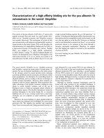
Fig. 1. Schematic representation of the EpoR showing the location of the
eight cytoplasmic tyrosine motifs used in this study (A) and the sequence
of the peptides used for SPR and precipitation assays (B). Phosphoryl-
ation of the tyrosine residue is indicated as (pY), unphosporylated
tyrosines as (Y).
Ó FEBS 2002 SOCS-3 binds to the pY429pY431 motif of Epo-R (Eur. J. Biochem. 269) 2517
chip surface at a flow rate of 20 lLÆmin
)1
for 1 min. Before
injection of SOCS protein, the sensor chip was flushed with
bovine serum albumin (0.1 mgÆmL
)1
)ataflowrateof
20 lLÆmin
)1
for 1 min. For measurement of the K
d
value
the flow rate was enhanced to 100 lLÆmin
)1
in order to
obtain higher resolution of kinetics. For this type of
experiment SOCS-3 was injected for 3 min, dissociation
time was 5 min, regeneration of the chip between the
measurements in all experiments performed was done at
20 lLÆmin
)1
with 1
M
NaCl in 50 m
M
NaOH for 30 s.
Binding curves were analyzed by using
BIAEVALUATION
software v3.0.1 (Biacore). To correct for nonspecific binding
events, an empty sensor surface without peptide was
analyzed in parallel during protein injection. Additionally,
thioredoxin was injected at high concentrations (3.5 l
M
)to
rule out nonspecific interactions of the fusion protein of
SOCS-3. Curves were plotted with subtracted nonspecific
binding. Determination of the dissociation constant was
carried out by Scatchard analysis [22].
Peptide precipitation assay and immunoblot analysis
Approximately 0.15 lmol of the biotinylated peptides were
immobilized by incubation with 2.5 mg NeutrAvidin-
coupled agarose (Pierce, Bonn, Germany). For SOCS-3
precipitation cells were lysed in 500 lL lysis buffer (50 m
M
Tris/HCl, pH 8; 150 m
M
NaCl; 10% glycerol; 0.5% NP-40;
0.1 m
M
EDTA) supplemented with NaF (50 m
M
), pepst-
atin A (2 lgÆmL
)1
), leupeptin (5 lgÆmL
)1
), aprotinin
(5 lgÆmL
)1
), phenylmethanesulfonyl fluoride (1 m
M
)and
Na
3
VO
4
(1 m
M
). Equal amounts of cellular protein and
expressed SOCS-3 in each sample were obtained by mixing
the total cell lysates prior to the precipitation experiment.
SOCS-3 was precipitated by incubation of the total cell
lysates with the immobilized peptides at 4 °Covernight.
Precipitates were then washed three times with 500 lLlysis
buffer. The precipitated proteins were resolved by SDS/
PAGE and transferred to an Immobilon poly(vinylidene
difluoride) membrane (Millipore, Eschborn, Germany)
using a semidry electroblotting apparatus. Human SOCS-
3 was detected with a polyclonal antibody kindly provided
by J. A. Johnston (Queen’s University, Belfast, Northern
Ireland). A polyclonal goat anti-rabbit horse-radish per-
oxidase-conjugated secondary Ig (DAKO, Hamburg,
Germany) was used to visualize the immunoreactive bands
by ECL techniques.
Molecular modeling of the human SOCS-3 SH2 Domain
For molecular modeling and graphic representation of the
protein structures, the programs
WHATIF
[23] and
GRASP
[24]
were used on an Indigo2 SGI computer. Energy minimiza-
tions were performed under vacuum conditions with the
GROMOS
program library (W. F. van Gunsteren, distributed
by BIOMOS Biomolecular Software B.V., Laboratory of
Physical Chemistry, University of Groningen, the Nether-
lands).
The following SH2 domain sequences and structures
were used as templates: human c-src protein-tyrosine
kinase, Brookhaven data bank entry codes 1hcs, 1a1b
and 1shd [25–27]; human phosphatidylinositol 3-kinase
p85 subunit, code 1pic [28], bovine phospholipase C-c1,
code 2pld [29]; human Bcr-abl protein-tyrosine kinase,
code 2abl [30]; murine SHP2 protein-tyrosine phosphatase,
accession no. 1AYA [31]. Initial amino-acid sequence
alignments were performed by the use of the
BLAST
programme [32]. Modifications were then introduced to
meet structural requirements derived from the known SH2
structures. The sequential alignment of the known struc-
tures is based on the direct superposition of their backbone
coordinates.
RESULTS
SOCS-3 binds to the phosphotyrosine motifs pY343,
pY401 and pY429pY431 of the EPO-receptor
We and others have recently shown that SOCS-3 exerts its
inhibitory activity on IL-6 signaling by binding to phos-
photyrosine 759 of gp130 [16,17], which is also the
recruitment site for the phosphotyrosine phosphatase
SHP2 [33]. Moreover, it has been shown that SOCS-3 also
binds to the recruitment site for SHP2 of the erythropoetin
receptor [20]. To determine the binding affinities of SOCS-3
to the EpoR we investigated SOCS-3 binding to tyrosine-
phosphorylated and nonphosphorylated peptides of all
EpoR tyrosine motifs. Figure 1 shows the sequences of the
peptides used in this study. Peptides with two proximate
tyrosine residues were presented as double-phosphorylated
or mutually substituted with phenylalanine to check syner-
gistic effects on SOCS-3 binding. The N-terminal biotinyl-
ated peptides were captured on a SA Biosensor chip and the
interaction with SOCS-3 was analyzed. As control, binding
of SOCS-3 to an unloaded sensor surface was measured in
parallel. Additionally, in all experiments 3.5 l
M
thioredoxin
was injected to rule out nonspecific binding of the fusion
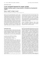
protein. As shown in Fig. 2, we confirmed the binding of
SOCS-3 to phosphotyrosine pY401 recently reported by
Sasaki et al. [20]. We also found that SOCS-3 weakly binds
to a peptide containing pY343, a binding site for STAT5
[34]. Interestingly, SOCS-3 showed high affinity binding to a
phosphopeptide encompassing pY429 and pY431 of the
EpoR (Fig. 2A). Both tyrosines Y429 and Y431 are
phosphorylated after stimulation with Epo [34]. The inter-
action between SOCS-3 and this peptide is phosphoryla-
tion-dependent as a nonphosphorylated peptide Y429Y431
failed to recruit SOCS-3 (Fig. 2A). When either of these two
tyrosines were substituted with phenylalanine SOCS-3
binding was significantly reduced (Table 1). Apart from
the peptides pY343, pY401 and pY429pY431 none of the
other EpoR-tyrosine motifs showed binding to SOCS-3
(data not shown).
SOCS-3 binds with higher affinity to pY429 pY431
than to pY401
As SOCS-3 was recruited by both pY401 and pY429pY431
we determined the affinities of the binding of SOCS-3 to
these receptor motifs. We found that SOCS-3 binds with
ninefold higher affinity to pY429pY431, which contained
two proximate phosphotyrosines, than to pY401, which had
a single phosphotyrosine residue (Table 1). Figure 2B
illustrates the concentration dependent binding of human
SOCS-3 to immobilized pY401 peptide in the range of
0.275–8.8 l
M
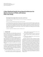
. In Fig. 3A and B, Scatchard plots used to
assess the binding affinity of SOCS-3 to the pY401 and
2518 M. Ho
¨
rtner et al. (Eur. J. Biochem. 269) Ó FEBS 2002
pY429pY431 motifs are shown. Scatchard analysis revealed
that the dissociation constant K
d
is around 9.5 l
M
for the
binding of SOCS-3 to pY401 whereas the pY429pY431
motif bound with a K
d
of 1.1 l
M
(Table 1).
SOCS-3 needs a double phosphorylated Y429Y431
motif for highest affinity binding
The motif pY429pY431 of the EpoR contains two tyrosine
phosphorylation sites, spaced only by one amino-acid
residue (Fig. 1). In order to differentiate between these
two tyrosines in the context of SOCS-binding we deter-
mined binding affinities of SOCS-3 to peptides containing
only phosphotyrosine pY429 or pY431, as well as a double-
phosphorylated peptide pY429pY431. The SPR measure-
ments demonstrated that highest affinity binding of SOCS-3
occurred only if both tyrosine 429 and tyrosine 431 were
phosphorylated (Fig. 3B–D and Table 1).
SOCS-3 specifically binds to the receptor motifs
encompassing tyrosines pY401 and pY429pY431
in COS7 cells
In order to investigate whether SOCS-3 binds to the
receptor motifs containing pY401 and pY429pY431, we
performed a peptide precipitation assay with the biotinyl-
ated EpoR-peptides, which have been shown to interact
with SOCS-3 in the SPR experiments. The nonphosphor-
ylated peptide Y429Y431 was used as control. Equal
amounts of whole cell extracts of COS7 cells expressing
SOCS-3 were incubated with the different EpoR-peptides
immobilized on NeutrAvidin-coupled agarose. Subse-
quently, precipitated SOCS-3 was analyzed by Western
blotting (Fig. 4). SOCS-3 was found to specifically interact
with the tyrosine motifs pY401 and pY429pY431.
pY429pY431 was more potently recruiting SOCS-3 than
pY401, reflecting the high affinity binding determined by
SPR. The nonphosphorylated peptide Y429Y431 failed to
precipitate SOCS-3 as did the phosphopeptide containing
pY343 of the EpoR. This shows that the interaction is
phosphorylation- and sequence-dependent. Single phos-
phorylated peptides of the motif encompassing tyrosines
Y429 and Y431 in which one of the tyrosines has been
exchanged to phenylalanine (pY429F431 and F429pY431)
also readily precipitated SOCS-3, although to a lesser extent
than the double phosphorylated peptide. The single phos-
phorylated peptides were found to precipitate SOCS-3 with
similar efficiency. The peptide precipitation assay suggests
that both phosphotyrosines take part in the interaction with
SOCS-3 and act synergistically.
Model structure of the human SOCS-3 SH2 domain
To understand the binding of the different receptor peptides
to the SOCS-3 SH2 domain at the molecular level and to
explain the distinct binding affinities, we generated a model
structure of the human SOCS-3 SH2 domain based on
solved structures of other SH2 domains. Figure 5 shows an
alignment of the SOCS-3 SH2 domain with the sequences of
the template structures that were used for model building.
The sequence similarity between the SOCS-3 SH2 domain
and the aligned SH2 domains varies between 37 and 41%
and reflects the sequence similarity between the structurally
characterized SH2 domains like Src and SHP2 (40%) or Src
and PLCc (39%) for example. For evaluation of the binding
specificities of the SOCS-3 SH2 domain, we modelled the
complex of the SOCS-3 SH2 domain and the receptor
peptides pY401 and pY429pY431 (Fig. 6). For comparison,
Fig. 2. Comparison of SOCS-3 binding to pY343, pY401 and
pY429pY431 of the human EpoR (A) and sensogram showing the
interaction of serial dilutions of SOCS-3 and peptide pY401 (B).
(A) Biotinylated peptides were immobilized on SA chips, the concen-
tration of SOCS-3 was 8.8 l
M
. (B) SOCS-3 was diluted twofold from
8.8 l
M
to 275 n
M
. Purified thioredoxin was taken as control for
specific binding. Steady state binding values were taken for Scatchard-
analysis for the determination of K
d
values.
Table 1. Calculated K
d
values of the EpoR peptides as determined by
Scatchard analysis. ND, not determined due to a lack of interaction
with SOCS-3.
Peptide K
d
(l
M
)
pY343 > 30
pY401 9.5 ± 0.12
pY429pY431 1.1 ± 0.03
F429pY431 4.6 ± 0.02
pY429F431 4.9 ± 0.02
Y429Y431 ND
pY443 ND
pY461pY464 ND
Y461pY464 ND
pY461Y464 ND
pY479 ND
Ó FEBS 2002 SOCS-3 binds to the pY429pY431 motif of Epo-R (Eur. J. Biochem. 269) 2519
we also considered the binding of the receptor peptide
corresponding to the SOCS-3 recruitment site pY759 of
gp130.
Figure 6A shows the binding of the SOCS-3 SH2 domain
to the peptide pY401 (SFEpYTILDPSS; rod model). The
SOCS-3 SH2 domain is represented as electrostatic potential
map. The phosphotyrosine pY401 is embedded in the
positively charged binding pocket (blue) of the SH2 domain
containing R71 of SOCS-3. In the model structure, phenyl-
alanine at position Y)2 of the peptide contacts G53.
Threonine Y+1 can undergo a hydrophobic contact with
bC of K91 as well as a hydrogen bond with the backbone
NH-group of asparagine N92. The leucine residue at
position Y+3 inserts into a hydrophobic pocket made up
of tyrosine Y127, leucines L93 and L104 and phenylalanine
F136. Furthermore, the proline at position Y+5 is in close
proximity to P108 of SOCS-3. Thus the model suggests that
the major contributions to the specific binding of SOCS-3 to
pY401 originate from amino-acid residues at the positions
Y)2, Y+1 and Y+3.
SOCS-3 binding to the peptide pY429pY431 is represen-
tedinFig.6B.ThepredictedcontactswithintheSH2
domain for the residues at positions Y)2(L)andY+3(L)
of the peptide are similar to pY401. The leucine at Y+1 is
predicted to undergo a hydrophobic contact with the side
chain of K91. In addition, the valine residues at positions
Y+4 and Y+5 contact F136 and P108, respectively. The
Y+2 residue in SH2/peptide interactions is usually exposed
to the solvent and does not contribute to the binding
[35–37]. Most interestingly, in our model the phosphotyr-
osine at Y+2 is able to form a salt bridge with the positively
charged R94 (contact is shown by a red Ô±Õ symbol in
Fig. 6B). This explains our observation that the double
phosphorylated peptide pY429pY431 binds with higher
affinity than a peptide in which pY431 is substituted by
phenylalanine. In addition the side chain of R94 is able to
build up a hydrophobic contact with the aromatic ring of
the phosphotyrosine at position Y+0 (contact shown as a
red ÔhÕ in Fig. 6B). Taken together, the contributions of the
positions Y+2, Y+4 and Y+5 appear to account for most
Fig. 3. Scatchard analysis of SOCS-3 interaction with EpoR peptides pY401 (A), pY429pY431 (B), pY429F431 (C), and F429pY431 (D). Plateau
values of the binding curves with serial dilutions of SOCS-3 (30, 15, 7.5, 3.75, 1.9 and 0.9 l
M
) were taken for calculation of the K
d
values.
Fig. 4. SOCS-3 selectively binds to tyrosine-phosphorylated peptides
corresponding to the pY401 and pY429pY431 motifs of EpoR. COS7
cells were transfected with an expression vector for human SOCS-3
(5 lg). Thirty-six hours after transfection cellular extracts were pre-
pared and incubated with biotinylated peptides corresponding to the
EpoR motifs encompassing Y343, Y401 and Y429Y431 immobilized
on NeutrAvidin-coupled agarose. After precipitation the proteins were
subjected to Western blot analysis using a polyclonal anti-(SOCS-3) Ig
to detect coprecipitated SOCS-3.
2520 M. Ho
¨
rtner et al. (Eur. J. Biochem. 269) Ó FEBS 2002
of the higher affinity of pY429pY431 to SOCS-3 in
comparison to the peptide pY401. The SOCS-3 residues
important for the binding of the different peptide residues
are highlighted in Fig. 6A.
As we and others have recently shown that SOCS-3 binds
to gp130 through its SH2 domain [16,17], we have also
modeled a peptide encompassing the phosphotyrosine 759
of gp130 to the SOCS-3 SH2 domain (Fig. 6C). The model
suggests that this peptide binds in a way very similar to
pY429pY431 with the residues Y)2, Y+3, Y+4 and Y+5
building up hydrophobic contacts to the SOCS-3 SH2
domain. Serine at position Y+1 forms a hydrogen bond
with the backbone of asparagine N92.
In order to check the reliability of our model structure we
generated several point mutations within the SOCS-3 SH2
domain and performed a peptide precipitation assay using
the phosphorylated peptides pY429pY431, pY429F431 and
F429pY431 (Fig. 7). Total cell lysates (TCL) of 293T cells
expressing wild-type SOCS-3 or SOCS-3 mutants (R94E,
L93A, L58A and G53V) were incubated with the biotinyl-
ated peptide pY429pY431 immobilized on NeutrAvidin-
coupled agarose. Subsequently, precipitated SOCS-3 was
analyzed by Western blotting. Figure 7A shows that the
SOCS-3 mutant L58A, which we predicted not to affect
peptide binding, can be precipitated with pY429pY431 to
the same extent as wild-type SOCS-3. The point mutations
R94E, L93A and G53V that we expected to play a role in
peptide recognition all impair SOCS-3 precipitation with
R94E and G53V most strongly affecting the interaction
between SOCS-3 and pY429pY431 (Fig. 7A).
To better assess the binding mode of the peptides
pY429pY431, pY429F431 or F429pY431, we performed a
peptide precipitation assay with wild-type SOCS-3 or
the SOCS-3 R94E mutant (Fig. 7B). Again we find that
the mutation of arginine 94 to glutamic acid strongly affects
the interaction of SOCS-3 with the double phosphorylated
peptide pY429pY431 (pYpY). In comparison, the muta-
tion only marginally reduces the interaction with the
single phosphorylated peptides pY429F431 (pYF) and
F429pY431 (FpY) suggesting that both phosphotyrosines
bind to the phosphotyrosine binding pocket of the SH2
domain, with R94 only playing a minor role in the
binding of these peptides.
DISCUSSION
The cytoplasmic part of the EpoR contains eight tyrosine
residues that serve as recruitment sites for a number of SH2
domain containing proteins. Among these are the protein-
tyrosine phosphatases SHP1 and SHP2 [38,39], the Jak2
and PI3 kinases [40,41] as well as STAT5, CIS and SOCS-3
[4,20,42]. In order to study binding of SOCS-3 to the EpoR
we used a biochemical approach by means of SPR
measurements. For this purpose, tyrosine phosphorylated
and nonphosphorylated peptides of all eight tyrosine motifs
of the human EpoR were immobilized on a sensor chip and
the interaction with SOCS-3 was analyzed. To further
validate the SPR data obtained, in vitro binding assays in
eukaryotic cells were performed.
The results from our SPR experiments show that SOCS-3
binds to pY343, pY401 and pY429pY431 with different
affinities (Table 1). The phosphotyrosine peptides of all
other EpoR tyrosine motifs did not show significant binding
to SOCS-3. In the SPR experiments the weakest interaction
of SOCS-3 was observed with peptide pY343, a motif that
has been shown to recruit STAT5 [41]. The dissociation
constant for this binding event was greater than 30 l
M
.In
this case the exact K
d
value was not assessed by Scatchard
analysis because the highest SOCS-3 concentration was
30 l
M
and a calculation by the
BIAEVALUATION
software
Fig. 5. Alignment of the SOCS-3 SH2 domain with other SH2 domains. The sequence of the human SOCS-3 SH2 domain was aligned with the SH2
domains of the human c-src protein-tyrosine kinase [25–27], the human phosphatidylinositol 3-kinase p85 subunit [28], the bovine phospholipase
C-c [29], the human Bcr-abl protein-tyrosine [30] as well as with the N-terminal SH2 domains of murine SHP1 and SHP2 protein-tyrosine
phosphatases [31,50]. Secondary structure characteristics are given on top following the common nomenclature [37]. SOCS-3 amino-acid numbers
(italic) precede the sequence. The sequence homology (%) between the SOCS-3 SH2 domain and the aligned sequences is indicated in parentheses.
Residues that are highly conserved within the represented sequences are highlighted (bold characters). Blue and red characters indicate residues
conserved in SH2 domains to at least 30% or 80%, respectively (software:
MULTALIN
v5.4.1 [51]). Residues interacting with the phosphotyrosine as
suggested by the model structure are represented by closed circles. The open arrowhead highlights the amino acid in the aA helix that contacts the
residue Y)2. The amino acids postulated to interact with the peptide residues Y+1, Y+2, Y+3 Y+4 and Y+5 are indicated by the numbers 1, 2,
3, 4 and 5, respectively.
Ó FEBS 2002 SOCS-3 binds to the pY429pY431 motif of Epo-R (Eur. J. Biochem. 269) 2521
was not possible because the sensograms could not be fitted
to an ideal binding model. Confirming results were obtained
from peptide precipitation assays, as we were not able to
precipitate SOCS-3 out of COS7 cells overexpressing
human SOCS-3 (Fig. 4). This indicates that the pY343
motif does not play a role with respect to SOCS-3
recruitment. In the context of IL-6 signaling, SOCS-3 has
been found to act as potential competitor to SHP2 for the
binding of the same tyrosine Y759 in the gp130 receptor
subunit [16,17]. Additionally, it was recently shown that the
binding site of SHP2 in the EpoR, Y401 also recruits
SOCS-3, which results in the down-regulation of the Epo
signaling [20]. In our experiments, we confirm binding of
SOCS-3 to pY401, with a calculated K
d
for this interaction
in the range of 9.5 l
M
(Table 1). Concerning EpoR
signaling, SHP2 is suggested to positively regulate prolifer-
ation [43]. As we found that SOCS-3 binds to the same
phosphotyrosine of gp130 as SHP2, which negatively
regulates IL-6 signaling [16], we asked whether SOCS-3
would likewise compete with another negative regulator in
the EpoR context, namely SHP1. SHP1 is closely related to
SHP2 and is recruited to the pY429pY431 motif of the
EpoR after stimulation, whereas pY429 is the higher affinity
binding site for the phosphatase [38]. Both pY429 and
pY431 are phosphorylated after stimulation with Epo [34].
Fig. 6. Model structure of the SOCS-3 SH2 complexed with phospho-
tyrosine peptides. Electrostatic potential maps of the model structure of
the human SOCS-3 SH2 domain complexed with peptides corres-
ponding to the pY401 and the pY429pY431 motifs of the EpoR as well
as the pY759 motif of gp130. Red and blue-coloured regions on the
structure surface of the SH2 domain indicate negative and positive
charges, respectively. Bound phosphopeptides are represented as rod
models with nitrogen, oxygen, carbon, and phosphorous atoms being
coloured in blue, red, white, and yellow, respectively. The N- and
C-termini of the bound peptides are indicated in italic. (A) Interaction
of the SOCS-3 SH2 domain with the SFE(pY401)TILDPSS motif of
EpoR (B) with the EpoR motif HLK(pY429)L(pY431)LVVSS and
(C) with the TVQ(pY759)STVVHSG motif of gp130. The positions of
the amino acids of SOCS-3 relevant for the interaction with the peptide
is indicated. The hydrophobic contact (h) between the side chain of
R94 and pY429 as well as the salt bridge (±) between R94 and pY431
are highlighted in red.
Fig. 7. The SOCS-3 point mutations G53V, L93A and R94E affect the
binding to phosphotyrosine peptides. 293T cells were transfected with an
expression vector for wild-type SOCS-3 or SOCS-3 mutants (5 lg).
36 h after transfection cellular extracts were prepared and incubated
with biotinylated peptides immobilized on NeutrAvidin-coupled
agarose. After precipitation the proteins were subjected to Western
blot analysis using a polyclonal SOCS-3 antibody to detect copreci-
pitated SOCS-3. Detection of total cell lysates (TCL) with a SOCS-3
antibody was used to check the expression levels of the different
mutants. (A) Precipitation of SOCS-3 WT or the SOCS-3 point
mutations G53V, L58A, L93A and R94E with the peptide
pY429pY431 (pYpY). (B) Precipitation of SOCS-3 WT or SOCS-3
R94E with the peptides pY429pY431 (pYpY), pY429F431 (pYF) and
F429pY431 (FpY).
2522 M. Ho
¨
rtner et al. (Eur. J. Biochem. 269) Ó FEBS 2002
In Epo signal transduction, SHP1 has been reported to
negatively regulate proliferation and differentiation of
Ba/F3 or SKT6 cells [38,44]. Interestingly, we found a
peptide encompassing the double phosphorylated tyrosine
motifpY429pY431tobindSOCS-3withaK
d
of 1.1 l
M
,a
ninefold higher affinity than determined for pY401. Single
phosphorylated peptides pY429 and pY431 revealed K
d
values in the range of 5 l
M
. We were able to confirm this
finding by the use of a peptide precipitation assay. SOCS-3
was coprecipitated with both the double phosphorylated
peptide pY429pY431 as well as the single phosphorylated
motifs, with pY429pY431 precipitating SOCS-3 most
potently (Fig. 4). As the proximity of pY431 to pY429
impedes the simultaneous recruitment of two SOCS-3 SH2
domains to this double tyrosine motif, the two phospho-
tyrosine residues must contact the same SH2 domain,
thereby both contributing to the high affinity binding.
In order to better evaluate the results obtained in the SPR
experiments and the peptide precipitation assay, we gener-
ated a model structure of the human SOCS-3 SH2 domain
complexed with peptides corresponding to the receptor
motifs pY401 and pY429pY431 of the EpoR as well as to
the SOCS-3 recruiting motif pY759 of gp130 (Fig. 6).
Critical positions for specific binding of SH2 domains to
phosphotyrosine motifs are the amino acids surrounding
the phosphotyrosine residues. The C-terminal amino-acid
residues at positions Y+1 to Y+5 of bound peptides have
been shown to be important for the interaction with SH2
domains, with positions Y+1 and Y+3 having the greatest
impact on the binding [35,37]. Table 2 shows the sequences
of several receptor phosphotyrosine motifs that have been
shown to bind SOCS-3 [16–20] (and this study). Based on
the model structure, we determined the residues involved in
specific binding to the SOCS-3 SH2 domain (Table 2).
Positions Y)2, Y+1, Y+3, Y+4 as well as Y+5 all
contribute to the interaction with residues Y)2, Y+1 and
Y+3 being most crucial for specific binding. This is
supported by a recent report investigating the binding of
SOCS-3 to the gp130 tyrosine motif pY759 where these
amino-acid residues have also been found to contribute to
the specific recruitment of SOCS-3 [17]. In regard to the
overlapping binding specificities of SOCS-3 and the two
phosphatases SHP1 and SHP2, the position Y)2ofthe
interacting phosphotyrosine motif seems to play an import-
ant role. Although the two phosphatases bind to different
tyrosine motifs within the EpoR, they are recruited to the
same phosphotyrosine pY612 of the common b chaininthe
context of IL-3 signaling [45]. A common feature of SHP1
and SHP2 recruiting motifs is a hydrophobic residue at
position Y)2 of the binding phosphotyrosine sequence. It
has been shown that this residue is filling a gap created by a
glycine in helix aA within the SH2 domain of the
phosphatase [31,46–48]. The glycine is conserved in the
N- and C-terminal SH2 domains of both SHP1 and SHP2
and is required for the unusual involvement of the residue
Y)2 of the binding phosphotyrosine motif [47]. Most
interestingly, the glycine residue in helix aA of the SH2
domain is conserved in SOCS-3 and has recently been
shown to contribute to the binding of SOCS-3 to gp130 [17].
As illustrated in Table 2, all receptor tyrosine motifs that
have been shown to bind SOCS-3 contain a hydrophobic
residue at position Y)2. The model structure of the SOCS-3
SH2 domain shows that this residue can easily be fitted into
a gap created by G53 of SOCS-3. In regard to the position
Y+1 of the interacting motifs, we suggest a hydrophobic
residue contacting the side chain of K91 or alternatively a
small polar residue like serine or threonine making a
hydrogen bond with the backbone of the b strand D to be
most favourable for peptide recognition. For the positions
Y+3 to Y+5, a hydrophobic residue seems optimal for
SOCS-3 recruitment with Y+3 contributing most to high
affinity binding.
In order to check the reliability of our model structure we
generated several point mutations within the SH2 domain
of SOCS-3. We mutated L58, which we predicted not to be
involved in peptide binding, as well as the residues G53, L93
and R94, which according to our model contact the peptide
positions Y)2, Y+3 and Y+2, respectively. A peptide
precipitation assay confirms the reliability of our model
structure (Fig. 7). Whereas L58A does not affect peptide
binding, the point mutation G53V strongly impairs the
interaction between SOCS-3 and the peptide. According to
our model structure the valine prevents optimal binding of
the peptide by sterically interfering with the hydrophobic
residue at position Y)2 of the phosphotyrosine peptide. L93
is part of a hydrophobic pocket also involving Y127, L104
and F136 that accommodates the peptide position Y+3.
The fact that the mutation of leucine 93 to alanine reduces
the interaction with pY429pY431 further confirms our
model structure. We propose arginine 94 to provide a
double contact with the peptide pY429pY431. First, the side
chain makes a hydrophobic contact with the aromatic ring
of pY429 (contact shown as a red ÔhÕ in Fig. 6B) and thereby
contributes to the binding of pY429 into the phospho-
tyrosine binding pocket of the SH2 domain. This interaction
is likely to occur for every phosphotyrosine that is
embedded in the classical phosphotyrosine binding pocket
of the SOCS-3 SH2 domain and can also be found in other
SH2 domains as demonstrated by the solved structures of
Src [25] and SHP2 [31], for example. Second, we postulate
the positively charged R94 to form a salt bridge with the
negatively charged phosphotyrosine at position Y+2 of
the pY429pY431 motif (contact shown as red Ô±Õ in
Fig. 6B). The point mutation R94E (which should only
marginally affect the interaction ÔhÕ but would impede
the contact Ô±Õ) drastically affected SOCS-3 binding to the
double phosphorylated peptide pY429pY431 in our peptide
Table 2. Sequence comparison of receptor phosphotyrosine motifs
known to recruit SOCS-3. Bold characters highlight residues favour-
able for selective binding to the SOCS-3 SH2 domain. h, hydrophobic
residue.
Receptor pY location sequence
h-gp130 pY759
STV Q pY S T VVHSG
h-EpoR pY401 ASF E pY T I L D P SS
h-EpoR pY429 PHL K pY L pY L V V SD
m-LeptinR pY985 PSV K pY A T LVSND
m-LeptinR pY1077 KSV C pY L G V TSVN
Consensus
sequence
h X pY h X Lhh
SV
T
Position
relative
to pY
)20+1 +2 +3+4+5
Ó FEBS 2002 SOCS-3 binds to the pY429pY431 motif of Epo-R (Eur. J. Biochem. 269) 2523
precipitation assay (Fig. 7A,B). In contrast the binding of
the single phosphorylated peptides pY429F431 and
F429pY431 is only weakly affected by the mutation of
arginine 94 to glutamic acid (Fig. 7B). This indicates that
for both peptides the phosphotyrosine residue binds into the
classical phosphotyrosine binding pocket. The binding of
the peptide F429pY431 thus involves a shift of two residues
when compared to the binding mode of pY429pY431 with
pY431 binding into the classical pY-binding pocket and
F429 (position Y)2) filling the gap created by G53 of the
SH2 domain. In the context of the activated EpoR (and
peptide pY429pY431), where both tyrosines are phosphor-
ylated, this binding mode would be unfavourable because of
the presence of pY429 at position Y)2. The peptide
precipitation assay with the pY429pY431 motif (Fig. 7A,B)
supports the idea that arginine 94 plays an important role in
the recognition of the double phosphorylated motif by
forming a salt bridge with phosphotyrosine pY431.
Based on the identified binding motifs for SOCS-3 and our
model structure, we propose a consensus motif h-X-pY-h/S/
T-X-L/V-h-h (with h ¼ hydrophobic) optimal for SOCS-3
recruitment (see also Table 2). Remarkably, in the case of the
EpoR motif pY429pY431, we find pY431 at position Y+2
contributes to SOCS-3 binding. Similar co-operative effects
of two proximal phosphotyrosine residues have been repor-
ted to increase the binding of the platelet-derived growth
factor (PDGF) b-receptor to the SH2 domain of the Src
family kinases [49]. Mori et al. found a double phosphor-
ylated tyrosine motif encompassing tyrosines Y579 and
Y581 to recruit and activate the kinases of the Src family
more potently than the corresponding single phosphorylated
motifs. The authors discuss the phosphorylation of tyrosine
Y581 creating a more favourable conformation of the
sequence surrounding the tyrosines Y579 and Y581, thereby
increasing binding affinity. Interestingly, the EpoR contains
a similar phosphotyrosine arrangement pattern. The plas-
mon resonance studies, peptide precipitation assays as well
as the model structures presented in this report, suggest that
phosphotyrosine pY429 binds into the classical phospho-
tyrosine binding pocket of the SOCS-3 SH2 domain between
helix aA and the central b sheet. pY431 appears to increase
the binding affinity by providing an additional contact with
the SH2 domain of SOCS-3 involving R94. A conforma-
tional change induced by the phosphorylation of tyrosine
Y431 may also contribute to the increase in binding affinity
compared to the single phosphorylated peptide. The posi-
tively charged residue (R94 in SOCS-3) in b strand D is
conserved (R/K) in a large number of SH2 domains. As the
members of the Src family also carry a positively charged
residue at this position, we favour the idea that the enhanced
binding of Src family kinases to the pY579pY581 motif of
the PDGF b-receptor reported by Mori et al. [49] may be
due to the formation of a salt bridge between pY581 and the
lysine residue in b strand D of the SH2 domain of the Src
kinases. The co-operative binding mode that we describe
may thus represent a more general binding mechanism by
which SH2 domains achieve high affinity binding to motifs
with proximal phosphotyrosine residues.
Our data strongly suggest that SOCS-3 binds to more
than one binding site to the EpoR. As shown by SPR
measurements as well as in vitro binding assays the double
phosphorylated motif pY429pY431 in the EpoR seems to
be the preferred binding site for SOCS-3. The implications
of the regulatory proteins SOCS-3, SHP2 and SHP1 sharing
recruitment sites on the EpoR will be subject to further
investigations.
ACKNOWLEDGEMENTS
We thank Joachim Gro
¨
tzinger for valuable advice concerning the
generation of the model structures, James A. Johnston for providing
the polyclonal SOCS-3 antibody and Fred Schaper for helpful
discussions. This work was supported by grants from the Deutsche
Forschungsgemeinschaft (Bonn, Germany) and the Fonds der
Chemischen Industrie (Frankfurt/Main, Germany).
REFERENCES
1. Lai, P.H., Everett, R., Wang, F.F., Arakawa, T. & Goldwasser, E.
(1986) Structural characterization of human erythropoietin.
J. Biol. Chem 261, 3116–3121.
2. Krantz, S.B. (1991) Erythropoietin. Blood 77, 419–434.
3. Wojchowski, D.M., Gregory, R.C., Miller, C.P., Pandit, A.K. &
Pircher, T.J. (1999) Signal transduction in the erythropoietin
receptor system. Exp Cell Res. 253, 143–156.
4. Yoshimura, A., Ohkubo, T., Kiguchi, T., Jenkins, N.A., Gilbert,
D.J., Copeland, N.G., Hara, T. & Miyajima, A. (1995) A novel
cytokine-inducible gene CIS encodes an SH2-containing protein
that binds to tyrosine-phosphorylated interleukin 3 and erythro-
poietin receptors. EMBO J. 14, 2816–2826.
5. Starr, R., Willson, T.A., Viney, E.M., Murray, L.J., Rayner, J.R.,
Jenkins, B.J., Gonda, T.J., Alexander, W.S., Metcalf, D., Nicola,
N.A. & Hilton, D.J. (1997) A family of cytokine-inducible
inhibitors of signalling. Nature 387, 917–921.
6. Endo, T.A., Masuhara, M., Yokouchi, M., Suzuki, R., Sakamoto,
H., Mitsui, K., Matsumoto, A., Tanimura, S., Ohtsubo, M.,
Misawa, H., Miyazaki, T., Leonor, N., Taniguchi, T., Fujita, T.,
Kanakura, Y., Komiya, S. & Yoshimura, A. (1997) A new protein
containing an SH2 domain that inhibits JAK kinases. Nature 387,
921–924.
7. Naka,T.,Narazaki,M.,Hirata,M.,Matsumoto,T.,Minamoto,
S., Aono, A., Nishimoto, N., Kajita, T., Taga, T., Yoshizaki, K.,
Akira, S. & Kishimoto, T. (1997) Structure and function of a new
STAT-induced STAT inhibitor. Nature 387, 924–929.
8. Hilton, D.J., Richardson, R.T., Alexander, W.S., Viney, E.M.,
Willson, T.A., Sprigg, N.S., Starr, R., Nicholson, S.E., Metcalf, D.
& Nicola, N.A. (1998) Twenty proteins containing a C-terminal
SOCS box form five structural classes. Proc. Natl Acad. Sci. USA
95, 114–119.
9. Masuhara, M., Sakamoto, H., Matsumoto, A., Suzuki, R.,
Yasukawa, H., Mitsui, K., Wakioka, T., Tanimura, S., Sasaki, A.,
Misawa, H., Yokouchi, M., Ohtsubo, M. & Yoshimura, A. (1997)
Cloning and characterization of novel CIS family genes. Biochem.
Biophys. Res. Commun 239, 439–446.
10. Minamoto, S., Ikegame, K., Ueno, K., Narazaki, M., Naka, T.,
Yamamoto, H., Matsumoto, T., Saito, H., Hosoe, S. & Kishi-
moto, T. (1997) Cloning and functional analysis of new members
of STAT induced STAT inhibitor (SSI) family: SSI-2 and SSI-3.
Biochem. Biophys. Res. Commun 237, 79–83.
11. Zhang, J.G., Farley, A., Nicholson, S.E., Willson, T.A., Zugaro,
L.M., Simpson, R.J., Moritz, R.L., Cary, D., Richardson, R.,
Hausmann, G., Kile, B.J., Kent, S.B., Alexander, W.S., Metcalf,
D., Hilton, D.J., Nicola, N.A. & Baca, M. (1999) The conserved
SOCS box motif in suppressors of cytokine signaling binds to
elongins B and C and may couple bound proteins to proteasomal
degradation. Proc. Natl Acad. Sci. USA 96, 2071–2076.
12. Okabe, S., Tauchi, T., Morita, H., Ohashi, H., Yoshimura, A. &
Ohyashiki, K. (1999) Thrombopoietin induces an SH2-containing
protein, CIS1, which binds to Mpl: involvement of the ubiquitin
proteosome pathway. Exp. Hematol. 27, 1542–1547.
2524 M. Ho
¨
rtner et al. (Eur. J. Biochem. 269) Ó FEBS 2002
13. Matsumoto, A., Masuhara, M., Mitsui, K., Yokouchi, M.,
Ohtsubo, M., Misawa, H., Miyajima, A. & Yoshimura, A. (1997)
CIS, a cytokine inducible SH1 protein, is a target of the JAK-
STAT5 pathway and modulates STAT5 activation. Blood 89,
3148–3154.
14. Verdier, F., Chretien, S., Muller, O., Varlet, P., Yoshimura, A.,
Gisselbrecht, S., Lacombe, C. & Mayeux, P. (1998) Proteasomes
regulate erythropoietin receptor and signal transducer and
activator of transcription 5 (STAT5) activation. Possible
involvement of the ubiquitinated CIS protein. J. Biol. Chem 273,
28185–28190.
15. Sasaki, A., Yasukawa, H., Suzuki, A., Kamizono, S., Syoda, T.,
Kinjyo, I., Sasaki, M., Johnston, J.A. & Yoshimura, A. (1999)
Cytokine-inducible SH2 protein-3 (CIS3/SOCS3) inhibits Janus
tyrosine kinase by binding through the N-terminal kinase inhibi-
tory region as well as SH2 domain. Genes Cells 4, 339–351.
16. Schmitz, J., Weissenbach, M., Haan, S., Heinrich, P.C. & Schaper,
F. (2000) SOCS3 exerts its inhibitory function on interleukin-6
signal transduction through the SHP2 recruitment site of gp130.
J. Biol. Chem. 275, 12848–12856.
17. Nicholson, S.E., De Souza, D., Fabri, L.J., Corbin, J., Willson,
T.A., Zhang, J.G., Silva, A., Asimakis, M., Farley, A., Nash,
A.D., Metcalf, D., Hilton, D.J., Nicola, N.A. & Baca, M. (2000)
Suppressor of cytokine signaling-3 preferentially binds to the
SHP-2-binding site on the shared cytokine receptor subunit gp130.
Proc. Natl Acad. Sci. USA 97, 6493–6498.
18. Bjørbaek, C., Lavery, H.J., Bates, S.H., Olson, R.K., Davis, S.M.,
Flier, J.S. & Myers, M.G. Jr (2000) SOCS3 mediates feedback
inhibition of the leptin receptor via Tyr985. J. Biol. Chem 275,
40649–40657.
19. Eyckerman, S., Broekaert, D., Verhee, A., Vandekerckhove, J. &
Tavernier, J. (2000) Identification of the Y985 and Y1077 motifs
as SOCS3 recruitment sites in the murine leptin receptor. FEBS
Lett. 486, 33–37.
20. Sasaki, A., Yasukawa, H., Shouda, T., Kitamura, T., Dikic, I. &
Yoshimura, A. (2000) CIS3/SOCS-3 suppresses erythropoietin
(EPO) signaling by binding the EPO receptor and JAK2. J. Biol.
Chem 275, 29338–29347.
21. Sambrook, J. & Russell, D.W. (2001) Molecular Cloning:
a Laboratory Manual. Cold Spring Harbor Laboratory Press,
Cold Spring Harbor, New York.
22. Payne, G., Shoelson, S.E., Gish, G.D., Pawson, T. & Walsh, C.T.
(1993) Kinetics of p56lck and p60src Src homology 2 domain
binding to tyrosine-phosphorylated peptides determined by a
competition assay or surface plasmon resonance. Proc. Natl Acad.
Sci. USA 90, 4902–4906.
23. Vriend, G. (1990) WHAT IF: a molecular modeling and drug
design program. J. Mol Graph. 8, 52–56.
24. Nicholls, A., Sharp, K.A. & Honig, B. (1991) Protein folding and
association: insights from the interfacial and thermodynamic
properties of hydrocarbons. Proteins 11, 281–293.
25. Xu, R.X., Word, J.M., Davis, D.G., Rink, M.J., Willard, D.H.J.
& Gampe, R.T.J. (1995) Solution structure of the human pp60c-
src SH2 domain complexed with a phosphorylated tyrosine pen-
tapeptide. Biochemistry 34, 2107–2121.
26. Charifson, P.S., Shewchuk, L.M., Rocque, W., Hummel, C.W.,
Jordan, S.R., Mohr, C., Pacovsky, G.J., Peel, M.R., Rordriguez,
M., Sternbach, D.D. & Consler, T.G. (1997) Peptide ligands of
PP60 (C-SRC) SH2 domains: a thermodynamic and structural
study. Biochemistry 36, 6283.
27. Gilmer, T., Rodriguez, M., Jordan, S., Crosby, R., Alligood, K.,
Green,M.,Kimery,M.,Wagner,C.,Kinder,D.&Charifson,P.
(1994) Peptide inhibitors of src SH3–SH2–phosphoprotein inter-
actions. J. Biol. Chem. 269, 31711–31719.
28. Breeze,A.L.,Kara,B.V.,Barratt,D.G.,Anderson,M.,Smith,
J.C., Luke, R.W., Best, J.R. & Cartlidge, S.A. (1996) Structure of
a specific peptide complex of the carboxy-terminal SH2 domain
from the p85 alpha subunit of phosphatidylinositol 3-kinase.
EMBO J. 15, 3579–3589.
29. Pascal, S.M., Singer, A.U., Gish, G., Yamazaki, T., Shoelson,
S.E., Pawson, T., Kay, L.E. & Forman-Kay, J.D. (1994) Nuclear
magnetic resonance structure of an SH2 domain of phospholipase
C-gamma1complexedwithahighaffinitybindingpeptide.Cell
77, 461–472.
30. Nam, H.J., Haser, W.G., Roberts, T.M. & Frederick, C.A. (1996)
Intramolecular interactions of the regulatory domains of the Bcr-
Abl kinase reveal a novel control mechanism. Structure 4, 1105–
1114.
31. Lee, C H., Kominos, D., Jacques, S., Margolis, B., Schlessinger,
J., Shoelson, S.E. & Kuriyan, J. (1994) Crystal structures of pep-
tide complexes of the amino-terminal SH2 domain of the SYP
tyrosine phosphatase. Structure 2, 423.
32. Altschul, S.F., Gish, W., Miller, W., Myers, E.W. & Lipman, D.J.
(1990) Basic local alignment search tool. J. Mol Biol. 215, 403–410.
33. Fuhrer, D.K., Feng, G.S. & Yang, Y.C. (1995) Syp associates with
gp130 and Janus kinase 2 in response to interleukin-11 in 3T3-L1
mouse preadipocytes. J. Biol. Chem. 270, 24826–24830.
34. Klingmu
¨
ller, U., Bergelson, S., Hsiao, J.G. & Lodish, H.F. (1996)
Multiple tyrosine residues in the cytosolic domain of the erythro-
poietin receptor promote activation of STAT5. Proc. Natl Acad.
Sci. USA 93, 8324–8328.
35. Songyang, Z., Shoelson, S.E., Chaudhuri, M., Gish, G., Pawson,
T.,Haser,W.G.,King,F.,Roberts,T.,Ratnofsky,S.,Lechleider,
R.J. et al. (1993) SH2 domains recognize specific phosphopeptide
sequences. Cell 72, 767–778.
36. Songyang, Z., Shoelson, S.E., McGlade, J., Olivier, P., Pawson,
T.,Bustelo,X.R.,Barbacid,M.,Sabe,H.,Hanafusa,H.&Yi,T.
(1994) Specific motifs recognized by the SH2 domains of Csk,
3BP2, fps/fes, GRB-2, HCP, SHC, Syk, and Vav. Mol. Cell Biol.
14, 2777–2785.
37. Eck, M.J., Shoelson, S.E. & Harrison, S.C. (1993) Recognition of
a high-affinity phosphotyrosyl peptide by the Src homology-2
domain of p56lck. Nature 362, 87–91.
38. Klingmu
¨
ller, U., Lorenz, U., Cantley, L.C., Neel, B.G. & Lodish,
H.F. (1995) Specific recruitment of SH-PTP1 to the erythropoietin
receptor causes inactivation of JAK2 and termination of pro-
liferative signals. Cell 80, 729–738.
39. Tauchi, T., Feng, G.S., Shen, R., Hoatlin, M., Bagby, G.C. Jr,
Kabat, D., Lu, L. & Broxmeyer, H.E. (1995) Involvement of SH2-
containing phosphotyrosine phosphatase Syp in erythropoietin
receptor signal transduction pathways. J. Biol. Chem. 270, 5631–
5635.
40. Witthuhn, B.A., Quelle, F.W., Silvennoinen, O., Yi, T., Tang, B.,
Miura, O. & Ihle, J.N. (1993) JAK2 associates with the ery-
thropoietin receptor and is tyrosine phosphorylated and activated
following stimulation with erythropoietin. Cell 74, 227–236.
41. Damen, J.E., Cutler, R.L., Jiao, H., Yi, T. & Krystal, G. (1995)
Phosphorylation of tyrosine 503 in the erythropoietin receptor
(EpR) is essential for binding the P85 subunit of phosphatidyl-
inositol (PI) 3-kinase and for EpR-associated PI 3-kinase activity.
J. Biol. Chem 270, 23402–23408.
42. Damen,J.E., Wakao,H., Miyajima,A., Krosl,J., Humphries,R.K.,
Cutler, R.L. & Krystal, G. (1995) Tyrosine 343 in the erythro-
poietin receptor positively regulates erythropoietin-induced cell
proliferation and Stat5 activation. EMBO J. 14, 5557–5568.
43. Tauchi, T., Damen, J.E., Toyama, K., Feng, G.S., Broxmeyer,
H.E. & Krystal, G. (1996) Tyrosine 425 within the activated ery-
thropoietin receptor binds Syp, reduces the erythropoietin
required for Syp tyrosine phosphorylation, and promotes mito-
genesis. Blood 87, 4495–4501.
44. Sharlow, E.R., Pacifici, R., Crouse, J., Batac, J., Todokoro, K. &
Wojchowski, D.M. (1997) Hematopoietic cell phosphatase nega-
tively regulates erythropoietin-induced hemoglobinization in ery-
throleukemic SKT6 cells. Blood 90, 2175–2187.
Ó FEBS 2002 SOCS-3 binds to the pY429pY431 motif of Epo-R (Eur. J. Biochem. 269) 2525
45. Bone, H., Dechert, U., Jirik, F., Schrader, J.W. & Welham, M.J.
(1997) SHP1 and SHP2 protein-tyrosine phosphatases associate
with bc after interleukin-3-induced receptor tyrosine phosphory-
lation. Identification of potential binding sites and substrates.
J. Biol. Chem. 272, 14470–14476.
46. Huyer, G., Li, Z.M., Adam, M., Huckle, W.R. & Ramachandran,
C. (1995) Direct determination of the sequence recognition
requirements of the SH2 domains of SH-PTP2. Biochemistry 34,
1040–1049.
47. Huyer, G. & Ramachandran, C. (1998) The specificity of the
N-terminal SH2 domain of SHP-2 is modified by a single point
mutation. Biochemistry 37, 2741–2747.
48. Beebe, K.D., Wang, P., Arabaci, G. & Pei, D. (2000) Determi-
nation of the binding specificity of the SH2 domains of protein
tyrosine phosphatase SHP-1 through the screening of a combi-
natorial phosphotyrosyl peptide library. Biochemistry 39, 13251–
13260.
49. Mori, S., Ronnstrand, L., Yokote, K., Engstrom, A., Court-
neidge, S.A., Claesson-Welsh, L. & Heldin, C.H. (1993) Identifi-
cation of two juxtamembrane autophosphorylation sites in the
PDGF beta-receptor; involvement in the interaction with Src
family tyrosine kinases. EMBO J. 12, 2257–2264.
50. Plutzky, J., Neel, B.G. & Rosenberg, R.D. (1992) Isolation of a src
homology 2-containing tyrosine phosphatase. Proc. Natl Acad.
Sci. USA 89, 1123–1127.
51. Corpet, F. (1988) Multiple sequence alignment with hierarchical
clustering. Nucleic Acids Res. 16, 10881–10890.
2526 M. Ho
¨
rtner et al. (Eur. J. Biochem. 269) Ó FEBS 2002