Báo cáo Y học: Substrate selectivity and sensitivity to inhibition by FK506 and cyclosporin A of calcineurin heterodimers composed of the a or b catalytic subunit potx
Bạn đang xem bản rút gọn của tài liệu. Xem và tải ngay bản đầy đủ của tài liệu tại đây (323.14 KB, 9 trang )
Substrate selectivity and sensitivity to inhibition by FK506
and cyclosporin A of calcineurin heterodimers composed
of the a or b catalytic subunit
Brian A. Perrino
1
, Andrew J. Wilson
2
, Patricia Ellison
3
and Lucie H. Clapp
2
1
Department of Physiology & Cell Biology, University of Nevada School of Medicine, Reno, NV, USA;
2
Center for Clinical
Pharmacology, University College London, UK;
3
Department of Biochemistry, University of Nevada School of Medicine,
Reno, NV, USA
The calcineurin (CaN) a and b catalytic subunit isoforms are
coexpressed within almost all cell types. The enzymatic
properties of CaN heterodimers comprised of the regulatory
B subunit (CnB) with either the a or b catalytic subunit were
compared using in vitro phosphatase assays. CaN containing
the a isoform (CnAa)haslowerK
m
and higher V
max
values
than CaN containing the b isoform (CnAb) toward the PO
4
-
RII, PO
4
-DARPP-32(20–38) peptides, and p-nitrophenyl-
phosphate (pNPP). CaN heterodimers containing the a or b
catalytic subunit isoform displayed identical calmodulin
dissociation rates. Similar inhibition curves for each CaN
heterodimer were obtained with the CaN autoinhibitory
peptide (CaP) and cyclophilin A/cyclosporin A (CyPA/CsA)
using each peptide substrate at K
m
concentrations, except for
a five- to ninefold higher IC
50
value measured for CaN
containing the b isoform with p-nitrophenylphosphate as
substrate. No difference in stimulation of phosphatase
activity toward p-nitrophenylphosphate by FKBP12/FK506
was observed. At low concentrations of FKBP12/FK506,
CaN containing the a isoform is more sensitive to inhibition
than CaN containing the b isoform using the phosphopep-
tide substrates. Higher concentrations of FKBP12/FK506
are required for maximal inhibition of b CaN using PO
4
-
DARPP-32(20–38) as substrate. The functional differences
conferred upon CaN by the a or b catalytic subunit isoforms
suggestthatthea:b and CaN:substrate ratios may determine
the levels of CaN phosphatase activity toward specific sub-
strates within tissues and specific cell types. These findings
also indicate that the a and b catalytic subunit isoforms give
rise to substrate-dependent differences in sensitivity toward
FKBP12/FK506.
Keywords: calcineurin; calmodulin; dephosphorylation; Ser/
Thr protein phosphatase.
Calcineurin (CaN) is a ubiquitously expressed Ca
2+
/CaM-
dependent protein phosphatase that is a critical component
of several Ca
2+
-dependent signaling pathways. CaN
regulates a number of transcription factors and ion
channels and is involved in the regulation of T-cell
activation, long-term depression of postsynaptic potential,
synaptic vesicle recycling, and cardiac and skeletal muscle
hypertrophy [1,2]. CaN is a heterodimer of a catalytic A
subunit (CnA) (58–61 kDa) and a Ca
2+
-binding B subunit
(19 kDa). Three CnA isoforms (a, b, c) have been
described. The expression of CnAc is restricted to testis,
while the CnAa and CnAb isoforms are present in all
tissues examined [3]. The physiological significance of the
expression of multiple CaN catalytic subunits within
the same cell or tissue is unknown. Overall, the amino-
acid sequences of CnAa and CnAb are 81% identical [4].
However, the amino-acid sequence identity is 90% within
the core catalytic region, the CnB-binding helix, the
CaM-binding domain, and the autoinhibitory domain [4].
Mammalian CnAa or CnAb subunits exhibit extensive
sequence homologies, with only one or five amino-acid
changes between human and rat CnAa and CnAb,
respectively [4]. The most striking differences between the
CnAa and CnAb catalytic subunit isoforms are the 12 Pro
residues within the first 24 amino-acid residues of CnAb
and multiple amino-acid differences C-terminal of the
autoinhibitory domain [4].
It has been proposed that the two isoforms may exhibit
substrate preferences and may also be selectively targeted to
distinct subcellular locations [2]. Variations in the amount
and ratio of CnAa:CnAb have been noted within and
between tissues [3]. For example, although CnAa is more
abundant than CnAb in mammalian brain, the CnAa/CnAb
ratio in the striatum is 4 : 1, while in the cerebellum the ratio
is 2.5 : 1 [5]. Similarly, CnAa is more abundant in kidney,
but its expression is restricted to the tubules, while CnAb
expression was observed only in the glomerular region [6].
In contrast, CnAb is more abundant in T and B cells [6]. In
addition, in hepatocytes and some neurons, CnAa is found
in the cytoplasm and nucleus, while CnAb is found only in
the cytoplasm [7,8]. Together these findings raise the
possibility of substrate-dependent functional differences
between the a and b CaN catalytic subunit isoforms. To
determine whether the CnA a and b isoforms impart
functional differences to CaN phosphatase activity, we have
Correspondence to B. A. Perrino, Department of Physiology & Cell
Biology, Anderson Medical Bldg. MS352, University of Nevada
School of Medicine, Reno, Nevada, 89557,
Tel.: + 1 775 784 6396, Fax: + 1 775 784 6903,
E-mail:
Abbreviations: CaN, calcineurin; CnB, calcineurin regulatory
B subunit.
(Received 12 March 2002, revised 16 May 2002,
accepted 10 June 2002)
Eur. J. Biochem. 269, 3540–3548 (2002) Ó FEBS 2002 doi:10.1046/j.1432-1033.2002.03040.x
initiated in vitro studies of the enzymatic characteristics of
CaN heterodimers composed of either CnAa or CnAb.
CaNa or CaNb heterodimers were obtained by coexpress-
ing each CnA isoform with CnB in the Sf21/baculovirus-
expression system. CaM binding to each CnA isoform
within the CaN heterodimer was measured using stopped-
flow techniques. We compared K
m
and V
max
values
obtained from assays of the phosphatase activities of both
CaN heterodimers towards two peptide substrates, and
pNPP. We also compared the inhibition of CaN phospha-
tase activity toward the two peptide substrates by the CaN
autoinhibitory peptide, and the FKBP12/FK506 complex.
The activation of CaN phosphatase activity towards pNPP
by the FKBP12/FK506 complex was also measured. Our
results are the first indication that CaN heterodimers
composed of either CnAa or CnAb exhibit differences in
substrate selectivity and sensitivity to immunophilin/immu-
nosuppressant inhibition.
EXPERIMENTAL PROCEDURES
Materials
Rabbit anti-CnAa IgG, rabbit anti-CnAb IgG, and
horseradish peroxidase-conjugated goat anti-(rabbit IgG)
Ig were purchased from Chemicon. T-4 DNA ligase,
restriction enzymes, Grace’s supplemented insect cell
medium, antibiotic/antimycotic solution, Pluronic F-68,
and bacterial culture media, were obtained from Gibco/
BRL. Fetal bovine serum was purchased from Atlanta
Biologicals. CaM–Sepharose was purchased from Amer-
sham Pharmacia. Antibiotics, FKBP12, and pNPP were
obtained from Sigma. Cyclophilin A, cyclosporine A, and
FK506 were obtained from Calbiochem. Phospho-RII
peptide, CaP, and BioMol Green reagent were purchased
from BioMol. Phospho-DARPP-32(20–38) [LDPRQVE-
MIRRRRPT(PO
4
)PAML] was purchased from American
Peptide Company. Human CnAb cDNA was generously
provided by M. M. Lai and S. Snyder (The Johns Hopkins
University School of Medicine [9]). All other materials and
reagents were of the highest quality available commercially.
Recombinant CaNa and CaNb expression and purification
The 1.6 kb Sal1-Not1 CnAb fragment was ligated into SalI-
NotI cut pSE420 (InVitrogen). The pSE420/CnAb con-
struct was restriction digested with EcoRI–NotIandthe
EcoRI–NotICnAb cDNA ligated into EcoRI–NotIcut
pVL1393 (InVitrogen). Sf21 cells were transfected with the
pVL1393/CnAb construct using the Bac-N-Blue Transfec-
tion kit from InVitrogen. Recombinant CnAb baculovirus-
es were screened by plaque assay and Western blotting using
anti-CnAb Ig, and amplified and titered by plaque assay as
described [10]. The coinfection, expression and purification
of baculovirus-expressed CaN containing the rat brain
CnAa subunit and rat brain CnB, or the human CnAb
subunit and rat brain CnB was carried out as described,
except that monolayer cultures of Sf21 cells were used for
CaN expression [11]. The phosphatase activities of the
purified CaN heterodimers were not further stimulated by
the addition of purified CnB, indicating that the CaN
heterodimers are composed of a 1 : 1 molar ratio of CnA/
CnB (data not shown) [10].
Phosphatase assays
Dephosphorylation of PO
4
-RII peptide, PO
4
-DARPP-
32(20–38) peptide, and pNPP by CaN was carried out at
30 °Cin50lL reaction volumes in duplicate. The assays
were carried out in CaN assay buffer (40 m
M
Tris/HCl,
pH 7.5, 6 m
M
Mg(C
2
H
3
O
2
)
2
,8m
M
ascorbic acid, 100 m
M
NaCl, 0.1 m
M
CaCl
2
,0.5m
M
MnCl
2
,0.5m
M
dithiothrei-
tol, 0.1 mgÆmL
)1
bovine serum albumin). The reactions
were initiated by addition of substrate, and the peptide
dephosphorylation assays terminated by the addition of
100 lL of BioMol Green reagent, while the pNPP
dephosphorylation assays were terminated by the addition
of 2 lL of 65% K
2
HPO
4
[12]. The assay times are indicated
in the Figure legends. The concentrations of CaN, CaM,
CaP, FKBP12, and substrates are indicated in the Figure
legends. The K
m
and V
max
values were determined by linear
regression analysis (
PRISM
software) of inverse plots of the
data from phosphatase assays in which the concentrations
of substrates were varied. The FK506 or CsA stocks (1 m
M
in dimethylsulfoxide) were diluted 80-fold in H
2
0inaglass
tube before being added to the samples. The final dimethyl-
sulfoxide concentration of 0.05% in the assays had no effect
on CaN phosphatase activity. FKBP12 and FK506 or
CyPA and CsA were preincubated together on ice for
10 min, followed by incubation with CaN in assay buffer
for 10 min prior to the start of the phosphatase assays. The
amount of phosphate released from the peptide substrates
was determined by comparing the A
620
values obtained
from the experimental samples to the values generated from
the K
2
HPO
4
standard curve according to the manufac-
turer’s (BioMol) instructions. Dephosphorylation of pNPP
was monitored by measuring the A
410
values [13]. The data
were best fit to a second-order polynomial equation by
nonlinear regression analysis.
Rate constant measurements
The Lys75 to Cys CaM mutant (CaM C75) was labeled at
Cys75 with the fluorescent probe acrylodan (Molecular
Probes) essentially as described by Waxham et al. [14,15].
Dissociation rates of CaM from CaN isoforms were
determined using a temperature-controlled stopped-flow
fluorimeter (Hi-Tech SF 61-DX-2) equipped with a 150-
Watt Hg-Xe lamp. The excitation was at 365 nm and
emission was monitored using a 399-nm cut-off filter.
Acrylodan-labeled CaM(C75) [CaM(C75)
ACR
](0.1l
M
)
and either (0.3 l
M
)CaNAa or CaNAb in 25 m
M
Mops,
pH 7.0, 150 m
M
KCl, 0.5 m
M
CaCl
2
were rapidly mixed
with native CaM (10 l
M
) in the same buffer at 20 °C. Rate
constants were derived by fitting the experimental data
using the Kinetasyst software supplied with the Hi-Tech
stopped-flow fluorimeter. In both cases, the best fit was
obtained to a double-exponential model, where each rate
accounted for approximately 50% of the observed ampli-
tude change.
RESULTS
Expression and purification of CaNa and CaNb
CaN heterodimers composed of the Ca
2+
-binding B
subunit and either the a or b catalytic subunit isoform were
Ó FEBS 2002 Enzymatic characteristics of calcineurin isoforms (Eur. J. Biochem. 269) 3541
generated by coinfecting Sf21 cells with recombinant CnB
baculovirus and either recombinant CnAa or CnAb bacu-
loviruses. The CaN heterodimers were obtained by CaM–
Sepharose chromatography as described in Experimental
Procedures, and analyzed by SDS/PAGE and Western
blotting. The purified CaNa and CaNb heterodimers are
90–95% pure as indicated by the Coomassie stained SDS-
polyacrylamide gel shown in Fig. 1A. The CnAb subunit
has a slightly slower mobility in SDS/PAGE, consistent
with its higher molecular mass (59 kDa) compared to CnAa
(57.6 kDa). Immunoblotting the purified CaN heterodimers
with isoform-specific antibodies confirm that CnAa and
CnAb proteins are expressed by the appropriate recombin-
ant CnAa and CnAb baculoviruses (Fig. 1B,C).
Kinetic assays of CaNa and CaNb phosphatase activity
Kinetic analyses of the in vitro phosphatase activities of
CaNa and CaNb were carried out to determine their K
m
and V
max
values toward three different substrates; namely
PO
4
-RII peptide, PO
4
-DARPP-32(20–38), and pNPP. The
PO
4
-RII peptide and pNPP have been extensively used to
characterize the phosphatase activity of CaN [10,11,16,17].
The PO
4
-DARPP-32(20–38) peptide contains amino-acid
residues 20–38 of DARPP-32, which is a physiological
substrate of CaN [18]. PO
4
-Thr34 of the DARPP-32(20–38)
peptide is dephosphorylated by CaN with K
m
and V
max
values similar to the values obtained with native DARPP-32
[18]. In agreement with previous reports, the results in Fig. 2
show that CaN phosphatase activity is characterized by
different K
m
and V
max
values toward different substrates
[19]. However, the results also indicate that the phosphatase
activities of CaN heterodimers containing the CnAa or
CnAb catalytic subunit are characterized by different K
m
and V
max
values toward the same substrate. For each
substrate tested, CaN heterodimers containing the CnAa
catalytic subunit are characterized by lower K
m
and higher
V
max
values compared to CaN heterodimers containing the
CnAb catalytic subunit. For both phosphopeptide sub-
strates, the K
m
values of CaN heterodimers containing the
CnAb catalytic subunit are approximately threefold higher
than the K
m
values of CaN heterodimers containing the
CnAa catalytic subunit (Fig. 2A,B). With pNPP as sub-
strate, the difference in K
m
values is only twofold (Fig. 2C).
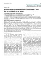
Fig. 1. SDS/PAGE and Western blot analysis of baculovirus expressed
CaN composed of CnAa or CnAb catalytic subunit isoforms. CaN
heterodimers were expressed in Sf21 cells using recombinant baculo-
viruses, purified as described in Experimental procedures, and ana-
lyzed by SDS/PAGE (15%) and Western blotting. Lane 1, CaN
heterodimer containing the CnAa catalytic subunit isoform (5 lg);
lane 2, CaN heterodimer containing the CnAb catalytic subunit iso-
form (3 lg). (A) Purified CaN heterodimers were separated by SDS/
PAGE and stained with Coomassie Brilliant Blue. (B) Immunostaining
of purified CaN heterodimers using anti-CnAa Ig. (C) Immunostain-
ing of purified CaN heterodimers using anti-CnAb Ig.
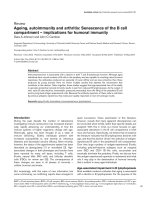
Fig. 2. Kinetic analyses of CaN heterodimers composed of CnAa or CnAb catalytic subunit isoforms. (A) Dephosphorylation of PO
4
-RII peptide.
CaNa or CaNb were each present at a final concentration of 5 n
M
. CaM was present at a final concentration of 15 n
M
. The reactions were allowed
to proceed for 7 min at 30 °C. The concentrations of PO
4
-RII peptide used were 25 l
M
,50l
M
,75l
M
,100l
M
, and 150 l
M
. (B) Dephospho-
rylation of PO
4
-DARPP-32(20–38). CaNa or CaNb were each present at a final concentration of 50 n
M
. CaM was present at a final concentration
of 150 n
M
. The reactions were allowed to proceed for 10 min at 30 °C. The concentrations of PO
4
-DARPP-32(20–38) used were 7 l
M
,12l
M
,
17 l
M
,and25l
M
. (C) Dephosphorylation of pNPP. CaNa or CaNb were each present at a final concentration of 50 n
M
. CaM was present at a
final concentration of 150 n
M
. The reactions were allowed to proceed for 20 min at 30 °C. The concentrations of pNPP used were 10 m
M
,15m
M
,
20 m
M
,and30m
M
,50m
M
, and 100 m
M
. The results shown are representative of three assays performed in triplicate for each CaN heterodimer.
CaNa, d;CaNb, s.
3542 B. A. Perrino et al. (Eur. J. Biochem. 269) Ó FEBS 2002
CaN heterodimers containing the CnAa catalytic subunit
are characterized by approximately twofold higher V
max
values toward the three substrates tested. Together these
results indicate that CaN heterodimers used in these
experiments containing the CnAa catalytic subunit are
characterized by higher levels of phosphatase activity
toward these three substrates.
Inhibition of CaNa and CaNb phosphatase activity by CaP
The CaN crystal structure shows that in the inactive state,
the CaN autoinhibitory domain lies over the catalytic site
[20]. The amino-acid sequences of the CnAa and CnAb
catalytic domains are 90% identical, and the autoinhibitory
domain amino-acid sequences are 89% identical, suggesting
that CaN heterodimers containing the CnAa or CnAb
catalytic subunit would be equally inhibited by CaP, which
contains the autoinhibitory domain from CnAa [4,21].
However, because of the differences in K
m
and V
max
values
obtained with the CaN heterodimers used in these experi-
ments containing the CnAa or CnAb catalytic subunit
toward the same substrate, we examined the inhibition of
CaNa or CaNb phosphatase activity by CaP toward the
three substrates. It has previously been reported that CaP
inhibits bovine brain CaN or baculovirus-expressed rat
brain CaNa with IC
50
values between 12 l
M
and 18 l
M
,
using
32
PO
4
-RII peptide as substrate [10,11]. As shown in
Fig. 3A, the phosphatase activities of CaN heterodimers
containing the CnAa or CnAb catalytic subunit toward
PO
4
-RII peptide are equally inhibited by CaP. The IC
50
values (10 l
M
-12 l
M
) and final extent of inhibition (90%
inhibition of phosphatase activity by 90 l
M
CaP) obtained
are similar to the previously reported values using
32
P-RII
peptide as substrate [10,11]. Similar kinetics of inhibition
were also obtained for CaP with CaN heterodimers
containing the CnAa or CnAb catalytic subunit using
PO
4
-DARPP-32(20–38) as substrate (Fig. 3B), giving IC
50
values of 15 l
M
and 25 l
M
, respectively. In addition, CaN
phosphatase activity is 85% inhibited by 90 l
M
CaP. It has
been reported that bovine brain CaN phosphatase activity is
50% inhibited by 35 l
M
CaP using pNPP as substrate [21].
Using CaN heterodimers containing the CnAa or CnAb
catalytic subunit and pNPP as substrate, we measured IC
50
values of 20 l
M
for CaNa and 90 l
M
for CaNb.Further-
more, in contrast to the results obtained using the
phosphopeptide substrates, the phosphatase activities of
CaNa and CaNb are only 70%, and 50% inhibited by
90 l
M
CaP, respectively.
Inhibition of CaNa and CaNb phosphatase activity
by FKBP12/FK506 or CypA/CsA
The structurally unrelated immunophilin/immunosuppres-
sant complexes of FKBP12/FK506 or CypA/CsA inhibit
CaN noncompetitively by binding to the CnB-binding helix,
CnB, and one side of the substrate-binding cleft of the
catalytic site to alter the active-site geometry [16,17,20,22].
As the mechanism of inhibition of CaN by the immuno-
philin/immunosuppressant complexes is different from that
of CaP, we examined the inhibition of CaN heterodimers
containing the CnAa or CnAb catalytic subunit by
FKBP12/FK506 or CyP/CsA. As shown in the dose–
response curves of Fig. 4, using the two different phospho-
peptide substrates, CaNa is more sensitive to inhibition by
FKBP12/FK506 than CaNb.WithPO
4
-RII peptide as
substrate, 50% inhibition of CaNa activity was achieved
with 73 n
M
FKBP12 (in the presence of 500 n
M
FK506),
compared to 50% inhibition of CaNb activity by 120 n
M
FKBP12. As expected, the high concentration of FKBP12
(200 n
M
) resulted in 90% inhibition of CaNa and CaNb
with PO
4
-RII peptide as substrate (Fig. 4A). Similarly, 50%
inhibition of CaNa activity was achieved with 60 n
M
FKBP12, compared to 50% inhibition of CaNb activity
by 117 n
M
FKBP12 using PO
4
-DARPP-32(20–38) as
substrate. 200 n
M
FKBP12 resulted in 90% inhibition of
CaNa using PO
4
-DARPP-32(20–38) as substrate. However,
90% inhibition of CaNb phosphatase activity was achieved
by 1 l
M
FKBP12 using PO
4
-DARPP-32(20–38) as
Fig. 3. Inhibition of CaN heterodimers composed of CnAa or CnAb catalytic subunit isoforms by CaP. (A) The reactions proceeded for 10 min at
30 °Cusing32 l
M
and 91 l
M
PO
4
-RII peptide for CaNa or CaNb, respectively. CaNa or CaNb were each present at a final concentration of 5 n
M
.
CaM was present at a final concentration of 15 n
M
. (B) The reactions proceeded for 10 min at 30 °Cusing6 l
M
and 21 l
M
PO
4
-DARPP-32(20–38)
for CaNa or CaNb, respectively. CnAa or CnAb were each present at a final concentration of 50 n
M
. CaM was present at a final concentration of
150 n
M
. (C) The reactions proceeded for 20 min at 30 °Cusing45m
M
and 83 m
M
pNPP for CaNa or CaNb, respectively. CaNa or CaNb were
each present at a final concentration of 50 n
M
. CaM was present at a final concentration of 150 n
M
. The results shown are the averages ± SD from
three assays in triplicate for each CaN heterodimer. CaNa, d;CaNb, s.
Ó FEBS 2002 Enzymatic characteristics of calcineurin isoforms (Eur. J. Biochem. 269) 3543
substrate. These findings indicate that the CaNb used in
these experiments is less sensitive than CaNa to inhibition
by FKBP12/FK506 when PO
4
-DARPP-32(20–38) is used
as substrate.
With CyPA/CsA and PO
4
-RII peptide as substrate 50%
inhibition of CaNa activity was achieved with 342 n
M
CyPA (in the presence of 2 l
M
CsA), compared to 50%
inhibition of CaNb activity by 456 n
M
CyPA. A high
concentration of CyPA (1000 n
M
)resultedin80%-90%
inhibition of CaNa and CaNb with PO
4
-RII peptide as
substrate (Fig. 4B). Similar IC
50
values were obtained for
CyPA/CsA inhibition of CaNa and CaNb using PO
4
-
DARPP-32(20–38) as substrate and 80–90% inhibition of
phosphatase activity was attained with 1000 n
M
CyPA
(Fig. 4D). Using two different phosphopeptide substrates,
these findings indicate that CyPA/CsA results in similar
inhibition of both CaNa and CaNb. These findings also
indicate that FKBP12/FK506 is a more potent inhibitor of
both CaNa and CaNb than CyPA/CsA.
Activation of CaNa and CaNb phosphatase activity
by FKBP12/FK506 toward pNPP
In contrast to the inhibition of CaN phosphatase activity
toward phosphopeptide and phospho-protein substrates by
FKBP12/FK506, the phosphatase activity of CaN toward
the small organic compound pNPP is increased two- to
fourfold by FKBP12/FK506 [16,23]. These observations are
consistent with the findings that the FKBP12/FK506
complex alters the conformation of the active site [20]. To
examine the possibility that FKBP12/FK506 may have
different effects on the activities of CaNa and CaNb toward
pNPP, we examined the activation of CaNa and CaNb
phosphatase activities toward pNPP by FKBP12/FK506.
As shown in Fig. 5, a twofold increase in CaN phosphatase
activity was observed with 200 n
M
FKBP12 and 500 n
M
FK506, in agreement with previous studies [16,23]. How-
ever, there was essentially no difference in the dose-
dependent activation of CaNa or CaNb phosphatase
activities toward pNPP by FKBP12/FK506.
Determination of dissociation rates between
CaM(C75)
ACR
and CaNa or CaNb
Because of the highly conserved amino-acid sequences of
the CnAa and CnAb catalytic, CaM-binding, and CnB-
binding domains, it was surprising to find differences in the
phosphatase activities between CaNa and CaNb toward the
same substrate. The results of the kinetic analyses, and
inhibition studies are summarized in Table 1. Although the
amino-acid sequences of the CnAa and CnAb CaM-
binding domains are identical, adjacent functional domains
can influence the CaM-binding properties of CaM-binding
a helices [24]. Thus, stop-flow analyses of Ca
2+
/CaM off
rates were carried out in order to determine whether there
are any differences in the Ca
2+
/CaM-binding properties of
CaNa and Canb. The dissociation rates of CaNa or CaNb
from CaM(C75)
ACR
were determined by monitoring the
rates of fluorescence decrease as CaM(C75)
ACR
bound to
CaNa or CaNb is exchanged for excess unlabeled CaM
(Fig. 6). The data were best fit by a double-exponential
model and yielded two essentially identical fast and slow
Fig. 4. Inhibition of CaN heterodimers
composed of CnAa or CnAb catalytic subunit
isoforms by FKBP12/FK506 or CyPA/CsA.
FK506 or CsA were each present at a final
concentration of 2 l
M
,andtheFKBP12or
CyPA concentrations varied as indicated in
the figure legends. The reactions proceeded for
20 min at 30 °Cusing32l
M
(A) and 91 l
M
(B) PO
4
-RII peptide for CaNa or CaNb,
respectively. CaNa or CaNb were each present
at a final concentration of 5 n
M
.CaMwas
present at a final concentration of 15 n
M
.The
reactions proceeded for 20 min at 30 °Cusing
6 l
M
(C) and 21 l
M
(D) PO
4
-DARPP-32(20–
38) for CaNa or CaNb, respectively. CaNa or
CaNb were each present at a final concent-
ration of 5 n
M
. CaM was present at a final
concentration of 15 n
M
. The results shown are
the averages ± SD from three assays in
triplicate for each CaN heterodimer. CaNa,
d;CaNb, s.
3544 B. A. Perrino et al. (Eur. J. Biochem. 269) Ó FEBS 2002
CaM dissociation constants for both CaNa and CaNb.Fast
and slow rates of 4 s
)1
and 0.4 s
)1
,and3.9s
)1
and 0.4 s
)1
were obtained for CaNa and CaNb, respectively. These
results indicate that there are essentially no differences in
Ca
2+
/CaM-dissociation from the CnAa and CnAb cata-
lytic subunit isoforms. The mechanistic basis for the two
rate constants is not clear, although Ca
2+
/CaM-binding to
the CaM-binding domain of CnA is modulated by Ca
2+
-
binding to CnB [25,26].
DISCUSSION
Previous studies of the CaN a and b catalytic subunit
isoforms have mainly focused on their tissue and subcellular
distributions [5–8,27]. These reports have provided import-
ant information demonstrating regional differences in
expression levels within tissues and differences in the
subcellular distribution of CnAa and CnAb. Although it
has been generally assumed that CaNa and CaNb
dephosphorylate the same set of substrates, the differences
in CnAa and CnAb localization and expression have been
proposed to reflect differences in substrate selectivity
between CaNa and CaNb [2,4]. However, in the absence
of information concerning enzymatic differences between
these two CaN catalytic subunit isoforms, the physiological
significance of their differential distribution is unclear. To
address this question, we have examined the enzymatic
properties of CaN heterodimers containing either the CnAa
or CnAb catalytic subunit. In agreement with previous
findings using purified mammalian brain CaN, we found
that CaNa or CaNb heterodimers are characterized by
different K
m
and V
max
values toward different substrates
[19]. However, our results also indicate that CaNa and
CaNb heterodimers exhibit differences in phosphatase
activity toward the same substrate, as indicated by the
different K
m
and V
max
values obtained. The results from the
kinetic assays show that the CaN heterodimers containing
the CnAa subunit have higher levels of phosphatase activity
toward all three substrates tested, as indicated by lower K
m
and higher V
max
values (Table 1). These findings indicate
that the CaN catalytic subunits are characterized by
different rates of phosphatase activity towards their sub-
strates, and suggest that the CaNa:CaNb ratiowithinacell
or tissue is an important determinant of CaN phosphatase
activity toward specific substrates.
The use of CnAa knockout mice has provided evidence
that CaNa and CaNb exhibit selective phosphatase activity
toward specific substrates within a cell or tissue. Tau
proteins are hyperphosphorylated in the brains of CnAa –/–
mice [28]. Similarly, hippocampal depotentiation is abol-
ished while long-term depression and long-term potentia-
tion are unaffected in CnAa –/– mice [29]. Furthermore,
CnAa –/– mice have impaired antigen-specific T-cell
responses in vivo [30]. Two possible interpretations of these
findings are (a) CaNa selectively dephosphorylates tau
proteins, and also specifically dephosphorylates substrates
required for hippocampal depotentiation and antigen-
induced T-cell responses, or (b) CaNa and CaNb have no
substrate preferences, but the residual CaNb phosphatase
activity is insufficient to dephosphorylate tau or participate
in hippocampal depotentiation and antigen-induced T-cell
responses. The findings that CnAb is the predominant
isoform present in T-cells argues against impaired antigen-
specific T-cell responses in CnAa –/– mice being due to
insufficient CaN phosphatase activity [6]. Our findings that
the CnAa and CnAb catalytic subunits confer differences in
substrate affinity and phosphatase activity (as shown by
Fig. 5. Stimulation of CaN phosphatase activity toward pNPP by
FKBP12/FK506. FK506 was present at a final concentration of
500 n
M
, and the FKBP12 concentration varied as indicated in the
figure legend. (A) The reactions proceeded for 45 min at 30 °Cusing
45 m
M
and 83 m
M
pNPP for CaNa or CaNb, respectively. CaNa or
CaNb were each present at a final concentration of 5 n
M
.CaMwas
present at a final concentration of 15 n
M
. The results shown are the
averages ± SD from three assays in triplicate for each CaN het-
erodimer. CaNa, d;CaNb, s.
Table 1. Summary of kinetic parameters of CaNa and CaNb phosphatase activities toward three substrates. The K
m
and V
max
values, CaP IC
50
values, FKBP12 IC
50
and CyPA IC
50
values were obtained from the inverse plots of V vs [S], the CaP dose–response experiments, and the FKBP12
and CyPA dose–response experiments, respectively.
PO
4
-RII peptide (l
M
)PO
4
-DARPP-32 pNPP
CaNa CaNb CaNa CaNb CaNa CaNb
K
m
32 l
M
91 l
M
6.2 l
M
21 l
M
45 m
M
83 m
M
V
max
(lmolÆmin
)1
Æmg
)1
) 4.1 2.8 0.384 0.224 5.8 3.1
CaP IC
50
(l
M
) 101215252290
FKBP12 IC
50
(n
M
) 74 120 60 114 – –
CyPA IC
50
(n
M
) 342 451 303 251 – –
Ó FEBS 2002 Enzymatic characteristics of calcineurin isoforms (Eur. J. Biochem. 269) 3545
different K
m
and V
max
values) toward the same substrate
support the conclusion that CaN substrates are differen-
tially dephosphorylated by CaNa and CaNb in vivo.
The differences in phosphatase activity and sensitivity to
FKBP12/FK506 or CyPA/CsA inhibition between CaNa
and CaNb that we observed may be due to subtle differences
in how the CnAa and CnAb catalytic subunits interact with
CnB. The regulation of enzyme activity by two EF-hand
Ca
2+
-binding proteins is unique to CaN [4]. Similar to
Ca
2+
/CaM-dependent kinases, Ca
2+
/CaM-binding to CnA
activates the enzyme by relieving inhibition due to an
autoinhibitory domain, which is evidenced by an increase in
V
max
[11,31]. Conversely, Ca
2+
-binding to CnB activates
CaN primarily by affecting the affinity of the catalytic site
for substrate, as evidenced by the decrease in K
m
[11,31].
Circular dichroism analysis has shown that CnB and CnA
both undergo conformational changes upon Ca
2+
binding
to CnB [32]. It appears that in the absence of Ca
2+
the
catalytic core is in an inactive conformation and that Ca
2+
-
binding to CnB changes the conformation of the catalytic
core to allow substrate binding [25,33]. The CaN crystal
structure shows that the CnB-binding helix is immediately
C-terminal of the b14 strand of one half of the b sandwich
forming the catalytic core, providing a mechanism for
transmission of Ca
2+
-induced conformational changes in
CnB to the active site [22]. In fact, the catalytic activity of
CaN is sensitive to the amino-acid composition of the region
linking the CnB-binding helix to the b14 strand of the
catalytic core [34,35]. Mutations S373P, H375L, and L379S
decrease CaN activity, indicating the importance of this
linker region to the activation of CaN by Ca
2+
-binding to
CnB [34]. Calcium binding to CnB also influences the
affinity of Ca
2+
/CaM for its binding domain on CnA
[25,26]. CnB has two low affinity and two high affinity EF-
hand Ca
2+
-binding loops [36]. In the absence of Ca
2+
-
binding to the low affinity sites, the CaM-binding domain
interacts with the exposed side of the CnB-binding helix [26].
Calcium binding to the low affinity sites on CnB disrupts the
interaction between the CaM-binding domain and the CnB-
binding helix and increases the affinity of the CaM-binding
domain for Ca
2+
/CaM [25,26]. The regulation of the CaM-
binding domain by CnB may partly account for the slow
and fast dissociation constants we measured using stopped-
flow analysis.
The FKBP12/FK506 complex inhibits CaN noncompeti-
tively using PO
4
-RII and PO
4
-DARPP-32 as substrates [20].
These findings are consistent with crystallographic data
showing the active site and substrate-binding cleft are not
directly blocked by the FKBP12/FK506 complex, support-
ing the conclusion that alterations in the active site
conformation affect the substrate-binding cleft and are
responsible for the inhibition by FKBP12/FK506 [20,22].
This mechanism of inhibition would account for the
findings that CaN phosphatase activity toward the small
organic molecule pNPP is increased by the FKBP12/FK506
complex (Fig. 5) [16,23]. However, the structural and
enzymatic studies of the interactions between the
FKBP12/FK506 complex and CaN have been carried out
with CaN containing the CnAa catalytic subunit
[16,20,22,23]. The results presented here are the first direct
studies of the inhibition of CaN containing the CnAb
catalytic subunit by FKBP12/FK506 or CyPA/CsA. For
both immunophilin/immunosuppressant complexes, the
IC
50
values are higher than previously reported [23,37].
Differences in CaN preparations and methods of enzyme
activity assays may account for the differences in IC
50
values. For these studies the CaN activity was assayed in the
presence of ascorbic acid, which results in higher activity
compared to activity in the absence of antioxidants [38,39].
With both peptide substrates CaNa was more sensitive to
inhibition by FKBP12/FK506 than CaNb (Fig. 4). How-
ever, CaNa and CaNb were both 90% inhibited by 200 n
M
FKBP12 using PO
4
-RII at K
m
concentrations. Similarly,
Fig. 6. Measurement of CaM dissociation rate constants from CaNa or
CaNb associated with CaM (C75)
ACR
using stopped-flow kinetics. The
time course for CaM dissociation from CaNa (A) or CaNb (B) from
CaM(C75)
ACR
as determined using a stopped-flow fluorimeter.
CaM(C75)
ACR
(0.1 l
M
) and either (0.3 l
M
)CaNa (or CaNb)in
25 m
M
Mops, pH 7.0, 150 m
M
KCl, 0.5 m
M
CaCl
2
were rapidly mixed
with native CaM (10 l
M
)inthesamebufferat20°C. The excitation
was at 365 nm and emission was monitored using a 399-nm cut-off
filter. Each curve represents the average of four exchange reactions.
3546 B. A. Perrino et al. (Eur. J. Biochem. 269) Ó FEBS 2002
CaNa was 90% inhibited by 200 n
M
FKBP12 using PO
4
-
DARPP-32(20–38) at the K
m
concentration. In contrast,
only 60% inhibition of CaNb by 200 n
M
FKBP12 was
measured using PO
4
-DARPP-32(20–38) as substrate, and
90% inhibition required 1000 n
M
FKBP12. Our results
using the two different phosphopeptide substrates show that
CsA is a less potent inhibitor of both CaNa and CaNb than
FK506. These results are consistent with the findings that
in vivo, CsA and FK506 are equally effective, but FK506 is
10-fold more potent in inhibiting CaN activity and IL-2
gene activation [37]. Although FK506 and CyPA are
structurally unrelated compounds, biochemical and muta-
tional studies indicate that FKBP12/FK506 and CyPA/CsA
bind to a common site on the CaN heterodimer composed
of the CnB-binding helix, CnB and part of the substrate-
binding cleft of CaN [35,40]. However, differences in the
interactions between these structurally dissimilar immuno-
philin/immunosupressant complexes and CaN will likely
contribute to differences in their inhibitory potency.
As the substrate-binding cleft geometry is affected by
CnB, the interaction between CnA and CnB may affect how
the FKBP12/FK506 and CyPA/CsA complexes affect the
substrate-binding cleft. Thus, both the catalytic subunit and
substrate may influence the degree of inhibition of CaN
phosphatase activity by FKBP12/FK506 and CyPA/CsA.
The differences in phosphatase activity and sensitivity to
FKBP12/FK506 inhibition between CaNa and CaNb are
likely not due to differences in the linker region, as the
amino-acid sequences of the CnB-binding helix, and the
linker region of CnAa and CnAb are identical [41].
However, proteolysis of the CnA N-terminus results in loss
of CaN activity, suggesting that the CnA N-terminus is
involved in enzyme activation [42]. Indeed, as shown in the
crystal structure, the N-terminus of CnA interacts with the
C-terminal half of CnB as part of a CnB-binding cleft,
suggesting that the interaction of the CnA N-terminus with
CnB is involved in Ca
2+
CnB-dependent activation of the
enzyme [20,22]. As noted previously, the N-terminus of
CnAb is different from CnAa, containing 12 Pro residues
within the first 24 amino acids [41]. Information regarding
the interaction of the CnAb N-terminus with CnB is lacking
because only CaN containing CnAa has been crystalized
[20,22]. However, the presence of 11 consecutive Pro
residues in the N-terminus of CnAb suggests that the CnAa
and CnAb N-termini interact with CnB differently, giving
rise to the different K
m
and V
max
values measured for CaNa
and CaNb using the same substrate.
Polyproline motifs are involved in protein–protein inter-
actions [43]. Molecular modeling indicates that an 11-
residue type II polyproline helix exactly spans the length of
the central helix of CaM, and led to the proposal that the 11
consecutive Pro residues of CnAb may modulate the
interaction of CaM with the CaM-binding domain of CnAb
[41]. However, as noted previously, the crystal structure
subsequently showed that the N-terminus of CnAa forms
extensive contacts with CnB. Interestingly, the central helix
of CnB differs in length from the central helix of CaM by
only one amino-acid residue [44]. These findings suggest
that the 11 Pro residues within the first 24 amino-acid
residues of CnAb may instead interact with the central helix
of CnB and affect its interaction with CnAb.AsCa
2+
-
binding to CnB regulates the catalytic activity of CnA, this
proposal provides a potential mechanistic basis for the
differences in K
m
and V
max
values between CaNa and CaNb
obtained using the same substrate.
ACKNOWLEDGEMENTS
This work was supported by National Institutes of Health Grants NS-
36318, DK-57168 (B.A.P), and The Medical Research Council, UK
(G117/440) (L.H.C). L.H.C. is a MRC Senior Fellow in Basic Science.
We thank M. Neal Waxham (University of Texas Medical School at
Houston) for the generous gift of CaM (C75), and Michael M. Lai and
Solomon H. Snyder (The Johns Hopkins University School of
Medicine) for providing the human CnAb cDNA.
REFERENCES
1. Olson, E.N. & Williams, R.S. (2000) Remodeling muscles with
calcineurin. Bioessays 22, 510–519.
2. Perrino, B.A. & Soderling, T.R. (1998) Biochemistry and phar-
macology of calmodulin-regulated phosphatase calcineurin. In
Calmodulin and Signal Transduction (VanEldik, L. & Watterson,
D.M., eds), pp. 169–236. Academic Press, New York.
3. Rusnak, F. & Mertz, P. (2000) Calcineurin: form and function.
Physiol. Rev. 80, 1483–1521.
4. Guerini, D. & Klee, C.B. (1991) Structural diversity of calcineurin,
aCa
2+
and calmodulin-stimulated protein phosphatase. Adv.
Prot. Phosphatases 6, 391–410.
5. Kuno, T., Mukai, H., Ito, A., Chang, C.D., Kishima, K., Saito, N.
& Tanaka, C. (1992) Distinct cellular expression of calcineurin Aa
and Ab in rat brain. J. Neurochem. 58, 1643–1651.
6. Jiang, H., Xiong, F., Kong, S., Ogawa, T., Kobayashi, M. & Liu,
J.O. (1997) Distinct tissue and cellular distribution of two major
isoforms of calcineurin. Mol Immunol. 34, 663–669.
7.Bosser,R.,Aligue,R.,Guerini,D.,Agell,N.,Carafoli,E.&
Bachs, O. (1993) Calmodulin can modulate protein phospho-
rylation in rat liver cells nuclei. J. Biol. Chem. 268, 15477–15483.
8. Usuda, N., Arai, H., Sasaki, H., Hanai, T., Nagata, T.,
Muramatsu, T., Kincaid, R.L. & Higuchi, S. (1996) Differential
subcellular localization of neural isoforms of the catalytic subunit
of calmodulin-dependent protein phosphatase (calcineurin) in
central nervous system neurons: immunohistochemistry on for-
malin-fixed paraffin sections employing antigen retrieval by
microwave irradiation. J. Histochem. Cytochem. 44, 13–18.
9. Lai, M.M., Burnett, P.E., Wolosker, H., Blackshear, S. & Snyder,
S.H. (1998) Cain, a novel physiologic protein inhibitor of calci-
neurin. J. Biol. Chem. 273, 18325–18331.
10. Perrino, B.A., Fong, Y.L., Brickey, D.A., Saitoh, Y., Ushio, Y.,
Fukunaga, K., Miyamoto, E. & Soderling, T.R. (1992) Char-
acterization of the phosphatase activity of a baculovirus-expressed
calcineurin A isoform. J. Biol. Chem. 267, 15965–15969.
11. Perrino, B.A., Ng, L.Y. & Soderling, T.R. (1995) Calcium reg-
ulation of calcineurin phosphatase activity by its B subunit and
calmodulin: role of the autoinhibitory domain. J. Biol. Chem. 270,
340–346.
12. Pallen, C.J. & Wang, J.H. (1983) Calmodulin-stimulated depho-
sphorylation of p-nitrophenyl phosphate and free phosphotyro-
sine by calcineurin. J. Biol. Chem. 258, 8550–8553.
13. Li, H C. (1984) Activation of brain calcineurin phosphatase
towards nonprotein phosphoesters by Ca
2+
, calmodulin, and
Mg
2+
. J. Biol. Chem. 259, 8801–8807.
14. Putkey, J.A. & Waxham, M.N. (1996) A peptide model for cal-
modulin trapping by calcium/calmodulin-dependent protein kin-
ase II. J. Biol. Chem. 271, 29619–29623.
15. Waxham, M.N., Tsai, A. & Putkey, J.A. (1998) A mechanism for
calmodulin (CaM) trapping by CaM-kinase II defined by a family
of CaM-binding peptides. J. Biol. Chem. 273, 17579–17584.
16. Liu, J., Farmer, J.D. Jr, Lane, W.S., Friedman, J., Weissman, I. &
Schreiber, S.L. (1991) Calcineurin is a common target of
Ó FEBS 2002 Enzymatic characteristics of calcineurin isoforms (Eur. J. Biochem. 269) 3547
cyclophilin-cyclosporin A and FKBP-FK506 complexes. Cell 66,
807–815.
17. Liu, J., Albers, M.W., Wandless, T.J., Luan, S., Alberg, D.G.,
Belshaw, P.J., Cohen, P., MacKintosh, C., Klee, C.B. & Schreiber,
S.L. (1992) Inhibition of T-cell signaling by immunophilin-ligand
complexes correlates with loss of calcineurin phosphatase activity.
Biochemistry 31, 3896–3901.
18. Hemmings, H.C. Jr, Nairn, A.C., Elliot, J.I. & Greengard, P.
(1990) Synthetic peptide analogs of DARPP-32 (Mr 32,000
dopamine- and cAMP-regulated phosphoprotein), an inhibitor of
protein phosphatase-1. J. Biol. Chem. 265, 20369–20376.
19. Klee, C.B., Draetta, G.F. & Hubbard, M.J. (1988) Calcineurin.
Adv. Enzymol. 61, 149–200.
20. Kissinger, C.R., Parge, H.E., Knighton, D.R., Lewis, C.T.,
Pelletier, L.A., Tempczyk, A., Kalish, V.J., Tucker, K.D., Show-
alter, R.E., Moomaw, E.W., Gastinel, L.N., Habuka, N., Chen,
X., Maldonado, F., Barker, J.E., Bacquet, R. & Villafranca, J.E.
(1995) Crystal structures of human calcineurin and the human
FKBP12-FK506 complex. Nature 378, 641–644.
21. Hashimoto, Y., Perrino, B.A. & Soderling, T.R. (1990) Identifi-
cation of an autoinhibitory domain in calcineurin. J. Biol. Chem.
265, 1924–1927.
22. Griffith, J.P., Kim, J.L., Kim, E.E., Sintchak, M.D., Thomson,
J.A., Fitzgibbon, M.J., Fleming, M.A., Caron, P.R., Hsiao, K. &
Navia, M.A. (1995) X-ray structure of calcineurin inhibited by the
immunophilin-immunosuppressant FKBP12-FK506 complex.
Cell 82, 507–522.
23. Swanson, S.K., Born, T., Zydowski, L.D., Cho, H., Chang, H.Y.,
Walsh, C.T. & Rusnak, F. (1992) Cyclosporin-mediated inhibition
of bovine calcineurin by cyclophilins A and B. Proc. Natl Acad.
Sci. USA 89, 3741–3745.
24. Kwiatkowski, A.P. & McGill, J.M. (2000) Alternative splice
variant of c-calmodulin-dependent protein kinase II alters acti-
vation by calmodulin. Arch. Biochem. Biophysics 378, 377–383.
25. Feng, B. & Stemmer, P.M. (2001) Ca
2+
binding site 2 in cal-
cineurin-B modulates calmodulin-dependent calcineurin phos-
phatase activity. Biochemistry 40, 8808–8814.
26. Yang, S.A. & Klee, C.B. (2000) Low affinity Ca
2+
-binding sites of
calcineurin B mediate conformational changes in calcineurin A.
Biochemistry 39, 16147–16154.
27. Strack, S., Wadzinski, B.E. & Ebner, F.F. (1996) Localization of
the calcium/calmodulin-dependent protein phosphatase, cal-
cineurin, in the hindbrain and spinal cord of the rat. J. Comp.
Neurol. 375, 66–76.
28. Kayyali,U.S.,Zhang,W.,Yee,A.G.,Seidman,J.G.&Potter,H.
(1997) Cytoskeletal changes in the brains of mice lacking calci-
neurin A alpha. J. Neurochem. 68, 1668–1678.
29. Zhuo, M., Zhang, W.,Son, H., Mansuy, I., Sobel, R.A., Seidman, J.
& Kandel, E.R. (1999) A selective role of calcineurin Aa in
synaptic depotentiation in hippocampus. Proc. Natl Acad. Sci.
USA 96, 4650–4655.
30. Zhang, W., Zimmer, G., Chen, J., Ladd, D., Li, E., Alt, F.W.,
Wiederrecht, G.J., Cryan, J., O’Neill, E.A., Seidman, C.E., Abbas,
A.F. & Seidman, J.G. (1996) T-cell responses in calcineurin
Aa-deficient mice. J. Exp. Med. 183, 413–420.
31. Stemmer, P.M. & Klee, C.B. (1994) Dual calcium regulation of
calcineurin by calmodulin and calcineurin B. Biochemistry 33,
6859–6866.
32. Wolff, D.J. & Sved, D.W. (1985) The divalent cation dependence
of bovine brain calmodulin-dependent phosphatase. J. Biol. Chem.
260, 4195–4202.
33. Wei, Q. & Lee, E.Y.C. (1997) Mutagenesis of the L7 loop con-
necting b strands 12 and 13 of calcineurin: evidence for a structural
role in activity changes. Biochemistry 36, 7418–7424.
34. Jiang, B. & Cyert, M.S. (1999) Identification of a novel region
critical for calcineurin function in vivo and in vitro. J. Biol. Chem.
274, 18543–18551.
35. Kawamura, A. & Su, M.S.S. (1995) Interaction of FKBP12-
FK506 with calcineurin A at the B subunit-binding domain.
J. Biol. Chem. 270, 15463–15466.
36. Feng, B. & Stemmer, P.M. (1999) Interactions of calcineurin A,
calcineurin B, and Ca
2+
. Biochemistry 38, 12481–12489.
37. Fruman, D.A., Klee, C.B., Bierer, B.E. & Burakoff, S.J. (1992)
Calcineurin phosphatase activity in T lymphocytes is inhibited by
FK 506 and cyclosporin A. Proc. Natl Acad. Sci. USA 89, 3686–
3690.
38. Mitsuhashi, S., Shima, H., Kikuchi, K., Yazawa, M., Hatsuse, R.,
Maeda, K., Yazawa, M., Murayama, T., Okuma, Y. & Nomura,
Y. (2000) Development of an assay method for activities of
serine/threonine protein phosphatase type 2B (calcineurin) in
crude extracts. Anal. Biochem. 278, 192–197.
39. Sommer, D., Fakata, K.L., Swanson, S.K. & Stemmer, P.M.
(2000) Modulation of the phosphatase activity of calcineurin by
oxidants and antioxidants in vitro. Eur. J. Biochem. 267, 2312–
2322.
40. Milan, D., Griffith, J., Su, M., Price, E.R. & McKeon, F.D. (1994)
The latch region of calcineurin B is involved in both
immunosuppressant-immunophilin complex docking and phos-
phatase activation. Cell 79, 437–447.
41. Guerini, D. & Klee, C.B. (1989) Cloning of human calcineurin A:
Evidence for two isozymes and identification of a polyproline
structural domain. Proc.NatlAcad.Sci.USA86, 9183–9187.
42. Hubbard, M.J. & Klee, C.B. (1989) Functional domain structure
of calcineurin A: mapping by limited proteolysis. Biochemistry 28,
1868–1874.
43. Dalgarno, D.C., Botfield, M.C. & Rickles, R.J. (1997) SH3
domains and drug design: Ligands, structure, and biological
function. Biopolymers 43, 383–400.
44. Klee, C.B. & Cohen, P. (1988) The calmodulin-regulated protein
phosphatase. In Calmodulin (Klee, C.B. & Cohen, P., eds), pp.
225–248. Elsevier, Amsterdam.
3548 B. A. Perrino et al. (Eur. J. Biochem. 269) Ó FEBS 2002