Báo cáo Y học: Supermolecular organization of photosystem II and its associated light-harvesting antenna in Arabidopsis thaliana docx
Bạn đang xem bản rút gọn của tài liệu. Xem và tải ngay bản đầy đủ của tài liệu tại đây (4.64 MB, 9 trang )
Supermolecular organization of photosystem II and its associated
light-harvesting antenna in
Arabidopsis thaliana
Alevtyna E. Yakushevska
1
, Poul E. Jensen
2
, Wilko Keegstra
1
, Henny van Roon
3
, Henrik V. Scheller
2
,
Egbert J. Boekema
1
and Jan P. Dekker
3
1
Department of Biophysical Chemistry, Groningen Biomolecular Sciences and Biotechnology Institute, University of Groningen,
Nijenborgh, Groningen, the Netherlands;
2
Plant Biochemistry Laboratory, Department of Plant Biology, The Royal Veterinary and
Agricultural University, Copenhagen, Denmark;
3
Faculty of Sciences, Division of Physics and Astronomy, Vrije Universiteit, Amsterdam,
the Netherlands
The organization of Arabidopsis thaliana photosystem II
(PSII) and its associated light-harvesting antenna (LHCII)
was studied in isolated PSII–LHCII supercomplexes and
native membrane-bound crystals by transmission electron
microscopy and image analysis. Over 4000 single-particle
projections of PSII – LHCII supercomplexes were analyzed.
In comparison to spinach supercomplexes [Boekema, E.J.,
van Roon, H., van Breemen, J.F.L. & Dekker, J.P. (1999)
Eur. J. Biochem. 266, 444–452] some striking differences
were revealed: a much larger number of supercomplexes
from Arabidopsis contain copies of M-type LHCII trimers.
M-type trimers can also bind in the absence of the more
common S-type trimers. No binding of
L-type trimers could
be detected. Analysis of native membrane-bound PSII
crystals revealed a novel type of crystal with a unit cell of
25.6 Â 21.4 nm (angle 778), which is larger than any of the
PSII lattices observed before. The data show that the unit
cell is built up from C
2
S
2
M
2
supercomplexes, rather than
from C
2
S
2
M supercomplexes observed in native membrane
crystals from spinach [Boekema, E.J., Van Breemen, J.F.L.,
Van Roon, H. & Dekker, J.P. (2000) J. Mol. Biol. 301,
1123–1133]. It is concluded from both the single particle
analysis and the crystal analysis that the M-type trimers bind
more strongly to PSII core complexes in Arabidopsis than in
spinach.
Keywords: photosystem II; Arabidopsis thaliana; electron
microscopy; supercomplex; light-harvesting.
Photosystem II (PSII) is a pigment-protein complex
embedded in the thylakoid membrane of higher plants,
algae and cyanobacteria. It catalyses a sunlight-driven
process, splitting water into protons and molecular oxygen.
This multisubunit protein complex consists of more than 25
structurally and functionally distinct subunits organized
hierarchically [1,2]. First is the PSII core complex, of
which the chlorophyll-containing CP43 and CP47 proteins
and the reaction center are the most important components.
The latter consists of D1 and D2 proteins, which generate
the redox potential required to drive the water splitting
reaction. The structure of the core complex has been solved
at 3.8 A
˚
resolution by the X-ray diffraction of three-
dimensional crystals of PSII from Synechococcus elongatus
[3]. The core complex, which normally exists as a dimer,
contains extrinsic proteins attached to the lumenal surface.
These proteins, which arise from the oxygen-evolving
complex (OEC), have an apparent molecular mass of 33
(Mn-stabilizing protein), 23 and 17 kDa, respectively, and
are necessary for maintaining the water oxidation process.
The recently reported structure of PSII from Synecho-
coccus elongatus revealed, in detail, the spatial organization
of the protein subunits and pigment molecules within
PSII [3]. The arrangement of all the large subunits and
most of the small subunits has been assigned, as well as
the position of cytochrome b
559
and cytochrome c
550
and
the localization of the manganese cluster that catalyzes
oxidation of water. This knowledge will also aid under-
standing of the structure of the green plant core PSII, which
has a very similar structure. Several small PSII core
subunits such as PsbL, PsbK, and PsbW have been proposed
to be involved in the dimerization of the PSII [4,5]. The first
two have been tentatively assigned, but other small
components required for stabilizing dimeric PSII, such as
phosphatidyl glycerol [6], cannot be solved at the present
resolution.
Second is the peripheral antenna, which is involved in the
absorption of light and subsequent energy transfer to the
reaction center. In higher plants and algae it consists of
various pigment-containing proteins encoded by the ‘cab’
gene family [7]. The major light-harvesting complex II
(LHCII) complex is trimeric, consists of the Lhcb1-3
proteins and contains chlorophyll a/b and carotenoids. Its
structure was solved by electron crystallography at 3.4 A
˚
resolution [8]. The minor complexes, Lhcb4 (CP29), Lhcb5
(CP26), Lhcb6 (CP24) are monomeric and also contain
chlorophyll a/b and carotenoids. They are involved in
mediating excitation energy transfer from the LHCII trimers
to the PSII core complex.
Correspondence to E. J. Boekema, Department of Biophysical
Chemistry, Groningen Biomolecular Sciences and Biotechnology
Institute, University of Groningen, Nijenborgh 4, 9747 AG Groningen,
the Netherlands. Fax: 1 31 50 363 4800, Tel.: 1 31 50 363 4225,
E-mail: boekema.chem.rug.nl
(Received 25 May 2001, revised 30 July 2001, accepted 10 September
2001)
Abbreviations: PSII, photosystem II; PSI, photosystem I; LHCII,
light-harvesting antenna; OEC, oxygen evolving complex; S, strongly
bound; M, moderately bound; L, loosely bound; a-DM, n-dodecyl-
a-
D-maltoside.
Eur. J. Biochem. 268, 6020–6028 (2001) q FEBS 2001
The components mentioned above together comprise the
PSII–LHCII supercomplexes [1,9]. Biochemical evidence
suggests that on average, about eight trimers of LHCII are
present per PSII core dimer [10], but only a part of the
LHCII complexes are directly bound to the PSII core
complexes. Various LHCII trimers have been distinguished
by the strength of binding to the core complex. The
following classification was proposed based on the strength
of binding of LHCII to the core part of the supercomplex: S,
strongly bound LHCII; M, moderately bound LHCII and L,
loosely bound LHCII [11,12]. The number of constituents is
indicated after the corresponding letter; thus C
2
represents a
dimeric core complex and C
2
S
2
represents a dimeric core
with two strongly bound LHCII trimers.
The structural organization of the core PSII complexes
and multiple types of associations of the core complex with
light-harvesting antennae (supercomplexes) have been
investigated by electron microscopy (EM) [9,11–13] and
cross-linking studies [14]. A three-dimensional structure of
a supercomplex from spinach, calculated at 24 A
˚
resolution
by EM and single particle analysis of ice-embedded
preparations [13], revealed the position and the tetrameric
structure of the OEC, as well as the location of the
extrinsic OEC proteins on the lumenal surface of the
membrane. Cross-linking studies suggested the specific
location of the three minor antenna complexes Lhcb-4,
Lhcb-5 and Lhcb-6 [14].
It is not known in detail how these supercomplexes,
nonbound LHCII trimers, and possibly the cytochrome b
6
/f
complex are arranged in the grana membrane or how these
proteins from adjacent grana membranes interact to get
membrane stacks. The LHCII trimers serve as the main
stabilizers in the formation of appressed grana membranes.
However, chlorina mutants from barley, despite the greatly
reduced levels of these proteins, still have grana. It was
consequently concluded that PSII core proteins can also
induce membrane oppression [15]. Numerous freeze-etch
and freeze-fracture EM studies indicate that PSII complexes
have a tendency to form ordered domains within the grana
membrane, although noncrystalline domains usually seem
to dominate in wild-type plants [16]. However, these
techniques do not precisely reveal the type of PSII complex,
because of the lack of resolution [1]. Crystallinity can be
studied in greater detail in isolated, paired inside-out grana
membranes purified by gel-filtration chromatography.
Investigation of negatively stained paired membrane
fragments from spinach by EM and image analysis revealed
the motif of two different types of lattices [17]. The most
dominant one consisted of C
2
S
2
M supercomplexes, the
other contained C
2
S
2
supercomplexes. Crystalline domains
of two adjacent layers also show some specificity in their
association, as preferential angles between two lattices are
frequently observed.
In this report, we present details on the structural
organization of PSII from wild type Arabidopsis thaliana by
EM and image analysis. Two approaches were followed: the
periodic approach (analysis of crystalline membrane
fragments) indicated a new type of crystal lattice composed
of C
2
S
2
M
2
PSII–LHCII supercomplexes; and the a-periodic
approach (analysis of 4000 projections of single PSII
particles) indicated the presence of large numbers of
PSII–LHCII supercomplexes with attached M-type LHCII
trimers.
MATERIALS AND METHODS
Isolation of inside-out grana and single particles
PSII–LHCII supercomplexes and native grana membranes
were isolated from thylakoid membranes from A. thaliana,
essentially as described previously for spinach thylakoid
membranes [18], and prepared immediately for EM. For the
PSII–LHCII supercomplexes, thylakoid membranes at a
chlorophyll concentration of 1.4 mg
:
mL
21
were partially
solubilized with n-dodecyl-a-
D-maltoside (a-DM) at a final
concentration of 0.6% at 4 8C in a buffer containing 20 m
M
Bistris (pH 6,5) and 5 mM MgCl
2
. After centrifuging for
3 min at 9000 r.p.m. in an Eppendorf table-top centrifuge
the supernatant was filtered through a 0.45-mm filter in order
to remove large fragments. The solubilized fractions were
purified by gel-filtration chromatography, using a super-
dex 200 HR 10/30 column as described previously [18]. The
fractions eluting at 20 min were used for single particles
analysis. For the native grana membranes, the thylakoid
membranes were solubilized as above, but with only 0.3%
a-DM. The first fraction with green material, eluting at
17 min, was used for EM analysis.
Electron microscopy and image analysis
Samples of purified single particles and membranes were
negatively stained using the droplet method with 2% uranyl
acetate and imaged using a Philips CM10 electron
microscope at 52 000 Â magnification. Negatively stained
specimens were prepared on glow discharged carbon-coated
copper grids as described previously [11]. Electron
micrographs were digitized with a Kodak Eikonix Model
1412 CCD camera with a step size of 25 mm, corresponding
to a pixel size of 0.485 nm at the specimen level. Particles
were extracted from negatives and analyzed with
IMAGIC
software [19] and GRONINGEN IMAGE PROCESSING (‘GRIP’)
software (W. Keegstra, unpublished results). A total of
20 000 single particle images from 105 negatives were
obtained by selecting all discernable particles. The analysis
of these images started with multireference alignment
[20,21], followed by multivariate statistical analysis [20]
and classification [21]. The data set was then split into 90
classes, rejecting 15% of the particles. Only classes
containing PSII particles were selected for further analysis.
Classes containing similar particles were pooled together.
This procedure resulted in eight classes containing 4249
PSII particles of various types. The quality of these eight
data sets was ascertained by subsequent alignment and
classification. The best 40 – 50% of particles from each data
set were finally summed. The resolution of the images was
measured as described previously [22].
From 29 digitized negatives with paired inside-out
membranes with crystalline arrays, we selected 656 partially
overlapping fragments with a size of 144 Â 144 pixels (or
700 Â 700 nm). The crystal fragments were analyzed
following an a-periodic procedure, as for single particles,
in a similar way as for spinach crystals [17]. To examine the
positions of individual PSII complexes in adjacent layers,
the layers were separated and individually filtered with
standard Fourier-peak filtering. The resolution of final sums
was evaluated from the IQ values of structure factors
q FEBS 2001 Supermolecular organization of photosystem II (Eur. J. Biochem. 268) 6021
obtained by two-dimensional Fourier transformation, as
previously described [23].
RESULTS
Analysis of single, isolated PSII–LHCII supercomplexes
In order to investigate the supermolecular organization of
PSII from A. thaliana the thylakoid membranes were
solubilized with a-DM under stacking conditions, basically
as described before for spinach thylakoids by Van Roon et al.
[18]. In spinach, this approach resulted in a selective
solubilization of all components from the stromal and
margin membranes, leaving the grana virtually intact as
paired membranes with an average diameter of about
360 nm [17,18]. In Arabidopsis, however, a solubilization
procedure with even a two times smaller amount of
detergent resulted in almost complete disruption of the
membranes, as indicated by a strongly decreased content of
large aggregates eluting at 17 min in the gel filtration
chromatography and a strongly increased content of
particles eluting at 20 min, where PSII–LHCII complexes
are expected [24], together with photosystem I monomers
(PSI-200) and aggregates of PSI [18]. This interpretation
was confirmed by EM and image analysis.
To investigate the structure and numbers of various types
of supercomplexes, a data set of 20 000 single particle
projections was selected from the fractions eluting at
20 min. After repeated cycles of multireference alignment,
multivariate statistical analysis and classification of the
20 000 projections, 15% were automatically rejected during
the last classification step. From the remaining 85%, < 54%
of the projections could be assigned, either to PSI monomers
and aggregates (33%; not shown) or to PSII (21%; see
below). Some of the nonassigned projections were assumed
to represent wrong aligned particles. A total of 4249
projections assigned to PSII represent dimeric PSII core
complexes (‘C
2
’) to which variable numbers of two different
types of LHCII trimers (the ‘S’ type and ‘M’ type) are
associated, forming so-called PSII–LHCII supercomplexes
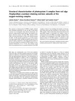
[9,11,12]. Seven main types of PSII–LHCII supercom-
plexes were found (Fig. 1); their relative abundance is listed
in Table 1. The resolution of the images was < 2.6 nm.
Four out of these seven types of supercomplexes have
previously been found in spinach [11,12,25]. They consist of
C
2
S
2
M
2
,C
2
S
2
M, C
2
SM and C
2
S
2
configurations and are
depicted in Fig. 1A–D, respectively. An interesting result in
the present data set was the presence of three new types of
PSII–LHCII particles (Fig. 1E –G). They together com-
prised < 10% of the PSII data set. Image analysis revealed
that they basically consist of a C
2
core part (as in Fig. 1H)
and S-type and M-type trimers (as in Fig. 1A), but that some
of these M-trimers could be attached without the presence of
neighboring S-trimers. For instance, the supercomplex of
Fig. 1F lacks a S-trimer in the left upper part of the complex.
We named it MC
2
S, to discriminate from the C
2
SM
supercomplex (Fig. 1C). The projection of Fig. 1E is similar
but has an additional M-trimer attached to the lower left tip.
A rather small number of supercomplexes has no S-trimers
attached at all (Fig. 1G). These three types of supercomplex
particles were not previously observed in spinach. To
exclude the possibility that they had been previously
overlooked, we re-examined the data sets of complexes
obtained from solubilized spinach PSII membranes by using
those novel types of particles (Fig. 1E–G) each as a
reference. There were no particles matching these
references, indicating their total absence from spinach. On
the other hand, there is no detectable amount of
L-type
LHCII trimers attached to the Arabidopsis supercomplexes,
in contrast to spinach, where low numbers were found [12].
Moreover, no particles from the type I and type II
megacomplexes were found. Finally, we only detected
three particles of the type III megacomplex [12]. The
absence of megacomplexes could in part be caused by the
fractions that were analyzed; they are probably more
abundant in earlier fractions.
Fig. 1. Results of multireference alignment and classification of top-view projections of PSII complexes from Arabidopsis wild type. (A)
Average of the best 218 C
2
S
2
M
2
projections. (B) Average of 290 C
2
S
2
M projections. (C) Average of 175 C
2
SM projections. (D) Average of 300 C
2
S
2
projections. (E) Average of 164 C
2
SM
2
projections. (F) Average of 190 MC
2
S projections. (G) Average of 34 C
2
M
2
projections. (H) Average of 100
C
2
core complexes. Twofold rotational symmetry was imposed on the images of A, D and G. The scale bar is 10 nm.
6022 A. E. Yakushevska et al. (Eur. J. Biochem. 268) q FEBS 2001
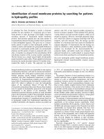
Conformational differences between supercomplex
projections
A more detailed investigation of the particle projections
from Arabidopsis shows some unexpected changes in the
association of the M-type trimers, if neighboring S-type
trimers were absent. Absence of such a trimer can lead to a
significant rearrangement in the binding position the M-type
trimer, as demonstrated in Fig. 2B –D, which shows the
contoured versions of the new types of projections. The
rearrangements are visualized by comparing the density
maximums of the C
2
S
2
M
2
particle (Fig. 2A) with the
positions of these densities in the new types of super-
complexes. At the site of the tentative CP24 subunit, the
positions of these density maximums differ substantially
from the observed positions on the C
2
SM
2
,MC
2
S and C
2
M
2
supercomplexes (indicated by yellow dots in Fig. 2B–D).
The shifts are also obvious if the place of the cleft between
the S-type and M-type trimers, as indicated by the red arrow
in Fig. 2A, is superposed on the supercomplexes of
Fig. 2B–D. In the C
2
M
2
particle (Fig. 2D) there is an
inward shift of CP24 of < 2.7 nm and the M-trimer is also
simultaneously shifted in an upward direction (see red arrow
in Fig. 2D). In the case of the C
2
SM
2
particle, the density
attributed to the CP24 subunits moved < 3.4 nm inward and
an even stronger shift of the M-trimer was visible. However,
the shifts in the MC
2
S particle are less pronounced
(Fig. 2C). These differences might indicate one or more
Table 1. Relative occurrence of PSII–LHCII supercomplexes and megacomplexes in solubilized PSII membranes from A. thaliana and
spinach. Taken from [12].
Complex No. assigned projections
% Projections
Arabidopsis Spinach
Total 4249 100 100
C
2
S
2
1134 26.7 70.6
C
2
SM 828 19.5 7.2
C
2
S
2
M 1142 26.9 14.6
C
2
S
2
M
2
747 17.6 0.9
C
2
M
2
37 0.9 0
C
2
SM
2
164 38 0
MC
2
S 194 4.5 0
Mega I (C
4
S
4
M
2-4
) 0 0 2.8
Mega II (C
4
S
4
M
2-4
) 0 0 1.3
Mega III (C
4
S
4
) 3 0.1 0.3
Total S 7232 85.2 100
Total M 3665 43.2 16.5
Total L 0 0 0.7
Fig. 2. Comparison of positions of M-type
trimers in various types of Arabidopis and
spinach PSII– LHCII supercomplexes. (A)
Contoured version of the image of Fig. 1A. (B–D)
Contoured versions of the images of Fig. 1E,G. (E)
contoured image of Fig. 1C. (F) Contoured image
from 200 spinach C
2
SM supercomplexes
(generated from data sets in [12]). The position of
some density maxima in the C
2
S
2
M
2
projection of
(A) have been indicated by red dots. These
positions have been overlaid on the other images, if
present. This has also been performed for the
position of the red arrow, which indicates in (A) the
position of the interface between the upper S-type
and M-type trimers. The yellow dots in (B) to (D)
indicate the place where the density, marked in (A)
by the upper right red dot, is actually observed and
thus point to larger or smaller changes in the
position of the upper M-trimer upon absence of an
adjacent S-type trimer. The black arrow in (E) and
(F) indicates the site where the C
2
SM
supercomplexes from Arabidopsis and spinach
have their largest difference. The scale bar is
10 nm.
q FEBS 2001 Supermolecular organization of photosystem II (Eur. J. Biochem. 268) 6023
successive stages of an increasing displacement. On the
other hand, no differences between the upper tips of the
images of Fig. 2B,C can be expected if the subunit
composition is the same. Unfortunately, the causes for the
discrepancies in shifts cannot be solved by further
classification, given the limited numbers of these
projections.
In addition to these differences in the peripheral antennae,
there are smaller differences between the core parts of
Arabidopsis and spinach. Because the resolution in the C
2
core complexes is limited, these differences are only well
observed in the C
2
SM type of PSII particle (Fig. 2E,F). The
red dots in Fig. 2E,F near the black arrows mark the place
with the largest difference.
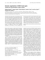
Analysis of two-dimensional crystals
The study of negatively stained intact inside-out paired
membranes can give interesting insight into the packing and
ordering of PSII supercomplexes, because the row-like
arranged PSII core parts can be easily averaged due to their
strongly protruding extrinsic subunits. In comparison to
spinach grana membranes, however, the grana membranes
of Arabidopsis appeared to be more easily disrupted by
a-DM. At a detergent concentration of 0.3%, which is
four times less than used to prepare spinach paired
membranes, most observed membranes were seen to be
severely damaged. On the other hand, almost all of the
remaining intact membranes showed strong periodicity in
the arrangement of PSII complexes (Fig. 3A,B). The
crystallinity was present in both superimposed layers
(Fig. 3A,B). For calculating a two-dimensional projection
map, information of 29 recorded crystals was combined.
The standard approach for crystals, which is the Fourier
analysis, was not used (see below). Instead, a noncrystallo-
graphic approach was used, as it better handles small
crystals with severe lattice imperfections [17]. The crystals
were divided in 656 overlapping small fragments for
analysis by repeated alignments, multivariate statistical
analysis and classification, in a very similar way as carried
out previously with spinach inside-out paired membranes
[17]. To extract as much information as possible from both
layers, the fragments were additionally processed in their
mirrored version. In this way, information from the upper
layer could also be extracted, although this layer was usually
less well negatively stained and preserved than the lower
layer that sticks to the carbon support film. This addition
was not performed in the analysis of the spinach crystals,
where crystallinity in one specific part of a layer did not
strongly correlate to crystallinity in the other one [17]. After
a final classification step (not shown) of the 1312 fragments,
a final group of 450 fragments from the best classes was
obtained (Fig. 4A). In this group, < 68% of the fragments
originated from the lower layer and 32% from the mirrored
upper layer. The final group of 450 crystal fragments shows
a handedness that is opposite to the handedness of the single
particles, where the extrinsic subunits are positioned away
from the carbon support film, but otherwise shows very
similar structural features, especially in the core part region.
The unit cell or repeating motif of the crystal was
Fig. 3. Electron micrographs and filtering of a selected crystalline area of PSII supercomplexes in paired inside-out grana membranes. (A,B)
Membranes negatively stained with 2% uranyl acetate. (C) Selected area of image (B), used for Fourier-peak-filtering of the two superimposed layers.
(D,E) Images of separated layers after filtering. The black dots in (E) mark the centers of individual core complexes and indicate a dislocation in the
crystal packing of one particular layer. The arrow in (A) points to a single C
2
S
2
M
2
supercomplex. The space bars represent 100 nm.
6024 A. E. Yakushevska et al. (Eur. J. Biochem. 268) q FEBS 2001
25.6 Â 21.4 nm with an angle of 778 between the crystal
axes (Fig. 4A). Fourier-transformation indicated that
structural details were present up to 2.5 nm in resolution
[23].
Relative positions of supercomplexes in inside-out paired
membranes
If analysis of crystalline arrays of paired membranes was
focussed on the superposition of both layers, it became clear
that the interaction of the rows of PSII complexes of
adjacent layers was to some extend specific. Of 58 crystals
examined, the majority showed that the angle between rows
of PSII complexes from the superposed layers fell into a
small range; it was either < 328^ 28 (mean ^ SD, n ¼ 16)
or < 588^ 38 (n ¼ 33). Crystalline arrays showing different
angles had an overall smaller degree of ordering. Examples
of crystals with overlapping rows of PSIl complexes
differing in rotation by 328 and 588 are presented in Fig. 3A
and B, respectively. However, image analysis of these and
several other crystals showed that the restricted rotational
freedom in attachment does not imply that the super-
complexes become associated to each other in a similar way
all over the crystal. The largest crystals, such as the one
shown in Fig. 3A, show this very clearly. It can be seen that
the Moire
´
pattern, which is the resulting motif from the
overlap of two crystals making an angle, gradually changes.
The upper and lower part of the crystal show about the same
type of Moire
´
pattern (and thus of the type of overlap),
which differs substantially from the central part. In the
center of this crystal, the PSII core parts of both layers are
more strongly overlapping in projection. A similar pattern
with overlapping core complexes in the center of the paired
membranes was observed in other crystals of the 328-type.
Another aspect of the lateral freedom in the interaction of
the two layers is demonstrated by the crystal of Fig. 3B.
This was shown by Fourier filtering, a technique that can
separate two superposed layers differing in rotational
position by selecting only those peaks in the Fourier-
transformed image that belong to one specific layer. The
processed selected central part of this crystal (Fig. 3C)
shows this very well. After Fourier filtering, which reduces
noise and separates the layers, a crystal defect could be
detected in one of the layers resulting in a translational shift
with a magnitude of about half a unit cell (Fig. 3E). Similar
types of imperfections within one layer were observed
in other analyzed inside-out paired of membranes of the
588-type.
DISCUSSION
In this report, we present an analysis of membrane-bound
crystalline PSII and isolated PSII–LHCII supercomplexes
from Arabidopsis. One of the ultimate aims of such an
analysis is the detection of subunits crucial to regulation
mechanisms, such as PsbS [26], by comparing the sets of
Fig. 4. Final result of image analysis of two-
dimensional crystalline and comparison. (A)
Sum of 450 aligned crystal fragments from
Arabidopsis; the unit cell has been indicated; (B)
sum of 900 aligned crystal fragments from spinach
(from [17]) with the central PSII dimer in the same
position as in (A), the unit cell has been indicated;
(C) Difference image between (A) and (B), arrows
point to the site of strongest difference. (D) Image
form (A) with contours; positions of S-type and
M-type LHCII trimers indicated in yellow and
positions of CP29, CP26 and CP24 indicated in
green. (E) Image of the C
2
S
2
M
2
supercomplex
(from Fig. 1A) in equivalent position, with the
dimeric part (‘c’), the S-type and M-type LHCII
trimers (‘s’and ‘m’) indicated in yellow, the CP29,
CP26 and CP24 areas in green and the strongest
difference area from image (C) indicated in red;
(F) image of type III megacomplex (from [11]) in
similar rotational position as in (A). The blue dots
mark the centers of two adjacent PSII complexes as
in (A) and the green areas indicate two CP26
positions as in (A). The images of (A) to (D) have
been mirrored after image analysis, to facilitate
comparison with the single particle averages. The
space bar represents 20 nm.
q FEBS 2001 Supermolecular organization of photosystem II (Eur. J. Biochem. 268) 6025
isolated supercomplexes or complete (crystalline) mem-
branes from wild-type and mutants lacking such subunits.
Furthermore, a close comparison to the spinach data
[1,9,11–13,25] is useful for detecting specific structural
features of Arabidopsis PSII. With the appearance of the first
complete genomic sequence of a plant, Arabidopsis [27],
new perspectives for further investigation of all individual
photosynthetic proteins, becomes available.
Analysis of the Arabidopsis crystals shows the presence
of only one crystal form (Fig. 4A), which has unit cell
dimensions of 25.6 Â 21.4 nm (angle 778). It covers a
surface area of 534 nm
2
. The crystal is larger than any of the
PSII lattices observed previously. The projection map
clearly reveals the positions of two LHCII trimers flanking
each side of a dimeric PSII core complex (Fig. 4A). Because
the position of these trimers strongly resembles the positions
of S- and M-type LHCII trimers as found in the single
C
2
S
2
M
2
supercomplex projection (Fig. 4E), we fitted these
trimers, together with the remaining peripheral antenna
components CP29, CP26 and CP24, into the crystal lattice
(Fig. 4D). From this fitting it can be concluded that the
crystal arrays consist of C
2
S
2
M
2
particles. Further evidence
comes from the observation that only C
2
S
2
M
2
single
particles can be frequently found around the crystals
(Fig. 3A). In contrast, the previously analyzed large-sized
crystals from spinach [17] had a differently shaped unit cell
of 27.3 Â 18.3 nm (Fig. 4B), which is also 11% smaller in
surface. We previously noticed that the spinach crystals were
too small to contain a C
2
S
2
M
2
particle and it was concluded
that they were composed of C
2
S
2
M particles [17]. The present
analysis of Arabidopsis crystals, where M-type trimers are
directly observed, confirms this assignment.
The Arabidopsis crystals show a marked preference in the
way the two layers are attached. The rows of core complexes
in the two layers make an angle of either < 328 or 588.A
rotational preference was also observed in the spinach
crystals: preferential angles of 38 or 468 were found [17]. As
the PSII complexes from which the two different types of
crystals are built up only differ in the peripheral antennae,
we speculate that the LHCII trimers, rather than the core
complexes, determine the way of interaction between the
two layers, which leads to the preference in rotational
position. If the LHCII trimers strive to optimize association
and overlap, this would automatically lead to strong overlap
of core complexes in at least a part (the center) of the paired
membranes. This is essentially also the case in the
previously studied spinach crystals, but in that case large
domains of LHCII trimers without attached core complexes
form an additional type of variation [17]. Although the
LHCII trimers appear to be important to induce preferential
stacking of crystalline membranes, other factors might be
relevant as well, such as the lipid composition of the
membranes and the protein/lipid ratio.
The analysis of isolated Arabidopsis PSII–LHCII super-
complexes showed the presence of relatively high amounts
of M-type LHCII trimers. This is evident from a comparison
of the relative numbers of those types of supercomplexes,
which were also present in the previously analyzed 12755
spinach supercomplexes (Table 1 [12]); the number of
C
2
S
2
M
2
projections is significantly (20 times) larger.
Furthermore, we noticed that the numbers of C
2
S
2
and
C
2
S
2
M particles are about equal, whereas in spinach the
larger part was formed by the C
2
S
2
particles. The analysis of
isolated Arabidopsis PSII–LHCII supercomplexes also
Fig. 5. Modeling of subunit densities in supercomplex projection maps. (A,B) The C
2
SM
2
and C
2
S
2
supercomplexes of Arabidopsis. (C) The
C
2
S
2
supercomplex from spinach (from [12] with core subunit densities from [5]). The position of the upper M-type LHCII trimer in (A) indicates a
major shift for the attached minor LHC subunit (possibly a CP24 subunit), but otherwise the core parts and the lower half of the peripheral antennas in
(A–C) are rather similar. The difference areas from Fig. 4E are reproduced in (B) and overlap of these densities suggests that these areas, assigned to
the extrinsic subunits occupy a larger space than the extrinsic 33 and 17/23 kDa subunit areas as shown in (C) (from [12]). The space bar is 10 nm.
6026 A. E. Yakushevska et al. (Eur. J. Biochem. 268) q FEBS 2001
showed for the first time the presence of M-type trimers in
the absence of adjacent S-type trimers (Fig. 2). Such
particles were completely absent in the spinach data sets.
The best resolved particle was the C
2
SM
2
supercomplex.
Fitting of subunits densities into this projection (Fig. 5A)
revealed that the upper M-type trimer occupies the correct
amount of expected space, but that the adjacent minor LHC
subunit, possibly CP24, must have rearranged its position
dramatically because otherwise no space is left over.
Apparently, the absence of the adjacent S-type trimer
strongly influences the binding of the more peripheral
M-type trimers. This situation is reminiscent to our
experiments with the specific removal of the extrinsic
subunits, which had a profound influence on the position of
the S-type trimer and CP29 [25]. There is, however, no
evidence that the Arabidopsis single particles have a
reduced number of S-type trimers because of the loss or
absence of extrinsic subunits. On the contrary, a close
comparison of the features from the central supercomplex
particles in the averaged native crystals from Arabidopsis
and spinach indicates additional masses for Arabidopsis at
the two sites where the S-type trimers are attached to the
core complex (Figs 4C and 5B). The fact that these masses
seem to overlap with the S-type trimers suggests that they
should be considered as extrinsic subunits. These masses,
which were previously assigned as the 17 and 23 kDa
extrinsic subunits [28]), are easily lost in spinach but are
apparently stronger when attached to Arabidopsis super-
complexes. This is also reflected in the comparison of
density profiles of isolated C
2
SM supercomplexes
(Fig. 2E,F). The averaged Arabidopsis C
2
S
2
M supercom-
plex shows more density at about the same site as the
spinach supercomplex, as became clear from the crystal
difference map (Fig. 4C). If indeed the Arabidopsis PSII
complexes have stronger bound extrinsic subunits, as
suggested from the crystal difference map (Fig. 4C), then
it is unlikely that they are responsible for the overall lower
stability of bound S-type trimers in Arabidopsis.
Another difference between the isolated Arabidopsis and
spinach supercomplexes has to do with the further
association into megacomplexes. No particles of the type I
and II megacomplexes were observed in Arabidopsis, and
only a few type III complexes were present (Table 1),
although we did not examine the fractions where such
particles tend to accumulate. The observation of only
type III megacomplexes could be of coincidence, but we
would like to point out that in the latter type of
megacomplex, the packing of the supercomplex is very
similar to the packing in the crystals, as is shown in the
fitting of crystal features into the spinach type III
megacomplex (Fig. 4F). Finally, the CP26 area in all
Arabidopsis supercomplexes was somewhat the same. No
variation at the CP26 position was detected (Fig. 1A–F). On
the other hand, a substantial number of the spinach
projections lacked this tip completely, although this absence
did not influence the supercomplex structure [12,25].
The analysis of Arabidopsis PSII–LHCII supercom-
plexes has contributed further to the evidence for the
structural complexity of the association of the peripheral
antenna of PSII. Some of this variation could originate from
slight differences in the solubilization and purification
process. Nevertheless we can conclude that both the single
particle analysis and the crystal analysis indicate the
presence of larger numbers of associated M-type LHCII
trimers. A strong argument for the in vivo presence of
larger amounts of the M-type LHCII trimers is the fact
that in contrast to spinach, where C
2
S
2
M as well as
C
2
S
2
-containing crystals were found, Arabidopsis seems to
exclusively contain C
2
S
2
M
2
crystals. The finding that native
Arabidopsis plants contain a higher degree of M-type LHCII
trimers, and thus an overall larger antenna associated to
PSII–LHCII supercomplexes, is of interest for further
studies focussed on subunits important for state transitions
and nonphotochemical quenching, such as PsbS [26]. As
there is evidence that PsbS is not present in the C
2
S
2
supercomplexes [29], larger supercomplexes, as found in
Arabidopsis, might be more suited for structural localization
of PsbS.
ACKNOWLEDGEMENTS
Support from the Dutch Scientific foundation NWO/ALW to E. J. B
and J. P. D and from the Danish National Research Foundation to
P. E. J. and H. V. S. is gratefully acknowledged.
REFERENCES
1. Hankamer, B., Barber, J. & Boekema, E.J. (1997) Structure and
membrane organization ofphotosystem II in green plants. Annu.
Rev. Plant Physiol. Plant Mol. Biol. 48, 641– 671.
2. Hankamer, B., Morris, E.P. & Barber, J. (1998) Revealing the
structure of the oxygen-evolving core dimer ofphotosystem II by
cryoelectron crystallography. Nat. Struct. Biol. 6, 560– 564.
3. Zouni, A., Witt, H T., Kern, J., Fromme, P., Krauss, N., Saenger,
W. & Orth, P. (2001) Crystal structure ofphotosystem II from
Synechoccus elongatus at 3.8 A
˚
resolution. Nature 409, 739– 743.
4. Zheleva, D., Sharma, J., Panico, M., Morris, H.R. & Barber, J.
(1998) Isolation and characterization of monomeric and dimeric
CP47-reaction center photosystem II complexes. J. Biol. Chem.
273, 16122– 16127.
5. Shi, L X., Lorkovic, Z.J., Oelmu
¨
ller, R. & Schro
¨
der, W.P. (2000)
The low molecular mass PsbW protein is involved in the
stabilization of the dimeric photosystem II complex in Arabidopsis
thaliana. J. Biol. Chem. 275, 37945–37950.
6. Kruse, O., Hankamer, B., Konczak, C., Gerle, C., Morris, E.,
Radunz, A., Schmid, G.H. & Barber, J. (2000) Phosphatidylgly-
cerol is involved in the dimerisation of photosystem II. J. Biol.
Chem. 275, 6509–6514.
7. Jansson, S. (1994) The light-harvesting chlorophyll a/b-binding
proteins. Biochim. Biophys. Acta 1184, 1 –19.
8. Ku
¨
hlbrandt, W., Wang, D.N. & Fujiyoshi, Y. (1994) Atomic model
of plant light-harvesting complex by electron microscopy. Nature
367, 614– 621.
9. Boekema, E.J., Hankamer, B., Bald, D., Kruip, J., Boonstra, A.F.,
Barber, J. & Ro
¨
gner, M. (1995) Supramolecular structure of the
photosystem II complex from green plants and cyanobacteria. Proc.
Natl Acad. Sci. USA 92, 175– 179.
10. Peter, G.F. & Thornber, J.P. (1991) Biochemical composition and
organization of higher plant photosystem II light-harvesting
pigment-proteins. J. Biol. Chem. 266, 16745–16754.
11. Boekema, E.J., Van Roon, H., Calkoen, F., Bassi, R. & Dekker, J.P.
(1999) Multiple types of association of photosystem II and its light-
harvesting antenna in partially solubilized photosystem II
membranes. Biochemistry 38, 2233– 2239.
12. Boekema, E.J., Van Roon, H., Van Breemen, J.F.L. & Dekker, J.P.
(1999) Supramolecular organization of photosystem II and its light-
harvesting antenna in partially solubilized photosystem II
membranes. Eur. J. Biochem. 266, 444–452.
13. Nield, J., Orlova, E.V., Morris, E.P., Gowen, B., Van Heel, M. &
q FEBS 2001 Supermolecular organization of photosystem II (Eur. J. Biochem. 268) 6027
Barber, J. (2000) 3D map of the plant photosystem II supercomplex
obtained by cryoelectron microscopy and single particle analysis.
Nat. Struct. Biol. 7, 44–47.
14. Harrer, R., Bassi, R., Testi, M.G. & Scha
¨
fer, C. (1998) Nearest-
neighbour analysis of a photosystem II complex from Marchantia
polymorpha L. (liverworth) which contains reaction centre and
antenna proteins. Eur. J. Biochem. 255, 196–205.
15. Simpson, D.J. (1979) Freeze-fracture studies on barley plastid
membranes III. Location of the light-harvesting chlorophyll
protein. Carlsberg Res. Commun. 44, 305–336.
16. Staehelin, L.A., Armond, P.A. & Miller, K.R. (1977) Chloroplast
membrane organization at the supermolecular level and its
functional implication. Brookhaven Symposia Biol. 28, 278–315.
17. Boekema, E.J. & Van Breemen, J.F.L., Van Roon, H. & Dekker J.P.
(2000) Arrangement of photosystem II in crystalline macrodomains
within the thylakoid membrane of green plant chloroplasts. J. Mol.
Biol. 301, 1123–1133.
18. Van Roon, H. Van Breemen, J.F.L., De Weerd, F.L., Dekker J.P. &
Boekema, E.J. (2000) Solubilization of green plant thylakoid
membranes with n-dodecyl-a,
D-maltoside. Implications for the
structural organization of the photosystem II, photosystem I ATP
synthase and cytochrome b
6
/f complexes. Photosynth. Res. 64,
155–166.
19. Harauz, G., Boekema, E. & Van Heel, M. (1988) Statistical image
analysis of electron micrographs of ribosomal subunits. Methods
Enzymol. 164, 35–49.
20. Van Heel, M. & Frank, J. (1981) Use of multivariate statistics in
analysing images of biological macromolecules. Ultramicroscopy
6, 187– 194.
21. Van Heel, M. (1989) Classification of very large electron
microscopical image data sets. Optik 82, 114–126.
22. Van Heel, M. (1987) Similarity between images. Ultramicroscopy
21, 95– 100.
23. Henderson, R.J., Baldwin, J.M., Downing, K.H., Lepault, J. &
Zemlin, F. (1986) Structure of purple membrane from Halobacter-
ium halobium: recording, measurement and evaluation of electron
micrographs at 3.5 A
˚
resolution. Ultramicroscopy 19, 147–178.
24. Boekema, E.J., van Roon, H. & Dekker, J.P (1998) Specific
association of photosystem II and light-harvesting complex II in
partially solubilizedphotosystem II membranes. FEBS Lett. 424,
95–99.
25. Boekema, E.J., van Breemen, J.F.L., van Roon, H. & Dekker, J.P.
(2000) Conformational changes in photosystem II superomplexes
upon removal of extrinsic subunits. Biochemistry 39,
12907–12915.
26. Li, X P., Bjo
¨
rkman, O., Shih, C., Grossman, A.R., Rosenquist, M.,
Jansson, S. & Niyogi, K. (2000) A pigment-binding protein
essential for regulation of photosynthetic light harvesting. Nature
403, 391– 395.
27. The Arabidopsis Genome Initiative (2000) Sequence and analysis
of the flowering plant Arabidopsis thaliana. Nature 408, 796–815.
28. Boekema, E.J., Nield, J., Hankamer, B. & Barber, J. (1998)
Localization of the 23 kDa subunit of the oxygen evolving complex
of photosystem II by electron microscopy. Eur. J. Biochem. 252,
268–276.
29. Nield, J., Funk, C. & Barber, J. (2000) Supermolecular structure of
photosytem II and location of the PsbS protein. Phil. Trans. R. Soc.
London 29, 1337–1344.
6028 A. E. Yakushevska et al. (Eur. J. Biochem. 268) q FEBS 2001