Báo cáo Y học: Isolation and characterization of MUC15, a novel cell membrane-associated mucin pot
Bạn đang xem bản rút gọn của tài liệu. Xem và tải ngay bản đầy đủ của tài liệu tại đây (301.23 KB, 9 trang )
Isolation and characterization of MUC15, a novel
cell membrane-associated mucin
Lone T. Pallesen, Lars Berglund, Lone K. Rasmussen, Torben E. Petersen and Jan T. Rasmussen
Protein Chemistry Laboratory, Department of Molecular and Structural Biology, University of Aarhus, Denmark
The present work reports isolation and characterization of a
highly glycosylated protein from bovine milk fat globule
membranes, known as PAS III. Partial amino-acid sequen-
cing of the purified protein allowed construction of degen-
erate oligonucleotide primers, enabling isolation of a
full-length cDNA encoding a protein of 330 amino-acid
residues. N-terminal amino-acid sequencing of derived
peptides and the purified protein confirmed 76% of the
sequence and demonstrated presence of a cleavable signal
peptide of 23 residues, leaving a mature protein of 307 amino
acids. Database searches showed no homology to any other
proteins. A survey of the human genome indicated the
presence of a corresponding gene on chromosome band
11p14.3. Isolation and sequencing of the complete cDNA
sequence of the human homologue proved the existence of
the gene product (334 amino-acid residues). This novel
mucin-like protein was named MUC15 by appointment of
the HUGO Gene Nomenclature Committee. The deduced
amino-acid sequences of human and bovine MUC15 dem-
onstrated structural hallmarks characteristic for other
membrane-bound mucins, such as a serine, threonine, and
proline-rich extracellular region with several potential
glycosylation sites, a putative transmembrane domain, and a
short cytoplasmic C-terminal. We have shown the presence
of O-glycosylations, identified N-glycosylations at 11 of 15
potential sites in bovine MUC15, and a splice variant
encoding a short secreted mucin. Finally, analysis of human
and bovine cDNA panels and libraries showed MUC15 gene
expression in adult human spleen, thymus, prostate, testis,
ovary, small intestine, colon, peripheral blood leukocyte,
bone marrow, lymph node, tonsil, breast, fetal liver, bovine
lymph nodes and lungs of both species.
Keywords: MUC15; amino-acid sequence; bovine and
human cDNA; splice variant; N-glycosylation.
Mucins are a heterogeneous family of high molecular mass
proteins that are broadly defined by their high content of
carbohydrates (50–90%), which are mainly O-linked but in
some cases also N-linked. These glycoproteins are major
constituents of the mucus covering the surfaces of epithelial
organs and they provide selective physical barriers protect-
ing the underlying epithelium. Mucins are known to be
expressed in various epithelia. Nevertheless, the overall
expression patterns have not been completely elucidated
(reviewed in [1,2]). To date 15 human mucin genes encoding
epithelial mucin type proteins have been identified: MUC1,
-2, -3A, -3B, -4, -5AC, -5B, -6, -7, -8, -9, -11, -12, -13, and -16
[3–15]. In addition, two mouse mucin genes, MUC10 and
MUC14, have been isolated ([16], GenBank accession
number NM_016885). Mucins can be divided into at least
two structurally and functionally distinct classes, the
secreted (gel-forming or nongel-forming) mucins and the
membrane-associated mucins.
Four of the secreted mucins are encoded by a cluster of
genes (MUC2, MUC5AC, MUC5B and MUC6) contained
within a 400-kb genomic DNA fragment on chromo-
some 11 band p15.5 [17]. The MUC7, MUC8 and MUC9
are relatively small mucins expressed in the salivary gland,
respiratory tissue and fallopian tube, respectively. The
family of epithelial membrane-associated mucins includes
MUC1,-3,-4,-12,-13and-16.MUC3,MUC11,and
MUC12 have been located to chromosome 7q22 suggesting
the presence of yet another cluster of mucin genes. It should,
however, be noted that only partial sequences are known for
the MUC11 and MUC12 genes and that it is possible that
they are produced as a result of alternative splicing of a
single, large mucin gene [13]. Human MUC1 was the first
mucin to be cloned and is to date probably the best
characterized of the mucins. Generally, MUC1 is expressed
on the apical cell surface of nearly all polarized epithelial
tissues that line ducts and glands, e.g. mammary gland [18].
MUC1 is found to be a major constituent of human and
bovine milk fat globule membranes (MFGM) surrounding
the lipid droplets secreted from the mammary gland
epithelial cells [19,20].
Bovine MFGM has been shown to contain another
heavily glycosylated mucin-like glycoprotein with high
molecular mass, named PAS III. Glycoprotein C, Glyco-
protein 4, Component II and PAS3, are alternative names
that have been used for this glycoprotein as well [21]. This
poorly characterized glycoprotein has been named accord-
ing to its mobility upon separation by SDS/PAGE and
ability to stain with periodic acid-Schiff’s reagent (PAS) [22].
The protein appears heterodisperse with apparent molecular
Correspondence to J. Trige Rasmussen, Protein Chemistry
Laboratory, Gustav Wieds Vej 10C, 8000 Aarhus C, Denmark.
Fax: + 45 86136597, Tel.: + 45 89425093,
E-mail:
Abbreviations: PAS, periodic acid-Schiff’s; MFGM, milk fat globule
membrane; MTC, multiple tissue cDNA.
Note: reported nucleotide sequences are available from the EMBL
Nucleotide Sequence Database under the accession numbers
AJ417816, AJ417817 and AJ417818.
(Received 5 December 2001, revised 13 March 2002,
accepted 22 April 2002)
Eur. J. Biochem. 269, 2755–2763 (2002) Ó FEBS 2002 doi:10.1046/j.1432-1033.2002.02949.x
mass ranging from 95 to over 100 kDa in polyacrylamide
gels. Antibody staining of sections from bovine prelactating
and lactating mammary gland using monoclonal and
polyclonal antibodies has shown that PAS III is largely
concentrated on apical surfaces of the mammary epithelial
cells [23].
The present work was initiated in order to isolate and
characterize the bovine mucin-like glycoprotein PAS III. A
purification method has been established, together with a
determination of the complete amino-acid sequence enco-
ded by the corresponding cDNA. In addition, the cDNA
encoding the human homologue has been isolated and
sequenced, thereby identifying a novel human transmem-
brane mucin gene named MUC15 by appointment of the
HUGO Gene Nomenclature Committee. Presence of
O-glycosylation and sites of N-glycosylation in bovine
MUC15/PAS III have been determined, and a splice variant
encoding a short secreted mucin was identified. Finally,
PCR on cDNA panels revealed MUC15 expression in a
variety of human tissues.
MATERIALS AND METHODS
Purification of bovine MUC15
MFGM was prepared as described by Hvarregaard et al.
[24] using the cream fraction of freshly collected unpasteur-
ized bovine milk samples. Bovine MUC15 was purified
from MFGM using a method essentially as the one used for
isolation of bovine MUC1 [20]. Briefly, MFGM proteins
were extracted from the membranes using the nonionic
detergent Triton X-100. Extracted proteins were subjected
to cation- and anion-exchange chromatography on CM-
Sepharose and DEAE-Sepharose columns, respectively
(Amersham Pharmacia Biotech, Uppsala, Sweden).
MUC15 containing fractions were dialyzed, freeze-dried,
and finally subjected to further purification by reverse-phase
chromatography using a 1-mL Resource RPC column
(Amersham Pharmacia Biotech) with a gradient of
2-propanol in 20% formic acid. MUC15 containing sam-
ples appearing at 48% 2-propanol were collected and freeze-
dried. Standard procedures were employed analysing pro-
tein samples by SDS/PAGE using 18% polyacrylamide
gels, and for the staining of proteins using Coomassie
Brilliant Blue R-250 and PAS reagent.
Peptide mapping of bovine MUC15
Bovine MUC15 peptides were generated by enzymatic
cleavage of the purified protein with trypsin (Worthington
Biochemical Corp., Lakewood, NJ, USA) for 4 h at 37 °C.
Resulting peptide mixtures were separated by RP-HPLC on
aVydacC18column(4· 250 mm, Vydac, Hesperia, CA)
using a linear gradient of acetonitrile (0–80%) in 0.1%
trifluoroacetic acid. Selected peptide fractions were further
purified by reverse-phase chromatography on a Sephacil C8
SC 2.1/10 column (Amersham Pharmacia Biotech) using
the same gradient. Additional peptides were produced
treating unmodified or deglycosylated MUC15 with five
different proteases independently [Staphylococcus aureus V8
protease, (Worthington Biochemical Corp.), endopeptidase
LysC (Roche, Basel, Switzerland), thermolysin, chymotryp-
sin or elastase (Sigma, St Louis, MO, USA)] and
successively purifying generated peptides by RP-HPLC as
described above. Deglycosylation of bovine MUC15 was
achieved by an initial treatment with neuraminidase (Roche)
in 50 m
M
ammonium acetate, pH 5.0 at 37 °C for 18 h.
After that, N-linked oligosaccharides were removed with
peptide-N
4
-(acetyl-b-glucosaminyl)-asparagine amidase
(PNGase F; Roche) in 50 m
M
sodium phosphate, pH 7.5,
0.5% SDS, 5 m
M
dithioerythritol, 2% octyl-glycopyrano-
side for 18 h at 37 °C. Finally, a part of this material was
treated with endo-a-N-acetylgalactosaminidase (O-glycosi-
dase; Sigma) in 50 m
M
sodium citrate, pH 6.0 at 37 °Cfor
20 h. Purified MUC15 and resolved peptide fragments were
subjected to N-terminal amino-acid sequencing by automa-
ted Edman degradation by means of an ABI 477 A/120 A
Protein Sequencer (Applied Biosystems, Foster City, CA,
USA) with online identification of the phenylthiohydantoin
derivatives. N-glycosylation sites were assigned to aspara-
gine residues lacking an identifiable phenylthiohydantoin
derivative during amino-acid sequencing of glycosylated
samplesorshowingupasasparticacidinPNGaseFtreated
MUC15 peptides.
Cloning of the bovine MUC15 cDNA by PCR
with degenerate primers
Isolation of total RNA from the mammary gland of a
lactating Danish Holstein cow was performed by means of
an RNeasy kit (Qiagen, Hilden, Germany). Synthesis of
cDNA was performed by oligo(dT) primed reverse tran-
scription of the isolated total RNA using M-MLV Reverse
Transcriptase (Life Technologies, Inc., Gaithersburg, MD,
USA) in accordance with the manufacturer’s instructions.
Six degenerate oligonucleotides were synthesized corres-
ponding to partial bovine MUC15 amino-acid sequences
obtained by peptide mapping and N-terminal sequencing of
the mature protein (DNA Technology, Aarhus, Denmark):
P1, 5¢-GARGARGGICARAARAC-3¢ (forward), corres-
ponding to the amino-acid sequence E(24)EGQKT(29)
(residues underlined in Fig. 1B); P2, 5¢-AARACNATGGA
RAAYCA-3¢ (forward), K(40)TMENQ(45); P3, 5¢-TCYT
TRTCISWIGTIARRTT-3¢ (reverse), N(54)LTSDKE(60);
P4, 5¢-GGYTCRTTICKRTCRTCRTA-3¢ (reverse), Y(271)
DDRNEP(277); P5, 5¢-CATRTCRTAIGGYTCIGGNG
C-3¢ (reverse), A(284)PEPYDM(290); P6, 5¢-GCNGTIGG
RTTRTARTA-3¢ (reverse), Y(297)YNPTA(302); where
R ¼ AorG,Y¼ CorT,K¼ GorT,S¼ Cor
G, W ¼ AorT,N¼ A, G, C or T, and I ¼ deoxyin-
deoxyinosine. The degenerate primers were employed in
PCR amplifications of cDNA performed in a total volume
of 25 lL containing 0.4 m
M
dNTPs, 2.5 lL10· PCR
buffer, 2.5 U of HotStarTaq polymerase (HotStarTaq
Master Mix Kit, Qiagen), 5 lL first-strand cDNA and
4 l
M
each of the forward and reverse degenerate primers.
After a 15-min, 95 °C activation step of the HotStarTaq
DNA polymerase, amplification was performed as follows:
five cycles of denaturation at 94 °C for 45 s, annealing at
46 °C for 45 s and extension at 72 °C for 120 s, followed by
35 amplification cycles with an annealing temperature of
50 °C. Obtained PCR products were cloned into pCR 2.1-
TOPO cloning vectors using the TOPO TA Cloning Kit
(Invitrogen, Groningen, the Netherlands). Sequencing
inserts from 50 positive clones a single was found to contain
a MUC15 fragment of 62 nucleotides generated with the P2
2756 L. T. Pallesen et al. (Eur. J. Biochem. 269) Ó FEBS 2002
and P3 primers. From the obtained MUC15 nucleotide
sequence a single specific oligonucleotide primer was
designed: P7, 5¢-CAATCTGTCCCTTTAGA-3¢ (forward).
The major part of the coding sequence was then cloned and
sequenced using PCR as described above with 0.4 l
M
and
4 l
M
of the specific P7 primer and degenerate primers
(P4-P6), respectively. The cDNA sequence of the bovine
MUC15wasextendedinboth5¢ and 3¢ directions by PCR
screening of an oligo(dT) primed mammary gland Uni-ZAP
cDNA library, derived from a lactating Holstein cow
(Stratagene, La Jolla, CA, USA), using MUC15-specific
and library vector primers. The full-length cDNA was
obtained sequencing overlapping clones and PCR products
derived by RT-PCR on the isolated RNA from the Danish
Holstein cow. The bovine MUC15 cDNA was sequenced
on both strands using a BigDye Sequencing kit and an ABI
PRISM 310 Genetic Analyser (Applied Biosystems).
Identification of the human MUC15 cDNA
The bovine MUC15 nucleotide sequence was employed in a
BLASTn search of the human genome database at NCBI,
and a match was found on a Ôchromosome 11 working draft
sequence segmentÕ (GenBank accession number
NT_008952). Identified partial sequences of the putative
human homologue were examined and specific PCR
primers were designed enclosing the coding sequence of
the bovine protein. To investigate the presence of MUC15
expression in epithelial cells of the human mammary gland,
we proceeded to isolate the cellular fraction of human milk
samples obtained from four lactating women at different
stages in the lactation. Samples were collected immediately
after milking and stored on ice. Milk cells were harvested by
centrifugation at 3200 g for 20 min at 4 °C, and the cellular
fraction was washed in NaCl/P
i
buffer and processed for
total RNA purification using a RNeasy Blood kit (Qiagen).
Synthesis of cDNA was performed by oligo(dT) primed
reverse transcription of the mRNA isolated from milk cells
using M-MLV Reverse Transcriptase. Using the specific
primers in RT-PCR a 1501 base pair cDNA composite of
the human gene was obtained. RT-PCR products were
purified using a Jetquick PCR Purification Spin Kit
(Genomed, Bad Oeynhausen, Germany) and sequenced as
described for the bovine counterpart.
Detection of an alternatively spliced MUC15 variant
First-strand cDNA was prepared from the mammary gland
RNA of a Danish Holstein cow using M-MLV Reverse
Transcriptase as described above. Specific forward and
reverse primers were designed to produce a PCR product of
513 bp containing the transmembrane domain: 5¢-CATCC
ATAGCAGATAACAGTC-3¢ (forward) and 5¢-TCCCA
AAGCTCATGTCATAAG-3¢ (reverse) corresponding to
amino-acid residues S(123)SIADNSL(130) and P(287)YD
MSFGN(294), respectively (see below). The PCR products
were subjected to DdeI restriction enzyme treatment
(Roche) following standard procedures, and a second round
of PCR was performed using the same primers. Obtained
PCR products were ligated into pCR 2.1-TOPO cloning
vectors and sequenced on both strands.
MUC15 expression analysis
MUC15 mRNA expression was examined in a variety of
tissues and cell types by PCR screening. The screening
analysis was performed using commercial multiple tissue
cDNA (MTC) panels of fetal and adult human tissues
(human MTC Panel II, Cat. # K1421-1 and human
Immune System MTC Panel, Cat. # K1426-1, Clontech,
Palo Alto, CA, USA). The panels contained normalized,
first-strand cDNA preparations generated from each of the
following human tissues and cell types: spleen, thymus,
prostate, testis, ovary, small intestine, colon, peripheral
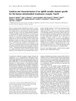
Fig. 1. Purification of bovine MUC15 and obtained tryptic peptidemap.
(A) RP-HPLC chromatography of bovine milk fat globule membrane
proteins eluted from the DEAE column. Separation was performed on
a 1-mL Resource RPC column with a linear gradient of 0–80%
2-propanol in 20% formic acid at 40 °C (dotted line). Proteins were
monitored at 278 nm (solid line). The peaks containing MUC1 and
MUC15 are indicated. (B) RP-HPLC separation of peptides generated
by trypsin digestion of bovine MUC15. Peptides were eluted from a
Vydac C18 column using a linear gradient from 0 to 80% acetonitrile
in 0.1% trifluoroacetic acid (dotted line), and monitored at 226 nm
(solid line). Amino-acid sequences of labelled peaks are shown.
Underlining indicates amino-acid residues used for design of degen-
erate oligonucleotide primers.
Ó FEBS 2002 MUC15, a novel membrane-associated mucin (Eur. J. Biochem. 269) 2757
blood leukocyte, bone marrow, fetal liver, lymph node, and
tonsil. Further tissue specific studies were performed by
PCR screening of oligo(dT) primed cDNA libraries of
bovine lymph node, bovine lung, human lung (Stratagene),
and human breast tissue (Clontech). Specific bovine and
human MUC15 primer sets were employed in the PCR
screening reactions. PCR products were separated by
electrophoresis on 1% agarose gels, visualized with ethi-
dium bromide and finally sequenced.
RESULTS
Purification of bovine MUC15
Bovine MUC15 copurifies with MUC1 during the initial
steps of the protocol designed for isolation of the latter
mucin from MFGM [20]. Complete separation was
achieved by RP-HPLC on a Resource RPC column with
a gradient of 2-propanol in 20% formic acid (Fig. 1A). The
bovine MUC15 eluted at approximately 48% 2-propanol,
and the purity of this fraction was confirmed by SDS/
PAGE (Fig. 2). N-terminal amino-acid sequencing of the
isolated mature bovine MUC15 was performed and
revealed a segment of 30 residues (EEGQKTXTTESTAED
LKTMENQSVPLESKA), which did not show similarity to
any known sequences as revealed by
BLASTP
and
FASTA
3
homology searching of databases accessed through the
NCBI and EBI, respectively.
Sequence description of bovine MUC15
To obtain sequence information from peptide mapping,
purified bovine MUC15 was subjected to enzymatic diges-
tion with trypsin. Generated tryptic peptides were separated
by RP-HPLC, and subjected to N-terminal amino-acid
sequencing (Fig. 1B). To enable deduction of the complete
amino-acid sequence of bovine MUC15 by cDNA cloning,
six degenerate oligonucleotide primers were designed from
the acquired partial amino-acid sequences. After RT-PCR
on mammary gland mRNA of a Danish Holstein cow a
single MUC15 fragment was cloned and a specific primer
was constructed. By additional use of degenerate and
specific MUC15 primers, a full-length cDNA sequence was
obtained. Reported nucleotide sequence data are available
from the EMBL Nucleotide Sequence Database under the
accession number AJ417816.
Analysis of the obtained full-length cDNA sequence
(3125 nucleotides in total) showed the presence of an open
reading frame encoding a protein of 330 amino-acid
residues (Fig. 3). Approximately 76% of the cDNA-enco-
ded amino-acid sequence was confirmed by N-terminal
sequencing of the mature protein and enzymatic generated
peptides (Fig. 3, underlined residues). The proposed trans-
lational start codon (ATG) follows a 5¢ untranslated
sequence of 120 nucleotides. The translational stop codon
(TAA), positioned at residues 1111–1113, is followed by a
3¢ untranslated sequence of 1994 nucleotides, including a
polyadenylation signal (AATAAA) (position 3085–3090)
Fig. 2. SDS/PAGE analysis of purified bovine MUC15. Analysis was
performed on 18% Tris/glycine polyacrylamide gels. Positions of
molecular mass standards are indicated to the left. Gels were stained
with periodic acid-Schiff’s reagent (PAS). Lane 1, bovine milk fat
globule membrane proteins (MFGM); lane 2, fraction from the
Resource RPC column containing purified bovine MUC15; lane 3,
neuraminidase and O-glycosidase treated bovine MUC15; lane 4,
PNGase F treated bovine MUC15; lane 5, neuraminidase treated
bovine MUC15.
Fig. 3. Alignment of the deduced amino-acid sequences of bovine and human MUC15. Fully conserved residues are indicated with black boxes.
Amino-acid sequence obtained by peptide mapping and Edman degradation of the bovine protein is underlined. Identified bovine N-glycosylation
sites are marked with asterisks and arrows indicate the signal peptide and transmembrane region. The alignment was performed using the
BIOLOGY
WORKBENCH
3.2, San Diego Supercomputer Center, University of California, San Diego. EMBL Accession Numbers: bovine (AJ417816), human
(AJ417818).
2758 L. T. Pallesen et al. (Eur. J. Biochem. 269) Ó FEBS 2002
and a poly(A) tail of 18 nucleotides. Two alternative
poly(A) signals [A(1259)TAAA and A(1430)ATTAAA]
giving rise to poly(A) tails were observed by PCR-screening
of the bovine mammary gland cDNA library.
The N-terminal amino-acid sequencing of purified bovine
MUC15 revealed Glu24 as the initial residue of the mature
protein, showing that the preceding 23 residues comprise a
cleavable signal peptide. Computer analysis of the transla-
ted protein sequence suggested presence of a single mem-
brane-spanning domain (residues 234–256, Fig. 3), giving
rise to a type 1 integral membrane protein spanning the
plasma membrane once. The protein appears to be oriented
with an intracellular C-terminal region of 74 residues
(residues 257–330) and an extracellular N-terminal part
(amino acids 24–233, Fig. 4). The N-terminal region of
MUC15, rich in serine, threonine and proline residues,
contains 15 consensus motifs for N-glycosylation and
numerous potential O-glycosylation sites.
N- and O-glycosylation of bovine MUC15
The calculated average molecular mass of the mature
MUC15 at 33 317 Da is quite distant from the approxi-
mately 100 kDa extrapolated from the electrophoretic
mobility (Fig. 2). The heavy glycosylation, suggested by
the staining behaviour of the protein, might explain at least
a part of this discrepancy. The carbohydrate might thereby
constitute up to 67% of the relative molecular mass,
although the massive glycosylation most likely affects the
electrophoretic migration of the protein. Removal of sialic
acid by neuraminidase resulted in a slight decrease in the
mobility of bovine MUC15 in SDS/PAGE (Fig. 2). Pres-
ence of O-linked glycans was shown by incubating neura-
minidase treated protein with O-glycosidase, which reduced
the relative molecular mass (Fig. 2). This indicates the
presence of core-1 O-linked glycans, as O-glycosidase
specifically liberates Galb1–3GalNAc from serine and
threonine residues. Upon PNGase F treatment, the appar-
ent molecular mass of MUC15 shifted from 100 kDa to
approximately 80 kDa (Fig. 2), demonstrating ample pres-
ence of N-linked glycans. Hydrolysis of the Asn-oligosac-
charide linkage by PNGase F leads to deamination of
asparagine to aspartic acid [25]. This facilitates identification
of N-glycosylation sites during amino-acid sequencing, as
an Asp-phenylthiohydantoin derivative is seen instead of
the unidentifiable glycosylated asparagine derivative. Fol-
lowing sequence analysis of the generated peptides, 11 of the
15 possible sites in bovine MUC15 showed to contain
N-linked glycosylations (marked with asterisks in Fig. 3).
Identification and cloning of the human MUC15 cDNA
In order to investigate the existence of a human MUC15
homologue, the bovine MUC15 nucleotide sequence was
employed in a search of the human genome database, and a
similar sequence was located. The milk cell fraction of
lactating tissue contains bud-off epithelial cells, enabling
performance of an indirect assay for expression of this
possible human homologue in mammary epithelium.
RT-PCR was performed on the RNA isolated from the
cellular fractions of milk obtained from four lactating
women, and expression of a human MUC15 mRNA
transcript was shown in all samples. Examination of the
obtained cDNA sequence (1501 nucleotides in total, EMBL
accession number AJ417818) showed the presence of an
open reading frame encoding a protein of 334 amino-acid
residues (Fig. 3).
Analysis of the coding sequence of human MUC15
suggested that it contains a signal peptide (amino acids 1–
23), an extracellular Ser, Thr, Pro, Leu and Asn rich area
(residues 24–237) containing 10 N-glycosylation motifs and
numerous possible O-glycosylation sites, a transmembrane
domain (residues 238–260), and a short cytoplasmic
C-terminal (residues 261–334). Thus, the mature human
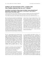
Fig. 4. Schematic representation of MUC15. (A) Schematic representation of the human MUC15 gene. Nucleotide positions (in AJ417818) are
indicated by numbers. Exons and introns are indicated by E and I, respectively. Intron sizes are given in parentheses. Shaded boxes represent the
coding regions whereas white boxes indicate the noncoding regions. (B) Schematic representation showing the organization of the bovine MUC15
protein: The 23 amino-acid signal peptide (SP), the extracellular Ser, Thr, and Pro rich region, the transmembrane domain (TM), and the
cytoplasmic C-terminal (CYT). Positions of the domains are indicated with amino-acid numbers. Identified N-glycosylation sites are marked with
hexagons. The protein is oriented with an exoplasmic N-terminal and a cytoplasmic C-terminal tail. The 50-amino-acid region skipped in the
MUC15/S splice variant is shown.
Ó FEBS 2002 MUC15, a novel membrane-associated mucin (Eur. J. Biochem. 269) 2759
MUC15 is proposed to comprise 311 amino acids with a
calculated average mass of 33 875 Da.
Alignment of the bovine and human MUC15 sequences
showed 67% similarity (Fig. 3). The majority of the differ-
ences exist in the extracellular part where similarity with the
bovine mucin is only 59%. The similarity rises to 87% in the
transmembrane domain and cytoplasmic area, suggesting
that these regions may be of functional importance.
By comparison of the human MUC15 cDNA sequence
with the working draft sequence version of the human
genome, available from the NCBI, homologous sequences
were located on chromosome 11 (p14.3 region). With two
minor exceptions, the derived and genomic sequences were
identical. These differences correspond to nucleotide vari-
ations observed at positions 495 (a–g polymorphism) and
827 (t–c polymorphism), the latter causing an amino-acid
change from Ile to Thr (residue 202 in Fig. 3). Comparing
the obtained human MUC15 cDNA and the genomic
sequence revealed the boundaries of five exons and four
introns (Fig. 4A). The signal peptide and the major part of
the extracellular part are encoded by a single exon (exon 3),
which is followed by a 150-bp exon encoding the trans-
membrane domain (exon 4). Nucleotides encoding the
cytoplasmic domain span exons 4 and 5, which also contain
the stop codon as well as a 274-bp 3¢ untranslated region.
Alternatively splicing and expression pattern of MUC15
Database searches showed that MUC15 is widely expressed,
as numerous human EST clones have been isolated from
fetal liver and spleen, fetal ear, placenta, lung, pancreas and
kidney (e.g. accession numbers; H53268, BI491080,
BG434403, BG485125, AA386131, BG425830). By PCR
screening of human MTC panels using MUC15-specific
primers we have also demonstrated human MUC15 mRNA
expression in a wide range of tissues; adult human spleen,
thymus, prostate, testis, ovary, small intestine, colon,
peripheral blood leukocyte, bone marrow, lymph node,
tonsil, and fetal liver. Furthermore, PCR screening of
bovine and human cDNA libraries showed the presence of
MUC15 mRNA in human breast, bovine mammary gland,
bovine lymph nodes and lungs of both species (Table 1). Of
the identified ESTs a single clone, isolated from the human
lung (GenBank accession number BG485125), appeared to
have been derived from an alternative splicing event. In
agreement with this, 11 of the 19 PCR screening experi-
ments revealed a smaller and weaker band in addition to the
expected product (Table 1). Therefore, to investigate the
possible existence of an alternatively spliced mRNA variant
of MUC15, RT-PCR experiments were performed on total
RNA extracted from the mammary gland of a Holstein
cow. Using MUC15-specific primers flanking the region
containing the potential splice site, a major band of 513 bp
was amplified by RT-PCR, along with a second shorter and
weaker band. To specifically amplify the shorter variant in a
second round of PCR, the products were subjected to
specific enzymatic cleavage with the DdeIenzyme,which
should only cut generated products comprising the trans-
membrane region. Isolation and sequencing of a clone
corresponding to the short variant confirmed the presence
of an alternatively spliced form of bovine MUC15. The
isolated variant (EMBL accession number AJ417817)
Table 1. Expression of MUC15 in human and bovine tissues and cell types. MUC15 mRNA expression was examined by PCR screening of
commercial multiple tissue cDNA panels and oligo(dT) primed cDNA libraries and by RT-PCR on RNA isolated from the mammary gland of a
Holstein cow and the cellular fraction of human milk samples. ND, not detected; NI, not investigated.
Tissue Template MUC15 mRNA MUC15/S mRNA
Human
Colon
a
cDNA Panel + +
Ovary
a
–++
Peripheral blood leukocyte
a
–++
Prostate
a
–++
Small intestine
a
–++
Spleen
a
–++
Testis
a
–++
Thymus
a
–++
Bone marrow
a
–+ND
Fetal liver
a
–++
Lymph node
a
–+ND
Tonsil
a
–+ND
Breast
b
cDNA library +
d
NI
Lung
b
–+
d
NI
Milk cells
a
cDNA +
d
ND
Bovine
Mammary gland
c
cDNA +
d
+
d
Mammary gland
c
cDNA library +
d
+
Lung
c
–+
d
ND
Lymph node
c
–+
d
ND
a
Primer pair: 5¢-AATACCAAAGAAGCCTACAATG-3¢ and 5¢-GTACGAAGTGGAGGTATGTCATC-3¢.
b
Primer pair: 5¢-GCCATTT
TAGGTGCTATTCTGG-3¢ and 5¢-TATTTTCTTTATCTGAGTTTA-3¢.
c
Primer pair: 5¢-CATCCATAGCAGATAACAGTC-3¢ and 5¢-T
CCCAAAGCTCATGTCATAAG-3¢.
d
Generated PCR products have been additionally verified by nucleotide sequencing.
2760 L. T. Pallesen et al. (Eur. J. Biochem. 269) Ó FEBS 2002
showed deletion of a segment of 150 nucleotides, corres-
ponding to the entire exon 4 of the human homologue
encoding the transmembrane domain. This variant was
called MUC15/S in analogy with the secreted short variant
of human MUC1 (GenBank accession number AF348143).
Thus, bovine MUC15/S encodes a potential secreted mucin
of 257 amino acids with a calculated molecular mass of
27 842 Da.
DISCUSSION
The present paper describes the purification and character-
ization of a hitherto unknown bovine membrane-associated
mucin-like glycoprotein, MUC15, and cloning of a human
homologue. The mature protein contains a single trans-
membrane domain, and is proposed to be oriented with a
small intracellular C-terminal part and an extracellular
N-terminal comprising numerous N- and O-glycosylation
sites (Fig. 4). Furthermore, database searches performed to
look for other proteins with significant sequence similarity
turned out fruitless.
Several features of the isolated bovine glycoprotein
suggest that it is a mucin-type molecule; a high molecular
mass, a high content of carbohydrate, and third expression
at the apical surface in epithelial cells of the mammary
gland. Likewise, the deduced amino-acid sequences of both
bovine and human MUC15 resemble the mucins in having
serine, threonine and proline as the predominant amino
acids, however, their high contents of leucine and aspara-
gine is a characteristic shared only with the MUC8, MUC9,
MUC13, and MUC16. Like the membrane-associated
members of the mucin family, MUC15 appears to be
derived from a precursor sequence including a signal
peptide, a serine/threonine/proline-rich extracellular region,
a hydrophobic transmembrane domain and a cytoplasmic
tail. Although most structural elements of the membrane-
associated mucins turned out to be present in MUC15, it is
unique in its short extracellular domain and lack of
repetitive segments with the typical mucin tandem repeats.
However, lack of tandem repeats is also seen in the mucin-
like glycoproteins mouse MUC14, endomucin-1, and
endomucin-2 [26]. Nevertheless, the extracellular region of
MUC15 and traditional mucin tandem repeat domains
share the same characteristics with long extended sequences
devoid of secondary structure and great potential for
extensive glycosylation.
TreatmentofbovineMUC15withO-glycosidase dem-
onstrated presence but not extend of O-glycosylation. Until
now no specific motif for O-glycosylation has been identi-
fied, however, proline is preferentially positioned in prox-
imity to the glycosylation site and especially in the )1 and/or
+3 positions [27]. According to the NetOGlyc server,
predicting mucin-type O-glycosylations using the algorithm
of Nielsen et al. [28], the extracellular region of bovine and
human MUC15 offer 22 and 14 O-glycosylation sites,
respectively. The majority of these potential O-glycosylation
sites are positioned in the central part of the extracellular
region, which also contains 10 predicted N-glycosylation
motifs in human MUC15 and 15 in the bovine counterpart.
Interestingly, doubly glycosylated Asn-Xaa-Ser/Thr motifs
have been reported, illustrating that N-glycosylations do not
hinder O-glycosylation of the surrounding serine and
threonine residues [29]. There is limited information avail-
able regarding the actual presence of N-linked oligosaccha-
rides in mucins. So far, N-glycans have only been identified
on bovine MUC1 together with human MUC2 and
MUC5AC [20,30,31]. Moreover, N-glycosylations are likely
to be present on human MUC3, MUC4, MUC7, MUC12,
MUC13, and MUC16 [10,13–15,32,33]. The present inves-
tigation shows that bovine MUC15 is N-glycosylated in 11
out of 15 potential sites.
Localization of the human MUC15 to chromosome
11p14.3 on the human genome, showed the structure of the
gene (Fig. 4A). A cluster of four secreted gel-forming mucin
genes (MUC2, MUC5AC, MUC5B, and MUC6) has been
localized within a 400-kb genomic DNA fragment on
chromosome 11 band p15.5, and appears to have originated
from a common ancestral gene [34]. Despite the location of
the MUC15 gene close to the cluster of mucin genes, it does
not show the characteristics of this group of secreted gel-
forming mucins and therefore presumably has not evolved
from the same ancestral gene.
Two variant forms of MUC15 cDNA were found to be
expressed by the normal bovine mammary gland. The short
variant of bovine MUC15 (MUC15/S) arises from an
alternative splicing event in which a section of 150
nucleotides was spliced out of the mRNA transcript,
leading to the synthesis of a protein lacking a 50 amino-
acid residues long region covering the transmembrane
domain. Hence, MUC15/S may represent a secreted nongel
forming mucin-type molecule as it does not contain any
cysteine-rich regions characteristic for the gel forming
mucins [1]. Examination of a corresponding alternatively
spliced database EST clone of the human lung showed that
the missing region of this clone corresponds to exon 4
(Fig. 4A). Likewise, nucleotides absent in the bovine
MUC15/S variant correspond precisely to exon 4 of the
human homologue, indicating a conserved genomic struc-
ture of human and bovine MUC15, and exon skipping as a
possible explanation for the origin of the splice variant.
Interestingly, the appearance of alternative soluble variants
of membrane-associated mucins has previously been repor-
ted. Experiments have shown that the nascent RNA
transcripts of the MUC1, MUC3, and MUC4 genes, are
spliced in an alternative manner possibly forming soluble
molecules that are secreted rather than retained on the cell
surface [18,35,36]. Recently, the membrane-associated
mucin MUC16 was found to be secreted from ovarian
tumours and cell lines by an unknown mechanism, however,
obtained results indicated that an alternative spliced variant
without the transmembrane region might exist [15]. More-
over, immunohistochemistry studies have demonstrated the
MUC13 protein within goblet cell thecae, indicative of
secretion in addition to presence on the cell surface [14]. To
this point, conclusive data showing that the MUC3, MUC4,
MUC13 and MUC16 mucins exist in both membrane-
associated and nonmembrane soluble forms are still miss-
ing. Likewise, at present there is no documentation for the
existence of the splice variant of MUC15 at the protein level.
The significance of the potential coexistence of MUC15
splice variants is unclear. However, the MUC1/SEC secre-
ted form of MUC1, devoid of the transmembrane and
cytoplasmic domain, has been found to constitute a cognate
binding protein for MUC1/Y, which lacks the tandem
repeat region. MUC1/SEC interacts with the extracellular
domain of MUC1/Y, resulting in the phosphorylation of the
Ó FEBS 2002 MUC15, a novel membrane-associated mucin (Eur. J. Biochem. 269) 2761
cytoplasmic domain of MUC1/Y and a concomitant change
in cell morphology [37]. These results suggest a mechanism
whereby alternative splicing regulates the relative levels of
both the receptor and its secreted cognate binding protein,
generated from the one and same gene, and thereby also
control the biological effects elicited by the interaction of
these two isoforms. Alternatively, it could be speculated that
the secreted isoform of MUC15 may function as a protective
mucin, perhaps as a coconstituent with gel-forming mucins
in mucus, or it may act at the apical cell surfaces as a ligand
for other cell surface molecules.
The physiological role of MUC15 is not known, however,
hints might arise from gene expression profiles. PCR
screening of human MTC panels and additional cDNA
libraries demonstrated MUC15 and MUC15/S mRNA
expression in a wide range of tissues (Table 1), but at a level
lower than the housekeeping gene, glyceraldehyde-3-phos-
phate dehydrogenase (results not shown). The expression of
mucins is generally thought to be restricted to epithelial
cells. Surprisingly, the present data indicate no restriction of
the MUC15 cDNA expression to epithelial cells. In
contrast, expression in hematopoietic cells and tissues with
function in the immune system was seen. Thereby, it might
be difficult to discriminate between expression by transiting
leukocytes, penetrating vascular endothelium, and the tissue
specific cells. MUC1 expression, which is associated most
consistently with epithelial tissues, has also been reported at
mRNA and protein level in peripheral blood lymphocytes,
lymph node samples, bone marrow and in various hema-
topoietic cell lines [18,38,39]. In addition, the membrane-
bound MUC13, like the human MUC15, also appears to be
expressed at low levels in prostate, lung, liver, spleen,
peripheral blood leukocytes, lymph node, bone marrow,
testis, and ovary [14]. Apparently, although historically
characterized as epithelia-specific, some membrane-associ-
ated mucins are also expressed in immune and hematopoi-
etic cells.
ACKNOWLEDGEMENTS
We express our thanks to Margit Skriver Rasmussen, Parisa
Mabhout and Marian Dyrberg Andersen for technical assistance,
Arla Innovation Centre, Brabrand, Denmark, for supplying the
bovine milk samples, and Department of Pediatrics, Aarhus
University Hospital, Skejby, Denmark for establishing contact to
the human milk donors. This work is part of the FØTEK program
supported by the Danish Government and the Danish Dairy
Research Foundation.
REFERENCES
1. Moniaux, N., Escande, F., Porchet, N., Aubert, J.P. & Batra, S.K.
(2001) Structural Organization and Classification of the Human
Mucin Genes. Front. Biosci. 6, d1192–1206.
2. Gendler, S.J. & Spicer, A.P. (1995) Epithelial mucin genes. Annu.
Rev. Physiol. 57, 607–634.
3. Lan, M.S., Batra, S.K., Qi, W.N., Metzgar, R.S. & Hollingsworth,
M.A. (1990) Cloning and sequencing of a human pancreatic
tumor mucin cDNA. J. Biol. Chem. 265, 15294–15299.
4. Gum, J.R. Jr, Hicks, J.W., Toribara, N.W., Siddiki, B. & Kim,
Y.S. (1994) Molecular cloning of human intestinal mucin (MUC2)
cDNA. Identification of the amino terminus and overall sequence
similarity to prepro-von Willebrand factor. J. Biol. Chem. 269,
2440–2446.
5. Pratt,W.S.,Crawley,S.,Hicks,J.,Ho,J.,Nash,M.,Kim,Y.S.,
Gum, J.R. & Swallow, D.M. (2000) Multiple transcripts of
MUC3: evidence for two genes, MUC3A and MUC3B. Biochem.
Biophys. Res. Commun. 275, 916–923.
6. Porchet, N., Nguyen, V.C., Dufosse, J., Audie, J.P., Guyonnet-
Duperat, V., Gross, M.S., Denis, C., Degand, P., Bernheim, A. &
Aubert, J.P. (1991) Molecular cloning and chromosomal locali-
zation of a novel human tracheo-bronchial mucin cDNA con-
taining tandemly repeated sequences of 48 base pairs. Biochem.
Biophys. Res. Commun. 175, 414–422.
7. Guyonnet-Duperat, V., Audie, J.P., Debailleul, V., Laine, A.,
Buisine, M.P., Galiegue-Zouitina, S., Pigny, P., Degand, P.,
Aubert, J.P. & Porchet, N. (1995) Characterization of the human
mucin gene MUC5AC: a consensus cysteine-rich domain for
11p15 mucin genes? Biochem. J. 305, 211–219.
8. Desseyn, J.L., Guyonnet-Duperat, V., Porchet, N., Aubert, J.P. &
Laine, A. (1997) Human mucin gene MUC5B, the 10.7-kb large
central exon encodes various alternate subdomains resulting in a
super-repeat. Structural evidence for a 11p15.5 gene family.
J. Biol. Chem. 272, 3168–3178.
9. Toribara, N.W., Roberton, A.M., Ho, S.B., Kuo, W.L., Gum, E.,
Hicks,J.W.,Gum,J.R.Jr,Byrd,J.C.,Siddiki,B.&Kim,Y.S.
(1993) Human gastric mucin. Identification of a unique species by
expression cloning. J. Biol. Chem. 268, 5879–5885.
10. Bobek,L.A.,Tsai,H.,Biesbrock,A.R.&Levine,M.J.(1993)
Molecular cloning, sequence, and specificity of expression of the
gene encoding the low molecular weight human salivary mucin
(MUC7). J. Biol. Chem. 268, 20563–20569.
11. Shankar,V.,Pichan,P.,Eddy,R.L.Jr,Tonk,V.,Nowak,N.,Sait,
S.N., Shows, T.B., Schultz, R.E., Gotway, G., Elkins, R.C.,
Gilmore, M.S. & Sachdev, G.P. (1997) Chromosomal localization
of a human mucin gene (MUC8) and cloning of the cDNA
corresponding to the carboxy terminus. Am. J. Respir. Cell Mol.
Biol. 16, 232–241.
12. Lapensee, L., Paquette, Y. & Bleau, G. (1997) Allelic poly-
morphism and chromosomal localization of the human oviductin
gene (MUC9). Fertil. Steril. 68, 702–708.
13. Williams, S.J., McGuckin, M.A., Gotley, D.C., Eyre, H.J.,
Sutherland, G.R. & Antalis, T.M. (1999) Two novel mucin genes
down-regulated in colorectal cancer identified by differential dis-
play. Cancer Res. 59, 4083–4089.
14. Williams,S.J.,Wreschner,D.H.,Tran,M.,Eyre,H.J.,Sutherland,
G.R. & McGuckin, M.A. (2001) MUC13, a novel human cell
surface mucin expressed by epithelial and hemopoietic cells.
J. Biol. Chem. 276, 18327–18336.
15. Yin, B.W. & Lloyd, K.O. (2001) Molecular cloning of the CA125
ovarian cancer antigen: identification as a new mucin, MUC16.
J. Biol. Chem. 276, 27371–27375.
16. Melnick, M., Chen, H., Zhou, Y. & Jaskoll, T. (2001) An alter-
natively spliced Muc10 glycoprotein ligand for putative
L
-selectin
binding during mouse embryonic submandibular gland morpho-
genesis. Arch.OralBiol.46, 745–757.
17. Pigny, P., Guyonnet-Duperat, V., Hill, A.S., Pratt, W.S.,
Galiegue-Zouitina, S., d’Hooge, M.C., Laine, A., Van-Seuningen,
I., Degand, P., Gum, J.R. et al. (1996) Human mucin genes
assigned to 11p15.5: identification and organization of a cluster of
genes. Genomics 38, 340–352.
18. Gendler, S.J. (2001) MUC1, the renaissance molecule. J. Mam-
mary Gland Biol. Neoplasia 6, 339–353.
19. Shimizu, M. & Yamauchi, K. (1982) Isolation and characteriza-
tion of mucin-like glycoprotein in human milk fat globule mem-
brane. J. Biochem. 91, 515–524.
20. Pallesen, L.T., Andersen, M.H., Nielsen, R.L., Berglund, L.,
Petersen, T.E., Rasmussen, L.K. & Rasmussen, J.T. (2001) Pur-
ification of MUC1 from bovine milk-fat globules and character-
ization of a corresponding full-length cDNA clone. J. Dairy Sci.
84, 2591–2598.
2762 L. T. Pallesen et al. (Eur. J. Biochem. 269) Ó FEBS 2002
21. Mather, I.H. (2000) A review and proposed nomenclature for
major proteins of the milk-fat globule membrane. J. Dairy Sci. 83,
203–247.
22. Mather, I.H., Tamplin, C.B. & Irving, M.G. (1980) Separation of
the proteins of bovine milk-fat-globule membrane by electro-
focusing with retention of enzymatic and immunological activity.
Eur. J. Biochem. 110, 327–336.
23. Kaetzel, C.S., Banghart, L.R., Jackson, D.Y., Madara, P.J.,
Jarasch, E D. & Mather, I.H. (1987) Expression of a 95–100
kDa glycoprotein on the apical surfaces of bovine mammary
epithelial cells during lactation. Biochem. Soc. Trans. 15, 1117–
1118.
24. Hvarregaard, J., Andersen, M.H., Berglund, L., Rasmussen, J.T.
& Petersen, T.E. (1996) Characterization of glycoprotein PAS-6/7
from membranes of bovine milk fat globules. Eur. J. Biochem. 240,
628–636.
25. Tarentino, A.L., Gomez, C.M. & Plummer, T.H. Jr (1985)
Deglycosylation of asparagine-linked glycans by peptide:
N-glycosidase F. Biochemistry 24, 4665–4671.
26. Kinoshita, M., Nakamura, T., Ihara, M., Haraguchi, T.,
Hiraoka,Y.,Tashiro,K.&Noda,M.(2001)Identification
of human endomucin-1 and -2 as membrane-bound
O-sialoglycoproteins with anti-adhesive activity. FEBS Lett. 499,
121–126.
27. Hansen, J.E., Lund, O., Tolstrup, N., Gooley, A.A., Williams,
K.L. & Brunak, S. (1998) NetOglyc: prediction of mucin type
O-glycosylation sites based on sequence context and surface
accessibility. Glycoconj. J. 15, 115–130.
28. Nielsen, H., Engelbrecht, J., Brunak, S. & von Heijne, G. (1997)
Identification of prokaryotic and eukaryotic signal peptides and
prediction of their cleavage sites. Protein Eng. 10,1–6.
29. Christlet, T.H.T. & Veluraja, K. (2001) Database analysis of
O-glycosylation sites in proteins. Biophys. J. 80, 952–960.
30. Asker, N., Axelsson, M.A., Olofsson, S.O. & Hansson, G.C.
(1998) Dimerization of the human MUC2 mucin in the
endoplasmic reticulum is followed by a N-glycosylation-depen-
dent transfer of the mono- and dimers to the Golgi apparatus.
J. Biol. Chem. 273, 18857–18863.
31. Asker, N., Axelsson, M.A., Olofsson, S.O. & Hansson, G.C.
(1998) Human MUC5AC mucin dimerizes in the rough
endoplasmic reticulum, similarly to the MUC2 mucin. Biochem.
J. 335, 381–387.
32. Williams, S.J., Munster, D.J., Quin, R.J., Gotley, D.C. &
McGuckin, M.A. (1999) The MUC3 gene encodes a transmem-
brane mucin and is alternatively spliced. Biochem. Biophys. Res.
Commun. 261, 83–89.
33. Moniaux, N., Nollet, S., Porchet, N., Degand, P., Laine, A. &
Aubert, J.P. (1999) Complete sequence of the human mucin
MUC4: a putative cell membrane-associated mucin. Biochem.
J. 338, 325–333.
34. Desseyn, J.L., Aubert, J.P., Prochet, N. & Laine, A. (2000) Evo-
lution of the large secrete gel-forming mucins. Mol. Biol. Evol. 17,
1175–1184.
35. Crawley, S.C., Gum, J.R. Jr, Hicks, J.W., Pratt, W.S., Aubert,
J.P., Swallow, D.M. & Kim, Y.S. (1999) Genomic organization
and structure of the 3¢ region of human MUC3: Alternative spli-
cing predicts membrane-bound and soluble forms of the mucin.
Biochem. Biophys. Res. Commun. 263, 728–736.
36. Moniaux, N., Escande, F., Batra, S.K., Porchet, N., Laine, A. &
Aubert, J.P. (2000) Alternative splicing generates a family of
putative secreted and membrane-associated MUC4 mucins. Eur.
J. Biochem. 267, 4536–4544.
37. Baruch,A.,Hartmann,M.,Yoeli,M.,Adereth,Y.,Greenstein,S.,
Stadler, Y., Skornik, Y., Zaretsky, J., Smorodinsky, N.I., Keydar,
I. & Wreschner, D.H. (1999) The breast cancer-associated MUC1
gene generates both a receptor and its cognate binding protein.
Cancer Res. 59, 1552–1561.
38. Dent, G.A., Civalier, C.J., Brecher, M.E. & Bentley, S.A. (1999)
MUC1 expression in hematopoietic tissues. Am. J. Clin. Pasthol.
111, 741–747.
39. Brugger,W.,Buhring,H.J.,Grunebach,F.,Vogle,W.,Kaul,
S., Muller, R., Brummendorf, T.H., Ziegler, B.L., Rappold, I.,
Brossart, P., Scheding, S. & Kanz, L. (1999) Expression of
MUC-1 epitopes on normal bone marrow: implications for the
detection of micrometastatic tumor cells. J. Clin. Oncol. 17, 1535–
1544.
Ó FEBS 2002 MUC15, a novel membrane-associated mucin (Eur. J. Biochem. 269) 2763