Báo cáo Y học: Herbaspirillum seropedicae signal transduction protein PII is structurally similar to the enteric GlnK potx
Bạn đang xem bản rút gọn của tài liệu. Xem và tải ngay bản đầy đủ của tài liệu tại đây (449.58 KB, 8 trang )
Herbaspirillum seropedicae
signal transduction protein PII
is structurally similar to the enteric GlnK
Elaine Machado Benelli
1
, Martin Buck
2
, Igor Polikarpov
3
, Emanuel Maltempi de Souza
1
,
Leonardo M. Cruz
1
and Fa
´
bio O. Pedrosa
1
1
Department of Biochemistry, Universidade Federal do Parana
´
, Curitiba, Brazil;
2
Department of Biological Science,
Imperial College of Science, Technology & Medicine, Sir Alexander Fleming Building, Imperial College Road, London, UK;
3
Laborato
´
rio Nacional de Luz Sincrotron, Campinas, Brazil
PII-like proteins are signal transduction proteins found in
bacteria, archaea and eukaryotes. They mediate a variety of
cellular responses. A second PII-like protein, called GlnK,
has been found in several organisms. In the diazotroph
Herbaspirillum seropedicae, PII protein is involved in sensing
nitrogen levels and controlling nitrogen fixation genes. In
this work, the crystal structure of the unliganded H. sero-
pedicae PII was solved by X-ray diffraction. H. seropedicae
PII has a Gly residue, Gly108 preceding Pro109 and the
main-chain forms a bturn. The glycine at position 108 allows
a bend in the C-terminal main-chain, thereby modifying the
surface of the cleft between monomers and potentially
changing function. The structure suggests that the
C-terminal region of PII proteins may be involved in
specificity of function, and nonenteric diazotrophs are found
to have the C-terminal consensus XGXDAX(107–112). We
are also proposing binding sites for ATP and 2-oxoglutarate
based on the structural alignment of PII with PII-ATP/
GlnK-ATP, 5-carboxymethyl-2-hydroxymuconate iso-
merase and 4-oxalocrotonate tautomerase bound to the
inhibitor 2-oxo-3-pentynoate.
Keywords: nitrogen regulation; PII X-ray structure; crystal
packing, Herbaspirillum seropedicae;GlnK.
Control of nitrogen metabolism in many bacteria utilizes a
conserved mechanism of intracellular signalling to regulate
patterns of gene expression and enzyme activity necessary
for adapting to changes in the quality and abundance of
nitrogen sources. The NifA protein is the transcriptional
activator of nitrogen fixation (nif) genes in the majority of
diazotrophs within the Proteobacteria. In several of these
organisms, nifA expression is controlled by the general
nitrogen regulation Ntr system, which, in turn, is controlled
by the state of the glnB product, the PII protein. Under
nitrogen excess, PII interacts with NtrB resulting in the
dephosphorylation of the transcriptional activator NtrC-P
and diminished nifA expression. Under limiting nitrogen,
PII is uridylylated by GlnD and this allows NtrB to
phosphorylate NtrC. In the c-subdivision of the Proteo-
bacteria, nif gene expression is regulated by NifA and NifL:
under high ammonium or oxygen levels NifL inhibits NifA
activity, whereas under nitrogen limiting conditions and low
oxygen NifA is active. In K. pneumoniae GlnK, a paralogue
of PII, interacts with the NifL–NifA complex, to relieve
NifA inhibition by NifL [12,13,16,]. In Azotobacter vinela-
ndii only the GlnK protein is present and it controls the
activity of NifA by the interaction with NifL and the
complex NifL–NifA is sensitive to 2-oxoglutarate levels [20].
Although extensively studied in bacteria, PII-like proteins
are present in all three kingdoms of life. For recent reviews
see Ninfa & Atkinson [24], Thomas et al. [33] and Mag-
asanik [21].
In Herbaspirillum seropedicae, a member of the bsubdi-
vision of Proteobacteria, the glnAntrBC and glnB genes have
been identified [6,26], suggesting that an Ntr PII-dependent
signal transducer cascade senses the nitrogen levels in this
organism. In H. seropedicae, nifA expression is also
dependent on phosphorylated NtrC (NtrC-P), but NifL
has not been found. However, the activity of NifA is known
to be controlled by the PII protein, as in Azospirillum
brasilense, a member of the a subdivision of the Proteobac-
teria [2,3]. The mechanism involved in this control is not
known. Souza et al. [30] observed that the activity of a
H. seropedicae N-terminal domain-truncated NifA
(DNTD) was independent of ammonium levels, suggesting
that the N-terminal domain (NTD) plays a role in the
control of NifA activity by ammonium. Arsene et al.[3]
made a similar observation in A. brasilense and suggested
that PII-UMP may interact with the NTD of NifA to
change its activity. The residue Tyr18 from the NTD of
NifA seems to be involved in the interaction between PII
and NifA [2].
PII proteins interact directly with a variety of ligands,
including ATP and 2-oxoglutarate. The structure of the
EcPII protein and the paralogue EcGlnK have been solved
in the presence and absence of ATP [7,9,35,36]. Here we
report the crystal structure of unliganded H. seropedicae PII
(HsPII) at 2.1 A
˚
resolution and compare this with the
available structures from E. coli. Although in amino-acid
Correspondence to E. Machado Benelli, Department of Biochemistry,
Universidade Federal do Parana
´
, C. Postal 19046, Curitiba, Brazil.
E-mail:
Abbreviations: NtrB, nitrogen regulation protein B; NtrC, nitrogen
regulation protein C; GlnD, uridylylating enzyme; NifA, nitrogen
fixation protein A; NifL, nitrogen fixation protein L; EcPII, Escheri-
chia coli glnB product; EcGlnK, Escherichia coli glnK product; HsPII,
Herbispirillum seropedicae glnB product; KpPII, K. pneumoniae glnB
product; KpGlnK, K. pneumoniae glnK product.
(Received 29 January 2002, revised 15 February 2002,
accepted 22 May 2002)
Eur. J. Biochem. 269, 3296–3303 (2002) Ó FEBS 2002 doi:10.1046/j.1432-1033.2002.03011.x
sequence HsPII shows higher identity to EcPII than
EcGlnK, distinct structural differences are evident, placing
HsPII closer to the unliganded and ATPbound forms of
EcGlnK in three-dimensional structure. We suggest a
correlation of the structural differences with the specialized
functions of PII-like proteins in diazotrophs. It seems that
function may be related to conformational flexibility
exhibited by PII and GlnK proteins, as indicated by a
comparison of crystal packing arrangements seen in several
different crystal forms of PII-like proteins [7,9,35]. Changes
in EcPII structure associated with ATP binding support this
view and indicate that C-terminal structures can be ligand
dependent [35]. When EcPII is bound to ATP the
C-terminal structure is similar to that in unliganded
EcGlnK [36] and unliganded HsPII (this paper). We note
similarities in quaternary and subunit tertiary structure with
other proteins, unrelated to PII by amino-acid sequence,
that interact with a-ketoacids, suggesting the existence of a
family of a-ketoacid interacting proteins.
EXPERIMENTAL PROCEDURES
Protein purification
HsPII protein was overexpressed in E. coli RB9065kDE3, a
glnB glnD mutant background lysogenized with kDE3 for
T7 RNA polymerase production and purified as described
by Benelli et al. [5]. The purified HsPII protein was dialysed
in a buffer containing 10 m
M
Tris/HCl pH 8.0, 50 m
M
NaCl, 20% glycerol and 0.1 m
M
EDTA and concentrated
in a Centricon-3 filter prior to crystallization.
Crystallization
Crystallizations used either the sitting or hanging drop
vapour diffusion method at 18 °C in Limbro tissue culture
plates. An initial Hampton crystallization screen of both
native and N-terminal hexa histidine-tagged HsPII yielded
promising microcrystals. Conditions were optimized by
addition of a number of additives [10]. HsPII protein
(14 mgÆmL
)1
)andHis
6
–PII protein (13 mgÆmL
)1
)inTris/
HCl 10 m
M
pH 8.0, NaCl 50 m
M
, glycerol 20% and
EDTA 0.1 m
M
were used in crystallization experiments. A
tetragonal crystal form of native PII was grown from
hanging drops containing protein solution mixed in a 1 : 1
ratio with well solution (15.8% ethyleneglycol). A trigonal
crystal form was grown by vapour diffusion in sitting
drops. The reservoir solution contained 0.1
M
sodium
acetate pH 4.6, 30% methylpentadiol and 0.15 mgÆmL
)1
of
dextran sulfate. The drops contained 1 lLofprotein
solution and 1 lL of reservoir solution. The orthorhombic
crystal form grew, using the hanging drop method, in 30%
methylpentadiol, 0.1 m
M
sodium cacodylate pH 6.5 and
0.2 m
M
magnesium acetate. Initial tests on a copper
rotating anode revealed diffraction to 3 A
˚
from the
tetragonal and trigonal crystal forms (Table 1). Crystals
of His
6
–PII were obtained by the hanging drop method at
18 °C. The reservoir solution contained 0.5 mL of 0.1
M
sodium citrate pH 6 and 10% PEG 6K and the drop
contained 1 lL of protein solution (13 mgÆmL
)1
)and1lL
of reservoir solution. The His
6
–PII crystal form diffracted
to 6 A
˚
with the rotating anode source, and was not further
characterized.
Table 1. Summary of X-ray data collection and crystallographic refinement statistics.
Data collection
a,b
Space group P2
1
2
1
2
1
P3
2
21 P4
3
2
1
2
Unit cell dimensions a ¼ 78.41 A
˚
,b¼ 82.36 A
˚
,a¼ b ¼ 121.74 A
˚
,c¼ 65.24 A
˚
a ¼ b ¼ 88.81 A
˚
,c¼ 116.91 A
˚
c ¼ 100.95 A
˚
a ¼ b ¼ 90°, c ¼ 120°
Solvent content (%) 68 68 61
Max. resolution (A
˚
) 2.1 3.0 3.2
Unique reflections 36523 21170 8163
Redundancy 3 3 4
Completeness (%) 94 (95) 98 (99) 100 (100)
Average I/rI 13 (1) 12 (2) 14 (4)
R ¼ S|I ) <I>|/S|I| 0.057 0.078 0.187
Refinement in orthorhombic crystal form
c,d,e
Data range (A
˚
) 13.0–2.1
Reflections (F > 0) 36331
Completeness (%) 94.4
Reflections in free set 1820
Non-H atoms 4313
Residues 560
Rms bond lengths (A
˚
) 0.018
Rms bond angles (deg) 0.044
Rms B-factors for bonded atoms (A
˚
2
) 4.2
R
free
(%) 27.2
R
cryst
(%) 20.3
a
Values in parentheses correspond to the highest resolution shell; 2.15–2.10 A
˚
(2415 reflections) for the orthorhombic form; 3.05–3.00 A
˚
(815 reflections) for the trigonal form; 3.25–3.20 A
˚
(393 reflections) for the tetragonal form.
b
The resolution cut-off was defined so that 50%
of reflections in the highest resolution shell had I > 3 r.
c
Rms deviations in bond lengths and angles are given from ideal values.
d
R
cryst
¼
S||F
o
|–|F
c
||/S|F
o
|.
e
R
free
is as for R
cryst
but calculated for a test set comprising 1820 reflections not used in the refinement.
Ó FEBS 2002 Structural similarities between PII and GlnK (Eur. J. Biochem. 269) 3297
Data collection and processing
A summary of the data collection and refinement statistics is
given in Table 1. Diffraction data were collected from a
single crystal of each form at 120 K using a 30-cm MAR
imaging plate detector system on a RIGAKU RU-200B
generator with a copper anode and double focusing mirrors.
A2.1-A
˚
data set on the orthorhombic crystal form was
collected at 120 K using synchrotron radiation at a
wavelength of 1.38 A
˚
, using a MAR 345 imaging plate on
the protein crystallography beamline [28,29] at the Brazilian
National Synchrotron Laboratory (Campinas, Brazil).
The crystal initially diffracted to 1.9 A
˚
, but the high
resolution reflections gradually decayed during data collec-
tion. The diffraction data were consistent with space group
P2
1
2
1
2
1
, with the cell parameters a ¼ 78.41 A
˚
, b ¼ 82.36 A
˚
,
c ¼ 100.95 A
˚
. The data were integrated, reduced and
scaled using
DENZO
and
SCALEPACK
[25], respectively.
Intensities were then converted to structure factors using
the method of French & Wilson [11] as implemented in
TRUNCATE
[8].
Structure solution and refinement
The structure of HsPII was solved in three space groups by
molecular replacement in
AMORE
[23]. Selected crystallo-
graphic data are given in Table 1. The complete EcPII
monomer structure (PDB accession no. 2PII; [7]) and a
truncated model lacking the uridylylation site loop and the
C-terminal tail, residues 40–54 and 96–112, respectively,
were both used as search models to solve the trigonal crystal
form. Both monomer and trimer forms, generated by the
crystallographic threefold axis in space group P6
3
,wereused
as search models. All calculations performed used 10 to 4 A
˚
data. Only when the trimer was used as a search model did
the first peak in the cross rotation function correspond to
the correct solution. A solution could not be found with the
entire monomer structure, only with the truncated mono-
mer model. Initial refinement of the whole model included
noncrystallographic symmetry averaging and yielded a
crystallography R-factor of 37%, the electron density map
calculated at this stage indicated that residues 38–51 and
104–112 were not in correct positions. Model building was
subsequently carried out on the truncated model only. The
electron density for the rest of the protein was well defined;
therefore it was possible to substitute all EcPII residues with
the corresponding HsPII residues. Electron densities for
residues 38 and 39 were so poor that they both had to be
removed. Additional electron densities were apparent for
two residues preceding Asp54 and five after Val96. The
current model including residues 1–37 and 52–110 was
obtained after a few rounds of model adjustment followed
by refinement in
REFMAC
[22].
The tetragonal crystal form was solved using the trigonal
HsPII model after the first build in which all the amino acids
different from EcPII were changed. This model included
residues 1–37 and 54–96. The structure of the orthorhombic
crystal form was solved using the trigonal HsPII containing
residues 1–35 and 55–107. Molecular replacement, including
rotation and translation functions followed by rigid body
refinement, was carried out using 10 to 3.3 A
˚
data and
resulted in an R-value of 39.6% and correlation coefficient
of 60.3%.
Refinement was carried out using the program
REFMAC
[22] from the
CCP
4 suite of the program [8]. Eighty cycles of
positional and B-factor refinement of the molecular
replacement model against all the data between 10 A
˚
and
2.1 A
˚
resolution resulted in a model with R
cryst
30.0% and
R
free
36.1%. Model building was carried out using the
programe
O
[18]. The orthorhombic HsPII model was built
into 2F
o
) F
c
and F
o
) F
c
difference maps, residues were
placed in well defined 2 r electron density maps. Eleven
cycles of model building and refinement resulted in an R-
factor of 23.1%. and R
free
of 29.8% In the last cycle, 125
molecules of water were added and the R-factors dropped to
20.3 and 27.3%, respectively. The final model comprises
residues 1–37 and 51–112 (monomer A), 1–36 and 43–107
(monomer B), 1–36 and 57–112 (monomer C), 1–37 and
50–112 (monomer D), 1–35 and 57–105 (monomer E) and
1–35 and 57–112 (monomer F). The residue Lys68 is placed
as Ala in chains B, D and F because the electron density of
the lateral chain of Lys was not observed in these chains.
The stereochemical quality of the final model of the HsPII
protein was verified by
PROCHECK
[19]. The coordinates were
deposited in the Protein Data Bank as the code 1HWU.
RESULTS AND DISCUSSION
Overall structure
HsPII was overproduced and purified from E. coli and
found to be a trimer of 36 kDa in solution, as are the EcPII
and EcGlnK proteins [24]. The crystal structure was solved
by molecular replacement using EcPII as the search model
(see Materials and methods). Several different crystal forms
of HsPII were grown (Table 1). The structural model was
obtained from the orthorhombic crystals which diffracted at
2.1 A
˚
.
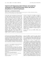
Monomers of the HsPII trimer are accommodated
around a central threefold axis (Fig. 1A). The core of the
HsPII monomer has a double bab motif (Figs 1B and 2A).
The structural scaffold (the b strands, the a helices and the
short B-loop) is well conserved in available PII-like struc-
tures (Fig. 1B). Major differences amongst structures are in
the T-loop (which contains the uridylylation site, Tyr51)
and C-loop. HsPII is similar to EcGlnK, EcGlnK-ATP and
EcPII-ATP in its C-loop (Figs 1B and 2A).
The b strands of the bab motif line the central cavity of
the HsPII trimer, with the a helices at the periphery of the
molecule (Fig. 1A). The bottom edge of the central cavity is
negatively charged (Fig. 2B, part i) owing to the presence of
Glu97 (Ala in EcPII and Gln in EcGlnK) and Glu95. The
entrance of the central cavity is partially restricted by Gln94
with lateral chains directed towards its interior. Gln94 is
substituted by Phe in EcPII and Ala in EcGlnK. The
entrance from the top is restricted by Thr31, whose lateral
chain is oriented to the interior of the cavity. The interior
wall of the cavity is largely hydrophilic. Most of the
intersubunit interactions that maintain the EcPII and
EcGlnK trimers occur between conserved residues and are
therefore also preserved in the HsPII structure, for example
between Lys34 and Glu32 and Lys60 and Glu62 or Asp62
(in EcGlnK) (Fig. 2A,B, part ii). The salt bridge between
residues Lys2 and Glu95 in EcPII appears to be substituted
by Lys2 and Asp97 in HsPII. Furthermore, the interaction
between residues Asp71 and Arg98 of different chains seen
3298 E. Machado Benelli et al. (Eur. J. Biochem. 269) Ó FEBS 2002
in EcPII does not exist in the HsPII structure. These residues
are substituted by Glu and Gln, respectively.
The lateral cleft created in the interface of each monomer
of HsPII (Figs 1A and 2B, part iii) is similar to that
observed in EcGlnK but smaller than in EcPII. In HsPII the
clefts are partially obstructed by C-terminal sequences. The
bend of the main chain at Gly108 pushes residues Pro109,
Asp110, Ala111 and Val112 into the cleft (Fig. 2B, part ii).
Around the lateral cleft in the HsPII protein there is a salt
bridge between Asp66 and Lys68 which does not appear in
EcPII or EcGlnK (Fig. 2B, parts i and iii). This bridge is
close to the C-terminal region and might mediate the
interactions between PII and its receptors. Single amino-
acid modifications of EcPII protein around the cleft
produced mutant proteins (residues Thr83, Gly89 and
Lys90) with impaired binding of the ligands 2-oxoglutarate
and ATP [17] (Fig. 4C). None of the C-terminal residues
seem to interact directly with ATP in the EcGlnK or EcPII
proteins, for which structures of the complexes with ATP
are available [35,36]. However these C-terminal residues are
closer to the lateral cleft in EcGlnK compared to EcPII and
might therefore influence binding of ATP indirectly. It is
reasonable to propose that the structure of the C-terminal
region is important for effector binding to PII, althought the
effector need not directly interact with the C-terminal
sequence (discussed below). The HsPII protein requires
2-oxoglutarate for uridylylation by the GlnD protein
whereas the EcPII requires both 2-oxoglutarate and ATP,
and the affinity constant for 2-oxoglutarate binding to
HsPII is considerably higher than that of EcPII [5].
The bottom face of the HsPII trimer comprises mainly
negatively charged residues (Fig. 2B, part i). Positive
charges are located around the B-loop, which is probably
involved in ATP interactions. In this region, HsPII Arg101
and Arg103 are separated by the lateral chain of Ile102
whereas in EcPII these residues are closer. This may explain
why in the presence of excess 2-oxoglutarate K
act
for ATP
binding to HsPII is higher (100 l
M
) than that for EcPII
( 3 l
M
) [5,17].
The T- and C-loops
The T-loop of PII-like proteins frequently includes a
tyrosine which is the site of uridylylation. Where structures
of the T-loop are available for EcPII and EcGlnK, crystal
packing contacts appear to stabilize the T-loop in an
artificially ordered conformation. In HsPII the part of
T-loop that could be built shows a high temperature factor,
and is exposed to the solvent. In the orthorhombic HsPII
crystal there are two PII trimers per asymmetric unit. As
Fig. 1. Ribbon diagrams of the trimeric HsPII
(A) and monomeric HsPII, EcPII, EcPII-
ATP, EcGlnK and EcGlnK-ATP (B). (A) A
ribbon diagram of the structure of the trimeric
HsPII, each chain in a different colour. The
b sheets of the bab motif line the central cavity
of the trimer with the a helices at the periph-
ery. (B) Ribbon diagrams of the monomers of
HsPII (i), EcPII (ii), EcPII-ATP (iii), EcGlnK
(iv) and EcGlnK-ATP (v). Secondary struc-
tures are colour coded: green b sheets, b1–4,
blue helices, a1–2 and 3
10
helix and orange
loops. The monomers share the same bab
motif with the major structural differences
residing in the loops T and C.
Ó FEBS 2002 Structural similarities between PII and GlnK (Eur. J. Biochem. 269) 3299
packing contacts are different for each monomer in these
two trimers, the monomers were refined independently. The
final rmsd values for the overlay of all atoms of the two
trimers was 0.42 A
˚
. Electron density in all HsPII monomers
to residues 38–53, which are within the T-loop, were not
completely visible and the C-loop could be built in four of
the six monomers (monomer A, C, D and F) present in unit
of cell (see Experimental procedures). Those residues of the
HsPII T-loop that can be traced represent a conformation
unaffected by crystal packing contacts and are presumably
in the preferred conformation of the T-loop as exists in the
absence of interacting ligands such as ATP and 2-oxoglut-
arate. The limited amount of HsPII T-loop that is structured
shows significant conformational differences compared to
those sequences ordered by packing contacts in the crystals
of EcPII and EcGlnK (Fig. 1B). This implies that changes
in conformation across much of the T-loop are possible
during the normal functioning of the PII-like proteins [1,35].
The K. pneumoniae glnK product (KpGlnK) and EcG-
lnK proteins function to relieve NifL inhibition of NifA
activity under nitrogen-limiting growth conditions. Arcon-
deguy et al. [1], investigated the importance of the KpGlnK
T-loop residues 43, 52 and 54 on the control of K. pneu-
moniae NifA activity. Both EcGlnK and KpGlnK proteins
have high sequence identity to EcPII. However, EcPII
expressed from the chromosome is unable to substitute for
the GlnKs with respect to NifLA [13,16]. Arcondeguy et al.
(2000) suggested that residue 54 is the most important
residue in the T-loop for distinguishing between PII and
GlnK in controlling NifL activity. Residue 54 in HsPII is
aspartate, as in K. pneumoniae glnB product (KpPII) and
EcPII. However HsPII differs functionally from EcPII and
KpPII, and is able to activate NifA in an E. coli background
containing NifL when expressed from a low copy number
plasmid, as does EcGlnK, but not EcPII or KpPII (A. C.
Bonatto, E. M. Souza, F. O. Pedrosa & E. M. Benelli,
unpublished results). This suggests that some determinants
of functionality that distinguish PII from GlnK must reside
outside the T-loop. Consistent with this a second HsPII-like
protein has been discovered, with the same T-loop sequence
as the HsPII studied here (L. Noindorf, M. B. Steffens,
E. M. Souza, F. O. Pedrosa & L. Chubatsu, unpublished
data). This protein was called GlnK because it has higher
identity to EcGlnK than EcPII and it is encoded by a glnK
gene which is located on the glnKamtB operon. The HsPII
and HsGlnK proteins are 78.6% identical and 93.75%
similar and one of the seven different amino acids is in the
C-terminal (Pro109 HsPII is substituted by Lys109
HsGlnK). Despite, the high homology between these
proteins they are functionally different. The H. seropedicae
glnB mutant has normal GS activity and biosynthesis but it
is unable to fix nitrogen, suggesting that in vivo HsGlnK is
unable to substitute HsPII [6].
The C-terminal structure of PII
The structure of the C-terminal region of HsPII could be
entirely built only for one of the monomers in the asymmetric
unit. In contrast to the C-terminal region of EcPII, which
contains a bsheet, the C-terminal of HsPII contains a turn of
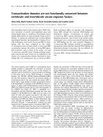
Fig. 2. Alignments of the HsPII with EcPII
and HsPII with EcGlnK amino-acid sequences
(A) and molecular surface of the HsPII trimer
(B). (A)AlignmentsoftheHsPIIwithEcPII
and HsPII with EcGlnK amino-acid
sequences. The identity (73%) and similarity
(86%) of HsPII to EcPII is higher than HsPII
to EcGlnK (67% and 76%, respectively).
Secondary structural elements are labelled
above and below the sequence. The a helix,
b strands, 3
10
helix and loops are coloured in
green, blue, dark green and black, respectively.
(B) Molecular surface of the HsPII trimer
colour-coded with acidic residue side-chains in
red, basic side-chains in blue and others in
white. T-loop residues 37–51 are not included.
Residuesreferredinthetextarelabelledonthe
monomer (i) The negatively charge bottom
face of the trimer; (ii) the top face of the trimer
and (iii) molecular surface of the lateral cleft of
the HsPII trimer. The salt bridge between
Asp66A and Arg68A, located close to the
C-terminal region, may mediate interaction
betweenPIIanditsreceptors.
3300 E. Machado Benelli et al. (Eur. J. Biochem. 269) Ó FEBS 2002
a3
10
helix as does EcGlnK, EcGlnk-ATP and PII-ATP
(Fig. 1B). Although the identity (73%) and similarity (83%)
of HsPII to EcPII is higher than that of HsPII to EcGlnK
(67% and 76%, respectively), HsPII is structurally closer to
EcGlnK or EcGlnK-ATP and EcPII-ATP (Figs 1B and
2A). The amino-acid sequence from residues 106–112,
encoding part of the C-loop of PII-like proteins is only
partly conserved (Fig. 3A). EcPII has a sequence of four
negatively charged amino acids in this region (residue 106–
109), EcGlnK three residues (106, 108 and 109), whereas
HsPII contains only two negatively charged amino acids at
positions 106 and 110. The rmsd values in Ca positions
obtained from superposition of the core (residues 1–35 and
56–95) were 0.58 A
˚
for HsPII-EcPII and 0.55 A
˚
for HsPII-
EcGlnK. The rmsd values in Ca obtained for the superpo-
sition of the C-terminal segments (residues 95–112) were
0.91 A
˚
and 0.43 A
˚
for EcPII and EcGlnK, respectively,
establishing that HsPII is structurally closer in this region to
the EcGlnK protein (Fig. 1B). The structural relatedness of
HsPII and EcGlnK in their C-terminal regions may explain
why HsPII is functionally similar to the KpGlnK and
EcGlnK proteins. HsPII and KpGlnK are involved in the
control of the NifA activity, as discussed above, whereas
EcPII and KpPII are not [6,12,1,16,20].
Recent structure determination of the EcPII protein with
ATP bound has shown that its C-terminal sequences can
adopt a conformation very close to that of the unliganded
EcGlnK [35,36] (Fig. 1B). The C-terminal part of unligan-
ded HsPII, preferentially adopts the structure seen in
unliganded EcGlnK (Fig. 1B). Although EcPII can adopt
two different structures in its C-terminus depending upon its
ligation state, we have no evidence for this in HsPII.
Nevertheless ligand induced structural changes may well
influence the functioning of the C-terminus of HsPII.
Amino-acid sequence alignment of a C-terminal region
(residues 106–112) (Fig. 3A) amongst PII proteins indicates
that this region is distinctly more conserved amongst
nonenteric diazotrophs (50% identity as opposed to 16%
between E. coli and H. seropedicae) suggesting similarity of
function. As with HsPII, the A. brasilense PII protein also
activates NifA [2,3]. Residues Gly108, Asp110 and Ala111
are present in the PII proteins of H. seropedicae, Rhodo-
bacter capsulatus, Rhodospirillum rubrum, Rhodobacter
sphaeroides, Bradyrhizhobium japonicum, A. brasilense,
Rhizobium leguminosarium and Azorhizobium caulinodans.
The residue Thr107 is present in the majority of these
organisms. On the other hand, the PII C-terminal sequences
are highly conserved between E. coli and K. pneumoniae.
These observations indicate that these proteins can be
divided into two classes. The enteric organisms share the
C-terminal sequence EDDAAI. In nonenteric diazotrophs
the C-terminal consensus is XGXDAX (Fig. 3A). It seems
the glycine at position 108 of the latter class allows a bend
in the C-terminal main-chain, thereby modifying the surface
of the intermonomer cleft and changing functionality. The
contribution of these residues to HsPII function is under
investigation.
Relationship of PII to GlnK
Jack et al. [16] aligned PII and parologue proteins from
several organisms and found five residues (positions 3, 5, 52,
54 and 64), which distinguish GlnK from PII proteins. PII
proteins contain Lys3, Glu5 or Asp5, Met52 or Val52,
Asp54 and Val64. In contrast, in GlnK proteins these amino
acids are: Leu3 or Ile3, Thr5, Met5 or Ile5, Ser52 or Ala52,
Ser54 or Asn54 and Ala64. In HsPII three of these residues
are identical to those of PII proteins: Met52, Asp54 and
Val64; one (Thr5) is found in GlnK proteins (Fig. 2A).
However, these alignments included only PII and paralogue
proteins of the organisms from the a) and c-subdivision of
the Proteobacteria. We have constructed a phylogenetic tree
of PII and paralogue proteins including HsPII and the PII
and paralogue proteins from Azoarcus, another member of
the b-subdivision of Proteobacteria using
CLUSTALX
[34]
(Fig. 3B). In this tree there are two groups of proteins: PII
and GlnK, separated according the Proteobacteria subdi-
visions. However, the HsPII was not included in either
group, which emphasizes the special nature of HsPII and is
consistent with the particular structural relationship HsPII
bears to EcPII and EcGlnK.
Structural alignment of
H. seropedicae
PII protein
to others proteins
Structural alignment of HsPII protein using the
DALI
program [14,15] showed that it has a relatedness to several
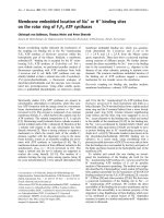
Fig. 3. Alignment of the C-loop residues of PII proteins from nonenteric
(the first nine sequences) and enteric bacteria PII and GlnK proteins (the
remaining four sequences) (A) and phylogenetic tree of the PII and
paralogue proteins in the proteobacteria (B). (A) The residues in blue
show the conserved residues of the XGXDA motif in the nonenteric
and enteric bacteria. Abbreviations are: Hs, H. seropedicae;Ab,Azo-
spirillum brasilense;Ac,Azorhozobium caulinodans;Bj,Bradyrhizobium
japonicum;Rl,Rhizobium leguminosarum;Rm,Rhizobium meliloti;Rr,
Rhodospirillum rubrum;Rs,Rhodobacter sphaeroides;Rc,Rhodobacter
capsulatus;Kp,K. pneumoniae;Ec,E. coli. (B) Phylogenetic tree of the
PII and paralogue proteins in the proteobacteria. This shows two
major groups with HsPII outlying these. The tree was calculated by the
CLUSTALX
program [34].
Ó FEBS 2002 Structural similarities between PII and GlnK (Eur. J. Biochem. 269) 3301
other proteins that possess a double bab fold, as nucleotide
diphosphate kinase, RNA binding protein, ribosomal
protein, allosteric domain of the regulatory subunit of
aspartate transcarbamylase, a viral transcriptional regulator
and procarboxypeptidase B [9]. Additionally, we found
HsPII aligned with the enzymes: 5-carboxymethyl-2-
hydroxymuconate isomerase and 4-oxalocrotonate tautom-
erase. These proteins are involved in the isomerization of
a-keto acids [31,32]. The superposition of HsPII with
4-oxalocrotonate tautomerase protein bound to 2-oxo-
3-pentynoate, an inhibitor of a-keto acid isomerization,
suggests that the 2-oxoglutarate may bind around the lateral
cleft region of PII (Lys90 and Arg101, from different
monomers). Comparisions between HsPII, EcGlnK-ATP
or EcPII-ATP and 4-oxalocrotonate tautomerase bound to
the inhibitor 2-oxo-3-pentynoate [32] suggests that although
the ATP and 2-oxoglutarate binding sites in HsPII are in the
lateral cleft, they are not superimposed (Fig. 4). The
suggested position of the 2-oxoglutarate binding-site is
consistent with biochemical data that show that mutations
in residues: G37, R38, Q39, K40, T83, G84, G89 and K90
affected the 2-oxoglutarate binding to EcPII [17]. Although,
Xu et al. [36], suggested that 2-oxoglutarate could bind
to the T-loop to stabilize this flexible loop, the present
model shows that it is possible that 2-oxoglutarate
can bind in the lateral cleft close to two Arg residues as
in 5-carboxymethyl-2-hydroxymuconate isomerase and
4-oxalocrotonate tautomerase. In the isomerases the
binding site also contains a proline residue involved in
the catalysis; this proline is not present in HsPII. It is
known that the affinity of E. coli PII for either ATP or
2-oxoglutarate is dependent on the other ligand, implying
that each ligand causes a conformational change to
increase acceptance of the second ligand [17].
ACKNOWLEDGEMENTS
This work in part was carried out at the Departments of Biology and
Biophysics, ICSTM and with Anne Harper, Madeleine H. Moore and
Johan P. Turkenburg in the Protein Structure Group, University of
York. We thank Silvia Onesti, Xiaodong Zhang and Marshall G. Yates
for their constructive suggestions and David Ollis for the coordinates of
the E. coli PII protein bound to ATP. This work was supported by
CNPg, PRONEX/MEC and BBRSC.
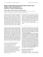
Fig. 4. Model to ATP and 2-oxoglutarate binding sites in HsPII protein. (A) Diagram of a Ca trace overlay of HsPII (orange) with CHMI (cyan)
[31]. A top view of the trimers similar in orientation to Figs 1A and 2Bii. The different views of the proposed 2-oxoglutarate and ATP-binding sites
in HsPII protein are shown in (B), (C) and (D). (B) Position of ATP and 2-oxo-3-pentynoate in the lateral cleft of HsPII. (C) ATP molecule and the
neighbouring amino-acid residues. (D) 2-oxo-3-pentynoate molecule and the neighbour amino-acid residues. Location of ATP and 2-oxo-3-
pentynoate was modelled using HsPII, EcGlnK-ATP, EcPII-ATP and 4-oxalocrotonate tautomerase-2-oxo-3-pentynoate structures using
DALI
and
LSQKAB
[8,14,15].
3302 E. Machado Benelli et al. (Eur. J. Biochem. 269) Ó FEBS 2002
REFERENCES
1. Arcondeguy, T., Lawson, D. & Merrick, M. (2000) Two residues
in the T-loop of GlnK determine NifL-dependent nitrogen control
of nif gene expression. J. Biol. Chem. 275, 38452–38456.
2. Arsene, F., Kaminski, P.A. & Elmerich, C. (1999) Control of
Azospirillum brasilense NifA activity by P (II): effect of replacing
Tyr residues of the NifA N-terminal domain on NifA activity.
FEMS Microbiol. Lett. 179, 339–343.
3. Arsene, F., Kaminski, P.A. & Elmerich, C. (1996) Modulation of
NifA activity by PII in Azospirillum brasilense: evidence for a
regulatory role of the NifA N-terminal domain. J. Bacteriol. 178,
4830–4838.
4. Benelli, E.M., Buck, M., Bonatto, A.C., Souza, E.M., Moore, M.,
Harper, A., Yates, M.G. & Pedrosa, F.O. (2000) Role of PII as a
signal of nitrogen levels in Herbaspirillum seropedicae PII protein
relieves NifA inhibition by NifL in a enteric background. Fourth
European Nitrogen Fixation Conference, pp. 247. Seville, Spain.
5. Benelli,E.M.,Buck,M.,Souza,E.M.,Yates,M.G.&Pedrosa,
F.O. (2001) Uridylylation of the P
II
protein from Herbaspirillum
seropedicae. Can J. Microbiol. 47, 309–314.
6. Benelli, E.M., Souza, E.M., Funayama, S., Rigo, L.U. & Pedrosa,
F.O. (1997) Evidence for two possible glnB-type genes in Her-
baspirillum seropedicae. J. Bacteriol. 179, 4623–4626.
7. Carr, P.D., Cheah, E., Suffolk, P.M., Vasudevan, S.G., Dixon,
N.E. & Ollis, D.L. (1996) X-Ray structure of the signal trans-
duction protein PII from Escherichia coli at 1.9 A
˚
. Acta Crystal-
logr. D52, 93–104.
8. CCP4 (1994) CCP4 suite: programs for protein crystallography
Collaborative Computational Project, Number 4. Acta Crystal-
logr. D50, 760–763.
9. Cheah,E.,Carr,P.D.,Suffolk,P.M.,Vasudevan,S.G.,Dixon,
N.E. & Ollis, D.L. (1994) Structure of the Escherichia coli signal
transducing protein PII. Structure 2, 981–990.
10. Cudney,R.,Patel,S.,Weisgraber,K.,Newhouse,Y.&McPher-
son, A. (1994) Screening and optimization strategies for macro-
molecular crystal growth. Acta Crystallogr. D50, 414–423.
11. French, G.S. & Wilson, K.S. (1978) On the treatment of negative
intensity observations. Acta Crystallogr. A34, 517.
12. He, L., Soupene, E. & Kustu, S. (1997) NtrC is required for
control of Klebsiella pneumoniae NifL activity. J. Bacteriol. 179,
7446–7455.
13. He, L., Soupene, E., Ninfa, A. & Kustu, S. (1998) Physiological role
for the GlnK protein of enteric bacteria: relief of NifL inhibition
under nitrogen-limiting conditions. J. Bacteriol. 180, 6661–6667.
14. Holm, L. & Sander, C. (1998) Dictionary of recurrent domains in
protein structures. Proteins 33, 88–96.
15. Holm, L. & Sander, C. (1998) Touring protein fold space with
Dali/FSSP. Nucleic Acids Res. 26, 316–319.
16. Jack, R., de Zamaroczy, M. & Merrick, M. (1999) The signal
transducer protein GlnK is required for NifL-dependent nitrogen
control of nif gene expression in Klebsiella pneumoniae. J. Bac-
teriol. 181, 1156–1162.
17. Jiang, P., Zucker, P., Atkinson, M.R., Kamberov, E.S., Tiraso-
phon, W., Chandran, P., Schefke, B. & Ninfa, A.J. (1997) Struc-
ture/function analysis of the PII signal transduction protein of
Escherichia coli: genetic separation of interactions with protein
receptors. J. Bacteriol. 179, 4342–4353.
18. Jones, T.A., Zou, J Y., Cowan, S. & Kjeldgaard, M. (1991)
Improving methods for building protein models in electron density
maps and the location of errors in these models. Acta Crystallogr.
A47, 110–119.
19. Laskowski, R.A., MacArthur, M.W., Moss, D.S. & Throton,
J.M. (1993) PROCHECK: a programme to check the sterio-
chemical quality of protein structure coordinates. J. Appl. Cryst.
A42, 140–149.
20. Little, R., Colombo, V., Leech, A. & Dixon, R. (2002) Direct
interaction of the NifL regulatory protein with the GlnK signal
transducer enables the Azotobacter vinelandii NifL-NifA reg-
ulatory system to respond to conditions replete for nitrogen.
J. Biol. Chem. 277, 15472–15481.
21. Magasanik, B. (2000) PII: a remarkable regulatory protein. Trends
Microbiol. 8, 447–448.
22. Murshudov, G.N., Vagin, A.A. & Dodson, E.J. (1997) Refine-
ment of macromolecular structure by the maximum-likelihood
method. Acta Crystallogr. D53, 240–255.
23. Navaza, J.A., (1994) MoRe: an automated package for molecular
replacement. Acta Crystallogr. A50, 157–163.
24.Ninfa,A.J.&Atkinson,M.R.(2000)PIIsignaltransduction
proteins. Trends Microbiol. 8, 172–179.
25. Otwinowski, Z. & Minor, W. (1997) Processing of X-ray diffrac-
tion data collected in oscillation mode. Methods Enzymol. 276,
307–326.
26. Pedrosa, F.O., Benelli, E.M., Yates, M.G., Wassem, R., Monte-
iro, R.A., Klassen, G., Steffens, M.B.R., Souza, E.M., Chubatsu,
L.S. & Rigo, L.U. (2001) Recent developments in the structural
organization and regulation of nitrogen fixation genes in Her-
baspirillum seropedicae. J. Biotechnol. 91, 189–195.
27. Persuhn, D.C., Steffens, M.B.R., Pedrosa, F.O., Souza, E.M.,
Yates, M.G. & Rigo, L.U. (2000) The transcriptional activator
NtrC controls the expression and activity of glutamine synthetase
in Herbaspirillum seropedicae. FEMS Microbiol. Lett. 192, 217–
221.
28. Polikarpov, I., Oliva, G., Castellano, E.E., Garratt, R., Arruda,
P., Leite, A. & Craievich, A.F. (1998) The protein crystallography
beamline at LNLS, the Brazilian National Synchrotron Light
Source. Nucleic Instrum. Methods A. 405, 159–164.
29. Polikarpov, I., Perles, L.A., de Oliveira, R.T., Oliva, G., Castel-
lano, E.E., Garratt, R. & Craievich. (1998) Set-up and Experi-
mental Parameters of the Protein Crystallography Beamline at the
Brazilian National Synchrotron Laboratory. J. Synch. Rad. 5,
72–76.
30. Souza, E.M., Pedrosa, F.O., Drummond, M.H., Rigo, L.U. &
Yates, M.G. (1999) Control of Herbaspirillum seropedicae NifA
activity by ammonium ions and oxygen. J. Bacteriol. 181, 681–
684.
31. Subramanya, H.S., Roper, D.I., Dauter, Z., Dodson, E.J.,
Davies, G.J., Wilson, G.J. & Wigley, D.B. (1996) Enzymatic
ketonization of 2-hydroxymuconate: specificity and mechanism
investigated by the crystal structures of two isomerases.
Biochemistry 35, 792–802.
32. Taylor, A.B., Czerwinski, R.M., Johnson, Jr, W.H., Whitman,
C.P. & Hackert, M.L. (1998) Crystal structure of 4-oxalocrotonate
tautomerase inactivated by 2-oxo-3-pentynoate at 2.4 A
˚
resolu-
tion: analysis and implications for the mechanism of inactivation
and catalysis. Biochemistry 37, 14692–14700.
33. Thomas, G. Coutts, G. & Merrick, M. (2000) The glnKamtB
operon. A conserved gene pair in prokaryotes. Trends Genet. 16,
11–14.
34. Thompson, J.D. Higgins, D.G. & Gibson, T.J. (1994) CLUSTAL
W: improving the sensitivity of progressive multiple sequence
alignment through sequence weighting, position specific gap
penalties and weight matrix choice. Nucleic Acids Res. 22, 4673–
4680.
35. Xu, Y. Carr, P.D. Huber, T. Vasudevan, S.G. & Ollis, D.L. (2001)
The structure of the PII-ATP complex. Eur. J. Biochem. 268,
2028–2037.
36. Xu, Y. E.Cheah, P.D. Carr, W.C. van Heeswijk, H.V. Westerhoff,
S.G. Vasudevan & D.L.Ollis. (1998) GlnK, a PII-homologue:
structure reveals ATP binding site and indicates how the T-loops
may be involved in molecular recognition. J. Mol. Biol. 282, 149–
165.
Ó FEBS 2002 Structural similarities between PII and GlnK (Eur. J. Biochem. 269) 3303