3D printing of shape-morphing and antibacterial anisotropic nanocellulose hydrogels
Bạn đang xem bản rút gọn của tài liệu. Xem và tải ngay bản đầy đủ của tài liệu tại đây (5.96 MB, 11 trang )
Carbohydrate Polymers 259 (2021) 117716
Contents lists available at ScienceDirect
Carbohydrate Polymers
journal homepage: www.elsevier.com/locate/carbpol
3D printing of shape-morphing and antibacterial anisotropic
nanocellulose hydrogels
Olivier Fourmann a, Michael K. Hausmann a, Antonia Neels b, Mark Schubert a,
ăm a, *, Tanja Zimmermann a, Gilberto Siqueira a, *
Gustav Nystro
a
b
Empa, Swiss Federal Laboratories for Materials Science and Technology, Cellulose and Wood Materials Laboratory, 8600, Dübendorf, Switzerland
Empa, Swiss Federal Laboratories for Materials Science and Technology, Center for X-ray Analytics, 8600, Dübendorf, Switzerland
A R T I C L E I N F O
A B S T R A C T
Keywords:
Cellulose nanocrystals
3D printing
Hydrogels
Alignment
Anisotropic actuation
Anti-bacterial properties
We report on a procedure for the preparation, printing and curing of antibacterial poly(N-isopropylacrylamide)
nanocellulose-reinforced hydrogels. These composites present a highly anisotropic microstructure which allows
to control and modulate the resulting mechanical properties. The incorporation of such nanoparticles enables us
to modify both the strength and the humidity-dependent swelling direction of printed parts, offering a fourthdimensional property to the resulting composite. Antibacterial properties of the hydrogels were obtained by
incorporating the functionalized peptide ε-polylysine, modified with the addition of a methacrylate group to
ensure UV-immobilization. We highlight the relevance of well-adapted viscoelastic properties of our material for
3D printing by direct ink writing of self-supporting complex structures reaching inclination angles of 45◦ . The
addition of cellulose nanoparticles, the overall ink composition and the printing parameters strongly determine
the resulting degree of orientation. The achieved control over the anisotropic swelling properties paves the way
to complex three-dimensional structures with programmable actuation.
1. Introduction
Hydrogels are materials with a hydrophilic character capable of
holding large amounts of water within their three dimensional network
of crosslinked polymers (Billiet, Vandenhaute, Schelfhout, Van Vlier
berghe, & Dubruel, 2012; Hoffman, 2012). In fact, hydrogels can swell
up to 1000-fold their initial volume when immersed in water whilst
retaining their form and some strength, thus enabling the design of
mechanical actuators (Cheng, Jia, & Li, 2020; Liu et al., 2016). Due to
some physico-chemical similarities with biological soft tissues, and the
ease of functional chemistry incorporation within their composition and
structure, hydrogels have attracted the attention of the medical field as
wound dressings (Gupta et al., 2020) and smart drug delivery systems
(Caballero-Aguilar, Silva, & Moulton, 2020).
A number of publications have shown that hydrogels and hydrogel
composites can be formulated as inks suitable for 3D printing by several
methods such as stereolithography or direct ink writing (DIW), facili
tating their use in a wide variety of applications (Billiet et al., 2012; Jang
et al., 2018; Koffler et al., 2019; Lee, Bristol, Preul, & Chae, 2020), while
continuous research develops on controlling their physicochemical
properties such as viscosity, dispersion of additives, size and shape
(Duan, Hockaday, Kang, & Butcher, 2013; Wüst, Godla, Müller, &
Hofmann, 2014). The ease with which the physical state of hydrogels
can be modified (as smart materials) by external factors such as pH,
humidity, temperature, light, or biochemical signals (Gaharwar, Peppas,
& Khademhosseini, 2014; Xu et al., 2008) further supports their
biomedical uses as e.g. in artificial muscles (Park & Kim), but has also
opened doors in the field of soft robotics (Han et al., 2018).
However, commonly used hydrogels have rather poor mechanical
properties when hydrated and this has led to intense research efforts to
develop tougher hydrogels. Among the different strategies explored, a
general trend tends to blend reinforcements materials (such as clays
(Gao, Du, Sun, & Fu, 2015) or oxides (Erb, Sander, Grisch, & Studart,
2013; Li et al., 2013)) with the hydrogels to improve their mechanical
strength, stiffness and toughness. Alternatively, the incorporation of
bio-based materials, such as cellulose nanocrystals and cellulose nano
fibers revealed not only to increase the strength and stiffness of the
resulting hydrogels (both pre-and post-cure) but also to enable a better
control of the viscoelastic properties of the inks (Dai et al., 2019; Liu
et al., 2019). Additionally, because of the anisotropic nature of the
* Corresponding authors.
E-mail addresses: (G. Nystră
om), (G. Siqueira).
/>Received 30 September 2020; Received in revised form 22 January 2021; Accepted 23 January 2021
Available online 1 February 2021
0144-8617/© 2021 The Author(s). Published by Elsevier Ltd. This is an open access article under the CC BY license ( />
O. Fourmann et al.
Carbohydrate Polymers 259 (2021) 117716
reinforcement, it enables the introduction of properties varying with
orientations and at different length scales (Hausmann et al., 2020;
ă
Markstedt et al., 2015; Mỹller, Oztỹrk,
Arlov, Gatenholm, &
Zenobi-Wong, 2017; Sydney Gladman, Matsumoto, Nuzzo, Mahadevan,
& Lewis, 2016).
Usually, the response of smart hydrogels to external stimuli is an
isotropic change in volume, but the incorporation of an anisotropic
mechanical response to environmental stimuli would enable the design
of more complex ’smarter’ actuators, and allow a better mimicry of
biological structures (Sano, Ishida, & Aida, 2018; Sydney Gladman et al.,
2016). It has been shown that one way to introduce mechanical
anisotropy into hydrogels is to incorporate stiffer elements with a high
aspect ratio within the hydrogel structure. These high aspect ratio ele
ments then adopt a preferred orientation when experiencing the high
shear and extensional forces associated with passing through a nozzle
for 3D printing (Hausmann et al., 2018; Siqueira et al., 2017). Incor
porating programmable shape changes into synthetic hydrogels has to
date most commonly been achieved with inorganic materials (Erb et al.,
2013) or cellulose nanofibers combined with nano-clay (Sydney Glad
man et al., 2016). The use of pure oriented cellulose nanoparticles
without any other anisotropic building blocks (e.g. laponite, carbon fi
bers or alumina platelets) to give the directional reinforcement, used to
recreate the self-morphing strategy of natural materials, has to the best
of our knowledge not been previously reported.
As alluded to above, the polymer network of hydrogels can quite
easily be functionalized to have desired chemical/biological properties
such as antimicrobial activity (Mauri, Rossi, & Sacchetti, 2016; Yigit,
Sanyal, & Sanyal, 2011) which is required in many biomedical appli
cations, but especially for tissue scaffolds and wound dressings. A
number of ways of achieving antimicrobial action including the use of
antibiotics and antimicrobial particles such as silver and zinc-oxide
nanoparticles has been reported (Gupta et al., 2020; Li et al., 2018;
Stojkovska et al., 2014). However, the use of naturally occurring mol
ecules such as antimicrobial peptides and proteins (AMPs) has attracted
particular interest (Lei et al., 2019; Neves, Pereira, Araújo, & Barrias,
2018; Zhang & Gallo, 2016) because of their broad spectrum efficacy
even at low concentration, the ease with which they can be incorporated
into hydrogels and because they are often more durable against micro
organism adaptation than synthetic agents (Zhou et al., 2011). A
promising example of such AMPs is ε-polylysine (EPL). EPL is usually
derived from Streptomyces albulus and has found widespread use in food
additives as it is non-toxic, biodegradable and can be produced at low
cost (Shih, Shen, & Van, 2006). Being water-soluble, EPL is a good
candidate for covalent chemical modification of hydrogels, conferring
upon them good antimicrobial properties against fungi, gram-positive
and gram-negative microorganisms. The immobilization of EPL in
hydrogels or coatings is not expected to affect its antimicrobial efficacy
(Hyldgaard et al., 2014; Zhou et al., 2011).
In this report, we focus on the synthesis of functionalized polymerhydrogel inks reinforced with cellulose nanocrystals and nanofibers
appropriate for direct ink writing. Cellulose nanocrystals are the main
reinforcing elements (up to 35 wt%), while cellulose nanofibers,
employed at a much lower concentration (1 wt%) are included to
significantly enhance the shape retention and tune the rheological
properties of the inks. N-isopropyl acrylamide (NIPAM), a photopolymerizable monomer, was chosen to be chemically and physically
crosslinked with the nanocellulose particles to produce biocompatible
hydrogels. We chose to create inks suitable for DIW 3D printing because
of the lack of constraints on material composition (polymer and rein
forcing content) and because it is easier to control the local orientation
of stiff reinforcing elements by this approach than with other 3D
printing methods.
2. Experimental section
2.1. Materials
N-isopropylacrylamide (NIPAM) 97 %, photo initiator Irgacure 2959
(98 %), crosslinker ethylene glycol dimethacrylate (EGDMA) 98 %,
glucose (99.5 %), sodium bromide (NaBr ≥ 99 %) and sodium hydroxide
(NaOH ≥ 99 %) were purchased from Sigma-Aldrich (Buchs,
Switzerland). Glucose oxidase (high purity), 2,2,6,6-Tetramethyl-1piperidinyloxyl (TEMPO), sodium hypochlorite (NaClO) solutions
(12–14 % chlorine) and dimethylformamide DMF (≥ 99.8 %) were
purchased from VWR International. ε-poly-lysine (99.4 %) was bought
form Handary S.A.. Methacrylic acid MA (≥ 99 %) and N,N’-Dicyclo
hexylcarbodiimide – DCC (99 %) were purchased from Alfa Aesar. NHydroxy-succinimide NHS (≥ 99 %) was acquired from Merck. Cellulose
nanocrystals from sulfuric acid hydrolysis of eucalyptus pulp produced
at the USDA Forest Service – Forest Products Laboratory (Madison, WI)
were purchased from University of Maine as freeze-dried powder (zpotential − 47.3 mV – Supplementary Information). Never-dried
elemental chlorine free (ECF) cellulose fibers (81.3 % cellulose, 12.6
% hemicellulose, lignin 0% and ash 0.3 %) from bleached softwood pulp
(Picea abies and Pinus spp.) were obtained from Stendal GmbH (Berlin,
Germany) and used for the production of cellulose nanofibers (CNFs).
2.2. Methods
2.2.1. CNF preparation
Never dried cellulose fibers were oxidized following previously
established protocols from Saito and Isogai (2004) with slight modifi
cation. The cellulose fibers were suspended in water in order to form a
suspension with a concentration of 2 wt%. TEMPO and sodium bromide
(NaBr) were dissolved in water to concentrations of 0.1 and 1.0 mmol
per gram of cellulose pulp, respectively, and mixed with the fiber sus
pension. The pH of the suspension was adjusted to 10 with NaOH so
lution (1 mol L− 1). A concentration of 10 mmol NaClO was chosen per
gram of cellulose pulp. The TEMPO-oxidized cellulose fibers were
thoroughly washed until the conductivity was similar to that of distilled
water. The oxidized and purified cellulose fibers were dispersed in water
to a concentration of 2 % (w/w) and ground using a Supermass Colloider
(MKZA10-20 J CE Masuko Sangyo, Japan) to obtain cellulose nanofiber
suspension. The energy applied to the grinding process was 9 kW h/kg of
cellulose. The oxidized fibers presented COOH content, determined by
condutometric titration with NaOH, of 1.1 mmol/g, and z-potential of
− 53.2 ± 2.7 mV (Supplementary Information).
2.2.2. Preparation of inks
2.2.2.1. CNC-based inks. To prepare an ink containing 20 wt% of CNC,
4 g of cellulose nanocrystals CNCs were mixed with 14.1 g of deionized
water (bubbled with N2 for one hour to remove oxygen). A dispersion of
the CNCs in water with dissolution of NIPAM has been achieved by
mixing the ingredients with the speedmixer (SpeedMixer DAC 150.1
FVZ) at speeds of 1400, 2000, 2500 and 3500 rpm for 5 min each. After
complete dispersion of CNC, the photoinitiator Irgacure 2959 (0.1 g) the
crosslinker EGDMA (190 μl) and the oxygen scavenger glucose oxidase
(9.5 mg), and glucose (158 mg) were added to the suspension and mixed
at 1400 rpm for 5 min in the speed mixer. The same procedure has been
adopted for other CNC concentrations, just varying the initial CNC and
the water contents.
2.2.2.2. CNC/CNF-based inks. Similar protocol used in the preparation
of pure CNC-based inks was used to prepare the CNC/CNF inks. How
ever, prior to addition of NIPAM, photoinitiator, glucose oxidase and
glucose, the water dispersion of CNC/CNF was processed two times on a
three-roll mill (DSY-200, Bühler, Switzerland) to enhance the dispersion
2
O. Fourmann et al.
Carbohydrate Polymers 259 (2021) 117716
of CNF within the inks and to avoid clogging of the nozzles while
printing.
electron microscopy (FEI Nano SEM 230) using an accelerating voltage
of 5 kV and a working distance of 5 mm. A drop of 0.05 wt % CNF so
lution was deposited on mica support. Samples were coated with 5 nm
platinum to avoid surface charge.
2.2.3. Functionalization of ε-poly-lysine (EPL)
ε-poly-lysine was modified according to the procedure described
elsewhere (Zhou et al., 2011). Methacrylic acid – MA (0.63 g, 7.34
mmol) and N-Hydroxy-succinimide – NHS (0.93 g, 8.1 mmol) were
dissolved in 10 mL DMF [≥ 99.8 %, VWR] and cooled to 0 ◦ C. N,
N’-Dicyclohexylcarbodiimide – DCC (1.51 g, 7.34 mmol) dissolved in 10
mL DMF was added dropwise to the NHS-MA solution over a period of
20 min keeping the temperature at 0 ◦ C. The mixture was stirred for 2 h
at 0 ◦ C another 4 h at room temperature. After filtration the filtrate was
added to a solution of epsilon-poly-lysine – EPL (20 g, 6.67 mmol) in
water/DMF (200 mL: 100 mL) and stirred for 24 h at room temperature.
The solvent was then removed with a rotary evaporator and acetone was
added to the solid. After filtration, the remaining solid was dissolved in
water and the undissolved product was filtrated again. The sample was
vacuum-dried over night at 50 ◦ C and purified to remove contamination
of DMF. Next, EPL-MA-powder was re-dissolved in the lowest amount of
water possible and acetone was added in excess. After washing twice
with acetone, the excess solvent was removed and the remaining solid
was solved in water. The filtrate was vacuum-dried at 40 ◦ C over night
yielding EPL-MA (6.41 g, 2.08 mmol, 28 %) as a white powder with
minor amounts of DMF (<1 %) and an unidentifiable solvent.
2.4.3. Optical microscopy (OM)
Optical microscopy analyses of CNC/CNF-based inks were performed
on an Axioplan microscope from Zeiss equipped with cross-polarized
filters.
2.4.4. HNMR spectroscopy
The 1HNMR spectra of neat and functionalized ε-poly-lysine were
recorded on a Bruker AV III HD 400 MHz wide-bore NMR spectrometer.
40 mg ε-poly-lysine (EPL) or ε-poly-lysine-modified (EPL-MA) was dis
solved in 1 mL D2O. The NMR values of both EPL and EPL-MA are shown
in the Supplementary Information.
2.5. Physical characterization
2.5.1. Rheology of nanocellulose-NIPAM hydrogel inks
The rheological behavior of the CNC/CNF-based inks were deter
mined using an MCR 302 Anton Paar Rheometer with a 50 mm plateplate geometry, 0.5 mm of gap and at a constant temperature of 25
◦
C. Shear sweep tests were performed at shear rates ranging from by
changing rotational shear rate from 0.01 to 1000 s− 1 at logarithmically
spaced intervals with 4 points per decade. With the amplitude sweeps,
the elastic shear (G’) and viscous (G’’) moduli were measured using an
oscillatory logarithmic intervals at the frequency of 1 Hz (strain varia
tions from 0.01 to 1000 %). An aqueous solvent trap was utilized in all
experiments to mitigate drying effects. The parameters for the calcula
tion of the maximal shear stress τ experienced upon printing is presented
in Table S1 (Supplementary Information).
2.3. Preparation and characterization of composites
2.3.1. 3D printing
Nanocellulose-NIPAM hydrogels were printed using a direct ink
writing (DIW) equipment from EnvisionTEC (Bioplotter Manufacturing
Series, Germany). The hydrogels were filled in plastic cartridges and
extruded through uniform steel nozzles (H. Sigrist & Partner AG) with
compressed air at pressures in the range 1.0–3.5 bar, at 10 mm/s and at a
fixed temperature of 10 ◦ C. The extrusion needles were 12.7 mm long
and exhibited a non-tapered geometry with diameter of 0.41 mm, for
comparison of swelling properties and degree of alignment some sam
ples were printed with nozzles of 0.84 mm in diameter. The substrate
onto which the materials were printed was kept at 25 ◦ C. The nozzles
sizes were chosen considering the rheological properties of the inks
aiming at high resolution and high degree of alignment of the
nanoceluloses.
After printing, the materials were cured with UV light under nitrogen
(N2) atmosphere to avoid oxygen inhibition of the polymerization re
action. The printed structure was placed in a customized UV-curing
chamber prepared with 5 LEDs (15 W, 75 lm, 400–410 nm wave
length) positioned 10 mm above the sample. The curing time was set to
10 min. Samples were post-cured for 5 min under a 400 W high pressure
mercury lamp (DrHoenle, UVA-spot 400/T) at a distance of 10 cm from
the lamp.
Swelling experiments were performed on samples of 2.0 cm width,
5.0 cm length while their thickness vary according to the diameter of the
needle (e.g. 0.41 or 0.84 mm per layer).
2.5.2. Wide angle X-ray diffraction (WAXD)
Two-dimensional wide-angle X-ray diffraction (2D-WAXD; STOE
IPDS-II, 0.71073 Mo Kα radiation source) was used to study the degree
of CNC alignment within the printed nanocellulose-hydrogels and the
neat NIPAM-hydrogel. The equipment was operated at 40 mA and 50 kV
for 30 min using a beam diameter of 0.5 mm in transmission mode. The
samples were fixed on the goniometer head and then placed perpen
dicular to the beam to allow the X-rays to pass only through the spec
imen. The 2D-WAXD patterns were recorded on an Image Plate Detector
System with a 340 mm diameter placed at a distance of 200 mm from the
sample. For each sample position a full image was recorded covering a
2θ range from 3 to 40◦ . Azimuthal scans were integrated for the cellulose
(200) reflection. The patterns were corrected for air scattering and
background by subtracting a no-sample diffraction pattern from the raw
data. The degree of orientation and Herman’s order parameter were
calculated according to the methods described elsewhere (Siqueira
et al., 2017) and depicted in the Supplementary information (Table S2).
2.6. Mechanical properties of hydrogels
2.4. Microstructural characterization
2.6.1. Compression tests
3D printed cubic specimens (1.0 × 1.0 × 1.0 cm) were filled in
different directions (e.g. 0◦ , 45/135◦ and 0/90◦ ) were prepared using
nozzle and line distance of 0.41 mm. Prior to compression tests, the
samples were swollen in distilled water for 4 days until no further water
uptake could be observed. 3D printed hydrogels were tested using a
uniaxial mechanical tester (Zwick Roell - model Z010 Universal Testing
System) with a load cell of 200 N. stress data were recorded at
compression rate of 1 mm/min at temperature of 25 ◦ C and relative
humidity of ≈ 55 %. A pre-load of 0.05 N and 70 % of compression strain
were set. A minimum of 5 samples per filling pattern was used to
characterize each hydrogel.
2.4.1. Transmission electron microscopy (TEM)
The morphology of the CNC was characterized by transmission
electron microscopy (TEM, Jeol JEM-2200FS, USA Inc.) using an ac
celeration voltage of 200 kV. Plasma activated (30 s) carbon-coated
grids were used as a support onto which a drop of a 0.02 wt % sus
pension of the cellulose nanocrystals was deposited and stained with a 2
wt % solution of uranyl acetate for 30 s. The average length and diam
eter of the CNCs were determined using the measuring tool in Image J.
2.4.2. Scanning electron microscopy (SEM)
The morphological characteristics of CNF were accessed by scanning
3
O. Fourmann et al.
Carbohydrate Polymers 259 (2021) 117716
2.7. Functional properties characterization
3: strong growth and 4: contact area completely grown.
2.7.1. Swelling properties
The swelling capacity of the hydrogels was determined on 1.0 × 1.0
× 1.0 cm3 3D printed samples at the temperature of 25 ◦ C. With this
experiment, we can calculate the equilibrium moisture content (EMC)
starting with a fully dried sample of the hydrogel, as follow:
3. Results and discussion
EMC(%) =
Ws − Wd
∗ 100
Wd
3.1. General overview of ink preparation, printing and functionalization
of hydrogels
The manufacturing of complex-shaped NIPAM-based hydrogels with
high loadings of cellulose nanocrystals (CNC) was carried out using
three main steps: A) assuring the homogeneous dispersion of the ink
components using planetary and mechanical mixing procedures, B) 3D
printing of cellulose scaffold with textured cellular architecture by
highly aligning the anisotropic CNCs upon printing and C) UV curing the
printed scaffold. These steps are illustrated in Fig. 1 and described in
more detail below.
The dispersion of high loadings of nanoparticles in either aqueous or
non-polar solvents is, in general, non-trivial and requires laborious
work. Nevertheless, a good dispersion of the ink components, especially
the nanocelluloses, is a crucial step to successfully print such materials
using the direct ink writing (DIW) technique (Fig. 1A). The absence of
aggregates (corresponding to a good dispersion of ink components), was
evaluated using cross-polarized light microscopy. The resulting optical
microscopy images of nanocellulose-based inks are presented in Figs. S1
and S2 (Supplementary Information). The DIW technique consists in the
extrusion of a fluid through a nozzle and deposition onto a substrate as
depicted in Fig. 1B. After the printing step, the sample undergoes a postpolymerization step to ensure and tune its mechanical properties
(Fig. 1C). The polymerization of CNC-based hydrogels is achieved by
UV-curing the printed parts under nitrogen (N2) atmosphere to reduce
the oxygen inhibition of the monomer (NIPAM). In Fig. 1D(I–V) we
schematically represent the fabrication explored in this work from the
ink preparation to the realization of aligned CNC 3D printed PNIPAM
hydrogel with controllable shape-changes, mechanical and functional
properties.
All steps of the ink preparation, alignment of anisotropic nano
particles, swelling and final properties of the printed parts are discussed
in the following sections.
(1)
where Ws is the weight of the swollen hydrogel, and Wd is the weight of
the dry sample. The effect of reversible swelling was investigated by
repeating such drying and swelling procedure over several cycles.
2.7.2. Antimicrobial properties
The activity of the modified hydrogels against bacteria S. aureus,
S. arlettae, E. coli and P. fluorescens was evaluated by adapting the proư
ăny-Meyer, Schwarze, and
cedure developed by Schubert, Engel, Tho
Ihssen (2012), as follow. First, 3D printed samples (1.0 × 1.0 × 1.0 cm3)
of modified hydrogels containing EPL-MA in two different concentra
tions (1 and 2.5 wt%) and the control without EPL-MA were prepared.
The printing conditions were set as follow: pressure of 1.5 × 10⁵ Pa, at
10 mm/s, nozzle offset of 0.32 mm and nozzle diameter of 0.41 mm.
After curing, the samples were thoroughly washed with distilled
water using dialysis membrane over a period of 5 days. A minimum of 15
samples per group was used to characterize each hydrogel. After, excess
liquid was removed by placing the samples on sterile paper towels and
the hydrogel surface was inoculated with 40 μl of either a gram-positive
or -negative bacterial suspension diluted to an optical density of 600 nm
(OD600) of 0.1 in weak buffered complex medium (the respective com
plex medium diluted 1:5 in phosphate – buffered saline). The samples
were incubated in water-saturated atmosphere for 8 h at the optimal
temperature of 37 ◦ C. The inoculated hydrogel surface was then placed
on an agar plate. The growth of bacteria for 20–28 hours at optimal
conditions on agar plates was visually determined. Growth was deter
mined as follows: 0: no growth, 1: weak growth, 2: intermediate growth,
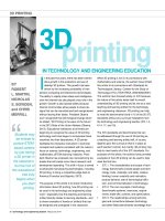
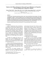
Fig. 1. Schematic illustration of the steps involved in the synthesis and 3D printing of functional cellulose-based hydrogels and the testing of their properties. A) Ink
formulation. B) Direct ink writing of cellulose-based polymer ink and effect of extrusion on the alignment of cellulose nanocrystals during the flow of the ink within
the nozzle. C) Post-treatment to cure the printed structure in functional parts. D – i–v) characteristics and properties evaluated for the inks and final hydrogels for
functional applications.
4
O. Fourmann et al.
Carbohydrate Polymers 259 (2021) 117716
3.2. Ink properties and rheological characterization
particles (CNC and CNF) play crucial roles in modifying the rheology of
the inks and as anisotropic reinforcements in the final printed hydrogels.
In our study, we optimized the ink properties to maximize the degree of
orientation of cellulose nanoparticles within the printed structures. The
flow-induced orientation of CNCs and CNFs is only possible if the
applied stress during printing exceeds the yield stress of the ink (i.e. the
differential-flow regime) (Siqueira et al., 2017). We evaluated the
rheological properties of inks with 15–35 wt% of CNCs, and also
developed inks containing 14 wt% CNC and 1 wt% CNF and inks with 19
wt% CNC and 1 wt% CNF. The rheological properties of the inks con
taining different amounts of CNC/CNF particles are presented in
Fig. S4A–D (Supplementary Information); they all show pronounced
shear thinning behavior. As expected from their rheological properties,
all of these inks are 3D printable, illustrating the versatility of the for
mulations and printing method. However, we decided to focus on three
ink formulations: containing 20 wt% or 25 wt% CNCs only, or 14 wt%
CNC and 1 wt% CNF. These formulations were chosen because they had
the best combination of rheological properties for ease of printing with
the best observed alignment of the nanoreinforcements.
The rheological behavior of these CNC-NIPAM hydrogel inks is
shown in Fig. 2E and F. The pure NIPAM ink (0 wt% CNC) exhibits a
constant viscosity (η) of 1.3 × 10− 3 Pa.s at shear rates higher than 10 s− 1
(Fig. 2e). This means that the pure NIPAM ink would freely flow though
the nozzles at modest pressures but it does not possess the ability to
support itself after being extruded from the printing needle. The addi
tion of nanocelluloses allows to transform the pure NIPAM ink into a
viscoelastic fluid (gel-like material) ready to print. In contrast with the
pure NIPAM ink, the CNC-NIPAM inks containing 20 and 25 wt% CNC
possess viscosities that decrease several orders of magnitude as the shear
rate increases from 0.001 to 50 s− 1 (Fig. 2E). Because of their high shear
thinning behavior, these inks exhibit viscosities ranging from 8.92 to
18.20 Pa.s at shear rate of 50 s− 1, which is a typical value applied during
DIW process.
The fabrication procedure of our nanocellulose-NIPAM hydrogel inks
is simple and easy to control. The hydrogel inks consist of rod-like stiff
cellulose nanocrystals (CNCs), or a combination of these with flexible
cellulose nanofibers (CNF), suspended in an aqueous solution of N,Nisopropylacrylamide
(NIPAM),
a
crosslinker
ethyleneglycoldymethylacrylate (EGDMA) and a photoinitiator (Irgacure 2959),
allowing the system to be polymerized after printing (Fig. 2A). To ach
ieve antibacterial properties in the hydrogel network, we functionalized
ε-polylysine (EPL) with methacrylic acid (MA) according to the pro
cedure developed by Zhou et al. (2011) and then added it to the
hydrogel ink prior to photopolymerisation (Fig. 2B). Glucose and
glucose oxidase were added to the system as oxygen scavengers to
ensure a sufficient UV curing under controlled nitrogen atmosphere. As
a first demonstration, a simple cubic structure has been printed with the
nanocellulose-NIPAM hydrogel ink (Fig. 2). This structure is composed
of 32 layers of 320 μm and demonstrates the precise fabrication of
multi-layered objects, with proper adhesion between the printed layers
without delamination of the filaments.
In previous work, we have demonstrated that the rheological prop
erties of similar inks (including shear-thinning behavior, rapid elastic
recovery, well-defined yield stress and elastic modulus) are the most
important parameters to ensure high shape fidelity, with no distortion of
single printed filaments, in the DIW process (Siqueira et al., 2017). Since
our main goal in this work is to achieve a high degree of alignment of the
nanocelluloses within the printed parts, we have formulated and printed
NIPAM-hydrogels with high CNC or CNC/CNF loadings of up to 35 wt%.
The CNCs have an average length of 115 nm with a diameter of 7.5 nm,
and thus possess an aspect ratio, s, of about 15 (Fig. 2D). The morpho
logical characteristics of TEMPO-CNFs, in particular their high aspect
ratio and the resulting entangled network structure, can be seen in
Fig. S3 (Supplementary Information). Both types of nanocellulose
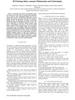
Fig. 2. Conceptual illustration of the hydrogels with nano-structured architectures used in this work, also showing the morphology of the wood pulp cellulose
nanocrystals that were used, and the rheological behaviour of the resulting CNC-PNIPAM hydrogels: A) CNC-PNIPAM hydrogels not modified with ε-polylysine (EPL)
and B) CNC-PNIPAM hydrogels modified with ε-polylysine (EPL-MA). C) 3D printed cubic structure of CNC-PNIPAM hydrogel loaded with 20 wt% of CNCs (1cm3).
D) Transmission electron image of anisotropic CNC particles (scale bar: 100 nm). e) Steady-shear and f) oscillatory rheological measurements (frequency (1 Hz) for
the PNIPAM-hydrogels with varied solid loading (20 and 25 wt% CNC).
5
O. Fourmann et al.
Carbohydrate Polymers 259 (2021) 117716
To assess the viscoelastic properties of the CNC-NIPAM hydrogel
inks, oscillatory measurements at low strains were carried out (Fig. 2F).
These experiments showed that the selected inks (20 or 25 wt% CNCs)
mainly exhibit elastic behavior at low shear rates (G’ > G’’) and a welldefined dynamic yield stress τy, at the crossover point between G’ and
G’’. The dynamic yield stress varies from 425 to 867 Pa for the inks
containing 20 and 25 wt% CNCs, respectively. Similar behaviors on the
rheological profiles were observed for the inks containing different CNC
contents or for the ones possessing 1 wt% of CNFs in their formulations
(see Fig. S4A–D in Supplementary Information).
the printing process and shows that a high degree of CNC alignment
within the printed filaments can be obtained as long as an appropriate
combination of needle diameter and nanocellulose concentration is
chosen. (The swelling behaviour of hydrogels for other nanocellulose
concentrations and needle diameters is shown in Supplementary Infor
mation Fig. S5).
To investigate the flow-induced orientation of anisotropic CNC par
ticles in the printed hydrogels, we carried out 2D wide-angle X-ray
scattering (2D-WAXS) measurements of the nanocellulose-NIPAM
hydrogels and the pure matrix (Fig. 3C–E, Table S2 and Fig. S6 - Sup
plementary Information). In agreement with our previous studies
(Hausmann et al., 2018), the results clearly show more pronounced CNC
alignment for the hydrogels (20 wt% CNC) printed with the 410 μm
nozzle (π = 86 %) as compared to the ones printed with 840 μm (π = 79
%) indicated by the full width at half maximum (FWHM) values
(Fig. 3D). The pure NIPAM matrix shows no preferential orientation,
whereas the printed CNC hydrogels show preferred orientation of CNCs
along a printed filament, regardless of the nozzle diameters.
The ink rheology combined with this high degree of alignment allows
the printing of 3D structures with intricate architectures, including freestanding components with angles of up to 45◦ , without the need for
rheological modifiers others than the nanocelluloses themselves (in
Fig. 3A).
Nanocelluloses are able to constrain the swelling and/or shrinkage of
the PNIPAM structures in the direction of reinforcement, similarly to
those observed in biological tissues such as in pine cones (Dawson,
3.3. Printed-induced and quantified nanocellulose alignment
To investigate the effects of flow-induced orientation we printed 3D
and 2D patterns (Fig. 3A and B) using the developed inks and observed
how these shapes changed when the hydrogels were allowed to swell in
water.
A quantification of the degree of alignment of the CNCs is necessary
to allow a reproducible tailoring of the (post-hydration) 3D structure of
printed objects with anisotropic actuation, and we used wide-angle Xray scattering to do this. In previous work on inks containing high
nanocellulose contents (Hausmann et al., 2018; Siqueira et al., 2017) we
determined the parameters for differential and plug flow regimes as a
function of the CNC concentration in gel-like inks and showed that
flow-induced CNC alignment is only possible if the applied stress ex
ceeds the yield stress of the inks. Fig. 3B illustrates our ability to control
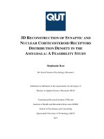
Fig. 3. CNC orientation within 3D printed NIPAM hydrogels. Simple 3D printed A) Complex and angled honeycomb 3D printed structure using nanocellulose-NIPAM
hydrogel (20 wt% CNC). B) Bilayer strips of CNC-NIPAM hydrogel. C) 2D-WAXS patterns of pure NIPAM matrix and 3D printed CNC-NIPAM hydrogels (20 wt%)
using 840 μm and 410 μm diameter nozzles respectively. D) Normalized 2D-WAXS azimuthal intensity distributions of the equatorial reflection (200) of 3D printed
CNC-NIPAM hydrogel (20 wt% CNC) focused on the axial direction of the printed filaments for printed structures with 840 μm and 410 μm diameter nozzles. The
inset image in D) shows the 3D printed grid and the location of the X-ray beam spot where the scattering measurements were performed. E) Dependence of hydrogel
swelling behaviour as a function of the degree of orientation and printing nozzle diameters.
6
O. Fourmann et al.
Carbohydrate Polymers 259 (2021) 117716
Vincent, & Rocca, 1997), Bauhinia variegate pods (Armon, Efrati, Kup
ferman, & Sharon, 2011), and wheat awns (Fratzl, Elbaum, & Burgert,
2008). As designed, the hydrogels engineered and printed to have highly
aligned CNCs, extend by 78 % in the transverse direction to the CNCs
alignment while the actuation in the longitudinal direction is only 22 %
(swelling ratio), for the samples containing 20 wt% CNCs (Fig. 3B). In
nature, especially in plants, the basic mechanism underlying the actu
ation by the swelling or shrinking (shape-changing) of cell walls is
achieved by the orientation of cellulose fibrils in a swellable natural
polymer matrix (Burgert & Fratzl, 2009a; Erb et al., 2013). We investi
gated the dependence of the anisotropic swelling of composites upon the
nozzle diameter used when printing (410 or 840 μm) (see Fig. 3E), the
CNC concentration (20 or 25 wt%) or nanocellulose morphology (CNC
and CNF) (see Supplementary Information Fig. S5). Both shear and
extensional flows impose orientation of anisotropic particles in fluids
(Håkansson et al., 2014; Nesaei, Rock, Wang, Kessler, & Gozen, 2017).
Considering only shear stresses, the use of the smaller nozzle diameter
(410 μm) results in higher shear forces from the walls of the nozzle than
larger diameter nozzles. As a result of this, a formulation containing 20
wt% CNC extruded through a needle of 410 μm in diameter requires a
pressure of about 1.5 bar to induce alignment of the cellulose nano
particles. However, when the larger needle diameter of 840 μm is used
with the same ink formulation, only 0.3 bar is necessary to enable the
extrusion of the 20 wt% CNC ink. Consequently, as the shear forces on
the wall of the nozzles are lower, the cellulose nanoparticles’ degree of
alignment and anisotropic swelling for the composites printed with 840
μm nozzles are inferior than the ones printed with the 410 μm nozzles.
The higher anisotropic swelling effect found for the ink containing 1 wt
% of CNF and 14 wt% CNCs is ascribed to the physical interactions
between the nanofibers. Such interactions, named entanglements,
contribute to an even higher reduction of swelling along their
orientation direction (Hausmann et al., 2018).
3.4. Soft actuation of printed bilayer structures
To assess the anisotropic swelling properties of the CNC-NIPAM
hydrogels, we printed bilayer strips with at least two different orienta
tions of filaments and observed their actuation over time as the inks
were hydrated. These experiments confirmed that the anisotropic
swelling of the nanocellulose-NIPAM hydrogels could lead to a macro
scopic programmable change in shape of the synthetic printed
structures.
To compensate the changes in rheology due to the presence of CNF,
we reduced the CNC concentration in the printed materials (Fig. 4A) to
14 wt% (overall 15 wt% of nanocellulose is present in the inks) aiming
for maximal swelling actuation and shape changes. Akin to the cellulose
fibrils microreinforcements in plant cell walls, in our system, shape
motion occurs because the nanocelluloses do not swell in their axial
direction (Burgert & Fratzl, 2009a, 2009b). On the contrary, swelling
will occur preferentially in the orthogonal direction to the nanocellulose
orientation within the printed filaments, which result in a highly
anisotropic deformation of the structure upon water uptake. Therefore,
the programmable shape change in our system is achieved due to the
orientation of stiff nanocellulose reinforcements within the hydrogels.
These aligned nanocelluloses create internal stresses when the structures
swell which can only be reduced by undergoing a deformation (Erb
et al., 2013; Le Ferrand et al., 2016). The transformation of the printed
bilayer from a flat to twist/bended or curled/bended configuration
follows the programmable designed direction. However the final
twisted, curled, or bended architectures of the swelled bilayer structures
are governed by the nanocellulose orientation in the upper layer as it is
less affected by misalignment, as proved by 2D-WAXS measurements
Fig. 4. 3D printed structures of nanocellulose-NIPAM hydrogels with swelling and anisotropic actuation behaviours. A) 3D printed bilayer structures of NIPAM
hydrogels with 14 wt% CNC and 1 % CNF after swelling in water. The schemes in the right and top show printing patterns (0/90◦ and 45/135◦ ). The lines drawn on
the top of printed structures indicate better the printing pattern and bending according to predictions. B) Evolution of water uptake of nanocellulose-based NIPAM
hydrogels on 9.2 × 9.2 × 10 mm samples. Error bars show standard deviation (n = 5). C) Combination of two bilayer strips produced by 3D printing of nanocelluloseNIPAM hydrogels (20 wt% CNC) leading to synthetic architectures that twist. On the left is the scheme of the printing pattern (45/135◦ filling) and on the left side,
pictures of the evolution of the anisotropic actuation of the printed structures.
7
O. Fourmann et al.
Carbohydrate Polymers 259 (2021) 117716
(Fig. 4A and Table S1). This is in contrast to the first printed layer which
has a lower degree of orientation of nanocelluloses due to the fact that
the filament is squeezed closer to the glass substrate to ensure proper
adhesion between this layer and the substrate. This extra pressure was
obtained by using a smaller nozzle offset for this layer than in the second
layer.
Time-dependent swelling tests were conducted to quantify the
maximal swelling capability of the hydrogels prior to mechanical testing
and actuation performance (Fig. 4B). The 3D printed cuboids (9.2 × 9.2
× 10 mm) produced with 0◦ filling pattern showed the highest swelling
rate in the beginning of the tests due to the high osmotic pressure be
tween water and the dried hydrogel. The equilibrium moisture content is
reached after 4 days when the osmotic pressure is equal to the retractile
forces of the stretching polymer chains (Buenger, Topuz, & Groll, 2012).
The reversible swelling allows the generation of composites with
shape-memory characteristics after drying cycles of printed hydrogels in
oven at 60 ◦ C (Video S1 and Fig. S7 Supplementary Information).
The twisting transformation is generally not possible in simple syn
thetic bilayer materials. To achieve such chiral twisting motions, the
reinforcing elements should be oriented with an angle of 45◦ or − 45◦
from the first to second layer (Erb et al., 2013). We investigated the
twisting motion on NIPAM-hydrogels reinforced with 20 wt% CNCs
(Fig. 4C and video S2– Supplementary Information) to evaluate if such
shape-morphing would also be possible with pure CNC-NIPAM-based
inks. For this test we kept the printing pressures and offset constant to
avoid or minimize possible misalignment of CNCs in the first printed
layer due to variations of printing parameters. The results show that the
two layers attempt to expand in perpendicular direction during hydra
tion thus resulting in helically twisting motion, similar to the natural
response found in plants as Bauhinia variegate (seedpod of orchid trees)
and climbing plants coil tendrils (Erb et al., 2013; Studart & Erb, 2014).
3.5. Structural characterization of printed materials
Control over the orientation of nanocellulose particles enables
tailoring of mechanical properties of 3D printed hydrogels in specific
directions (Fig. 5A–C). We investigated the effect of cellulose nano
crystals alignment on the mechanical behaviour of neat NIPAM and
CNC-NIPAM hydrogels by measuring the compressive mechanical
properties of specimens containing CNCs aligned in the longitudinal or
transverse direction relative to the applied load Fig. 5A–C). While the
hydrogel matrix alone has a soft and stretchable behaviour with a
Fig. 5. Enhanced mechanical properties of neat NIPAM hydrogels and CNC-NIPAM hydrogels, containing 20 and 25 wt% of cellulose nanocrystals, tested in
compression mode at longitudinal and transverse directions with different filling patterns (0◦ , 0/90◦ and 45/135◦ ). A) Representative stress vs. strain curves for neat
NIPAM matrix and its nanocomposites. B) Young’s modulus and C) ultimate stress of NIPAM hydrogels reinforced with CNCs tested under compression (70 % strain)
at longitudinal and transverse directions with 20 and 25 wt% of CNCs. Error bars show standard deviation (n = 6).
8
O. Fourmann et al.
Carbohydrate Polymers 259 (2021) 117716
Young’s modulus of only about 70 Pa (Fig. 5A), the reinforced sample
containing 20 wt% CNCs tested in the transverse direction had on
average a Young’s modulus of 13 (0◦ filling) to 16.6 kPa (for samples
filled at 45/135◦ and 0/90◦ ). This corresponds to an increase of the
Young’s modulus by a factor of 236 compared to the pure matrix.
Likewise, the reinforcing effect of CNCs on the mechanical properties of
NIPAM hydrogels is even more remarkable when comparing the prop
erties of the pure hydrogel matrix with the composites reinforced with
25 wt% CNCs, regardless of the filling pattern. The Young’s modulus of
composites loaded with 25 wt% CNCs, tested in the transverse direction
with filling patterns of 0/90◦ and 45/135◦ (Fig. 5B), is closer to 3-orders
of magnitude (650x) higher than that of the pure hydrogel matrix.
However, the Young’s modulus remains nearly the same for the 20 wt%
CNC-NIPAM hydrogels tested in the longitudinal and transverse di
rections. The increase in the elastic modulus with increased CNC con
centrations (from 20 to 25 wt%) is accompanied by a decrease of at least
13 % in the strain at rupture.
The average ultimate stress properties of the composites clearly
reveal a significant influence of the testing direction relative to the
orientation of the CNCs within the 3D printed filaments (Fig. 5C). Such
an effect is observed in composites reinforced with 20 wt% CNCs and it
is clear for all samples regardless of the filling pattern. However, the
enhanced mechanical properties of the composites, in the longitudinal
direction, become more pronounced for the samples tested at 0◦ filling
pattern due to the orientation of CNCs. Hence, in such condition, we
likely maximize the CNCs orientation with the probe direction. These
results illustrate our capability to precisely control the CNC orientations
and, therefore, the mechanical properties of the hydrogels by designing
inks with varied CNC loads and controlling the printing fillings and
parameters as needle sizes, pressure and speed.
3.6. Extended hydrogel functionalities
Combining natural antimicrobial peptides, such as ε-polylysine, with
the 20 wt% CNC-NIPAM hydrogels would allow to broaden the spectrum
of applications of our complex-shaped and textured materials (Fig. 6A).
To accomplish this, we functionalized ε-polylysine (NMR spectrum
Fig. S8- Supplementary Information) with methacrylic acid. The success
of this chemical modification, as shown by nuclear magnetic resonance
spectroscopy (NMR), is demonstrated in Fig. S9 (Supplementary Infor
mation). Antimicrobial properties of 3D printed materials were achieved
for contents of EPL-MA in the hydrogels varying from 1 to 2.5 wt%.
Recognized more than 30 years ago as antimicrobial agent, the mech
anism responsible for the antimicrobial activity of EPL is not completely
understood (Hyldgaard et al., 2014). However, it has been suggested
that such cationic polypeptide interacts with negatively charged cell
surface by ionic adsorption followed by microbial cell membrane
interaction, membrane disruption and ultimately cell lysis (Salom´e
Veiga & Schneider, 2013). Significant reduction of bacteria growth
compared to the control (Fig. 6B) was determined with the
Kruskal-Wallis-test and Mann-Whitney U test for pairwise comparison. A
significant deviation (P < 0.05) from the control (no EPL-MA) was
identified in all the samples where 1 or 2.5 wt% EPL-MA were added
(Fig. 6B II and III). The quantitative results presented in Fig. 6C also
indicate strong and significant reductions of both, gram positive and
gram negative, bacterial growth in the hydrogels prepared with EPL-MA
when compared to the biofilm formation in the control. This study re
veals the effectiveness of the antimicrobial properties added to the final
3D printed nanocellulose hydrogels given by EPL-MA, but other more
clinically relevant or specific antimicrobial agents could also be
considered.
To understand the effect of EPL on the final properties of the NIPAM
hydrogels we prepared inks containing 1 or 2.5 wt% EPL and measured
their rheological properties (steady-shear and oscillatory at the
Fig. 6. Antimicrobial properties of functionalized CNC-NIPAM hydrogels. A) Complex architecture with texturing effect of 3D printed CNC-NIPAM hydrogel
functionalized with ε-polylysine. B) Qualitative results of bacterial growth on the hydrogels functionalized with different concentrations of tryptone soya agar (TSA)
plates. I) control: no EPL-MA, II) 1 wt% EPL-MA and III) 2.5 wt% EPL-MA. C) Quantitative results of bacterial growth in the hydrogels before and after addition of
EPL-MA.
9
O. Fourmann et al.
Carbohydrate Polymers 259 (2021) 117716
frequency of 1 Hz) and compared these results with the ones of the ink
without EPL. The results (Fig. S12(A) - Supplementary Information)
indicate that all inks presented shear thinning, in which their viscosities
decrease by several orders of magnitude as the shear rate increases from
0.001 to 50 s− 1 (typically applied during DIW). It is also noticed that the
viscosities of the inks containing EPL are in the same range as the one
without EPL. The amplitude sweep tests (Fig. S12B- Supplementary In
formation) show that all inks present G’>G”, and dynamic yield stress in
the order of a few hundred Pa indicating that all of them can be
considered in the range of printable inks without the need for applying
prohibitive high printing pressures. Nevertheless G’ and G" are signifi
cantly higher for the inks containing EPL thus indicating possible dif
ferences in their final mechanical properties after polymerization,
however such properties were not investigated in the present work. EPLMA has the possibility to crosslink which may also lead to increased
mechanical properties of the hydrogels.
References
Armon, S., Efrati, E., Kupferman, R., & Sharon, E. (2011). Geometry and mechanics in the
opening of chiral seed pods. Science, 333(6050), 1726–1730.
Billiet, T., Vandenhaute, M., Schelfhout, J., Van Vlierberghe, S., & Dubruel, P. (2012).
A review of trends and limitations in hydrogel-rapid prototyping for tissue
engineering. Biomaterials, 33(26), 6020–6041.
Buenger, D., Topuz, F., & Groll, J. (2012). Hydrogels in sensing applications. Progress in
Polymer Science, 37(12), 1678–1719.
Burgert, I., & Fratzl, P. (2009a). Actuation systems in plants as prototypes for bioinspired
devices. Philosophical Transactions Series A, Mathematical, Physical, and Engineering
Sciences, 367(1893), 1541–1557.
Burgert, I., & Fratzl, P. (2009b). Plants control the properties and actuation of their
organs through the orientation of cellulose fibrils in their cell walls. Integrative and
Comparative Biology, 49(1), 69–79.
Caballero-Aguilar, L. M., Silva, S. M., & Moulton, S. E. (2020). 6 - Three-dimensional
printed drug delivery systems. In A. Seyfoddin, S. M. Dezfooli, & C. A. Greene (Eds.),
Engineering drug delivery systems (pp. 147–162). Woodhead Publishing.
Cheng, J., Jia, Z., & Li, T. (2020). A constitutive model of microfiber reinforced
anisotropic hydrogels: With applications to wood-based hydrogels. Journal of the
Mechanics and Physics of Solids, 138, Article 103893.
Dai, L., Cheng, T., Duan, C., Zhao, W., Zhang, W., Zou, X., & Ni, Y. (2019). 3D printing
using plant-derived cellulose and its derivatives: A review. Carbohydrate Polymers,
203, 71–86.
Dawson, C., Vincent, J. F. V., & Rocca, A.-M. (1997). How pine cones open. Nature, 390
(6661), 668.
Duan, B., Hockaday, L. A., Kang, K. H., & Butcher, J. T. (2013). 3D Bioprinting of
heterogeneous aortic valve conduits with alginate/gelatin hydrogels. Journal of
Biomedical Materials Research Part A, 101A(5), 1255–1264.
Erb, R. M., Sander, J. S., Grisch, R., & Studart, A. R. (2013). Self-shaping composites with
programmable bioinspired microstructures. Nature Communications, 4(1), 1712.
Fratzl, P., Elbaum, R., & Burgert, I. (2008). Cellulose fibrils direct plant organ
movements. Faraday Discussions, 139(0), 275–282.
Gaharwar, A. K., Peppas, N. A., & Khademhosseini, A. (2014). Nanocomposite hydrogels
for biomedical applications. Biotechnology and Bioengineering, 111(3), 441–453.
Gao, G., Du, G., Sun, Y., & Fu, J. (2015). Self-healable, tough, and ultrastretchable
nanocomposite hydrogels based on reversible polyacrylamide/montmorillonite
adsorption. ACS Applied Materials & Interfaces, 7(8), 5029–5037.
Gupta, A., Briffa, S. M., Swingler, S., Gibson, H., Kannappan, V., Adamus, G., &
Radecka, I. (2020). Synthesis of silver nanoparticles using curcumin-cyclodextrins
loaded into bacterial cellulose-based hydrogels for wound dressing applications.
Biomacromolecules.
Håkansson, K. M. O., Fall, A. B., Lundell, F., Yu, S., Krywka, C., Roth, S. V., &
Să
oderberg, L. D. (2014). Hydrodynamic alignment and assembly of nanofibrils
resulting in strong cellulose filaments. Nature Communications, 5(1), 4018.
Han, D., Farino, C., Yang, C., Scott, T., Browe, D., Choi, W., & Lee, H. (2018). Soft robotic
manipulation and locomotion with a 3D printed electroactive hydrogel. ACS Applied
Materials & Interfaces, 10(21), 17512–17518.
Hausmann, M. K., Rühs, P. A., Siqueira, G., Lă
auger, J., Libanori, R., Zimmermann, T., &
Studart, A. R. (2018). Dynamics of cellulose nanocrystal alignment during 3D
printing. ACS Nano, 12(7), 6926–6937.
Hausmann, M. K., Siqueira, G., Libanori, R., Kokkinis, D., Neels, A., Zimmermann, T., &
Studart, A. R. (2020). Complex-shaped cellulose composites made by wet
densification of 3D printed scaffolds. Advanced Functional Materials, 30(4), Article
1904127.
Hoffman, A. S. (2012). Hydrogels for biomedical applications. Advanced Drug Delivery
Reviews, 64, 18–23.
Hyldgaard, M., Mygind, T., Vad, B. S., Stenvang, M., Otzen, D. E., & Meyer, R. L. (2014).
The antimicrobial mechanism of action of epsilon-poly-l-lysine. Applied and
Environmental Microbiology, 80(24), 7758–7770.
Jang, T.-S., Jung, H.-D., Pan, H. M., Han, W. T., Chen, S., & Song, J. (2018). 3D printing
of hydrogel composite systems: Recent advances in technology for tissue
engineering. International Journal of Bioprinting, 4(1).
Koffler, J., Zhu, W., Qu, X., Platoshyn, O., Dulin, J. N., Brock, J., & Tuszynski, M. H.
(2019). Biomimetic 3D-printed scaffolds for spinal cord injury repair. Nature
Medicine, 25(2), 263–269.
Le Ferrand, H., Bolisetty, S., Demiră
ors, A. F., Libanori, R., Studart, A. R., & Mezzenga, R.
(2016). Magnetic assembly of transparent and conducting graphene-based functional
composites. Nature Communications, 7(1), 12078.
Lee, S., Bristol, R. E., Preul, M. C., & Chae, J. (2020). Three-dimensionally printed
microelectromechanical-system hydrogel valve for communicating hydrocephalus.
ACS Sensors.
Lei, J., Sun, L., Huang, S., Zhu, C., Li, P., He, J., & He, Q. (2019). The antimicrobial
peptides and their potential clinical applications. American Journal of Translational
Research, 11(7), 3919–3931.
Li, S., Dong, S., Xu, W., Tu, S., Yan, L., Zhao, C., & Chen, X. (2018). Antibacterial
hydrogels. Advanced Science (Weinheim, Baden-Wurttemberg, Germany), 5(5),
1700527.
Li, Z., Shen, J., Ma, H., Lu, X., Shi, M., Li, N., & Ye, M. (2013). Preparation and
characterization of pH- and temperature-responsive nanocomposite double network
hydrogels. Materials Science and Engineering C, 33(4), 1951–1957.
Liu, J., Gu, T., Shan, S., Kang, S. H., Weaver, J. C., & Bertoldi, K. (2016). Harnessing
buckling to design architected materials that exhibit effective negative swelling.
Advanced Materials, 28(31), 6619–6624.
4. Conclusions
In summary, complex shape morphing nanocellulose-based com
posites have been produced through direct ink writing 3D printing.
Alignment of high aspect ratio nanocellulose particles along the ink flow
direction occurs as a result of the shear and extensional forces in the
print nozzle, giving rise to anisotropic mechanical properties and
swelling behavior of the printed structures. The ability to produce
hydrogel based 3D printing inks in which both the nanocellulose content
(up to 35 wt%) and morphology (cellulose nanocrystals and/or cellulose
nanofibers) can be varied allows to tune the mechanical properties of the
printed structures along specific directions. Because of the high degree
of nanocellulose alignment upon printing, hydrogel structures with
complex architectures (angles and texture) and programmable selfshape actuation can be fabricated with these new inks. This is an
elegant method to synthetically create structures that can, upon hy
dration, bend or twist — resemble the mechanism in plants which use
the orientation of cellulose fibrils. The simplicity of the synthesis and
printing procedures demonstrated here mean that this approach has
great potential to be extended to similar materials such as hydrogels
used for wound healing. The antimicrobial properties provided by
functionalization of the hydrogels with modified ε-polylysine highlights
the potential use of this and related AMPs in biomedical applications of
composite hydrogels.
Author contributions
Experiments were designed and coordinated by G.S., T.Z., G.N. and
O.F., and were conducted by O.F. and M.H. The X-Ray analysis was
performed by A.N. Figure graphic designs were prepared by M.H. and O.
F. Antimicrobial tests were carried out by O.F. and M.S.. G.S. wrote the
manuscript with input from all coauthors. All authors reviewed and
commented on the manuscript.
Declaration of Competing Interest
The authors declare no conflict of interest.
Acknowledgements
We thank B. Fisher allowing us to use the mechanical testing
equipment and A. Huch for the SEM and TEM imaging. G.S., T.Z. and M.
H. greatly acknowledge the financial support from the Swiss National
Science Foundation (grant 200021_159906/1).
Appendix A. Supplementary data
Supplementary material related to this article can be found, in the
online version, at doi: />10
O. Fourmann et al.
Carbohydrate Polymers 259 (2021) 117716
Schubert, M., Engel, J., Thă
ony-Meyer, L., Schwarze, F. W. M. R., & Ihssen, J. (2012).
Protection of wood from microorganisms by laccase-catalyzed iodination. Applied
and Environmental Microbiology, 78(20), 7267–7275.
Shih, I.-L., Shen, M.-H., & Van, Y.-T. (2006). Microbial synthesis of poly(ε-lysine) and its
various applications. Bioresource Technology, 97(9), 1148–1159.
Siqueira, G., Kokkinis, D., Libanori, R., Hausmann, M. K., Gladman, A. S., Neels, A., &
Studart, A. R. (2017). Cellulose nanocrystal inks for 3D printing of textured cellular
architectures. Advanced Functional Materials, 27(12), Article 1604619.
ˇ Vukaˇsinovi´c-Sekuli´c, M., Miˇskovi´c-Stankovi´c, V.,
Stojkovska, J., Kosti´c, D., Jovanovi´c, Z.,
& Obradovi´c, B. (2014). A comprehensive approach to in vitro functional evaluation
of Ag/alginate nanocomposite hydrogels. Carbohydrate Polymers, 111, 305–314.
Studart, A. R., & Erb, R. M. (2014). Bioinspired materials that self-shape through
programmed microstructures. Soft Matter, 10(9), 1284–1294.
Sydney Gladman, A., Matsumoto, E. A., Nuzzo, R. G., Mahadevan, L., & Lewis, J. A.
(2016). Biomimetic 4D printing. Nature Materials, 15(4), 413–418.
Wüst, S., Godla, M. E., Müller, R., & Hofmann, S. (2014). Tunable hydrogel composite
with two-step processing in combination with innovative hardware upgrade for cellbased three-dimensional bioprinting. Acta Biomaterialia, 10(2), 630–640.
Xu, K., Wang, J., Chen, Q., Yue, Y., Zhang, W., & Wang, P. (2008). Spontaneous volume
transition of polyampholyte nanocomposite hydrogels based on pure electrostatic
interaction. Journal of Colloid and Interface Science, 321(2), 272–278.
Yigit, S., Sanyal, R., & Sanyal, A. (2011). Fabrication and functionalization of hydrogels
through “Click” chemistry. Chemistry – An Asian Journal, 6(10), 2648–2659.
Zhang, L.-j., & Gallo, R. L. (2016). Antimicrobial peptides. Current Biology, 26(1),
R14–R19.
Zhou, C., Li, P., Qi, X., Sharif, A. R. M., Poon, Y. F., Cao, Y., & Chan-Park, M. B. (2011).
A photopolymerized antimicrobial hydrogel coating derived from epsilon-poly-llysine. Biomaterials, 32(11), 2704–2712.
Liu, J., Sun, L., Xu, W., Wang, Q., Yu, S., & Sun, J. (2019). Current advances and future
perspectives of 3D printing natural-derived biopolymers. Carbohydrate Polymers,
207, 297–316.
´
Markstedt, K., Mantas, A., Tournier, I., Martớnez Avila,
H., Hă
agg, D., & Gatenholm, P.
(2015). 3D bioprinting human chondrocytes with nanocellulose–alginate bioink for
cartilage tissue engineering applications. Biomacromolecules, 16(5), 1489–1496.
Mauri, E., Rossi, F., & Sacchetti, A. (2016). Tunable drug delivery using chemoselective
functionalization of hydrogels. Materials Science and Engineering C, 61, 851857.
ă
Mỹller, M., Oztỹrk,
E., Arlov, ỉ., Gatenholm, P., & Zenobi-Wong, M. (2017). Alginate
sulfate–nanocellulose bioinks for cartilage bioprinting applications. Annals of
Biomedical Engineering, 45(1), 210–223.
Nesaei, S., Rock, M., Wang, Y., Kessler, M. R., & Gozen, A. (2017). Additive
manufacturing with conductive, viscoelastic polymer composites: Direct-ink-writing
of electrolytic and anodic poly(Ethylene oxide) composites. Journal of Manufacturing
Science and Engineering, 139(11).
Neves, S. C., Pereira, R. F., Araújo, M., & Barrias, C. C. (2018). 4 - Bioengineered peptidefunctionalized hydrogels for tissue regeneration and repair. In M. A. Barbosa, &
M. C. L. Martins (Eds.), Peptides and proteins as biomaterials for tissue regeneration and
repair (pp. 101–125). Woodhead Publishing.
Park, N., & Kim, J. Hydrogel-Based Artificial Muscles: Overview and Recent Progress.
Advanced Intelligent Systems, n/a(n/a), 1900135.
Saito, T., & Isogai, A. (2004). TEMPO-mediated oxidation of native cellulose. The effect
of oxidation conditions on chemical and crystal structures of the water-insoluble
fractions. Biomacromolecules, 5(5), 1983–1989.
Salom´
e Veiga, A., & Schneider, J. P. (2013). Antimicrobial hydrogels for the treatment of
infection. Peptide Science, 100(6), 637–644.
Sano, K., Ishida, Y., & Aida, T. (2018). Synthesis of anisotropic hydrogels and their
applications. Angewandte Chemie International Edition, 57(10), 2532–2543.
11