Effect of red seaweed sulfated galactans on initial steps of complement activation in vitro
Bạn đang xem bản rút gọn của tài liệu. Xem và tải ngay bản đầy đủ của tài liệu tại đây (2.14 MB, 9 trang )
Carbohydrate Polymers 254 (2021) 117251
Contents lists available at ScienceDirect
Carbohydrate Polymers
journal homepage: www.elsevier.com/locate/carbpol
Effect of red seaweed sulfated galactans on initial steps of complement
activation in vitro
E.V. Sokolova a, *, A.O. Kravchenko a, N.V. Sergeeva b, A.I. Kalinovsky a, V.P. Glazunov a, L.
N. Bogdanovich b, I.M. Yermak a
a
G.B. Elyakov Pacific Institute of Bioorganic Chemistry, Far East Branch of the Russian Academy of Sciences, Prospect 100-let Vladivostoku, 159, Vladivostok, 690022,
Russia
Medical Association of the Far East Branch of the Russian Academy of Sciences, Vladivostok, St. Kirova, 95, 690022, Russia
b
A R T I C L E I N F O
A B S T R A C T
Keywords:
Carrageenan
Agar
Heparin
Complement
Lipopolysaccharide
Plasmin
The research described here presents data on the effect of galactans of red algae, carrageenans (λ/μ/ν-, κ-, κ/β-,
and ι/κ-types), and agar on complement system activation in normal human serum. The experiments were based
on well surfaces coated with triggering agents for binding initiating complement components —C3 and C4. The
sulfated galactans inhibited C3 binding to lipopolysaccharide with direct dependence on the sulfation degree of
polysaccharides. Sulfation degree was also important in carrageenans’ capacity to reduce C4 binding to mannan.
However, C4 binding to antibodies was considerably activated by carrageenans, especially with 3,6-anhydroga
lactose. The gelling carrageenans were able to block antigen binding centers of total serum IgM and with more
intensity than non-gelling. No structural characteristics mattered in ameliorating C5 cleavage by plasmin in
extrinsic protease complement activation, but λ/μ/ν- and κ/β-carrageenans almost completely inhibited C5
cleavage. Thus, galactans participated in cell surface biology by imitating surface glycans in inhibition of C3
binding and mannose binding lectin, but as to the tthe heclassical pathway these substances stimulated com
plement, probably due to their structure based on carrabiose.
1. Introduction
Red algae contain considerable amounts of sulfated galactans, and
two groups of these polysaccharides, known as agars and carrageenans,
find wide practical application in gelling and stabilizing food com
pounds. These galactans usually have an unbranched backbone built of
alternating 3-linked β-D-galactopyranose and 4-linked α-galactopyr
anose residues. The latter has the L-configuration in the agar group of
polysaccharides and D-configuration in carrageenans. Additionally, 4linked residues may be present as 3,6-anhydro derivatives (Usov, 1998).
Carrageenans are composed of repeating units of [→3)-β-D-Galp(1→4)-α-D-Galp-(1→] (‘diads’ or ‘carrabiose’ disaccharides), mainly
substituted by sulfate groups (Stortz & Cerezo, 2002) and rarely with
other substituents (Chiovitti et al., 1998; Estevez, Ciancia, & Cerezo,
2004). Carrageenans are classified into families by the location of the
sulfate groups in the β-galactose moiety. Then, a particular name is
given to each structural disaccharide unit based on sulfate group loca
tions and presence or absence of the 3,6-anhydro sugar in the
α-galactose moiety. Carrageenans found in nature usually contain more
than one carrabiose unit, forming hybrid structures, and the number and
structure of diads varies with algal species and life stage (Cosenza,
Navarro, Ponce, & Stortz, 2017; Craigie, 1990). Some physico-chemical
characteristics of carrageenans with predominant λ-, κ-, or ι-diad con
tents enable their use as gelling and stabilizing agents, which are
properties carrageenans share with agars (Lahaye, 2001; Usov, 1998).
Carrageenans and agars also exhibit a wide spectrum of biological ac
tivities regarding human health (Koutsaviti, Ioannou, & Roussis, 2018;
Pereira & Critchley, 2020; Pereira, 2018). Sulfated galactans from red
algae have been observed to interact with the serine protease system
—the complement (Baker, Nicklin, & Miller, 1986; Davies, 1965) and
coagulation/fibrinolysis cascades (dos Santos-Fidencio, Gonỗalves,
Noseda, Duarte, & Ducatti, 2019; Opoku, Qiu, & Doctor, 2006).
Complement is the fluid-phase part of innate immunity contributing
to infectious and non-infectious diseases and is composed of cascading
proteases that assemble with almost immediate reactivity at abnormal
landscapes of foreign and altered host cell surfaces (Fig. 1) (Lubbers, Van
* Corresponding author.
E-mail address: (E.V. Sokolova).
/>Received 12 August 2020; Received in revised form 7 October 2020; Accepted 13 October 2020
Available online 21 October 2020
0144-8617/© 2020 Elsevier Ltd. All rights reserved.
E.V. Sokolova et al.
Carbohydrate Polymers 254 (2021) 117251
(PubChemCID: 101231952); κ-carrageenan (PubChemCID: 11966249);
β-carrageenan (PubChemCID: 102199626); λ-carrageenan (PubChem
CID: 101231953); LPS (PubChemCID: 11970143); heparin (PubChem
CID: 772); mannan (PubChemCID: 25147451).
2.1. Reagents
Commercial unfractionated heparin as sodium salt (cat no. 101931,
lot no. 2024H, St. Louis, Sigma, USA) and commercial LPS from the
bacterium E. coli 055:B5 (cat no. L2880, lot no. 025M4040 V, Sigma, St.
Louis, MO, USA) were purchased from Sigma, as was mannan from
Saccharomyces cerevisiae, prepared by alkaline extraction (cat no.
M7504, lot no. SLCC2157). Normal human IgG was manufactured by
Statens Serum Institute (007740, SSI, Denmark). Human plasmin was
from RENAM (cat no. FA-3, lot no. 0818, Moscow, Russia). Specific
enzyme-linked immunosorbent assay (ELISA) kit, used to measure C5a
concentrations, was purchased from Cytokine, Saint-Petersburg, Russia.
Human complement C4c was purchased from LeeBiosolutions (cat no.
194-41, lot no. 08D1609). Anti-human-C3 and C4 monoclonal antibody
(mAb) conjugated with horseradish peroxidase (HRP) were purchased
from Cytokine, Saint-Petersburg, Russia. Food agar of the first class,
brand 700 from Ahnfeltia tobuchiensis (Primorsky Krai, Russia) and
agarose (cat no. A9539, Sigma) were used for comparison in experi
ments of C3 binding to E. coli LPS and catalytic cleavage of C5 by
plasmin.
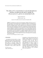
Fig. 1. A simple scheme of complement activation and major steps for further
proliferation of the complement cascade in tissues with all complement com
ponents. The main cleavage fragments of complement are responsible for many
of the host defense-mediated functions of complement, such as chemo
attraction, phagocytosis, and cell lysis.
Essen, Van Kooten, & Trouw, 2017; Ricklin, Mastellos, Reis, & Lambris,
2018). Almost immediate reactivity is achieved by a pivotal component
of the complement system—C3. C3 has an ability to cleave spontane
ously into C3a and C3b fragments and amplifies its own production by a
positive feedback loop. The activity of C3 loop on cell surfaces depends
on whether it encounters surfaces with complement stimulating factors
(e.g. antibodies, bacterial carbohydrates) or surfaces with absent re
ceptors against the C3/C3b attack (Harrison, 2018; Lachmann, 2018).
The C3 stimulating factors on surfaces are C4 and C2 converted to C3
convertase by pattern recognition receptor (PRR)-associated serine
proteases. Depending on PRRs, complement activation is divided into
‘lectin’ and ‘classical’ activation pathways. For the ‘lectin pathway,’ the
triggering PRRs are mannan-binding lectin (MBL), ficolins, and collec
tins detecting pathogen-associated molecular sugar patterns or altered
glycosylation patterns on abnormal host cells. In the ‘classical pathway’,
the PRR is C1q, activated upon recognition of the Fc portion of target cell
bound immunoglobulins or pentraxins (Lubbers et al., 2017). C3/C3b is
capable of covalently binding to the surface on its own, in the absence of
activity of other complement pathways. Such is the case when C3b/C3
(H2O) takes advantage of the surfaces lacking polyanions necessary for
the stabilization of Factor H (‘protected surface’) in the ‘alternative
pathway’ of complement activation. Factor H is a soluble PRR of lectin
nature, accelerating C3 convertase decay. Cells coated with bacterial
endotoxin (smooth lipopolysaccharides, LPS) may be the most impor
tant in vivo activator by this mechanism (Blaum, 2017; Lachmann,
2018).
Complement components can be directly cleaved by coagulation/
fibrinolytic factors, resulting in ‘extrinsic protease pathway’ (Amara
et al., 2010; Barnum, 2017). This non-canonical complement activation
pathway opens a possible link to why many complement disorders
feature pathologic thrombosis as a hallmark clinical manifestation
(Baines & Brodsky, 2017).
Since the earliest works on carrageenan and complement, our un
derstanding of complement organization and methods in the field have
drastically evolved. Initially, carrageenans’ action on complement was
limited only to classical and alternative pathways and was assayed with
the model based on the phenomenon of immune hemolysis (Baker et al.,
1986; Davies, 1965). This article describes the ability of red algal
polysaccharides to affect the human complement system in tissue con
taining all complement cascade proteins-serum by analyzing C3 binding
to well plate surfaces coated with Escherichia coli LPS, C4 binding to
wells coated with IgG or mannan molecules, and, finally, changes in C5a
concentration in human serum activated with plasmin.
2.2. Isolation and characterization of carrageenans
Red seaweeds Chondrus armatus (Gigartinaceae), Tichocarpus crinitus
(Tichocarpaceae), and Ahnfeltiopsis flabelliformis (Phyllophoraceae)
were collected along the Russian coast of the Japanese Sea in
2016–2017. Morphological and anatomic characteristics of the sea
weeds were determined according to Perestenko (1994) and identified
by light microscopy by Prof. E. Titlynov and Dr. Oksana Belous from the
A.V. Zhirmunsky National Scientific Center of Marine Biology, Far East
Branch of the Russian Academy of Sciences FEB RAS. According to the
identification, C. armatus was represented by male gametophyte and
T. crinitus and A. flabelliformis by female gametophytes with cystocarps.
The polysaccharides were extracted from dried algae (5 g) with hot
water (300 mL) at 80 ◦ C for 3 h, a total of three times, according to the
protocol (Yermak, Kim, Titlynov, Isakov, & Solov’eva, 1999). The sus
pensions were centrifuged (4000 rpm), residues recovered, and super
natants were filtered through a Vivaflow 200 membrane (Sartorius,
ăttingen, Germany) with a 100 kDa pore size to remove low molecular
Go
weight compounds. The polysaccharides were precipitated from solu
tions with a triple volume of 96 % ethanol. The precipitate was sepa
rated, washed with ethanol, suspended in hot water, and fractionated
into gelling and non-gelling fractions by 4 % KCl for C. armatus, 1 % KCl
for T. crinitus, and 4 % CaCl2 for A. flabelliformis total polysaccharides,
respectively. The structures of the obtained fractions were established
according to published protocols (Barabanova et al., 2005; Kravchenko
et al., 2016; Yermak et al., 1999).
To determine the content of 3,6-anhydrogalactose, total reductive
hydrolysis of the carrageenans and agar in 2 M Trifluoroacetic acid
(TFA) (100 ◦ C, 4 h) with 4-methylmorpholinborane was carried out, and
then, aldononitrile acetates were obtained (Usov & Elashvili, 1991).
Other monosaccharides (galactose, glucose, xylose) were determined as
alditol acetates according to a previously published protocol (Krav
chenko et al., 2020). The sulfate ester content of the polysaccharides was
determined by turbidimetry (Dodgson & Price, 1962). The protein
content of carrageenans and agar was determined according to the
Lowry method (Lowry, Rosebrough, Farr, & Randall, 1951).
To determine the configuration of 4-linked 3,6-anhydrogalactose in
food agar and soluble fraction of C. armatus, the polysaccharide samples
were subjected to partial acid hydrolysis as described by Kravchenko
et al. (2020). Agarose (Sigma-Aldrich, USA) and kappa-carrageenan
2. materials and methods
Chemical compounds studied in this article: ι-carrageenan
2
E.V. Sokolova et al.
Carbohydrate Polymers 254 (2021) 117251
μg mL− 1 normal human IgG or 0.1 μg mL− 1 mannan from S. cerevisiae in
from Kappaphycus alvarezii (Sigma-Aldrich, USA) were used as standards
for the production of aldononitrile acetates of agarobiose and
carrabiose.
Carrageenan viscosimetric molecular weights were calculated using
the Mark-Houwink equation: [η] = KMα, where [η] is the intrinsic vis
cosity, and K and α are empirical constants for carrageenans, being 3 ×
10− 3 and 0.95 at 25 ◦ C in 0.1 M NaCl, respectively (Rochas, Rinaudo, &
Landry, 1990). The viscosity of polysaccharide solution (1–2 mg mL-1 in
0.1 M NaCl) was measured with a modified Ubbelohde viscometer
(Design Bureau Puschino, Russia), and the intrinsic viscosity of the
polysaccharide sample was calculated by extrapolation of the depen
dence ln (η)rel/C to infinite dilution using the least squares method.
Infrared spectroscopy (IR) spectra of the polysaccharides (as films)
were recorded on a Vector 22 Fourier transform spectrophotometer
Equinox 55 (Bruker, USA) taking 120 scans with 4 cm–1 resolution.
Spectral regions of 1900–700 cm− 1 were scanned, and the baseline was
corrected for scattering. The spectra were normalized by mono
saccharide ring skeleton absorption at 1074 cm–1 (A1074 ≈ 1.0).
The polysaccharides (3 mg) were deuterium-exchanged twice with
heavy water (D2O, 0.6 mL) by freeze-drying prior to examination in a
solution of 99.95 % D2O, and the 1H and 13C Nuclear magnetic reso
nance (NMR) spectra were recorded using a DRX-500 (125.75 MHz)
spectrometer (Bruker, Hamburg, Germany) operating at 50 ◦ C. Chemical
shifts were described relative to the internal standard, acetone (δC
31.45, δH 2.25). The NMR data were acquired and processed using
XWIN-NMR 1.2 software (Bruker).
100 mM Na2CO3/NaHCO3, pH 9.6. After incubation overnight at room
temperature, residual protein-binding sites were blocked by the addition
of 200 μL of buffer containing 1 mg mL− 1 BSA, 10 mM Tris-Cl, and 145
mM NaCl (pH 7.4) for 1 h at 37 ◦ C. After each step, plates were washed
three times with 200 μL of TBS with 0.05 % (v/v) Tween 20 and 5 mM
CaCl2 (TBS/tw/Ca2+). After a final wash, the investigated poly
saccharide samples were added to the IgG- or mannan-coated plates (20
μL, C = 0.01, 0.1, 1.0, and 10.0 mg mL-1) and 80 μL of 1:200 diluted
serum in 20 mM Tris-HCl buffer with 10 mM CaCl2, 1 M NaCl, 0.05 %
v/v Triton X-100, and 0.1 % w/v BSA, pH 7.4. Wells receiving only
buffer were used as negative controls and heparin as positive controls.
All dilutions were added in duplicate. Following incubation overnight at
4 ◦ C and a wash using TBS/tw/Ca2+, C4b-depositing capacity was
assessed by adding 0.5 μg C4 in 100 μL of TBS/tw/Ca2+. After incubation
for 2 h at 37 ◦ C and a wash as described above, deposited C4b was
detected by anti-human-C4 mAb conjugated with HRP, followed by the
detection with TMB, according to the manufacturer’s instructions. The
absorbance was read at 450 nm on a microtiter plate reader. The tests
were carried out in triplicate in two independent experiments.
2.6. Determination of galactans ability to bind serum antibodies
A commercial diagnostic ELISA kit “Immunoscreen-G,M,A-ELISABEST” (ZAO Vector-Best, Russia) for the simultaneous determination of
the concentrations of total immunoglobulins of classes G, M, A in human
blood serum was used. The kit included three types of strips, which
differed in the specificity of antibodies immobilized on the inner surface
of the wells to heavy chains of IgG, IgM or IgA. At the first stage of
immunonalysis, 20 μL of 1:1500 serum diluted in PBS/Tween 20, 80 μL
of PBS/Tween 20, and 20 μL of polysaccharide (C = 2 mg mL− 1) were
incubated in the wells of all 3 strip types. The wells with control instead
of polysaccharide samples contained 20 μL of vehicle. Then the plate
was washed, treated with a conjugate of mAb to light chains of immu
noglobulins (kappa and lambda chains) with horseradish peroxidase.
The formed immune complexes were detected by the enzymatic reaction
of peroxidase with hydrogen peroxide in the presence of a chromogen
(TMB). The optical density of solutions in the wells after termination of
the reaction was measured at the main wavelength of 450 nm. The in
tensity of staining is proportional to the concentrations of IgG, IgM, IgA.
2.3. Human serum
The study protocol was approved by the medical ethical committee
of the local hospital (Vladivostok, Russia). Informed consent was ob
tained from all donors. To obtain human serum, blood was drawn in Clot
Activator Tubes (product code: 613060202, Improvacuter®, China).
Serum samples from 25 apparently healthy adult donors were pooled
and double centrifuged for 10 min, first at 3000 and then at 14,000 g.
The serum was subsequently aliquoted and frozen at 80 ◦ C for future
study, as recommended by Lachmann (2010).
2.4. Assessment of C3 binding to LPS-coated plates (alternative pathway)
Functional activity of the alternative pathway (AP) was assessed by
an ELISA-based assay with immobilized E. coli LPS as a ligand according
to a previous protocol with slight modifications (Damgaard et al., 2017).
To coat Nunc Maxisorb plates (Denmark) with LPS, LPS was dissolved in
phosphate buffered saline (PBS) at a concentration of 10 μg mL− 1 and
incubated for 16 h at room temperature. Residual binding sites were
blocked by 200 μL of 1 % bovine serum albumin (BSA) in PBS for 1 h at
37 ◦ C. The investigated polysaccharide samples were added to the
LPS-coated plate (20 μL, C = 0.1, 1.0, 5.0, and 10.0 mg mL− 1). Serum
samples were diluted in Tris-buffered saline (TBS) with 0.05 %
Tween-20, 9.5 mM ethylene glycol tetraacetic acid (EGTA), and 5 mM
Mg2+ (pH 7.5) to inhibit activity of the lectin and classical pathways (1:3
v/v) and added to the plate (80 μL per well), followed by incubation for
1 h at 37 ◦ C. Wells receiving only buffer were used as negative controls
and heparin as positive controls. Complement binding was assessed by
commercially available products (Cytokine, Saint-Petersburg, Russia)—
anti-human-C3 mAb conjugated with HRP, followed by the detection
with tetramethylbenzidine (TMB), according to the manufacturer’s in
structions. The absorbance was read at 450 nm on a microtiter plate
reader.
2.7. Effect of algal polysaccharides on complement in serum activated by
plasmin
The ability of the investigated polysaccharides to affect complement
activation induced by plasmin in human serum was investigated by
changes in the concentration of C5a anaphylatoxin. The generation of
C5a was assessed by ELISA (Cytokine, Saint-Petersburg, Russia) ac
cording to the manufacturer’s instructions. The only modification to the
protocol was on the step of 60 min incubation with first antibodies by
addition of plasmin (0.5 U mL− 1, final value) and the investigated
polysaccharides or heparin with varying concentrations (10, 100, and
1000 μg mL− 1, final value). Two controls were used, one with serum
only and a second with serum and plasmin. Concentration of generated
C5a was expressed in ng mL− 1 from triplicates of two independent
experiments.
2.8. Statistical analysis
All data are expressed as the means ± standard deviations. Statistical
analysis was performed using one-way repeated measures analysis of
variance (ANOVA) with Tukey post-hoc test. In tests with multiple
sample concentrations pairwise comparisons were calculated for the
highest concentration value. A probability value (P) less than 0.05 was
considered significant.
2.5. Complement deposition by classical and lectin pathway activity
The method was based on a protocol described elsewhere by
Petersen, Thiel, Jensen, Steffensen, & Jensenius (2001). Microtiter wells
(Maxisorb, Nunc, Kamstrup, Denmark) were coated with 100 μL of 0.1
3
E.V. Sokolova et al.
Carbohydrate Polymers 254 (2021) 117251
at 932 and 849 cm− 1 in IR spectra of insoluble fractions were charac
teristic of 3,6-anhydrogalactose (C–O vibration) and the secondary
axial sulfate group at C-4 of the 3-linked β-D-galactose residue, respec
tively (Fig. 2A–C). This made it possible to assign the polysaccharides to
κ-type carrageenans. The IR spectrum of the insoluble fraction of
A. flabelliformis also had a pronounced absorption band at 805 cm-1
(Fig. 2C), belonging to the secondary axial sulfate group at C-2 of a
4-linked 3,6-anhydro-α-D-galactose of ι-disaccharide unit (Pereira et al.,
2009). The absorption band at 890 cm-1 in the IR spectrum of the
insoluble fraction of T. crinitus (Fig. 2B) evidenced the presence of
non-sulfated β-D-galactose residues, typical for β-carrageenan (Renn
et al., 1993). There was no absorption band corresponding to 3,6-anhy
drogalactose in the IR spectrum of the soluble fraction of C. armatus
(Fig. 2D), consistent with chemical analysis (Table 1). On the contrary,
there was a wide absorption band at 815–830 cm− 1 corresponding to the
primary equatorial sulfate group at C-6 and the secondary equatorial
sulfate group at C-2 of 4-linked α-D-galactose, which were characteristic
of λ-carrageenan (Pereira et al., 2009). It should be noted that there was
an absorption band in this range in the IR spectra of ν- and μ-carra
geenans (the biosynthetic precursor of ι- and κ-carrageenan, respec
tively). So, the FTIR spectroscopy data indicated that soluble fraction
from C. armatus was likely represented by mixture of λ-, ν- and
μ-carrageenan types. According to partial reductive hydrolysis, soluble
fraction
of
C.
armatus
consisted
of
only
[→3)-β-D-Galp-(1→4)-α-D-Galp-(1→] disaccharide units (carrabiose).
Thus, FTIR spectroscopy data suggest that KCl-insoluble poly
saccharides from C. armatus were represented by κ-carrageenan (Yer
mak et al., 1999), whereas KCl-insoluble polysaccharides fractions from
T. crinitus and A. flabelliformis had hybrid structures and were identified
as κ/β-carrageenan (Barabanova et al., 2005) and ι/κ-carrageenan
respectively (Kravchenko et al., 2016).
In contrast to the IR spectra of carrageenans, the IR spectrum of agar
contained a weak absorption band at 1250 cm− 1 (Fig. 2E), which indi
cated a lower content of sulfate esters in this polysaccharide compared
to carrageenans that was consistent with chemical analysis (Table 1). As
3. Results
Polysaccharides were extracted from red seaweed C. armatus, T.
crinitus, and A. flabelliformis and fractionated by KCl or CaCl2 into
insoluble and soluble fractions, as described in the methods. In our
work, mainly insoluble or gelling fractions of polysaccharides and one
non-gelling or soluble fraction of C. armatus were used. Table 1 contains
structural characteristics and disaccharide repeating units of the carra
geenans, food agar from A. tobuchiensis and agarose (Sigma) used in the
current study. The molecular weights of these polysaccharides were
higher than 200 kDa. According to chemical analysis data, these poly
saccharides varied in the degree of sulfation and the amount of 3,6anhydrogalactose (Table 1). The non-gelling fraction of C. armatus is
characterized by the highest degree of sulfation and very low content of
3,6-anhydro derivative. The protein contents in polysaccharides did not
exceed 5 %. Agar and agarose differ from carrageenans by the lowest
degree of sulfation. The resulting sequence of sulfation degree of the
samples is λ/μ/ν > ι/κ > κ > κ/β > agar > agarose.
The structures of the obtained fractions were studied by Fourier
transform infrared (FTIR) and NMR spectroscopies, and the obtained
spectra were compared with spectra of polysaccharides isolated by us
from these species of algae, as detailed previously (Barabanova et al.,
2005; Kalitnik et al., 2015; Kravchenko et al., 2016; Yermak et al.,
1999). Absorption bands in the IR spectra and chemical shifts in the
NMR spectra were assigned via comparison to signals of known carra
geenan and agar structures (Kolender & Matulewicz, 2004; Miller &
Blunt, 2000; Pereira, Amado, Critchley, Van de Velde, & Ribeiro-Claro,
2009; Pereira, Gheda, & Ribeiro-Claro, 2013; Van de Velde, Knutsen,
Usov, Rollema, & Cerezo, 2002).
In this work, we present the IR spectra of the studied polysaccharides
and the 1H and 13C NMR spectra of the carrageenans. An intense ab
sorption band in the region of 1250 cm− 1 in the IR spectra of all studied
carrageenans (Fig. 2A–D) indicated the presence of a significant number
of sulfate groups (–S = O asymmetric vibration) (Pereira et al., 2009), in
agreement with results of chemical analysis (Table 1). Absorption bands
Table 1
The major disaccharide repeating unit structures of carrageenans from algae of the families Gigartinaceae, Tichocarpaceae, and Phyllophoraceae, commercial agar and
agarose.
Algal species/fraction
C. armatus
soluble
C. armatus
insoluble
T. crinitus
insoluble
A. flabelliformis
insoluble
A. tobuchiensis
commercial
Disaccharide repeating
unit structure
Composition, % of sample
weight
3-linked
4-linked
Gal
AnGal
SO3Na
λ/μ/ν-carrageenan
G2S
D2S,6S
26.8
0.5
κ-carrageenan
G4S
DA
32.8
κ/β-carrageenan
G4S/G
DA/DA
ι/κ-carrageenan
G4S/G4S
agar
agarose
G
G
Sample
Molar ratio Gal:AnGal: SO3Na
Polysaccharide molecular weight, kDa
31.0
1:0.02:1.8
200.0
22.0
23.8
1.0:0.8:1.1
560.0
39.5
27.5
18.7
1.0:0.8:0.7
328.0
DA2S/DA
31.6
15.6
30.2
1.0:0.6:1.5
330.0
LA
LA
43.7
54.5
33.5
52.4
14.3
1.0
1.0:0.9:0.5
1.0:1.0:0.02
Remarks: G: 3-linked β-D-galactopyranose; G2S: 3-linked β-D-galactopyranose 2-sulfate; G4S: 3-linked β-D-galactopyranose 4-sulfate; D2S,6S: 4-linked α-D-gal
actopyranose 2,6-disulfate; DA: 4-linked 3,6-anhydro-α-D-galactopyranose; DA2S: 4-linked 3,6-anhydro-α-D-galactopyranose 2-sulfate, with letter code nomenclature
by Knutsen, Myslabodski, Larsen, and Usov (1994).
4
E.V. Sokolova et al.
Carbohydrate Polymers 254 (2021) 117251
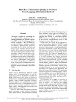
Fig. 2. IR spectra of κ- (A), κ/β- (B), ι/κ- (C), and λ/μ/ν- (D) carrageenans and agar (E).
in the case of gelling carrageenans (Fig. 2A–C), the IR spectrum of agar
(Fig. 2E) contained an absorption band at 932 cm− 1, typical for 3,6anhydrogalactose, as well as an intense absorption band at 890 cm− 1,
belonging to unsulfated 3-linked β-D-galactose (Pereira et al., 2013). The
partial reductive hydrolysis of food agar showed that the polysaccharide
contained only [→3)-β-D-Galp-(1→4)-α-L-AnGalp-(1→] disaccharide
units (agarobiose) without any [→3)-β-D-Galp-(1→4)-α-D-AnGalp-(1→]
disaccharide units (carrabiose). This distinction made classification as
agar possible.
FTIR spectroscopy data were confirmed by NMR spectroscopy anal
ysis, as the carrageenans were subjected to both 1H and 13C NMR ana
lyses. The spectra are presented as Supplementary materials. The two
signals at 103.1 ppm and 96.2 ppm in the anomeric carbon resonance
area of the both spectra of insoluble fractions (C. armatus and
A. flabelliformis) were assigned to C-1 of the 3-linked β-D-galactose res
idue (G4S) and C-1 of the 4-linked 3,6-anhydro-α-D-galactose (DA) of
κ-carrageenan, respectively (Supplementary 1). An intense signal at 92.9
ppm and less intense signal at 95.8 ppm, among the six signals observed
in the anomeric carbon resonance region of the 13C NMR spectrum of the
insoluble fraction from A. flabelliformis, were characteristic of C-1 of the
4-linked 3,6-anhydro-α-D-galactose-2-sulfate (DA2S) of ι-carrageenan
and C-1 of the 4-linked 3,6-anhydro-α-D-galactose (DA’) of β-carra
geenan, respectively (Supplementary 1B). There were poorly resolved
signals at 102.9, 103.1, and 103.2 ppm in the 13C NMR spectrum,
resulting from overlapping C-1 signals of the 3-linked β-D-galactose 4sulfate of the ι- (G4S’) and κ-carrageenans (G4S) and the 3-linked β-Dgalactose (G) of β-carrageenan, respectively (Usov & Shashkov, 1985).
The NMR spectroscopy data indicate that the content of the ι-type
disaccharide units in the polymer chain of ι/κ-carrageenan was pre
dominant. The ratio of ι- and κ-units was 2:1, and β-carrageenan was
present in minor quantities.
Well-resolved 1H and 13C NMR spectra of soluble fraction from
C. armatus could not be recorded, even at high temperature, because of
high polysaccharide viscosity and, probably, disordered macromolec
ular organization. However, we were able to identify some of the main
signals by comparing our spectra with literature data (Van de Velde
et al., 2002). There were four signals in the anomeric carbon resonance
area of the 13C NMR spectrum (Supplementary 2). Signals at 103.3 and
91.6 ppm were attributed to C-1 of 3-linked β-D-galactose 2-sulfate
(G2S-1) and 4-linked α-D-galactose 2,6-disulfate (D2S,6S-1), respec
tively, of λ-carrageenan (Van de Velde et al., 2002). The broad signal at
105.3 ppm was likely related to 3-linked β-D-galactose 4-sulfate of μ(G4S- 1) and ν- (G4S’-1) carrageenans (biosynthetic precursors of κ- and
ι-carrageenans, respectively). At the same time, a wide signal at 98.6
ppm was attributed to 4-linked α-D-galactose 6-sulfate (D6S-1) and
α-D-galactose 2,6-disulfate of μ- (D6S-1) and ν- (D2S,6S’-1) carra
geenans, respectively (Van de Velde et al., 2002). In addition, the
intense signal at 61.6 ppm in the upfield region of the 13C NMR spectrum
5
E.V. Sokolova et al.
Carbohydrate Polymers 254 (2021) 117251
was characteristic of the C-6 of 3-linked β-D-galactose of λ- (G2S-6), μ(G4S-6), and ν- (G4S’-6) carrageenans. A wide, poorly resolved signal at
69.3 ppm corresponded to 4-linked α-galactose sulfated at C-6 (D2S,6S,
D6S, D2S,6S’). At the same time, weak signal at 64 ppm can be attrib
uted to C-4 of 3-linked β-D-galactose 2-sulfate (G2S-4) of λ-carrageenan.
The 13C NMR data were consistent with the 1H NMR (not shown). There
was a broad signal at 5.52–5.59 ppm in the α-anomeric proton resonance
area, which was attributed to H-1 of the 4-linked α-D-galactose 2,6-disul
fate of λ- (5.59 ppm) and ν- (5.52 ppm) carrageenans. In addition, a
weak signal at 5.26 ppm in the spectrum suggested the presence of
μ-carrageenan (H-1 of 4-linked α-D-galactose 6-sulfate). Thus, the
non-gelling polysaccharide from C. armatus was a mixture of λ- μ- and
ν-carrageenans.
The 1H NMR spectrum of κ/β-carrageenan (Supplementary 3) con
tained four signals in the anomeric proton resonance area. The signals at
5.09 and 5.11 ppm were characteristic of the H-1 of 4-linked 3,6anhydro-α-D-galactose of β- (DA’) and κ-carrageenans (DA), respec
tively. The signals at 4.62 and 4.64 ppm were assigned to the H-1 of 3linked β-D-galactose (G) and 3-linked β-D-galactose 4-sulfate (G4S) of the
β- and κ-carrageenans, respectively (Kolender & Matulewicz, 2004; Van
de Velde et al., 2002).
3.1. The influence of red algal galactans on total functional complement
activation
The influence of the investigated galactans on binding C3 comple
ment component to plate wells coated with LPS was studied by an ELISAbased method. Results displayed in Fig. 3A revealed that, in general, the
investigated polysaccharides inhibited C3 binding to plate wells coated
with LPS. This capacity was dependent on the polysaccharide sample
and concentration. Heparin was the most potent inhibiting agent in this
assay, almost independent of concentration in the range of values in the
experiment, and the decrease by its action reached 59–68%, relative to
the negative control. Among the galactans, their effect decreased, as
follows: λ/μ/ν > κ/β > κ > ι/κ > agar. More precisely, at the highest
concentration (2 mg mL− 1), all carrageenans, on average, reduced C3
binding by 70 %, just like heparin, and agar and agarose by 40 and 20 %,
respectively. By lowering concentrations, the investigated samples, un
like heparin, gradually lost their inhibiting potential.
Regarding C4 binding to the mannan-coated surface (Fig. 3B), the
investigated samples were affected less efficiently. Heparin, again,
reduced C4 binding to the mannan-coated surface, depending on con
centration (35 % decrease at the highest concentration of 2 mg mL− 1).
The most active samples were λ/μ/ν- and κ-carrageenans, inhibiting C4
binding to mannan, on average, by 30 % within the concentration range
used in this test. The hybrid carrageenan structures of κ/β and ι/κ were
almost inactive. The wells containing agar and agarose gellified in C4
binding to mannan- and antibody-coated surfaces.
Another tendency was observed when we studied C4 binding to
antibody-coated surfaces (Fig. 3C). Heparin illustrated inhibiting po
tential at the two highest concentrations (0.2 and 2 mg mL− 1) by about
25–40 % and was inert at lower concentrations. Carrageenans stimu
lated C4 binding, especially at high concentrations. Of the poly
saccharides, κ/β- and κ-carrageenans’ actions at the highest
concentrations resulted in the most pronounced activity—a four-fold
increase in C4 binding to antibody-coated surfaces. λ/μ/ν-Carrageenan
was the least active one (two-fold increase at the highest concentration),
and ι/κ-type, independent of concentration, showed a two-fold increase
relative to the negative control (100 %).
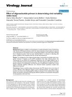
Fig. 3. Binding of C3 and C4 complement components to well surfaces coated
with E. coli LPS (A), human IgG (B), or S. cerevisiae mannan (C) in the presence
of carrageenan (λ/μ/ν-, κ-, κ/β-, and ι/κ-types) and agar (agar, agarose) groups
in varying concentrations. All concentrations are expressed in final values, as %
change in C3 or C4 concentration on the well surface relative to the vehicle
control (100%) in three replicates from two independent experiments. The
asterisk (*) indicates significant differences <0.05 by one-way ANOVA followed
by Tukey post hoc comparisons for the highest sample concentration value.
Table 2
Measured concentration of total serum IgM in the presence
of polysaccharides.
Sample
%
control
heparin
λ/μ/ν-carrageenan
κ-carrageenan
κ/β-carrageenan
ι/κ-carrageenan
100.0 ± 2.8
101.4 ± 3.9
91.2 ± 2.1*
82.4 ± 2.8*
81.0 ± 1.8*
76.8 ± 1.6*
The asterisk (*) indicates significant differences <0.05
relative to vehicle control (100 %).
3.2. Binding of red algal polysaccharides to serum immunoglobulins
The ability of the investigated samples to affect concentrations of the
total IgG, IgA, IgM of human serum was analyzed. Table 2 contains data
on total IgM measured in serum in the presence of the investigated
samples. The results revealed that the galactans were able to affect total
6
E.V. Sokolova et al.
Carbohydrate Polymers 254 (2021) 117251
serum IgM and insignificantly other types of serum Igs. The strongest
binding towards total serum IgM was observed for gelling carrageenans.
discrimination. The major leading factor in reading cell surface as self
is Factor H which fixates on surface polyanions (glycoproteins con
taining sialic acid residues, heparan sulfate, and other glycosamino
glycans) and moves the ongoing balance of complement
activation-inactivation on cells towards inactivation (Collins & Troe
berg, 2019; Langford-Smith, Day, Bishop, & Clark, 2015; Pangburn
et al., 2009). Our results demonstrated that, for C3 binding to
LPS-coated surfaces, i.e. without polyanions necessary for Factor H, the
galactans inhibited this process, although with less efficacy than heparin
(Fig. 3A). Influence of the sulfated galactans in C3 binding and visible
dependence on the sulfation degree allows us to assume they function as
surface polyanions. Some degree of C3 binding inhibition to LPS-coated
surfaces by the non-sulfated galactan agarose might be explained with
agarose’s ability to directly bind C3 but not stabilize Factor H on the
surface (Hetland & Eskhland, 1986). Thus, sulfated red algal galactans
should be capable of decreasing the inflammatory reaction by
strengthening surface readings as less non-self in the alternative
pathway and amplification loop because of their polyanion nature.
Factor H is not significant in the case of mannan-driven complement
attack, however, our data illustrated that carrageenans still can provide
cell surface protection but with far less efficacy than for C3 binding
(Fig. 3B). The only exception was observed for the most sulfated nongelling λ/μ/ν-type carrageenan sample, which had a comparable to
heparin effect. The C4 deposition on wells used in the assay reflected the
activity of serine protease circulating in complex with MBL (MBL-asso
ciated serine protease-2, MASP-2) (Petersen et al., 2001). Hence, car
rageenans probably inhibit MBL and/or MASP-2, up-regulating the
lectin pathway, and facilitate Factor H, down-regulating the alternative
pathway and amplification loop. The lectin pathway has an extensive
scope of therapeutic potential, especially in models of myocardial or
gastrointestinal ischemia-reperfusion injury. However, it has only been
actively studied for the last 10 years (Ricklin et al., 2018), so hypothe
sizing possible applications of algal sulfated polysaccharides at this
moment is difficult.
When wells are coated with antibodies, the classical pathway be
comes a leading force, allowing recognition of immune complexes by
C1q cleaving upon recognition into the homologous to MASP proteases
(C1r and C1s; Petersen et al., 2001). Our results revealed that, in gen
eral, carrageenans, contrary to heparin, augmented this pathway of
complement activation (Fig. 3C), which corresponds to the hemolytic
complement studies (Baker et al., 1986). In our experiment without
cells, the increasing C4 deposition onto well plates in the presence of
carrageenans must be connected with the increase in amount of anti
body during the incubation step with serum and samples. Blood serum
contains substantial amounts of an interesting variety of antibodies,
called natural/spontaneous antibodies (NA). The most prominent
functions of NAs are homeostatic (broadly reactive against self-antigens,
tumor-specific patterns, cell-surface-exposed structures of necrotic cells,
or plasma proteins leaking destroyed cells, etc.) and protective against
infections spreading hematologically. However, for protection, they act
as recognition proteins, like MBL and C-reactive protein; evoke strong
complement-mediated inflammatory response; and are capable of
recognizing evolutionarily fixed epitopes in foreign antigens (Holodick,
Rodríguez-Zhurbenko, & Hern´
andez, 2017; Lutz, Binder, & Kaveri,
2009; Ochsenbein & Zinkernagel, 2000). The most abundant NA in
humans (~1 % of the total serum immunoglobulins with major reactive
type being of IgG and especially IgM variety; McMorrow, Comrack,
Sachs, & DerSimonian, 1997) is directed against ‘α-gal epitope’ with the
structure α-Galp-(1→3)-β-Galp-(1→4)-GlcpNAc-R (2018, Galili, 2013,
2020). The investigated polysaccharides could bind NA of human serum
(EFSA Panel on Food Additives & Nutrient Sources added to Food (ANS)
et al., 2018) because the →4)-α-Galp-(1→3)-β-Galp-(1→ portion of the
xenoantigen is a disaccharide repeating unit of a carrageenan chain.
Structural features of the galactans in our study also matter because
polysaccharides containing 3,6-anhydrogalactose (κ, κ/β, ι/κ) were
more potent activators compared to the non-gelling type. This property
3.3. Influence of red algal galactans on the extrinsic protease pathway of
complement activation induced by plasmin
The effect of red algal polysaccharides on the extrinsic protease
pathway of complement by activating human serum with a component
of a coagulation system (plasmin) was studied (Fig. 4). The measure of
serum activation was determined by the concentration of a cleaved C5
component—C5a—in fluid phase by means of an ELISA method. Fig. 4
contains the control- and control + for human serum with and without
plasmin, showing activation by 50 % (from 43 to 62 ng mL− 1). Heparin
was inactive in this test, while the investigated samples illustrated some
degree of inhibition at the highest concentration, with λ/μ/ν- and
κ/β-carrageenans being the most impressive (almost to the level of
control-).
4. Discussion
As a dietary fiber, carrageenans encounter in human organisms only
the gastrointestinal tract (EFSA Panel on Food Additives & Nutrient
Sources added to Food (ANS) et al., 2018). To dampen the immune
response elicited by the presence of luminal antigens appears to be one
the main functions of the mucosal immunity (Brownlee, Dettmar, Stru
gala, & Pearson, 2006; Cummings et al., 2004). As a result the com
plement role there is dictated by location and is heavily inclined to
opsonization but not lysis of invading bacteria. In other words, the
complement composition is limited to C4, C3, factor B, and C1q, with
notably low or absent complement C5–C9 proteins composing mem
brane attack complex for cell lysis (Sina, Kemper, & Derer, 2018). The
experimental design of complement’s functional activity in the current
article was focused on the enzyme immunoassay method of C3 or C4
tethering to a suitable solid phase (Harboe, Thorgersen, & Mollnes,
2011). Heparin was used as a reference here because of its capacity to
inhibit complement (Weiler, Edens, Linhardt, & Kapelanski, 1992) and
because carrageenan’s ability to act in a similar manner to heparin, gives
a promising direction in the glycomimetic drug field (Buck et al., 2006;
Groult et al., 2019; Poupard et al., 2017).
All cell surfaces are coated with a layer of glycocalyx composed from
glycans in many different molecular forms (Ernst & Magnani, 2009).
Differences in cell surface glycans can serve as markers of a cell’s
identity (e.g. developmental state, tissue type, self versus non-self
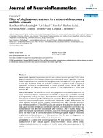
Fig. 4. C5a concentration in serum activated with plasmin (0.5 U mL− 1, final
value) in the presence of red algal galactans: carrageenan (λ/μ/ν-, κ-, κ/β-, and
ι/κ-types) and agar (agar, agarose) groups in varying concentrations. All con
centrations are expressed as final values. Control- is non-activated serum, and
control + is serum activated with plasmin. The results are expressed C5a con
centration (ng mL-1) from three replicates of two independent experiments. The
asterisk (*) indicates significant differences <0.05 by one-way ANOVA followed
by Tukey post hoc comparisons for the highest sample concentration value.
7
E.V. Sokolova et al.
Carbohydrate Polymers 254 (2021) 117251
of carrageenans to bind NA has been tested in our study (Table 2). The
data suggested an ability of carrageenans to connect with antigen
binding parts of total IgM of human serum leading as a result to a
decrease in number of IgM reacting with mAb against light chains of
immunoglobulins. The gelling types more actively bound IgM, corrob
orating the more substantial C4 binding to antibodies-coated surface in
the presence of carrageenans. Drawing conclusions about the degree of
influence by structural characteristics, like varying sulfate positions, was
difficult but could be connected with NAs’ property of polyreactivity,
accompanied with a degree of specificity (Bovin et al., 2012). The
former is for homeostatic functions and the latter mostly for protective
functions. The mucous layer of the gastrointestinal tract contains ho
meostatic polyreactive NAs, mostly of the IgA variety, with an innate
role to coat and contain the resident commensal microorganisms and
provide protection against detrimental ones (Bunker et al., 2017; Wells
et al., 2017). No reports of allergic reaction to carrageenan as a food
ingredient have been registered in humans (EFSA Panel on Food Addi
tives & Nutrient Sources added to Food (ANS) et al., 2018). However,
this complement activation in the presence of anti-Gal NAs has been
successfully explored in the accelerated wound healing model by
application of α-gal nanoparticles (Galili, 2013). Carrageenans, in turn,
have a long history of topical administration in tissue engineering and
wound healing (Ditta et al., 2020) for a variety of bioengineering ap
plications, and antiviral microbicides hydrogels (Yegappan, Selvapri
thiviraj, Amirthalingam, & Jayakumar, 2018) or other compounds. One
of the mechanisms of the antiviral action of carrageenans is due to their
negative charge which bind virus positively charged glycoproteins
responsible for attachment to a host cell (Damonte, Matulewicz, &
Cerezo, 2004). At the same time, anti-Gal-mediated neutralization and
complement-mediated lysis of the viruses after incubation of the viruses
expressing α-gal epitopes in human serum or, with purified anti-Gal
antibody had been shown, but no such effects for viruses lacking α-gal
epitopes (Galili, 2018).
With topical administration of red algal polysaccharides, one might
also consider useful knowledge of their influence on complement
through other homeostatic cascades by the ‘extrinsic protease pathway,’
encompassing complement interaction with the coagulation cascade and
fibrinolytic proteins. This interaction unlike canonical complement
activation is believed to take place on several host cell types with normal
surface landscapes, like platelets and endothelial cells, activated by
complement fragments (e.g. C4a protein released from C4 during acti
vation of the classical and lectin pathways) (Ricklin, 2018). Our very
simple experiment, without cells and surfaces imitating them, allowed
us to extricate onlygalactans’ effect on the reaction of complement
activation in solution by a fibrinolytic protein, plasmin (Fig. 4), the
strongest activator of C5 (Amara et al., 2010). Previously, heparin was
determined to be inert to plasmin (Andrade-Gordon & Strickland, 1986);
our data showed that heparin is also inert to plasmin-induced comple
ment activation in serum (Fig. 4). However, red algal polysaccharides
slightly retarded this process with little dependence on structural
characteristics and sulfate content, but two carrageenans with and
without κ-units at the highest concentration almost abolished C5
activation.
in extrinsic protease complement activation.
CRediT authorship contribution statement
E.V. Sokolova: Conceptualization, Methodology (Biological),
Funding acquisition, Writing - original draft, Investigation. A.O. Krav
chenko: Methodology (Chemical), Writing - review & editing (Chemical
part). N.V. Sergeeva: Resources. A.I. Kalinovsky: Methodology (NMR
spectroscopy data). V.P. Glazunov: Methodology (IR-spectroscopy
data). L.N. Bogdanovich: Resources. I.M. Yermak: Writing - original
draft (Chemical part), Writing - review & editing.
Acknowledgements
This study was supported by the Russian Science Foundation (RSF)
Grant 20-74-00006. The study was carried out on equipment from the
Collective Facilities Center, “The Far Eastern Center for Structural Mo
lecular Research (NMR/MS) PIBOC FEB RAS.” Ekaterina Sokolova
would like to express to PJL, an amazing person and no less amazing
scientist, her deepest respect and admiration.
Appendix A. Supplementary data
Supplementary material related to this article can be found, in the
online version, at doi: />References
Amara, U., Flierl, M. A., Rittirsch, D., Klos, A., Chen, H., Acker, B., et al. (2010).
Molecular intercommunication between the complement and coagulation systems.
The Journal of Immunology, 185(9), 5628–5636.
Andrade-Gordon, P., & Strickland, S. (1986). Interaction of heparin with plasminogen
activators and plasminogen: Effects on the activation of plasminogen. Biochemistry,
25(14), 4033–4040.
Baines, A. C., & Brodsky, R. A. (2017). Complementopathies. Blood Reviews, 31(4),
213–223.
Baker, K. C., Nicklin, S., & Miller, K. (1986). The role of carrageenan in complement
activation. Food and Chemical Toxicology, 24(9), 891–895.
Barabanova, A. O., Yermak, I. M., Glazunov, V. P., Isakov, V. V., Titlyanov, E. A., &
Solov’eva, T. F. (2005). Comparative study of carrageenans from reproductive and
sterile forms of Tichocarpus crinitus (Gmel.) Rupr (Rhodophyta, Tichocarpaceae).
Biochemistry (Moscow), 70(3), 350–356.
Barnum, S. R. (2017). Complement: A primer for the coming therapeutic revolution.
Pharmacology & Therapeutics, 172, 63–72.
Blaum, B. S. (2017). The lectin self of complement factor H. Current Opinion in Structural
Biology, 44, 111–118.
Bovin, N., Obukhova, P., Shilova, N., Rapoport, E., Popova, I., Navakouski, M., et al.
(2012). Repertoire of human natural anti-glycan immunoglobulins. Do we have
auto-antibodies? Biochimica et Biophysica Acta (BBA)-General Subjects, 1820(9),
1373–1382.
Brownlee, I. A., Dettmar, P. W., Strugala, V., & Pearson, J. P. (2006). The interaction of
dietary fibres with the colon. Current Nutrition and Food Science, 2(3), 243–264.
Buck, C. B., Thompson, C. D., Roberts, J. N., Müller, M., Lowy, D. R., & Schiller, J. T.
(2006). Carrageenan is a potent inhibitor of papillomavirus infection. PLoS
Pathogens, 2(7).
Bunker, J. J., Erickson, S. A., Flynn, T. M., Henry, C., Koval, J. C., Meisel, M., et al.
(2017). Natural polyreactive IgA antibodies coat the intestinal microbiota. Science,
358(6361).
Chiovitti, A., Bacic, A., Craik, D. J., Kraft, G. T., Liao, M. L., Falshaw, R., et al. (1998).
A pyruvated carrageenan from Australian specimens of the red alga Sarconema
filiforme. Carbohydrate Research, 310(1-2), 77–83.
Collins, L. E., & Troeberg, L. (2019). Heparan sulfate as a regulator of inflammation and
immunity. Journal of Leukocyte Biology, 105(1), 81–92.
Cosenza, V. A., Navarro, D. A., Ponce, N. M., & Stortz, C. A. (2017). Seaweed
polysaccharides: Structure and applications. In S. N. Goyanes, & N. B. D’Accorso
(Eds.), Industrial applications of renewable biomass products (pp. 75–116). Cham:
Springer.
Craigie, J. S. (1990). Cell wall. In K. M. Cole, & R. G. Sheath (Eds.), Biology of red algae
(pp. 221–258). New York: Cambridge University Press.
Cummings, J. H., Antoine, J. M., Azpiroz, F., Bourdet-Sicard, R., Brandtzaeg, P.,
Calder, P. C., et al. (2004). PASSCLAIM 1—Gut health and immunity. European
Journal of Nutrition, 43(2), ii118–ii173.
Damgaard, C., Reinholdt, J., Palarasah, Y., Enevold, C., Nielsen, C., Brimnes, M. K., et al.
(2017). In vitro complement activation, adherence to red blood cells and induction of
mononuclear cell cytokine production by four strains of Aggregatibacter
actinomycetemcomitans with different fimbriation and expression of leukotoxin.
Journal of Periodontal Research, 52(3), 485–496.
5. Conclusion
In summary, the red algal sulfated polysaccharides affected the
complement system and its interplay with fibrinolytic components.
These substances have the potential to participate in cell surface biology
by inhibiting C3 binding to the surface in a similar fashion as cell reg
ulators of the glycosaminoglycan family, depending on sulfation degree.
Sulfation degree was also important in carrageenans’ capacity to reduce
C4 binding in lectin complement activation. However, C4 binding in the
classical complement was considerably activated in the presence of
carrageenans with 3,6-anhydrogalactose. No structural characteristics
apparently mattered in ameliorating C5 cleavage by plasmin occurring
8
E.V. Sokolova et al.
Carbohydrate Polymers 254 (2021) 117251
Damonte, E. B., Matulewicz, M. C., & Cerezo, A. S. (2004). Sulfated seaweed
polysaccharides as antiviral agents. Current Medicinal Chemistry, 11(18), 2399–2419.
Davies, G. E. (1965). Inhibition of complement by carrageenin: Mode of action, effect on
allergic reactions and on complement of various species. Immunology, 8(3), 291.
Ditta, L. A., Rao, E., Provenzano, F., S´
anchez, J. L., Santonocito, R., Passantino, R., et al.
(2020). Agarose/κ-carrageenan-based hydrogel film enriched with natural plant
extracts for the treatment of cutaneous wounds. International Journal of Biological
Macromolecules, 164, 2818–2830. In press.
Dodgson, K. S., & Price, R. G. (1962). A note on the determination of the ester sulphate
content of sulphated polysaccharides. Journal of Biochemistry, 84, 106110.
dos Santos-Fidencio, G. C., Gonỗalves, A. G., Noseda, M. D., Duarte, M. E. R., &
Ducatti, D. R. (2019). Effects of carboxyl group on the anticoagulant activity of
oxidized carrageenans. Carbohydrate Polymers, 214, 286–293.
EFSA Panel on Food Additives and Nutrient Sources added to Food (ANS), Younes, M.,
Aggett, P., Aguilar, F., Crebelli, R., Filipiˇc, M., … Kuhnle, G. G. (2018). Re-evaluation
of carrageenan (E 407) and processed Eucheuma seaweed (E 407a) as food additives.
EFSA Journal, 16(4), Article e05238.
Ernst, B., & Magnani, J. L. (2009). From carbohydrate leads to glycomimetic drugs.
Nature Reviews Drug Discovery, 8(8), 661–677.
Estevez, J. M., Ciancia, M., & Cerezo, A. S. (2004). The system of galactans of the red
seaweed, Kappaphycus alvarezii, with emphasis on its minor constituents.
Carbohydrate Research, 339(15), 2575–2592.
Galili, U. (2013). Anti-Gal: An abundant human natural antibody of multiple
pathogeneses and clinical benefits. Immunology, 140(1), 1–11.
Galili, U. (2018). Why do we produce anti-gal: Evolutionary appearance of anti-gal in old
world primates. The Natural Anti-Gal Antibody As Foe Turned Friend In Medicine, 23.
/>Galili, U. (2020). Human natural antibodies to mammalian carbohydrate antigens as
unsung heroes protecting against past, present, and future viral infections.
Antibodies, 9(2), 25.
Groult, H., Cousin, R., Chot-Plassot, C., Maura, M., Bridiau, N., Piot, J. M., et al. (2019).
Λ-carrageenan oligosaccharides of distinct anti-heparanase and anticoagulant
activities inhibit MDA-MB-231 breast cancer cell migration. Marine Drugs, 17(3),
140.
Harboe, M., Thorgersen, E. B., & Mollnes, T. E. (2011). Advances in assay of complement
function and activation. Advanced Drug Delivery Reviews, 63(12), 976–987.
Harrison, R. A. (2018). The properdin pathway: An “alternative activation pathway” or a
“critical amplification loop” for C3 and C5 activation?. January. In Seminars in
Immunopathology (Vol. 40, pp. 15–35). Berlin Heidelberg: Springer. No. 1.
Hetland, G., & Eskhland, T. (1986). Formation of the functional alternative pathway of
complement by human monocytes in vitro as demonstrated by phagocytosis of
agarose beads. Scandinavian Journal of Immunology, 23(3), 301–308.
Holodick, N. E., Rodríguez-Zhurbenko, N., & Hern´
andez, A. M. (2017). Defining natural
antibodies. Frontiers in Immunology, 8, 872.
Kalitnik, A. A., Marcov, P. A., Anastyuk, S. D., Byankina Barabanova, A. O.,
Glazunov, V. P., Popov, S. V., Ovodov, Y. S., & Yermak, I. M. (2015). Gelling
polysaccharide from Chondrus armatus and its oligosaccharides: The structural
peculiarities and anti-inflammatory activity. Carbohydrate Polymers, 115, 768–775.
Knutsen, S. H., Myslabodski, D. E., Larsen, B., & Usov, A. I. (1994). A modified system of
nomenclature for red algal galactans. Botanica Marina, 37(2), 163–170.
Kolender, A. A., & Matulewicz, M. C. (2004). Desulfation of sulfated galactans with
chlorotrimethylsilane. Characterization of β-carrageenan by 1H NMR spectroscopy.
Carbohydrate Research, 339, 1619–1629.
Koutsaviti, A., Ioannou, E., & Roussis, V. (2018). Bioactive seaweed substances. Bioactive
seaweeds for food applications (pp. 25–52). Academic Press.
Kravchenko, A. O., Anastyuk, S. D., Sokolova, E. V., Isakov, V. V., Glazunov, V. P.,
Helbert, W., et al. (2016). Structural analysis and cytokine-induced activity of
gelling sulfated polysaccharide from the cystocarpic plants of Ahnfeltiopsis
flabelliformis. Carbohydrate Polymers, 151, 523–534.
Kravchenko, A. O., Anastyuk, S. D., Glazunov, V. P., Sokolova, E. V., Isakov, V. V., &
Yermak, I. M. (2020). Structural characteristics of carrageenans of red alga
Mastocarpus pacificus from Sea of Japan. Carbohydrate Polymers, 229, Article 115518.
Lachmann, P. J. (2010). Preparing serum for functional complement assays. Journal of
Immunological Methods, 352(1–2), 195–197.
Lachmann, P. J. (2018). Looking back on the alternative complement pathway.
Immunobiology, 223(8-9), 519–523.
Lahaye, M. (2001). Developments on gelling algal galactans, their structure and physicochemistry. Journal of Applied Phycology, 13(2), 173–184.
Langford-Smith, A., Day, A. J., Bishop, P. N., & Clark, S. J. (2015). Complementing the
sugar code: Role of GAGs and sialic acid in complement regulation. Frontiers in
Immunology, 6, 25.
Lowry, O. H., Rosebrough, N. J., Farr, A. L., & Randall, R. J. (1951). Protein
measurement with the Folin phenol reagent. The Journal of Biological Chemistry, 193,
265–275.
Lubbers, R., Van Essen, M. F., Van Kooten, C., & Trouw, L. A. (2017). Production of
complement components by cells of the immune system. Clinical and Experimental
Immunology, 188(2), 183–194.
Lutz, H. U., Binder, C. J., & Kaveri, S. (2009). Naturally occurring auto-antibodies in
homeostasis and disease. Trends in Immunology, 30(1), 43–51.
McMorrow, I. M., Comrack, C. A., Sachs, D. H., & DerSimonian, H. (1997). Heterogeneity
of human anti-pig natural antibodies cross-reactive with the gal (α1,3) galactose
epitope. Transplantation, 64(3), 501–510.
Miller, I. J., & Blunt, J. W. (2000). New 13C NMR methods for determining the structure
of algal polysaccharides, Part 1. The effect of substitution on the chemical shifts of
simple Diad galactans. Botanica Marina, 43, 239–250.
Ochsenbein, A. F., & Zinkernagel, R. M. (2000). Natural antibodies and complement link
innate and acquired immunity. Immunology Today, 21(12), 624–630.
Opoku, G., Qiu, X., & Doctor, V. (2006). Effect of oversulfation on the chemical and
biological properties of kappa carrageenan. Carbohydrate Polymers, 65(2), 134–138.
Pangburn, M. K., Rawal, N., Cortes, C., Alam, M. N., Ferreira, V. P., & Atkinson, M. A.
(2009). Polyanion-induced self-association of complement factor H. The Journal of
Immunology, 182(2), 1061–1068.
Pereira, L. (2018). Biological and therapeutic properties of the seaweed polysaccharides.
International Biology Review, 2(2), 1–50.
Pereira, L., Amado, A. M., Critchley, A. T., Van de Velde, F., & Ribeiro-Claro, P. J. A.
(2009). Identification of selected seaweed polysaccharides (phycocolloids) by
vibrational spectroscopy (FTIR-ATR and RT-Raman). Food Hydrocolloids, 30, 1–7.
Pereira, L., & Critchley, A. T. (2020). The COVID 19 novel coronavirus pandemic 2020:
seaweeds to the rescue? Why does substantial, supporting research about the
antiviral properties of seaweed polysaccharides seem to go unrecognized by the
pharmaceutical community in these desperate times? Journal of Applied Phycology,
32, 1875–1877. />Pereira, L., Gheda, S. F., & Ribeiro-Claro, P. J. A. (2013). Analysis by vibrational
spectroscopy of seaweed polysaccharides with potential use in food, pharmaceutical,
and cosmetic industries. International Journal of Carbohydrate Chemistry, 22, 7.
Perestenko, L. P. (1994). The red algae of the far eastern seas of Russia. St. Petersburg:
Nauka [In Russian].
Petersen, S. V., Thiel, S., Jensen, L., Steffensen, R., & Jensenius, J. C. (2001). An assay for
the Mannan-binding lectin pathway of complement activation. Journal of
Immunological Methods, 257(1-2), 107–116.
Poupard, N., Groult, H., Bodin, J., Bridiau, N., Bordenave-Juchereau, S., Sannier, F., et al.
(2017). Production of heparin and λ-carrageenan anti-heparanase derivatives using a
combination of physicochemical depolymerization and glycol splitting. Carbohydrate
Polymers, 166, 156–165.
Renn, D. W., Santos, G. A., Dumont, L. E., Parent, C. A., Stanley, N. F., Stancioff, D. J.,
et al. (1993). β-Carrageenan: Isolation and characterization. Carbohydrate Polymers,
22, 247–252.
Ricklin, D., Mastellos, D. C., Reis, E. S., & Lambris, J. D. (2018). The Renaissance of
complement therapeutics. Nature Reviews Nephrology, 14(1), 26.
Rochas, C., Rinaudo, M., & Landry, S. (1990). Role of the molecular weight on the
mechanical properties of kappa–carrageenan gels. Carbohydrate Polymers, 12,
255–266.
Sina, C., Kemper, C., & Derer, S. (2018). The intestinal complement system in
inflammatory bowel disease: Shaping intestinal barrier function. In Seminars in
Immunology, 37 pp. 66–73). Academic Press.
Stortz, C. A., & Cerezo, A. S. (2002). Novel findings in carrageenans, agaroids and
“hybrid” red seaweed galactans. Current Topics in Phytochemistry, 4, 121–134.
Usov, A. I. (1998). Structural analysis of red seaweed galactans of agar and carrageenan
groups. Food Hydrocolloids, 12(3), 301–308.
Usov, A. I., & Elashvili, M. Y. (1991). Quantitative-determination of 3,6-anhydrogalac
tose derivatives and specific fragmentation of the red algal galactans under reductive
hydrolysis conditions. Bioorganicheskaya Khimiya, 17(6), 839–848.
Usov, A. I., & Shashkov, A. S. (1985). Polysaccharides of algae XXXIV: Detection of iotacarrageenan in Phyllophora brodittei (Tum.) J Ag. (Rhodophyta) using 13C-NMR
spectroscopy. Botanica Marina, 28, 367–373.
Van de Velde, F., Knutsen, S. H., Usov, A. I., Rollema, H. S., & Cerezo, A. S. (2002). 1H
and 13C high resolution NMR spectroscopy of carrageenans: Application in research
and industry. Trends in Food Science & Technology, 13(3), 73–92.
Weiler, J. M., Edens, R. E., Linhardt, R. J., & Kapelanski, D. P. (1992). Heparin and
modified heparin inhibit complement activation in vivo. The Journal of Immunology,
148(10), 3210–3215.
Wells, J. M., Brummer, R. J., Derrien, M., MacDonald, T. T., Troost, F., Cani, P. D., et al.
(2017). Homeostasis of the gut barrier and potential biomarkers. American Journal of
Physiology-Gastrointestinal and Liver Physiology, 312(3), G171–G193.
Yegappan, R., Selvaprithiviraj, V., Amirthalingam, S., & Jayakumar, R. (2018).
Carrageenan based hydrogels for drug delivery, tissue engineering and wound
healing. Carbohydrate Polymers, 198, 385–400.
Yermak, I. M., Kim, Y. H., Titlynov, E. A., Isakov, V. V., & Solov’eva, T. F. (1999).
Chemical structure and gel properties of carrageenans from algae belonging to the
Gigartinaceae and Tichocarpaceae, collected from the Russian pacific coast. In
J. M. Kain, M. T. Brown, & M. Lahaye (Eds.), Sixteenth International seaweed
symposium (pp. 555–562). Dordrecht: Springer.
9