Advanced characterization of regioselectively substituted methylcellulose model compounds by DNP enhanced solid-state NMR spectroscopy
Bạn đang xem bản rút gọn của tài liệu. Xem và tải ngay bản đầy đủ của tài liệu tại đây (2.28 MB, 9 trang )
Carbohydrate Polymers 262 (2021) 117944
Contents lists available at ScienceDirect
Carbohydrate Polymers
journal homepage: www.elsevier.com/locate/carbpol
Advanced characterization of regioselectively substituted methylcellulose
model compounds by DNP enhanced solid-state NMR spectroscopy
ărthe Jakobi b, Michel Bardet a, c,
Pierrick Berruyer a, Martin Gericke b, Pinelopi Moutzouri a, Do
d
e
b,
Leif Karlson , Staffan Schantz , Thomas Heinze *, Lyndon Emsley a, *
a
Institut des Sciences et Ingénierie Chimiques, Ecole Polytechnique Fédérale de Lausanne (EPFL), CH-1015 Lausanne, Switzerland
Institute of Organic Chemistry and Macromolecular Chemistry, Friedrich Schiller University of Jena, Centre of Excellence for Polysaccharide Research, Humboldtstraße
10, D-07743 Jena, Germany
c
Univ. Grenoble Alpes, CEA, IRIG-MEM, Laboratoire de Résonance Magnétique, Grenoble 38000, France
d
Nouryon Functional Chemicals AB, SE-444 31 Stenungsund, Sweden
e
Oral Product Development, Pharmaceutical Technology & Development, Operations, AstraZeneca, Gothenburg, Sweden
b
A R T I C L E I N F O
A B S T R A C T
Keywords:
Cellulose ethers
Regioselectivity
Methylcellulose
Structure characterization
Solid state NMR
DNP enhancement
Dynamic Nuclear Polarization MAS NMR is introduced to characterize model methylcellulose ether compounds
at natural isotopic abundance. In particular an approach is provided to determine the position of the methyl ether
group within the repeating unit. Specifically, natural abundance 13C-13C correlation experiments are used to
characterize model 3-O-methylcellulose and 2,3-O-dimethylcellulose, and identify changes in chemical shifts
with respect to native cellulose. We also probe the use of through space connectivity to the closest carbons to the
CH3 to identify the substitution site on the cellulose ether. To this end, a series of methylcellulose ethers was
prepared by a multistep synthesis approach. Key intermediates in these reactions were 2,6-O-diprotected thex
yldimethylsilyl (TDMS) cellulose and 6-O-monoprotected TDMS cellulose methylated under homogeneous con
ditions. The products had degrees of substitution of 0.99 (3-O-methylcellulose) and 2.03 (2,3-Odimethylcellulose) with exclusively regioselective substitution. The approaches developed here will allow
characterization of the substitution patterns in cellulose ethers.
1. Introduction
Cellulose ethers are widely exploited as additives in a broad range of
applications, such as pharmaceutical formulations (Arca et al., 2018; Li,
Martini, Ford, & Roberts, 2005), paint and cement based building for
mulations (Karlson, Joabsson, & Thuresson, 2000; Patural et al., 2011),
food (Young, 2014), and drilling and mining processes (Wever, Pic
chioni, & Broekhuis, 2011). The overall molecular structure of cellulose
ethers determines the physical properties of their products, which in
return affect their efficacy. Some of the most important characteristics of
commercial cellulose ethers in this perspective include the average
molecular weight and the overall degree of substitution (DS) of the ether
substituents. Moreover, the distribution of substituents within the
repeating unit and the heterogeneity between the surface and bulk of the
material are key parameters. These characteristics can, to some extent,
be tuned by reaction conditions (molecular weight of the starting cel
lulose, amount of reagents, composition of the reaction medium, time,
temperature, etc.).
The cellulose repeating unit features three different hydroxyl groups
that can be functionalized, and therefore cellulose ethers with the same
DS might possess a different substitution pattern, e.g. 2-O-, 3-O-, 6-Osubstitution or non-regioselective substitution (Mischnick, 2018). For
several cellulose alkyl ethers, it has been reported that the distribution of
substituents within the repeating unit is an important characteristic that
has a strong influence on properties such as the self-aggregation
behavior (Heinze, Pfeifer, Sarbova, & Koschella, 2011; Sun et al.,
2009). The characterization of commercial cellulose ethers is therefore
crucial to understand their performance, and for example to ensure
constant batch-to-batch quality for the polysaccharide derivatives
themselves as well as the products in which they are employed. A range
of techniques is conventionally used to characterize cellulose ethers,
including liquid 1H and 13C NMR spectroscopy as well as HPLC and
GC-MS chromatography after degradation of the samples into mono- or
oligo-saccharide fragments (Kern et al., 2000). However, questions such
* Corresponding authors.
E-mail addresses: (T. Heinze), (L. Emsley).
/>Received 11 January 2021; Received in revised form 12 March 2021; Accepted 12 March 2021
Available online 15 March 2021
0144-8617/© 2021 The Authors.
Published by Elsevier Ltd.
This is an open
( />
access
article
under
the
CC
BY-NC-ND
license
P. Berruyer et al.
Carbohydrate Polymers 262 (2021) 117944
as the distribution of ether groups (i) within the repeating unit, (ii) along
the polymer chain, and (iii) within the bulk material as a whole, still
remain elusive and novel analytical tools are in high demand.
Solid-state magic angle spinning (MAS) NMR spectroscopy could
play a key role to address questions related to the molecular structure
characterization, as it can probe both local molecular environments, and
long-range order in materials, and has been widely used in polymers
(Reif, Ashbrook, Emsley, & Hong, 2021; Schmidt-Rohr & Spiess, 1999).
In the context of cellulose research, solid-state MAS NMR has been
particularly successful to distinguish the different crystalline and
amorphous domains in celluloses of different origins (Atalla & Vander
hart, 1984; Kono, Erata, & Takai, 2002; Kono, Numata, Erata, & Takai,
2004; Sparrman et al., 2019), but also to locate and estimate domain
sizes of different phases with 1H or 13C spin diffusion, notably in cellu
lose microfibrils or plant cells (Foston, 2014; Foston, Katahira, Gjersing,
Davis, & Ragauskas, 2012). During the last decade, Dynamic Nuclear
Polarization (DNP) has significantly increased the sensitivity of
solid-state MAS NMR (Berruyer, Emsley, & Lesage, 2018), and DNP
enhanced MAS NMR has emerged as a powerful tool to study materials,
including polymers (Mollica et al., 2014), and biomolecular assemblies
(Elkins, Sergeyev, & Hong, 2018; Gupta et al., 2019). In the context of
cellulose research, the high sensitivity provided by DNP MAS NMR
enabled the characterization of microcrystalline cellulose (Takahashi
et al., 2012), cellulose esters (Groszewicz et al., 2020), plant cell walls
(Wang et al., 2013; Wang, Yang, Kubicki, & Hong, 2016; Zhao et al.,
2021), biomass (Perras et al., 2017), lignin-polysaccharide interactions
(Kang et al., 2019), and the topology of wood fibers (Viger-Gravel et al.,
2019).
In this work, DNP MAS NMR is introduced to characterize model
methylcellulose ether compounds at natural isotopic abundance, and in
particular an approach is provided to determine the position of the
methyl ether group within the repeating unit. Specifically, natural
abundance 13C-13C correlation experiments are used to characterize the
backbone of 3-O-methylcellulose and 2,3-O-dimethylcellulose, and
identify changes in chemical shifts with respect to native cellulose. We
also probe the use of through space connectivity to the closest carbons to
the CH3 to identify the substitution site on the cellulose ether. The
approach will be useful to characterize cellulose ethers with an unknown
substitution pattern.
sulfur content). The silicon content was determined gravimetrically. The
samples (about 100 mg) were treated with fuming sulfuric acid in a
platinum cup. The liquid was removed by heating the open cup with a
Bunsen burner under a hood. After drying in an oven (500 ◦ C), the sil
icon content was calculated from the differential weights under the
assumption that silicon was converted into SiO2. The degree of substi
tution (DS) with thexyldimethylsilyl (TDMS) groups was determined
from the silicon content (Si %) according to formula 1. The DS with
methyl groups (DSMe) was determined from the 1H NMR spectra of
peracetylated samples according to formula 2 with I1 being the integral
from 1.9 to 2.3 ppm (peaks related to the methyl group in the acetyl
moiety) and I2 being the integral from 3.8 to 4.7 ppm (peaks related to
the H-1 and H-6 position within the cellulose backbone).
DSTDMS =
162.1 × Si%/100 %
142.2 × Si%/
100 %
28.1 −
DSMe = 3 −
I1
I2
(1)
(2)
2.3. DNP NMR spectroscopy
DNP Solid-State NMR was performed on a Bruker Avance III HD 400
MHz spectrometer equipped with a 263 GHz gyrotron or a 264 GHz
klystron outputting continuous μwaves for DNP. The main magnetic
field of the magnet was adjusted using the sweep coil to match the
maximum positive DNP enhancement of the AMUPOL DNP polarizing
agent. The μwaves power was optimized to get the highest DNP
enhancement on the cellulose signals via CP. The spectrometer was
equipped with a 3.2 mm LTMAS DNP probe in double mode configu
ration HX tuned to 1H and 13C. Typically, 15 mg of the cellulose ether
were impregnated with 15 μL of a 10 mM AMUPOL in D2O:H2O 9:1v/v
solution, and then transferred to a sapphire rotor sealed with a silicon
plug or a Teflon insert and closed with a zirconia drive cap. The typical
temperatures of the spinning samples under μwave irradiation are 105
K. The DNP enhancement is defined as the ratio of the signal area of the
spectrum recorded with μwaves to the one recorded without μwaves
irradiation. The error bars are estimated from the signal to noise ratio.
All experimental parameters are available from the supporting
information.
2. Experimental
2.4. Synthesis
2.1. Materials
2.4.1. Synthesis of 2,6-O-di-thexyldimethylsilyl cellulose
Cellulose (100 g) was dispersed in DMA (2.5 L) and the mixture was
stirred for 3 h at 130 ◦ C. After decreasing the temperature to 100 ◦ C, LiCl
(200 g) was added and the mixture was stirred until complete dissolu
tion occurred. Imidazole (200 g) was dissolved in the cellulose solution
and TDMS chloride (500 mL) was added in portions. After stirring for 24
h at 100 ◦ C, the reaction mixture was cooled to 25 ◦ C and poured into
water (3 L). The precipitate formed was removed by filtration, washed
two-times with water (600 mL each) and five-times with ethanol (300
mL each), and finally dried at 60 ◦ C under vacuum.
Yield: 272 g silylated product (98 % molar yield)
DSTDMS = 2.01 (based on a silicon content of 12.61 %)
Elemental analysis found: C% 59.23, H% 10.29, Si % 12.61; calcu
lated: C% 59.22, H% 10.31, Si% 12.61
13
C NMR (250 MHz, chloroform-d1): δ (ppm) = 102.0 (C-1), about
75.0 (C-2, C-4, C-5), 60.3 (C-6,), 34.1 (TDSM / c), 25.1 (TDSM / b)
20.5–18.5 (TDSM / d, e), -1.6 to -3.6 (TDSM / a)
N,N-Dimethylacetamide (DMA), dimethylsulfoxide (DMSO), and
pyridine of anhydrous grade were purchased from Acros Organics and
stored as received by the supplier. All other chemicals were obtained
from Sigma Aldrich and used as received. Microcrystalline cellulose
(Avicel PH-101) and LiCl was purchased from Sigma Aldrich and dried
prior to use under vacuum at 100 ◦ C and 130 ◦ C respectively. Deuterated
solvents were purchased from Cambridge Isotope Laboratories. The DNP
polarizing agent AMUPOL was obtained from Dr. Olivier Ouari (AixMarseille Universit´
e).
2.2. Measurements
Solution NMR spectra of polysaccharide derivatives were recorded at
25 ◦ C in methanol-d4, chloroform-d1, DMSO-d6 (with or without LiCl),
or mixtures therefrom at concentrations of ≥ 15 mg/mL (1H NMR) or ≥
for 60 mg/mL (13C NMR, HSQC-DEPT) with a Bruker Avance 250 MHz
or a Bruker Avance 400 MHz spectrometer. For the peak assignment,
carbon atoms were numbered consecutively starting from the cellulose
backbone (1–6) to the additional substituents (≥ 7) as displayed in each
figure. Carbon atoms of the TDMS substituent were labeled a to e.
A VARIO EL III CHNS analyzer (Elementaranalysensysteme GmbH)
was used for elemental analyses (carbon-, hydrogen-, nitrogen-, and
2.4.2. Synthesis of 6-O-thexyldimethylsilyl cellulose
Cellulose (60 g) was dispersed in N-methylpyrollidone (NMP; 250
mL) and stirred for 1 h at 80 ◦ C. Liquid ammonia (about 350 mL) was
condensed into a separate reaction vessel that was cooled to about − 77
◦
C using dry ice / isopropanol and NMP (250 mL) was added. The
2
P. Berruyer et al.
Carbohydrate Polymers 262 (2021) 117944
cellulose / NMP mixture was cooled to -25 ◦ C and combined with the
ammonia saturated NMP solution. After 1 h stirring at -25 ◦ C, TDMS
chloride (165 mL) was added in portions within 1 h and the reaction
mixture was stirred for 1 h at -25 ◦ C. Afterwards, the temperature was
raised gradually to 40 ◦ C within 5 h and the reaction was continued for
another 24 h at 80 ◦ C. The reaction mixture was poured into phosphate
buffer (pH-value of 7; 5 L) and the precipitate was removed, washed
five-times with water (5 L each) and five-times with ethanol (1.5 L each),
and finally dried at 60 ◦ C under vacuum.
Yield: 98 g silylated product (71 % molar yield)
DSTDSM = 0.97 (based on a silicon content of 9.13 %)
Elemental analysis found: C% 54.28, H% 8.95, Si % 9.13; calculated:
C% 55.15, H% 9.17, Si% 9.13
13
C NMR (250 MHz, chloroform-d1): δ (ppm) = 103.0 (C-1), about
75.0 (C-2, C-4, C-5), 60.3 (C-6,), 34.1 (TDSM / c), 25.1 (TDSM / b)
20.3–18.5 (TDSM / d, e), -3.7 (TDSM / a)
dissolved in DMA (20 mL) and 4-N,N-dimethylaminopyridine (20 mg)
followed by acetyl chloride (3 mL) were added. After stirring for 6 h at
50 ◦ C, the reaction mixture was cooled to 25 ◦ C and poured into ethanol
(150 mL). The precipitate was removed by filtration, washed three-times
with ethanol (50 mL), and dried at 60 ◦ C under vacuum. The interme
diate product was subsequently peracetylated with acetic anhydride in
pyridine as described above for cellulose ethers with a DSMe ≥ 1.0.
Yield: 210 mg product (71 % molar yield; determined for a DSMe of
0.99 and a DSAcetate of 2.01)
Elemental analysis found: C% 50.48, H% 6.36; calculated: C% 50,78,
H% 6.14
13
C NMR (250 MHz, chloroform-d1): δ (ppm) = 170.4–169.4 (acetyl
/ C-9) 100.9 (C-1), 82.5 (C-3methylated), 72.9–72.6 (C-2, C-5), 62.2 (C-6),
60.3 (methyl / C-7), 20.9 (acetyl / C-8)
2.4.3. Methylation of regioselectively protected cellulose derivatives (typical
example)
2,6-O-di-TDMS cellulose (13 g) was dissolved in tetrahydrofuran
(THF; 130 mL) and sodium hydride (7.6 g; 10 mol/mol modified
repeating unit) was added portion wise. The suspension was stirred for
30 min under inert conditions. Methyl iodide (18.5 mL; 10 mol/mol
modified repeating unit) was added and the reaction mixture was stirred
for 24 h at 25 ◦ C and subsequently for 4 d at 25 ◦ C. The product was
isolated by precipitation of the reaction mixture in water / acetic acid
mixture (30 : 1; 1500 mL), washed two-times with water (500 mL each)
and three-times with ethanol (500 mL each), and dried at 60 ◦ C under
vacuum.
Yield: 12.4 g product (91 % molar yield; determined for a DSTDMS of
2.01 and a DSMe of 0.99)
Elemental analysis found: C% 59.15, H% 10.25; calculated: C%
60.01, H% 10.43
13
C NMR (250 MHz, chloroform-d1): δ (ppm) = 101.4 (C-1), 86.2 (C3methylated), 76.5–74.0 (C-2, C-4, C-5), 61.5–60.8 (C-6, methyl / C-7),
34.0 (TDSM / c), 25.0 (TDSM / b) 20.5–18.5 (TDSM / d, e), -1.6 to -3.7
(TDSM / a)
3.1. Synthesis of reference samples
3. Results and discussion
The first goal of the present study was the synthesis of cellulose
methyl ethers with a well-defined molecular structure in terms of overall
degree of substitution with methyl groups (DSMe) and substitution
pattern. Two reference samples, 3-O-methylcellulose (DSMe = 1) and
2,3-O-dimethylcellulose (DSMe = 2) were targeted by a multistep syn
thesis approach (Fig. 1.). In the first step, cellulose was fully protected,
either at positions C-2 and C-6 or only at position C-6, using bulky
thexyldimethylsilyl (TDMS) moieties as protecting groups (Koschella &
Klemm, 1997; Koschella, Heinze, & Klemm, 2001). The resulting TDMS
cellulose ethers were methylated at the remaining free hydroxyl groups
to the desired DSMe. Finally, the TDMS protecting groups were cleaved
quantitatively from the polysaccharide backbone by conversion with
fluoride ions.
Two types of regioselectively protected cellulose derivatives were
prepared within this study. Conversion of cellulose dissolved in N,Ndimethylacetamide (DMA) / LiCl with TDMS chloride yielded a 2,6-OTDMS cellulose with a DSTDMS of 2.01. It has been demonstrated that
only positions 2 and 6 are converted to TDMS ethers under these ho
mogeneous reaction conditions (Koschella & Klemm, 1997; Koschella
et al., 2001). Thus, the cellulose silyl ether obtained by completely ho
mogeneous silylation is an ideal starting compound for the synthesis of
cellulose ethers with a selective 3-O-substitution pattern and a
maximum DSMe of 1.0. For the synthesis of regioselective 2,3-O-dime
thylcellulose, a starting cellulose derivative with a protecting group at
position 6 was desired. Tritylation, i.e., etherification of cellulose with
triphenylmethyl chloride or methoxy aryl analogues, has been described
as an approach towards 6-O-protected cellulose derivatives and 2,
3-O-substituted compounds prepared therefrom (Fox, Li, Xu, & Edgar,
2011; Kern et al., 2000; Kondo & Gray, 1991). In the present work,
TDMS protecting groups were preferred due to their higher regiose
lectivity and in order to simplify the final deprotection steps. A
6-O-TDMS cellulose with a DSTDMS of 0.97 was obtained in a heteroge
neous process using N-methylpyrollidone (NMP) and liquid ammonia as
the reaction medium. Under these conditions, position 6 is converted
selectively with TDMS protecting groups (Koschella & Klemm, 1997;
Koschella, Fenn, Illy, & Heinze, 2006).
Both TDMS celluloses were employed as starting materials for the
homogeneous methylation using tetrahydrofuran (THF) as solvent, so
dium hydride as base, and different amounts of methyl iodide as reagent.
In both cases the possibility of having a distribution of different sub
stitution patterns is avoided by design. The influence of the reaction
conditions during the methylation step was investigated comprehen
sively and a detailed report is given in Table S1. It was found that the
conversion of all the residual hydroxyl groups in 2,6-O-TDMS cellulose
and 6-O-TDMS cellulose was achieved within 5 days of reaction (1 d at
25 ◦ C and 4 d at 50 ◦ C). Thus, the desired 3-O-methylcellulose (DSMe =
2.03) and 2,3-O-dimethylcellulose (DSMe = 0.99) could be obtained after
2.4.4. Removal of silyl protecting groups (typical example)
The silylated cellulose ether (12 g; see 2.4.3) was dissolved in THF
(180 mL) and treated with tetrabutylammonium fluoride trihydrate
(TBAF x 3 H2O; 32.9 g) for 1 d at 50 ◦ C. The solution was poured into
ethanol (1 L) and the precipitate was removed by filtration, washed twotimes with ethanol (200 mL each), and dried in vacuum. The interme
diate product (4.5 g) was dissolved in dimethylsulfoxide (DMSO; 20 mL)
and treated again with TBAF x 3 H2O (9.0 g) for 1 d at 50 ◦ C. The so
lution was poured into ethanol (300 mL) and the precipitate was
removed by filtration, washed five-times with ethanol (50 mL), and
dried at 60 ◦ C under vacuum.
Yield: 4.5 g product (45 % molar yield; determined for a DSMe of
0.99)
Elemental analysis found: C% 44.28, H% 7.08; calculated: C% 47.72,
H% 6.81
13
C NMR (250 MHz, DMSO-d6): δ (ppm) = 103.4 (C-1), 85.4 (C3methylated), 77.4 (C-4), 75.6 (C-5), 74.5 (C-2), 60.8 (C-6), 59.7 (methyl /
C-7)
2.4.5. Peracetylation of cellulose ethers (typical example)
The cellulose ether (200 mg; see 2.4.4) was suspended in pyridine
(10 mL) and 4-N,N-dimethylaminopyridine (20 mg) followed by acetic
anhydride (10 mL) were added. After stirring for 24 h at 80 ◦ C, the re
action mixture was cooled to 25 ◦ C and poured into ethanol (400 mL).
The precipitate was removed by filtration, washed four-times with
ethanol (50 mL), and dried at 60 ◦ C under vacuum.
A modified two-step-procedure was employed for the peracetylation
of cellulose ethers with a low DSMe < 1.0. The samples (200 mg) were
3
P. Berruyer et al.
Carbohydrate Polymers 262 (2021) 117944
Fig. 1. Reaction scheme for the regioselective synthesis of 3-O-substituted and 2,3-O-substituted methylcelluloses.
deprotection. Moreover, methylcellulose model compounds with
regioselective substitution and defined DS < 1 (3-O-substitued) and DS
< 2 (2,3-O-substitued) were accessible as well by adjusting the molar
ratio. The products obtained in the methylation step were purified,
dissolved in THF and treated with an excess of tetrabutylammonium
fluoride (TBAF) since the affinity of silicon to fluoride leads to the
cleavage of the TDMS protecting groups from the polysaccharide back
bone and to the release of the hydroxyl groups. Here it should be noted
that the deprotection reaction always starts homogeneously and after
some time precipitation of an intermediate product occurs. This is due to
the fact that initially the methyl silyl mixed cellulose ethers are hydro
phobic and therefore soluble in rather non-polar solvents such as THF
and chloroform. However, upon partial cleavage of the hydrophobic
TDMS moieties the products become increasingly hydrophilic rendering
the polymers insoluble in the reaction medium but soluble in more polar
solvents such as dimethylsulfoxide (DMSO). It was found by solutionstate NMR that the desilylation reaction does not proceed once the in
termediate products precipitate. Therefore, they have to be isolated,
dissolved in DMSO, and treated with TBAF for a second time to ensure
complete removal of the TDMS protecting groups. A determination of
Fig. 2. (a) - (d) 13C NMR spectra of the reaction products obtained in the different steps of the regioselective synthesis of 3-O-methylcellulose, recorded in solution in
chloroform-d1 (2,6-protection and methylation) or DMSO-d6 with LiCl (1st and 2nd deprotection). (e) 13C NMR spectra of 2,3-O-dimethylcellulose, recorded in
solution in a mixture of methanol-d1 and chloroform-d1.
4
P. Berruyer et al.
Carbohydrate Polymers 262 (2021) 117944
DSMe from solution-state 1H NMR spectra of cellulose ethers is usually
not possible because the peaks related to the unmodified hydroxyl
groups overlap with the peaks related to the cellulose repeating unit.
Thus, a small portion of each product was peracetylated. This approach
enabled direct determination of the DSMe by solution-state 1H NMR
spectroscopy (Tezuka, Imai, Oshima, & Chiba, 1990).
through a rotor-synchronized dipolar dephasing experiment. They
found for the three samples, that the CH3 signal overlaps with the C-6
signal (see Fig. 2 for the atom labelling) with chemical shifts ranging
from 59.8 ppm (6-O-methylcellulose) to 62.3 ppm (2-O-methylcellu
lose), and 62.4 ppm (3-O-methylcellulose). The resolution of the 13C
solid-state NMR spectrum of cellulose is usually not sufficient to capture
such small chemical shift differences, and the authors attempted to use
the 13C T1 relaxation rate of the substitution site to identify the latter.
However, as we will show below, this relaxation rate approach may not
be reliable. Here, an alternative method to identity the substitution
pattern based on DNP NMR is proposed.
Although the chemical shift of the methyl function is not a discrim
inator for the substitution site (Karrasch et al., 2009), one can notice
from the solution-state NMR spectroscopy of methylcellulose above that
the chemical shifts of the carbon atoms within the cellulose repeating
unit itself are affected by the substitution. Thus, full assignment of the
chemical shifts of the 13C signals in the solid-state should be sufficient to
identify the substitution site. A simple and attractive approach to com
plete assignment of the carbon atoms in the cellulose repeating unit was
13
13 13
introduced
on
C
enriched
cellulose
using
C- C
CP-refocused-INADEQUATE experiments by Lesage et al.(Lesage, Bar
det, & Emsley, 1999). This approach has been used subsequently (Kono,
Erata, & Takai, 2003) but it suffers from low sensitivity and typically
requires days long acquisition times. To address this it was shown that
DNP can be adapted to NMR of powdered solids (Rossini et al., 2012)
and De Paăepe and co-workers showed that DNP can be efficiently used
on microcrystalline cellulose, with the report of 13C-13C DQ/SQ spectra
recorded with the POST-C7 sequence in 20 min (Takahashi et al., 2012).
In DNP MAS, the sample is typically impregnated with a solution
containing a polarizing agent, such as the bis-nitroxide AMUPOL used in
this work (Sauvee et al., 2013), frozen at a temperature around 100 K,
and then spun at the magic-angle under μwaves irradiation to induce
DNP hyperpolarization (Rossini et al., 2013). The reference 3-O-meth
ylcellulose sample was thus impregnated with a 10 mM AMUPOL so
lution in D2O:H2O 9:1v/v. As previously observed elsewhere (Kumar
et al., 2020; Viger-Gravel et al., 2019), no cryoprotectant was used in the
sample formulation, as the cellulose ether particles directly play this
role. Fig. 3a (black) reports the 1H-13C DNP CPMAS NMR spectrum of
the impregnated 3-O-methylcellulose. A 1H DNP enhancement of 16 was
measured on the cellulose sample through CP, which is consistent with
the fact that the sample has a high concentration of methyl moieties,
acting as polarization sinks (Zagdoun et al., 2013). Between 0 ppm and
40 ppm, low intensity signals are detected and have been presumed to be
impurities left from the synthesis (the full spectrum is shown in Fig S3).
Fig. 3a (red) shows the 1H-13C DNP CPMAS NMR spectrum with the
dipolar dephasing technique proposed in ref(Karrasch et al., 2009). In
essence, this filter allows to extract the CH3 signals of the methylcellu
lose ether. It consists of performing a 13C spin echo over a few milli
seconds following the CP transfer, without 1H-13C heteronuclear
decoupling. Because the T2’ of CH and CH2 are significantly more
reduced by the absence of decoupling than the T2’ of CH3, the latter is
the only one to survive the filter (Karrasch et al., 2009; Opella & Frey,
1979; Wu, Burns, & Zilm, 1994). Here it allows to efficiently select the
CH3 signal, which overlaps with C-6. Although it does not allow to
identify the substitution site, the experiment clearly corroborates that
the sample is a methylcellulose ether.
The DNP enhancement was high enough to achieve good sensitivity
and thus enable the recording of 13C-13C through bond (J coupling)
correlation at natural abundance using the CP-refocused-INADEQUATE
experiment (Lesage et al., 1999; Rossini et al., 2012). As depicted in
Fig. 3b, this experiment allows straightforward assignment of the
different carbons of the cellulose repeating unit of 3-O-methylcellulose.
The resulting 13C chemical shifts are provided in Table 1.
Comparing with the 13C assignment of native cellulose, one first
observes there is no co-existence of amorphous and crystalline regions in
the sample. The signal assignments match with the assignments
3.2. NMR spectroscopic characterization of regioselective
methylcelluloses in solution
All compounds synthesized were initially characterized using
solution-state NMR. In this context it should be noted that the methyl
cellulose model compounds showed poor solubility in water and dipolar
aprotic solvents, which is an indication for a very uniform molecular
structure that induces strong intermolecular interactions of the polymers
chain. DMSO-d6 with LiCl was employed as solvent for 3-O-methylcel
lulose and a 4:1 mixture of methanol-d4 and chloroform-d1 was used to
dissolve 2,3-O-dimethylcellulose. As shown in Fig. 2a, the 13C NMR
spectrum of TDMS cellulose showed the typical peaks of the cellulose
backbone between 60 and 110 ppm, as well as characteristic peaks in the
range of 35 to 20 ppm and at about -2 ppm that were assigned to the
carbon atoms of the TDMS group according to literature (Heinze, Wang,
Koschella, Sullo, & Foster, 2012; Ziegler, Bien, & Jurisch, 1998). In the
spectrum of the mixed alky silyl cellulose ether (Fig. 2b), two new peaks
are present. The one at about 59.7 ppm can be assigned to the carbon
atom of the newly introduced methyl group (Koschella, Fenn, & Heinze,
2006) and the second peak at about 85 ppm was attributed by means of
two-dimensional HSQC NMR to a cellulose carbon atom in position 3
bearing a methyl ether group. The intensity of the peaks related to the
TDMS moiety decreased significantly after the first deprotection step but
residues of the protecting group could still be detected and their com
plete removal was achieved after the second deprotection step.
The 13C NMR spectrum of the 3-O-methylcellulose with a DSMe of
0.99 showed seven individual peaks that were assigned to the six carbon
atoms within the cellulose repeating unit and to the one carbon atom of
the methyl group (Fig. 2d). This is a strong indication of the desired
uniform molecular structure of a regioselectively substituted cellulose
derivative. For the 2,3-O-dimethylcellulose (DS = 2.03), a similar 13C
NMR spectrum was recorded (see Fig. 2e). However, two peaks instead
of one were observed in the range of about 87 ppm, which corresponded
to the methylated C-2 and C-3 position. The peracetylated methylcel
luloses were also analyzed by solution state NMR spectroscopy (see
Fig. S1). It has been reported that the chemical shift of the peaks related
to the carbonyl carbon atom of the acetyl group is sensitive to the
location of the ester moiety within the repeating unit (Buchanan, Edgar,
Hyatt, & Wilson, 1991). The spectrum of the peracetylated 3-O-dime
thylcellulose sample featured only one peak in the carbonyl region
located at 170.5 ppm, which is characteristic for an acetyl group in
position 6. Two peaks in the carbonyl region could be identified in the
spectrum of the peracetylated 3-O-methylcellulose sample at 170.5 ppm
(acetyl group in position 6) and 169.5 ppm (acetyl group in position 2).
This indirectly confirms the methylation of cellulose at the positions C-3
as well as C-2 and C-3 in the respective methylcellulose model
compounds-.
3.3. NMR spectroscopic characterization of regioselective
methylcelluloses in the solid-state with DNP CP-INADEQUATE
To further characterize the materials and develop a method for solidstate characterization, which can be used to analyze industrial cellulose
ethers with unknown distribution patterns, the characterization of the
reference samples in the solid-state with DNP enhanced solid-state NMR
spectroscopy was studied. In 2009, Karrasch et al. studied uniform
samples of 2-O-, 3-O-, and 6-O-methylcellulose obtained by cationic ring
opening polymerization with solid-state NMR (Karrasch et al., 2009).
The authors showed that the methyl 13C signal can be efficiently selected
5
P. Berruyer et al.
Carbohydrate Polymers 262 (2021) 117944
Fig. 3. (a) 1H-13C DNP CPMAS NMR spectrum (black) and 1H-13C DNP CPMAS
NMR spectrum with CH3 selection (red), and (b) 13C-13C DNP CP-refocusedINADEQUATE NMR spectrum of 3-O-methylcellulose impregnated with 10
mM AMUPOL in D2O:H2O 9:1v/v, at 10 kHz MAS and at ca. 100 K. The red path
indicates the correlations assigning the 13C NMR spectrum. (c) 1H-13C DNP
CPMAS NMR spectrum (black) and 1H-13C DNP CPMAS NMR spectrum with
CH3 selection (red), and (d) 13C-13C DNP CP-refocused-INADEQUATE NMR
spectrum of 2,3-O-dimethylcellulose impregnated with 10 mM AMUPOL in
D2O/H2O, at 10 kHz MAS and at ca. 100 K. The red and green paths indicate the
correlations to assign the 13C spectrum. The red path is assigned to an amor
phous phase of 2,3-O-dimethylcellulose, whereas the green is proposed to
belong to a crystalline phase. The dashed green line denotes a possible uncer
tainty in the path.
obtained with liquid state NMR and previous reports in the literature
(Kono, Anai, Hashimoto, & Shimizu, 2015). In particular, the C-3 signal
is shifted to significantly higher chemical shift compared to native cel
lulose, giving a clear indication of the substitution site. More generally,
full spectral assignments of cellulose ethers seem to be a reliable
approach to assess the methylation site of the cellulose, as they provide
sufficient resolution to assign signals on the amorphous polymer.
Finally, we note that the origin of the signal at 50 ppm is still unclear,
and we are not able to provide an assignment. However, using con
ventional Solid-State NMR methods of the non-impregnated samples, we
have discarded the possibility of a side reaction due to the presence of
the DNP polarizing agent (see Fig. S2). We have observed similar signals
on different samples from different origins (not shown), suggesting that
it is not an impurity from the synthesis.
To benchmark the reliability of the identification of the methylation
site through the measurement of 13C chemical shifts with 13C-13C DNP
enhanced CP-refocused-INADEQUATE, we also analyzed the 2,3-Odimethylcellulose model compound with the same approach. Fig. 3c
reports the 1H-13C DNP CPMAS NMR spectrum of the 2,3-O-dime
thylcellulose (black) impregnated with 10 mM AMUPOL in D2O:H2O
9:1v/v and the 1H-13C DNP CPMAS NMR with CH3 selection (red).
Although the CH3 selection shows the presence of the methoxy groups in
the samples, their chemical shifts in a 1D experiment are too close to
allow individual identification of the methylation sites. Fig. 3d reports
the 13C-13C DNP enhanced CP-refocused-INADEQUATE of the 2,3-Odimethylcellulose. Two separate pathways of correlation have been
assigned and are reported on Fig. 3d (in green and red). Similarly, to
microcrystalline cellulose, we assign these two systems to the coexistence of a crystalline (green) and an amorphous (red) phase of
2,3-O-dimethylcellulose. The aspect of the C-1 signal (narrow peak at
107.5 ppm and broader peak at 103.5 ppm) is typical of the presence of
both crystalline and amorphous cellulose (Atalla & Vanderhart, 1984;
Foston, 2014; Wickholm, Hult, Larsson, Iversen, & Lennholm, 2001).
The 13C chemical shifts of the two spin systems are reported in Table 1.
Note the uncertainty of the assignments of crystalline-C-3 and
crystalline-C-4 is indicated using dashed green lines in Fig. 3d and italics
in Table 1. Compared to microcrystalline cellulose, we can see that both
C-2 and C-3 are shifted to higher chemical shifts, regardless of if they are
in the amorphous or crystalline phase. This clearly identifies the
methylation sites as C-2 and C-3. The fact that they both shifted is also a
clear indication that we are indeed looking at two phases of 2,
3-O-dimethylcellulose, and not 2,3-O-dimethylcellulose with an
unreacted cellulose impurity. Thus, this example illustrates nicely how
the sensitivity provided by DNP enables chemical shift assignments via
the 13C-13C CP-refocused-INADEQUATE experiment at natural abun
dance, and in consequence how the substitution sites can be simply
identified based on the difference of the 13C chemical shifts with respect
to native cellulose.
(caption on next column)
6
P. Berruyer et al.
Carbohydrate Polymers 262 (2021) 117944
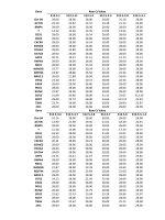
Table 1
13
C chemical shifts of the cellulose ethers 3-O-methylcellulose (3-O-MeC), and
2,3-O-dimethylcellulose (2,3-O-diMeC) determined from the 13C-13C DNP CPrefocused INADEQUATE NMR spectra and 1H-13C DNP CPMAS NMR spectrum
with CH3 selection. The 13C chemical shifts of microcrystalline cellulose (MCC)
are taken from the literature(Lesage et al., 1999). Chemical shifts for the crys
talline (c) and amorphous phase (a) of 2,3-O-diMeC are given. For MCC, the
amorphous/surface phase C-4 signal has been reported with lower chemical
shift, thus (c)/(a) chemical shifts are reproduced here. 13C chemical shifts are
provided using the tetramethylsilane reference scale.
Sample
3-O-MeC
2,3-O-diMeC
MCC
Chemical shift, ppm
c
a
c
a
C-1
C-2
C-3
C-4
C-5
C-6
CH3
103.1
107.5
103.5
105
75.4
86.6
84.4
73
85.8
87.5
87.3
74.5
78.7
85.4
83.2
87
≈ 82
75.4
74.6
75.5
74.5
59.1
59.2
61.6
62
63.1
62.2
–
3.4. NMR spectroscopic characterization of 3-O-methylcellulose in the
solid-state with DNP hNOE
While spectral assignments via DNP CP-INADEQUATE revealed the
substitution site of the cellulose ethers, we also explored the possibility
to directly correlate the methyl group with the nearby substitution site
on the cellulose backbone. In particular, CH3 groups are peculiar in the
DNP MAS context. Because of the fast rotation of the CH3 group, even at
100 K, the latter is known to (i) act as a relaxation sink which de
preciates the DNP enhancements, explaining why materials that contain
CH3 usually exhibit low DNP performance (Zagdoun et al., 2013), (ii)
1 13
H- C CP conditions of CH3 are very sensitive to the temperature in the
100 K range, this has been reported in polymers where the temperature
difference induced by the μwaves absorption can induce a bias in the
enhancement measurement (Mollica et al., 2014), and (iii) induce
spontaneous transfer from 1H to 13C through heteronuclear Nuclear
Overhauser Effect (hNOE) (Aladin & Corzilius, 2019; Daube et al., 2016;
Mao et al., 2019). In 2019, Corzilius and co-workers showed that uni
formly 13C-enriched amino acids can be hyperpolarized using hNOE
together with spontaneous 13C-13C spin diffusion. In the context of
characterization of methylcellulose, this is of particular interest, as
hNOE can selectively and spontaneously hyperpolarize the grafted CH3.
The strategy we propose here relies on the extension of DNP hNOE to
non-labelled cellulose ethers in order to localize the substitution site. As
pictured in Fig. 4a, 1H nuclei of the solvent are hyperpolarized directly
by the AMUPOL polarizing agent. Then, as established previously (Pinon
et al., 2017), the 1H hyperpolarization diffuses through the sample via
spontaneous 1H-1H spin diffusion and eventually reaches the 1H of the
cellulose including the CH3 moiety. Because of the hNOE effect, the 1H
hyperpolarization of the CH3 then spontaneously transfers to the 13C of
the CH3. The 13C hyperpolarization can then be further transmitted via
13 13
C- C spin diffusion. As we are working with 13C at natural abundance,
in principle 13C spin diffusion will only transfer polarization to the
closest carbons in space (including the substitution site) of the hyper
polarized CH3. Using the model reported in the literature (Bjorgvins
dottir, Walder, Pinon, & Emsley, 2018), it was possible to estimate that
the 13C spin diffusion will only probe from 4 to 9 Å with a transfer delay
of 1 to 5 s at 10 kHz MAS.
Fig. 4b shows the 13C DNP direct excitation spectra recorded with
and without μwaves on a sample of 3-O-methylcellulose impregnated
with 10 mM AMUPOL at different recycle delays. Identical phase
correction parameters were used to process all the spectra. Direct 13C
DNP will offer a positive DNP enhancement (Kaushik et al., 2016),
whereas hNOE is expected to provide a 13C negative DNP enhancement
(Daube et al., 2016). Thus, hNOE is the main active DNP transfer here.
Fig. 4c is the 1H-13C DNP CPMAS of the same sample at 5 kHz MAS,
displayed for comparison with the direct 13C spectra, one can observe
the predominance of the CH3 signals in 4b due to the direct hNOE DNP
Fig. 4. (a) Principle of substitution site identification for the reference sample
3-O-methylcellulose by combining DNP MAS with hNOE and 13C-13C spin
diffusion. (b) Direct 13C NMR spectra with (plain line) and without (dashed
line) μwave irradiation, and with recycle delays varying from 1 to 5 s and (c)
1
H-13C DNP CPMAS of 3-O-methylcellulose impregnated with 10 mM AMUPOL
in D2O:H2O 9:1v/v at 5 kHz MAS, at ca. 100 K. (d) 13C enhancement (from direct
13
C NMR) as function of the recycle delay and for the different resolved and
detectable signals.
enhancement route. As all signals are negative, and we can then expect
the process described in Fig. 4a to be predominant, i.e., 13C gets
hyperpolarized via 13C-13C spin diffusion from the CH3 functionality.
Fig. 4d reports the 13C signal enhancements of the different detectable
and resolved signals: CH3, C-1, C-3, and C-4/C-5 as a function of the 13C
spin diffusion delay. Note particularly that C-2 and C-6 signals are not
observable in the direct 13C spectra. As only C-1, C-3, and C-4/C-5 are
detectable, we can conclude that they are the closest carbons in space to
CH3. Among them, only C-3 can be etherified, and thus it implies that the
substitution site is C-3. Although the signal assignments of the
3-O-methylcellulose from the 13C-13C INADEQUATE already gives a
clear answer regarding the substitution site, the strategy based on hNOE
also seems to identify the substitution site.
7
P. Berruyer et al.
Carbohydrate Polymers 262 (2021) 117944
4. Conclusion
Heinze, T., Pfeifer, A., Sarbova, V., & Koschella, A. (2011). 3-O-propyl cellulose:
Cellulose ether with exceptionally low flocculation temperature. Polymer Bulletin, 66
(9), 1219–1229.
Heinze, T., Wang, Y., Koschella, A., Sullo, A., & Foster, T. J. (2012). Mixed 3-mono-Oalkyl cellulose: Synthesis, structure characterization and thermal properties.
Carbohydrate Polymers, 90(1), 380–386.
Kang, X., Kirui, A., Widanage, M. C. D., Mentink-Vigier, F., Cosgrove, D. J., & Wang, T.
(2019). Lignin-polysaccharide interactions in plant secondary cell walls revealed by
solid-state NMR. Nature Communications, 10.
Karlson, L., Joabsson, F., & Thuresson, K. (2000). Phase behavior and rheology in water
and in model paint formulations thickened with HM-EHEC: Influence of the chemical
structure and the distribution of hydrophobic tails. Carbohydrate Polymers, 41(1),
25–35.
Karrasch, A., Jager, C., Karakawa, M., Nakatsubo, F., Potthast, A., & Rosenau, T. (2009).
Solid-state NMR studies of methyl celluloses. Part 1: Regioselectively substituted
celluloses as standards for establishing an NMR data basis. Cellulose, 16(1), 129–137.
Kaushik, M., Bahrenberg, T., Can, T. V., Caporini, M. A., Silvers, R., Heiliger, J., et al.
(2016). Gd(III) and Mn(II) complexes for dynamic nuclear polarization: Small
molecular chelate polarizing agents and applications with site-directed spin labeling
of proteins. Physical Chemistry Chemical Physics, 18(39), 27205–27218.
Kern, H., Choi, S., Wenz, G., Heinrich, J., Ehrhardt, L., Mischnick, P., et al. (2000).
Synthesis, control of substitution pattern and phase transitions of 2,3-di-Omethylcellulose. Carbohydrate Research, 326(1), 67–79.
Kondo, T., & Gray, D. G. (1991). The preparation of O-methyl- and O-ethyl-celluloses
having controlled distribution of substituents. Carbohydrate Research, 220(0),
173–183.
Kono, H., Anai, H., Hashimoto, H., & Shimizu, Y. (2015). 13C-detection two-dimensional
NMR approaches for cellulose derivatives. Cellulose, 22(5), 2927–2942.
Kono, H., Erata, T., & Takai, M. (2002). CP/MAS 13C NMR study of cellulose and
cellulose derivatives. 2. Complete assignment of the 13C resonance for the ring
carbons of cellulose triacetate polymorphs. Journal of the American Chemical Society,
124(25), 7512–7518.
Kono, H., Erata, T., & Takai, M. (2003). Determination of the through-bond carboncarbon and carbon-proton connectivities of the native celluloses in the solid state.
Macromolecules, 36(14), 5131–5138.
Kono, H., Numata, Y., Erata, T., & Takai, M. (2004). 13C and 1H resonance assignment of
mercerized cellulose II by two-dimensional MAS NMR spectroscopies.
Macromolecules, 37(14), 5310–5316.
Koschella, A., & Klemm, D. (1997). Silylation of cellulose regiocontrolled by bulky
reagents and dispersity in the reaction media. Macromolecular Symposia, 120(1),
115–125.
Koschella, A., Heinze, T., & Klemm, D. (2001). First synthesis of 3-O-functionalized
cellulose ethers via 2,6-di-O-protected silyl cellulose. Macromolecular Bioscience, 1
(1), 49–54.
Koschella, A., Fenn, D., & Heinze, T. (2006). Water soluble 3-mono-O-ethyl cellulose:
Synthesis and characterization. Polymer Bulletin, 57(1), 33–41.
Koschella, A., Fenn, D., Illy, N., & Heinze, T. (2006). Regioselectively functionalized
cellulose derivatives: A mini review. Macromolecular Symposia, 244(1), 59–73.
Kumar, A., Durand, H., Zeno, E., Balsollier, C., Watbled, B., Sillard, C., et al. (2020). The
surface chemistry of a nanocellulose drug carrier unravelled by MAS-DNP. Chemical
Science, 11(15), 3868–3877.
Lesage, A., Bardet, M., & Emsley, L. (1999). Through-bond carbon-carbon connectivities
in disordered solids by NMR. Journal of the American Chemical Society, 121(47),
10987–10993.
Li, C. L., Martini, L. G., Ford, J. L., & Roberts, M. (2005). The use of hypromellose in oral
drug delivery. The Journal of Pharmacy and Pharmacology, 57(5), 533–546.
Mao, J. F., Aladin, V., Jin, X. S., Leeder, A. J., Brown, L. J., Brown, R. C. D., et al. (2019).
Exploring protein structures by dnp-enhanced methyl solid-state NMR spectroscopy.
Journal of the American Chemical Society, 141(50), 19888–19901.
Mischnick, P. (2018). Analysis of the substituent distribution in cellulose ethers - recent
contributions: Chemistry, analysis, and applications (pp. 143–173).
Mollica, G., Le, D., Ziarelli, F., Casano, G., Ouari, O., Phan, T. N. T., et al. (2014).
Observing apparent nonuniform sensitivity enhancements in dynamic nuclear
polarization solid-state NMR spectra of polymers. ACS Macro Letters, 3(9), 922–925.
Opella, S. J., & Frey, M. H. (1979). Selection of non-protonated carbon resonances in
solid-state nuclear magnetic-resonance. Journal of the American Chemical Society, 101
(19), 5854–5856.
Patural, L., Marchal, P., Govin, A., Grosseau, P., Ruot, B., & Dev`es, O. (2011). Cellulose
ethers influence on water retention and consistency in cement-based mortars. Cement
and Concrete Research, 41(1), 46–55.
Perras, F. A., Luo, H., Zhang, X. M., Mosier, N. S., Pruski, M., & Abu-Omar, M. M. (2017).
Atomic-level structure characterization of biomass pre- and post-lignin treatment by
dynamic nuclear polarization-enhanced solid-state NMR. The Journal of Physical
Chemistry A, 121(3), 623–630.
Pinon, A. C., Schlagnitweit, J., Berruyer, P., Rossini, A. J., Lelli, M., Socie, E., et al.
(2017). Measuring nano- to microstructures from relayed dynamic nuclear
polarization NMR. The Journal of Physical Chemistry C, 121(29), 15993–16005.
Reif, B., Ashbrook, S. E., Emsley, L., & Hong, M. (2021). Solid-state NMR spectroscopy.
Nature Reviews Methods Primers, 1(1), 2.
Rossini, A. J., Zagdoun, A., Hegner, F., Schwarzwalder, M., Gajan, D., Coperet, C., et al.
(2012). Dynamic nuclear polarization NMR spectroscopy of microcrystalline solids.
Journal of the American Chemical Society, 134(40), 16899–16908.
Rossini, A. J., Zagdoun, A., Lelli, M., Lesage, A., Coperet, C., & Emsley, L. (2013).
Dynamic nuclear polarization surface enhanced NMR spectroscopy. Accounts of
Chemical Research, 46(9), 1942–1951.
Using a multistep synthesis approach and protecting group strate
gies, two methylcellulose ethers with a well-defined molecular structure
were prepared, one with a regioselective 3-O-substitution (DSMe = 0.99)
and one with regioselective 2,3-O-substitution (DSMe = 2.03). These
model compounds were used to introduce and benchmark two new
solid-state NMR based approaches that allow to characterize the sub
stitution pattern in cellulose ethers. The first method uses the 13C
chemical shift assignments of the cellulose ether via 13C-13C DNP
enhanced refocused INADEQUATE. It gives a particularly clear identi
fication of the substitution site on both 3-O-methylcellulose and 2,3-Odimethylcellulose, and should be generalizable to any cellulose ether.
The assignments are made possible in the solid-state because of the
sensitivity enhancement due to the use of DNP MAS. The second method
is based on selective hyperpolarization of CH3 via hNOE and subsequent
transfer via 13C spin diffusion at natural abundance, which also allow to
confirm the postulated regioselectivity within the model compounds.
Author contributions
This work was produced through contributions of all authors to the
conception, implementation, analysis and writing of the paper.
Acknowledgment
This work was financially supported by the Swiss Innovation Agency
Innosuisse (Grant: 30819.1 IP-ENG).
Appendix A. Supplementary data
Supplementary material related to this article can be found, in the
online version, at doi: />References
Aladin, V., & Corzilius, B. (2019). Methyl dynamics in amino acids modulate
heteronuclear cross relaxation in the solid state under MAS DNP. Solid State Nuclear
Magnetic Resonance, 99, 27–35.
Arca, H. C., Mosquera-Giraldo, L. I., Bi, V., Xu, D., Taylor, L. S., & Edgar, K. J. (2018).
Pharmaceutical applications of cellulose ethers and cellulose ether esters.
Biomacromolecules, 19(7), 2351–2376.
Atalla, R. H., & Vanderhart, D. L. (1984). Native cellulose: A composite of two distinct
crystalline forms. Science, 223(4633), 283–285.
Berruyer, P., Emsley, L., & Lesage, A. (2018). DNP in materials science: Touching the
surface. Emagres, 7(4), 93–104.
Bjorgvinsdottir, S., Walder, B. J., Pinon, A. C., & Emsley, L. (2018). Bulk nuclear
hyperpolarization of inorganic solids by relay from the surface. Journal of the
American Chemical Society, 140(25), 7946–7951.
Buchanan, C. M., Edgar, K. J., Hyatt, J. A., & Wilson, A. K. (1991). Preparation of
cellulose [1-carbon-13]acetates and determination of monomer composition by
NMR spectroscopy. Macromolecules, 24(11), 3050–3059.
Daube, D., Aladin, V., Heiliger, J., Wittmann, J. J., Barthelmes, D., Bengs, C., et al.
(2016). Heteronuclear cross-relaxation under solid-state dynamic nuclear
polarization. Journal of the American Chemical Society, 138(51), 16572–16575.
Elkins, M. R., Sergeyev, I. V., & Hong, M. (2018). Determining cholesterol binding to
membrane proteins by cholesterol 13C labeling in yeast and dynamic nuclear
polarization NMR. Journal of the American Chemical Society, 140(45), 15437–15449.
Foston, M. (2014). Advances in solid-state NMR of cellulose. Current Opinion in
Biotechnology, 27, 176–184.
Foston, M., Katahira, R., Gjersing, E., Davis, M. F., & Ragauskas, A. J. (2012). Solid-state
selective (13)C excitation and spin diffusion NMR to resolve spatial dimensions in
plant cell walls. Journal of Agricultural and Food Chemistry, 60(6), 1419–1427.
Fox, S. C., Li, B., Xu, D., & Edgar, K. J. (2011). Regioselective esterification and
etherification of cellulose: A review. Biomacromolecules, 12(6), 1956–1972.
Groszewicz, P. B., Mendes, P., Kumari, B., Lins, J., Biesalski, M., Gutmann, T., et al.
(2020). N-hydroxysuccinimide-activated esters as a functionalization agent for
amino cellulose: Synthesis and solid-state NMR characterization. Cellulose, 27(3),
1239–1254.
Gupta, R., Zhang, H. L., Lu, M. M., Hou, G. J., Caporini, M., Rosay, M., et al. (2019).
Dynamic nuclear polarization magic-angle spinning nuclear magnetic resonance
combined with molecular dynamics simulations permits detection of order and
disorder in viral assemblies. The Journal of Physical Chemistry B, 123(24),
5048–5058.
8
P. Berruyer et al.
Carbohydrate Polymers 262 (2021) 117944
Sauvee, C., Rosay, M., Casano, G., Aussenac, F., Weber, R. T., Ouari, O., et al. (2013).
Highly efficient, water-soluble polarizing agents for dynamic nuclear polarization at
high frequency. Angewandte Chemie-International Edition, 52(41), 10858–10861.
Schmidt-Rohr, K., & Spiess, H. W. (1999). Multidimensional solid-state NMR and polymers
(third printing ed.). London etc: Academic Press.
Sparrman, T., Svenningsson, L., Sahlin-Sjovold, K., Nordstierna, L., Westman, G., &
Bernin, D. (2019). A revised solid-state NMR method to assess the crystallinity of
cellulose. Cellulose, 26(17), 8993–9003.
Sun, S. M., Foster, T. J., MacNaughtan, W., Mitchell, J. R., Fenn, D., Koschella, A., et al.
(2009). Self-association of cellulose ethers with random and regioselective
distribution of substitution. Journal of Polymer Science Part B-Polymer Physics, 47(18),
1743–1752.
Takahashi, H., Lee, D., Dubois, L., Bardet, M., Hediger, S., & De Paepe, G. (2012). Rapid
natural-abundance 2D 13C-13C correlation spectroscopy using dynamic nuclear
polarization enhanced solid-state NMR and matrix-free sample preparation.
Angewandte Chemie-International Edition, 51(47), 11766–11769.
Tezuka, Y., Imai, K., Oshima, M., & Chiba, T. (1990). Determination of substituent
distribution in cellulose ethers by 13C- and 1H-NMR. Studies of their acetylated
derivatives: O-(2-hydroxypropyl)cellulose. Carbohydrate Research, 196, 1–10.
Viger-Gravel, J., Lan, W., Pinon, A. C., Berruyer, P., Emsley, L., Bardet, M., et al. (2019).
Topology of pretreated wood fibers using dynamic nuclear polarization. The Journal
of Physical Chemistry C, 123(50), 30407–30415.
Wang, T., Park, Y. B., Caporini, M. A., Rosay, M., Zhong, L. H., Cosgrove, D. J., et al.
(2013). Sensitivity-enhanced solid-state NMR detection of expansin’s target in plant
cell walls. Proceedings of the National Academy of Sciences of the United States of
America, 110(41), 16444–16449.
Wang, T., Yang, H., Kubicki, J. D., & Hong, M. (2016). Cellulose structural polymorphism
in plant primary cell walls investigated by high-field 2D solid-state NMR
spectroscopy and density functional theory calculations. Biomacromolecules, 17(6),
2210–2222.
Wever, D. A. Z., Picchioni, F., & Broekhuis, A. A. (2011). Polymers for enhanced oil
recovery: A paradigm for structure–Property relationship in aqueous solution.
Progress in Polymer Science, 36(11), 1558–1628.
Wickholm, K., Hult, E. L., Larsson, P. T., Iversen, T., & Lennholm, H. (2001).
Quantification of cellulose forms in complex cellulose materials: A chemometric
model. Cellulose, 8(2), 139–148.
Wu, X. L., Burns, S. T., & Zilm, K. W. (1994). Spectral editing in CPMAS NMR - generating
subspectra based on proton multiplicities. Journal of Magnetic Resonance Series A, 111
(1), 29–36.
Young, N. W. G. (2014). Emulsifiers and stabilisers. In K. K. Rajah (Ed.), Fats in food
technology 2e (pp. 253–287). John Wiley & Sons, Ltd.
Zagdoun, A., Rossini, A. J., Conley, M. P., Gruning, W. R., Schwarzwalder, M., Lelli, M.,
et al. (2013). Improved dynamic nuclear polarization surface-enhanced NMR
spectroscopy through controlled incorporation of deuterated functional groups.
Angewandte Chemie-International Edition, 52(4), 1222–1225.
Zhao, W. C., Kirui, A., Deligey, F., Mentink-Vigier, F., Zhou, Y. H., Zhang, B. C., et al.
(2021). Solid-state nmr of unlabeled plant cell walls: High-resolution structural
analysis without isotopic enrichment. Biotechnology for Biofuels, 14(1).
Ziegler, T., Bien, F., & Jurisch, C. (1998). Chemoenzymatic synthesis of enantiomerically
pure alkene 1,2-diols and glycosides thereof. Tetrahedron: Asymmetry, 9(5), 765–780.
9