Digestibility of resistant starch type 3 is affected by crystal type, molecular weight and molecular weight distribution
Bạn đang xem bản rút gọn của tài liệu. Xem và tải ngay bản đầy đủ của tài liệu tại đây (3.49 MB, 10 trang )
Carbohydrate Polymers 265 (2021) 118069
Contents lists available at ScienceDirect
Carbohydrate Polymers
journal homepage: www.elsevier.com/locate/carbpol
Digestibility of resistant starch type 3 is affected by crystal type, molecular
weight and molecular weight distribution
C.E. Klostermann a, P.L. Buwalda a, b, H. Leemhuis b, P. de Vos c, H.A. Schols d, J.H. Bitter a, *
a
Biobased Chemistry and Technology, Wageningen University & Research, Bornse Weilanden 9, 6708 WG Wageningen, the Netherlands
Coă
operative AVEBE u.a., P.O. Box 15, 9640 AA Veendam, the Netherlands
c
Immunoendocrinology, Division of Medical Biology, Department of Pathology and Medical Biology, University of Groningen and University Medical Centre Groningen,
Groningen, Hanzeplein 1, 9700 RB Groningen, the Netherlands
d
Laboratory of Food Chemistry, Wageningen University & Research, Bornse Weilanden 9, 6708 WG Wageningen, the Netherlands
b
A R T I C L E I N F O
A B S T R A C T
Keywords:
Resistant starch type 3
Dietary fiber
α-glucan
Prebiotics
HPSEC
Resistant starch type 3 (RS-3) holds great potential as a prebiotic by supporting gut microbiota following in
testinal digestion. However the factors influencing the digestibility of RS-3 are largely unknown. This research
aims to reveal how crystal type and molecular weight (distribution) of RS-3 influence its resistance. Narrow and
polydisperse α-glucans of degree of polymerization (DP) 14–76, either obtained by enzymatic synthesis or
debranching amylopectins from different sources, were crystallized in 12 different A- or B-type crystals and in
vitro digested. Crystal type had the largest influence on resistance to digestion (A >>> B), followed by molecular
weight (Mw) (high DP >> low DP) and Mw distribution (narrow disperse > polydisperse). B-type crystals
escaping digestion changed in Mw and Mw distribution compared to that in the original B-type crystals, whereas
A-type crystals were unchanged. This indicates that pancreatic α-amylase binds and acts differently to A- or Btype RS-3 crystals.
1. Introduction
Resistant starch (RS) is starch that resists digestion in the small in
testine by human digestive enzymes and therefore ends up in the colon.
In the colon RS will be fermented and may even act as a prebiotic by
positively influencing beneficial gut microbiota (Fuentes-Zaragoza
et al., 2011; Haenen et al., 2013; Zaman & Sarbini, 2016). Recently, it
was shown that RS also may directly interact with the immune system to
activate several immune responses (Bermudez-Brito, Rosch, Schols,
Faas, & de Vos, 2015; L´epine et al., 2018). Five different types of RS
exist: physically inaccessible starch (RS-1), native starch granules
(RS-2), retrograded starch (RS-3), chemically modified starch (RS-4) and
amylose-lipid complexes (RS-5) (Birt et al., 2013; Fuentes-Zaragoza
et al., 2011). RS-3 is of interest as food ingredient since it is thermally
stable (Haralampu, 2000) and can easily be added to foods as dietary
fiber. RS-3 preparations can be made by debranching amylopectins to
short chain α-glucans followed by controlled crystallization (Cai & Shi,
2014). However, to be able to act as dietary fibre, RS-3 preparations
should be resistant to enzymatic digestion in the small intestine.
Recently, it was suggested that RS-3 may be resistant to digestion due to
slow enzyme binding of pancreatic α-amylase to the RS-3 crystals in
combination with slow catalytic hydrolysis (Dhital, Warren, Butter
worth, Ellis, & Gidley, 2017). However, it is not yet clear which physi
cochemical characteristics of RS-3 cause the resistance to digestion.
Differences in digestibility of RS-3 preparations might be caused by
characteristics like crystal type and molecular weight (distribution) of
the crystallized α-glucans.
Resistant starch type 3 (RS-3) preparations or so-called short chain
α-glucan crystals can be produced by gelatinizing starch at elevated
temperatures followed by slow cooling, which results in recrystallization
of the starch. The crystals formed by recrystallization can be recognized
as A-type or B-type, as measured by X-ray diffraction (Gidley & Bulpin,
1987; Kiatponglarp, Tongta, Rolland-Sabate, & Buleon, 2015; Nish
iyama, Putaux, Montesanti, Hazemann, & Rochas, 2010). Whether A- or
B-type crystals are formed depends on the chain length of the α-glucan,
concentration during crystallization and temperature of crystallization
(Buleon, Veronese, & Putaux, 2007; Creek, Ziegler, & Runt, 2006;
Kiatponglarp et al., 2015; Pfannemuller, 1987). In addition, RS-3
preparations differing in crystal type can be formed using different
solvents like acetone, ethanol or polyethylene glycol (Huang et al.,
* Corresponding author.
E-mail address: (J.H. Bitter).
/>Received 17 November 2020; Received in revised form 6 April 2021; Accepted 7 April 2021
Available online 16 April 2021
0144-8617/© 2021 The Author(s). Published by Elsevier Ltd. This is an open access article under the CC BY license ( />
C.E. Klostermann et al.
Carbohydrate Polymers 265 (2021) 118069
2019; Kobayashi, Kimura, Naito, Togawa, & Wada, 2015; Montesanti
et al., 2010). in vitro, native A-type starches are easier to digest
compared to native B-type starches (Martens, Gerrits, Bruininx, &
Schols, 2018). In contrast, research on digestibility of retrograded short
chain α-glucans has shown that retrograded A-type crystals are more
resistant to digestion than retrograded B-type crystals (Cai & Shi, 2013,
2014).
In addition to crystal type, average molecular weight also affects
resistance to digestion of RS-3 preparations. Most research on RS-3 is
performed by crystallization of debranched amylopectins resulting in a
wide range of short chain linear α-1,4 linked glucans (Cai & Shi, 2014;
Kiatponglarp et al., 2015). By choosing waxy starches of different
botanical sources, variations in average chain length (DPn) can be
achieved after debranching (Cai & Shi, 2010). For example, debranched
waxy maize starch has a DPn of 24, waxy wheat of DPn 22 and waxy
potato of DPn 32 (Cai & Shi, 2010). In addition, starches can be modified
by branching enzymes or by amylomaltases, due to which amylopectins
are produced that have very short chains or elongated chains, respec
tively (van der Maarel & Leemhuis, 2013). After digestion of RS-3
preparations made of debranched amylopectins of different botanical
sources, it was found that a higher DPn resulted in more resistance to
digestion (Cai & Shi, 2010).
However, it is largely unknown how molecular weight distribution
influences the resistance to digestion of RS-3 preparations. Such a broad
range of α-1,4 glucans can be obtained by debranching amylopectins, as
shown for waxy wheat amylopectin resulting in chain lengths of DP
6–66 with an average of DP 22 (Cai & Shi, 2010). When such a poly
disperse mixture is crystallized and subjected to digestion, it is not yet
clear how the presence of different chain lengths influences the crystal
formation and resistance to digestion. Previously, the effect of poly
dispersity on digestion was studied by debranching waxy and native rice
starch. Debranching waxy rice starch results in α-glucan chains with a
DP varying from 6 to 90, whereas debranching native rice starch also
includes the linear amylose part, which has a DP up to 1000 (Kiatpon
glarp et al., 2015). It was shown that crystals produced from relatively
narrow disperse debranched waxy rice starch are 10 % more resistant to
digestion than crystals produced from polydisperse debranched native
rice starch (Kiatponglarp, Rugmai, Rolland-Sabate, Buleon, & Tongta,
2016). Another study focussed on the fractionation of debranched waxy
rice starch (polydispersity index (PI) 2.2) (Hu et al., 2020). This frac
tionation caused narrowing of the molecular weight distribution to a PI
of 1.5 at most. After crystallization and digestion, it was shown that
these crystals made of relatively narrow disperse α-glucans were 10–20
% more resistant to digestion compared to the unfractionated poly
disperse crystals (Hu et al., 2020). However, the polydispersity index of
before mentioned debranched and fractionated starches is still relatively
high, making it hard to draw conclusions on the influence of poly
dispersity on crystal formation and subsequent digestibility.
In contrast to polydisperse α-1,4 glucans obtained by debranching
amylopectins, narrow disperse amyloses can be enzymatically synthe
sized by potato glucan phosphorylase from glucose-1-phosphate (G-1-P)
(Chang et al., 2018; Kobayashi et al., 2015; Roger, Axelos, & Colonna,
2000; Yanase, Takaha, & Kuriki, 2006). Potato glucan phosphorylase
uses glucose-1-phosphate as a substrate and transfers the glucose residue
to a primer molecule, being maltotetraose or an α-1,4 linked oligomer of
DP > 4 (Ohdan, Fujii, Yanase, Takaha, & Kuriki, 2006). The ratio be
tween the glucose-1-phosphate and primer molecule determines the DPn
at the end of the enzymatic synthesis. By choosing the right ratio, narrow
disperse equivalents of debranched amylopectins can be synthesized
that have a similar average molecular weight (Mw) but a lower poly
dispersity index. However, glucose-1-phosphate as substrate is quite
expensive. As an alternative, the combination of sucrose and sucrose
phosphorylase can be used to produce glucose-1-phosphate (Luley-
Goedl & Nidetzky, 2010; Qi, You, & Zhang, 2014). Using sucrose as
substrate also has shown to improve the yield of synthesis, compared to
using glucose-1-phosphate directly (Ohdan et al., 2006).
The present study focusses on the effect of crystal type, Mw and Mw
distribution on the resistance to digestion of RS-3 preparations. Different
resistant starches were produced by debranching amylopectins (poly
disperse) or through synthesis with the help of potato glucan phos
phorylase and sucrose phosphorylase (narrow disperse). The ratio of G1-P and sucrose was chosen to obtain α-1,4 linked glucans with a similar
average number molecular weight (Mwn) as the debranched amylo
pectins, but with a lower polydispersity index. The linear α-glucans were
crystallized at different concentrations and temperatures to obtain Aand B-type crystals. These RS-3 preparations were digested to study the
effect of crystal type, average Mw and Mw distribution on the resistance
to digestion.
2. Materials and methods
2.1. Materials
Waxy potato starch (Eliane100), amylomaltase modified potato
starch (Etenia 457) and highly branched starch of potato (Mw ±100
kDa, 8 % branch points) were provided by AVEBE (Veendam, The
Netherlands). Waxy rice starch (Remyline XS) was purchased from
Beneo (Mannheim, Germany). Isoamylase (EC 3.2.1.68) and maltote
traose were obtained from Megazyme (Bray, Wicklow, Ireland). Sucrose,
glucose, maltose, maltotriose, pancreatin, amyloglucosidase, Lennox B
(LB) medium, kanamycin sulphate, isopropyl β-D-1-thiogalactopyrano
side, glucose-1-phosphate potassium salt and imidazole of high purity
were obtained from Sigma-Aldrich (St. Louis, MO, USA). Bugbuster
(Novagen) and benzonase nuclease were purchased from Merck
(Darmstadt, Germany). MilliQ (MQ) water was used unless stated
otherwise (Arium mini essential UV Ultrapure water filter, Sartorius,
ăttingen, Germany).
Go
2.2. Production of potato glucan phosphorylase and sucrose
phosphorylase
The potato glucan phophorylase (PGP) (EC 2.4.1.1) and the Bifido
bacterium adolescentis sucrose phosphorylase (SP) (EC 2.4.1.7) (van den
Broek et al., 2004) were produced in Escherichia coli BL21 DE3 carrying
the pET28a expression vector. The genes encoding PGP and SP were
codon optimized for expression in E. coli, synthesized and cloned in
pET28a by GenScript (Leiden, the Netherlands). The E. coli cells con
taining the PGP plasmid were grown for 16 h at 37 ◦ C in LB medium that
contained 25 μg/mL kanamycin while shaking at 200 rpm. The culture
was transferred to 500 mL LB broth that contained 25 μg/mL kanamycin
and kept for 2− 3 h at 37 ◦ C, shaking at 200 rpm until OD600 = 0.5− 0.7.
The culture was cooled down on ice and 0.1 mM isopropyl β-D-1-thio
galactopyranoside was added after which the culture was incubated for
24 h at 18 ◦ C, 200 rpm. E. coli cells containing the SP plasmid were
grown similarly until the inducer was added. To the SP culture of OD600
= 0.5− 0.7 0.4 mM isopropyl β-D-1-thiogalactopyranoside was added
and incubation was continued for 4 h at 30 ◦ C, 200 rpm. Cells were
centrifuged for 10 min at 16,000 x g, 4 ◦ C. The cell pellets were resus
pended in Bugbuster, causing lysis of the E. coli cells, and supplemented
with benzonase nuclease, according to the company protocol. The lysed
cells were centrifuged for 10 min at 16,000 x g, 4 ◦ C. The supernatant
was decanted and stored for 30 min at 60 ◦ C. This suspension was
centrifuged and the supernatant was filtered over an 0.2 μm filter to
obtain a sterile cell-free enzyme extract. The enzymes were purified
using a His-Tag purification column, according to the company protocol
(GE Healthcare Life Sciences, Amersham, United Kingdom). Sample and
washing buffer contained 20 mM imidazole and elution of pure enzymes
was performed with 800 mM imidazole. The final PGP or SP concen
tration was determined by the Bradford protein assay (Bradford, 1976).
2
C.E. Klostermann et al.
Carbohydrate Polymers 265 (2021) 118069
2.3. Production of polydisperse α-1,4 linked glucans
overnight. The supernatants were inactivated and analysed as described
before.
Highly branched potato starch (HBPS), waxy potato starch (WPS),
amylomaltase modified potato starch (AMPS) and waxy rice starch
(WRS) were suspended in a 20 mM sodium acetate buffer of pH 5 and
autoclaved. The solutions were cooled to 40 ◦ C and isoamylase was
added (8 U/g). The amylopectins were debranched for 48 h at 40 ◦ C, 100
rpm and freeze dried to produce debranched HBPS (dHBPS), WPS
(dWPS), AMPS (dAMPS) and WRS (dWRS).
2.7. Molecular weight distribution of RS-3 preparations, before and after
digestion
RS-3 preparations of DP < 25 were suspended in MQ at 2.5 mg/mL
and dissolved by boiling. RS-3 preparations of DP > 32 were solubilised
in 1 M NaOH at 60 mg/mL sample. The samples were diluted to 2.5 mg/
mL and neutralized by addition of 1 M HCl. Samples were centrifuged at
19,000 x g for 10 min and the supernatant was analysed with a Dionex
Ultimate 3000 system (Sunnyvale, USA). Ten μL sample was injected on
a column set that consisted of three in series connected TSKgel SuperAW
columns (SuperAW4000 6.0 × 150 mm, 6 μm; SuperAW3000 6.0 × 150
mm, 4 μm; SuperAW2500 6.0 × 150 mm, 4 μm) (Tosoh Bioscience,
Tokyo, Japan) with a TSKgel guard column (SuperAW-L 4.6 × 35 mm, 7
μm). Elution was performed with 0.6 mL/min and 0.2 M NaNO3, at 55
◦
C. Detection was performed with a Shodex RI-101 detector (Showa
Denko, K.K., Kawasaki, Japan). Calibration of the column was per
formed with pullulan standards (Supelco, Bellefonte, USA).
From the HPSEC-RI results, DPn, DPm and PI were calculated using
pullulan calibration. Intensities were normalized and base-line cor
rected, after which Mn was calculated using formula 1. Mm was calcu
lated using formula 2 and PI was calculated by dividing Mm over Mn.
The retention time frame of each peak was taken into account to
calculate Mn and Mm.
∑
1) Mn =
Mwp ∗In
2.4. Enzymatic synthesis of narrow disperse α-1,4 linked glucans
For studying reaction dynamics of PGP and SP sucrose and dHBPS
were mixed at 105 mM in a molar ratio of 20/1 in a 30 mM sodium
phosphate buffer of pH 7.0. His-tag purified PGP and SP were added (25
μg/mL) and the mixtures were incubated at 50 ◦ C, 100 rpm in a shaking
incubator. After 0, 0.5, 1 and 4 h a 50 μL sample was taken for chemical
analysis (section 2.7) and heated for 15 min at 100 ◦ C to inactivate the
enzymes. For further incubations sucrose and dHBPS were mixed at 105
mM in a molar ratio of 2/1, 5/1, 20/1 and 65/1 in a 30 mM sodium
phosphate buffer of pH 7.0. His-tag purified PGP and SP were added
(6.25 μg/mL) and samples were incubated for 24 h at 50 ◦ C, 100 rpm in a
shaking incubator. After 24 h of incubation, the remaining samples were
freeze-dried and washed with cold MQ and 80 % ethanol to remove salts,
enzymes and small sugars and freeze-dried again to yield purified sG2
(2/1), sG5 (5/1), sG20 (20/1) and sG65 (65/1).
2.5. Crystallization of poly- and narrow disperse α-1,4 linked glucans
∑
Poly- and narrow disperse α-glucans of similar DPn were suspended
in MQ in different concentrations: dHBPS: 40 %w/w; sG2, dWRS and
sG5: 30 %w/w; dWPS and sG20: 10 %w/w; dAMPS and sG65: 5 % w/w.
The suspensions were autoclaved and stored at 80 ◦ C prior to crystalli
zation. Half of the dHBPS, sG2, dWRS and sG5 samples were stored for
24 h at 50 ◦ C to produce A-type crystals, according to Cai and Shi (2014).
The other half of dHBPS, sG2, dWRS and sG5 were immediately cooled
on ice and stored for 24 h at 4 ◦ C to produce B-type crystals, similar to
the method proposed by Cai and Shi (2014). In addition, dWPS, sG20,
dAMPS and sG65 were also immediately cooled on ice and stored for 24
h at 4 ◦ C, to produce B-type crystals. After 24 h storage, the samples were
centrifuged for 10 min at 7000 x g, 4 ◦ C and washed with cold MQ and
80 % ethanol. The supernatants were decanted and pellets containing
crystallized α-1,4 linked glucans were dried for 48 h at 40 ◦ C. Crystal
lization yield was calculated as (total mass after crystallization) / (mass
at start) * 100 %.
2) Mm =
Mw2p ∗In
Mn
In which Mn is the number based average Mw, whereas Mm is the
mass based average Mw, Mwp is the Mw based on pullulan calibration
and In is the normalised and base-line corrected intensity at retention
time x.
Samples were diluted to 0.25 mg/mL and centrifuged at 19,000 x g
for 10 min. The supernatant was analysed using an ICS 3500 HPAEC
system from Dionex, in combination with a CarboPac PA-1 (2 × 250
mm) column, with a CarboPac PA-1 guard column (Dionex). The de
tector used was an electrochemical Pulsed Amperometric detector from
Dionex. Ten μL of supernatant was injected on the column and eluted by
a gradient consisting of eluent A (0.1 M NaOH solution) and eluent B (1
M NaOAc in 0.1 M NaOH). The gradient used was 2.5–40 % B (0− 50
min), 40–100 % B (50− 65 min), 100 % B (65− 70 min), 2.5 % B (70− 85
min). Elution was performed with 0.3 mL/min at 25 ◦ C. A calibration
curve of 5− 10 μg/mL of malto-oligosaccharides (DP 1 – DP 7) was run
for quantification. Data analysis was performed with ChromeleonTM
7.2.6 software from Thermo Fisher Scientific (Waltham, Massachusetts,
USA).
2.6. Digestion of RS-3 preparations
Digestion was performed according to Martens et al. with minor
modifications (Martens et al., 2018). RS-3 preparations were suspended
in 100 mM sodium acetate buffer pH 5.9 at 20 mg/mL. Pancreatin so
lution was prepared according to Martens et al. (2018), without addition
of invertase. Samples were incubated for 0, 20, 60, 120 and 240 min and
enzymes were inactivated by heat treatment for 15 min at 100 ◦ C. After
360 min of incubation, the samples were centrifuged for 10 min at 19,
000 x g, 4 ◦ C and the enzymes in the supernatant were inactivated by
heat treatment for 15 min at 100 ◦ C. The remaining pellet was washed
twice with MQ and oven-dried at 40 ◦ C overnight. Free glucose content
in the heat-treated samples was measured with the GOPOD assay from
Megazyme. To study the effect of pancreatic α-amylase on the Mw dis
tribution of dWRS-A and dWRS-B crystals, a similar method was used as
described before, with some minor modifications. Pancreatin solution
was prepared according to Martens et al. (2018), without addition of
invertase and amyloglucosidase. Samples were incubated for 0, 20, 60
and 360 min and immediately centrifuged for 10 min at 19,000 x g, 4 ◦ C.
The pellets were washed twice with MQ and oven-dried at 40 ◦ C
2.8. Crystal type determination by X-ray diffraction
Wide angle X-ray scattering (WAXS) powder diffractograms of the
RS-3 preparations were measured on a Bruker Discover D2 diffractom
eter (Bruker corporation, Billerica, Massachusetts, USA) using Cu radi
ation (1.54 Å) in the reflection geometry in the angular range of 5–35
2◦ θ with a step size of 0.051◦ 2θ and 1 s per step in a rotating stage of
10◦ /min. Detection was performed with Lynxeye XE-T (Bruker corpo
ration). XRD diffractograms were background corrected and
normalized.
2.9. Scanning Electron Microscopy of RS-3 preparations
Crystal morphology was determined with Scanning Electron Micro
scopy (SEM) (Magellan 400, FEI, Eindhoven, The Netherlands) at the
3
C.E. Klostermann et al.
Carbohydrate Polymers 265 (2021) 118069
Wageningen Electron Microscopy Center (WEMC). The RS-3 prepara
tions were attached to sample holders containing carbon adhesive tabs
(EMS, Washington, USA) and coated with 12 nm tungsten (EM SCD 500,
Leica, Vienna, Austria). The crystals were analysed with a field emission
SEM at 2 kV and magnification of 10,000 times.
Table 1
Average chain length (DPn) and polydispersity index (PI) of synthesized α-glu
cans before and after purification after 24 h one-pot incubation of sucrose and
debranched HBPS in different molar ratios with potato glucan phosphorylase
and sucrose phosphorylase.
Sample
name
3. Results & discussion
3.1. Production of narrow disperse α-glucans
Narrow disperse α-glucans were enzymatically synthesized by potato
glucan phosphorylase (PGP) and sucrose phosphorylase (SP) using
debranched highly branched potato starch (dHBPS) as primer molecule
and sucrose as a substrate. The synthesis was followed over time and
analysed by HPAEC-PAD (Fig. 1).
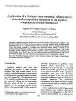
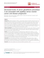
At t = 0 min, the chromatogram shows several peaks which can be
identified as malto-oligomers of dHBPS and G-1-P formed after enzy
matic hydrolysis of sucrose by SP (Fig. 1). The figure inset shows peaks
that can be identified as sucrose, fructose and glucose. Over time (t = 30,
t = 60, t = 240 min), the sucrose was hydrolysed and fructose formed,
showing activity of SP. The PAD signal of G-1-P increased and decreased
over time, whereas the malto-oligomers of dHBPS were elongated up to
at least DP 40 over time, indicating PGP activity. Due to this shift in
malto-oligomers towards higher DP’s over time, it can be stated that PGP
favoured to elongate the smallest malto-oligomer present (DP > 4).
Although literature states that based on the polydispersity index, enzy
matically synthesized α-glucans are narrow disperse (Kobayashi et al.,
2015; Ohdan et al., 2006), this result shows that still a rather broad
mixture of α-glucans was formed after enzymatic synthesis.
Sucrose and dHBPS were incubated at a ratio of 2/1, 5/1, 20/1 and
65/1 with PGP and SP to synthesize α-glucans of DP 14 (sG2), 18 (sG5),
32 (sG20) and 78 (sG65). The synthesis yields after 24 h of incubation
was between 65–85 %. The average Mw and polydispersity index (PI) of
the synthesized and purified α-glucans were analysed and calculated
after size exclusion chromatography (Table 1, Supplementary Fig. 1).
The results show that the Mw of the final α-glucan after enzymatic
synthesis increased with the sucrose/dHBPS ratio (Table 1). The higher
the sucrose/dHBPS molar ratio, the more G-1-P was available for the
reaction and thus the higher Mw α-glucans were formed, as stated in
literature previously (Ohdan et al., 2006). The DPn at the end of the
synthesis can be predicted by the choice of primer and the ratio between
substrate and primer (van der Vlist et al., 2008). The primer used in the
Sucrose/
dHBPS
sG2
2/1
sG5
5/1
sG20
20/1
sG65
65/1
DPnt=24
13.9 ±
0.1
15.9 ±
0.1
29.1 ±
0.3
74.7 ±
0.3
h
PIt=24 h
DPnpurified
PIpurified
1.40 ±
0.01
1.33 ±
0.01
1.20 ±
0.00
1.06 ±
0.01
16.3 ± 0.2
1.32 ±
0.01
1.25 ±
0.01
1.12 ±
0.01
1.08 ±
0.02
18.2 ± 0.3
30.7 ± 0.3
72.0 ± 0.3
present experiment was dHBPS which has a DPn of 12. The DP at the end
of synthesis can be calculated by:
DP = [sucrose]/[dHBPS] + 12
As the table shows, this equation matched quite well with the results
obtained. It should be noted that dHBPS has a PI of 1.51 and thus is not a
monodisperse α-glucan by itself (Fig. 1, t = 0).
After synthesis, the α-glucans were purified to remove left-over su
crose, G-1-P, salts, SP and PGP. The HPSEC profiles clearly show that
some small malto-oligomers were washed away during purification of
sG2 and sG5 (Supplementary Fig. 2).Therefore, this purification step
resulted in a lower PI, with a slightly higher DPn in case of low Mw
α-glucans (sG2, sG5) and a similar DPn in case of higher Mw α-glucans
(sG20, sG65).
In addition, the results show that the higher the Mw of the formed
α-glucan, the lower the PI found (Table 1; eg DPn 13.9, PI 1.40 vs DPn
74.7, PI 1.06). The PI of the synthesized α-glucans is quite high, espe
cially compared to literature that showed PI < 1.07 (Ohdan et al., 2006)
or PI < 1.17 (Roger et al., 2000). However, both studies focused on
synthesis of high Mw amyloses of DP >> 75, due to which lower PI
values were obtained. In addition, both studies used the monodisperse
primers maltotetraose (Ohdan et al., 2006) and maltohexaose (Roger
et al., 2000) whereas the present study used a polydisperse debranched
amylopectin as primer molecule. A previous study using glycogen
phosphorylase for enzymatic synthesis was able to synthesize α-glucans
of DPn 21 with a polydispersity index of 1.1, using maltopentaose as a
primer molecule (Kobayashi et al., 2015).
Fig. 1. HPAEC elution pattern of the one-pot incubation of sucrose and debranched HBPS (ratio 20/1) with potato glucan phosphorylase and sucrose phosphorylase
during 240 min of incubation. Abbreviations used: Glc = glucose, Fru = fructose, Suc = sucrose, G-1-P = glucose-1-phosphate, DP = degree of polymerization. The
inset shows the first 12 min of the chromatogram; a decrease of sucrose and an increase of fructose over time can be observed.
4
C.E. Klostermann et al.
Carbohydrate Polymers 265 (2021) 118069
Although our purified narrow disperse α-glucans of DPn 16 and 18
were still not fully monodisperse, it was decided that they were different
enough from their polydisperse equivalents and thus useful to study the
effect of Mw distribution on resistance to digestion in RS-3.
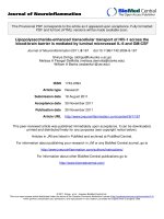
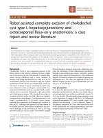
3.3. Morphology of narrow- and polydisperse RS-3 preparations
The RS-3 preparations were analysed on their morphology by scan
ning electron microscopy (Fig. 3). The images clearly show differences
between A- and B-type RS-3 crystals. The A-type RS-3 crystals seem to
consist of very tiny substructures that had been aggregated. The narrow
disperse B-type RS-3 crystals are regularly formed spherical particles,
except for sample sG65-B. sG65-B crystals seem to consist of smaller
particles, compared to the other narrow disperse B-type crystals. The
polydisperse B-type crystals show very different appearances: dHBPS-B
looks like sG2-B, which can be explained by a similar Mw and a rela
tively similar PI (Table 2). However, dWRS-B, dWPS-B and dAMPS-B,
which differ in Mw but all have a PI ≥ 1.50 do not show a regular
structure and seem to be more amorphous, although a clear crystal type
was confirmed by XRD (Fig. 2).
Previously, studies were performed on crystallization of debranched
amylopectins (Cai & Shi, 2013, 2014). The SEM images of the
debranched waxy maize starch spherulites showed similar morphology
as our narrow disperse B-type crystals (Fig. 3). In addition, Kiatponglarp
et al. (2016) studied crystallization of debranched native and waxy rice
starches (Kiatponglarp et al., 2016). These α-glucans all crystallized in a
B-type polymorph, but showed very different appearances. Their native
rice starch crystals showed a rough surface morphology, similar to our
sG65 crystals (Kiatponglarp et al., 2016). Also, Zeng, Zhu, Chen, Gao,
and Yu (2016)) studied morphology of crystallized α-glucans produced
by different drying methods (Zeng et al., 2016). Their air-dried
debranched waxy rice starch crystals greatly resembled our air-dried
dWRS crystals. In addition, narrow disperse α-glucans were previously
crystallized to A- and B-type crystals (Kobayashi et al., 2015). The B-type
crystals that had similar Mw values compared to the crystals in the
current study had the same morphology as we observed. However, the
previously produced A-type crystals showed a much more structured
morphology, which can be explained by precipitation with acetone
(Kobayashi et al., 2015) instead of self-assembly as in the present study.
It should be noted that our study focused on the retrogradation of
α-glucans from aqueous environment, mimicking resistant starch for
mation during cooking in a simplified way.
To summarize, 12 different RS-3 preparations were produced that
differed in crystal type (A/B), Mw (DPn ± 15, 20, 32 and 75) and PI (≤
1.25 or ≥1.35). These RS-3 preparations were used to study the effect of
crystal type, Mw and Mw distribution on resistance to digestion.
3.2. Crystallization of narrow- and polydisperse α-glucans
In order to produce RS-3 preparations differing in Mw, PI and crystal
type, the purified narrow disperse α-glucans were autoclaved and crys
tallized at 4 ◦ C or 50 ◦ C, according to Cai and Shi (2014), aiming at
B-type and A-type crystals, respectively. Different types of debranched
amylopectin were used as polydisperse equivalents of narrow disperse
synthesized sG2, sG5, sG20 and sG65, namely: debranched highly
branched potato starch (dHBPS), debranched waxy rice starch (dWRS),
debranched potato starch (dWPS) and debranched amylomaltase
modified potato starch (dAMPS), respectively. Crystallization was done
similarly to the narrow disperse α-glucans. The α-glucans of DP ≥ 32
were only stored at 4 ◦ C, since previous research showed that these al
ways crystallize in a B-type polymorph, irrespectively of crystallization
temperature (Cai & Shi, 2014). The crystal type of the α-glucans was
determined and their Mw distribution was analysed after solubilization
in NaOH (Table 2). The crystallization yield was calculated based on the
recovery of crystallized molecules (Table 2).
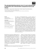
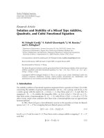
The results from X-ray diffraction show that crystallization at 4 and
50 ◦ C indeed resulted in the desired crystal polymorphs (Table 2).
Although differences in relative intensity of the peaks were observed
between the diffractograms of the crystallized α-glucans, still clear Aand B-type polymorphism could be recognized (Fig. 2). Previously, indepth studies were performed on identification of A- and B-type peak
positions of crystallized amylose (Kobayashi et al., 2015; Nishiyama
et al., 2010). The XRD patterns of our crystallized α-glucans match the
peak positions of Nishiyama et al. (2010), although differences in rela
tive intensities were observed.
Debranching of amylopectins of selected sources followed by crys
tallization resulted in crystals having similar Mw and crystal type
compared to their synthesized equivalents, but differing in poly
dispersity index. Despite large differences in PI and Mw distribution
(Supplementary Fig. 3), sG20-B and dWPS-B had a comparable Mw and
crystal type. The polydisperse equivalent of sG65-B (dAMPS) was found
to have a much lower average Mw compared to sG65-B (Table 2).
Therefore, these samples cannot be used to study the effect of PI on
resistance to digestion.
Crystallization yield was found to be highly dependent on DP and
crystallization temperature; at 50 ◦ C much lower yields were obtained
compared to crystallization at 4 ◦ C for α-glucans of the same Mw
(Table 2, A vs B-type crystals). In addition, the lower the DP, the lower
crystallization yields were found, although lower Mw α-glucans were
crystallized at higher concentrations (Table 2).
3.4. Digestibility of narrow and polydisperse RS-3 preparations
In order to investigate the effect of crystal type, Mw and Mw distri
bution on the resistance to digestion, the twelve narrow and poly
disperse RS-3 preparations were digested according to Englyst et al. and
Martens et al. (Englyst, Kingman, & Cummings, 1992; Martens et al.,
2018) (Fig. 4).
3.4.1. RS-3 A-type crystals are more resistant to digestion than B-type
crystals
Firstly, the results show that both dHBPS-A and dHBPS-B were
digested completely within 360 min and thus these RS-3 preparations
cannot be considered as RS-3, although being retrograded, insoluble and
showing a clear crystal type (Figs. 4A, 2). However, the results do show
that dHBPS-A (DPn 14) was slower digested than dHBPS-B (DPn 14),
indicating that B-type crystals were easier digested than A-type crystals.
Moreover, the narrow disperse A-type crystals (sG2-A, DPn 15) were
digested for 20 % during the first 60 min of digestion, whereas the
narrow disperse B-type crystals (sG2-B, DPn 15) were digested for 80 %
(Fig. 4A). Slower digestion of A-type crystals compared to B-type crys
tals was also observed for poly- and narrow disperse A- and B-type RS-3
preparations of DPn 18–22 (Fig. 4B). Therefore, it can be stated that Atype crystals were more resistant to digestion than B-type crystals,
comparing A- and B-type digestibility within one chain length. This
Table 2
Crystal type, Mw and Mw distribution and crystallization yield of purified nar
row and polydisperse RS-3 preparations.
α-glucan
Crystal type
DPncrystal
sG2-A
sG2-B
dHBPS-A
dHBPS-B
sG5-A
sG5-B
dWRS-A
dWRS-B
sG20-B
dWPS-B
sG65-B
dAMPS-B
A
B
A
B
A
B
A
B
B
B
B
B
15.6 ± 0.3
15.2 ± 0.1
14.3 ± 0.1
14.0 ± 0.1
18.0 ± 0.2
18.0 ± 0.0
21.4 ± 1.9
21.9 ± 0.5
31.6 ± 0.3
39.9 ± 0.7
75.6 ± 0.9
53.0 ± 2.3
PIcrystal
1.23 ±
1.25 ±
1.33 ±
1.35 ±
1.21 ±
1.21 ±
1.59 ±
1.50 ±
1.14 ±
2.11 ±
1.07 ±
1.67 ±
0.01
0.00
0.01
0.00
0.01
0.00
0.01
0.01
0.00
0.02
0.00
0.03
Crystallization
yield (%)
35 ± 1
86 ± 0
21 ± 1
45 ± 3
60 ± 2
89 ± 2
63 ± 2
79 ± 1
87 ± 8
78 ± 10
97 ± 3
86 ± 4
5
C.E. Klostermann et al.
Carbohydrate Polymers 265 (2021) 118069
Fig. 2. XRD profiles of narrow and polydisperse RS-3 preparations.
aligns with previous research showing that retrograded A-type crystals
of similar chain length were more resistant to digestion than B-type
crystals (Cai & Shi, 2014).
disperse α-glucans were more resistant to digestion than RS-3 prepara
tions made of polydisperse α-glucans, although no major differences
were found for most samples (Fig. 4A (dHBPS-B vs sG2-B), B, Table 3).
A-type crystals with a low PI and low Mw were found to be more
resistant than their polydisperse equivalent (dHBPS-A vs sG2-A or
dWRS-A vs sG5-A, Table 3). Interestingly, sG2-A was much more resis
tant to digestion than its polydisperse counterpart dHBPS-A (23 vs 100
% digestible, respectively). This, although their Mw and PI only differed
slightly from each other (Table 2). We hypothesize that a lower limit of
DPn 15 is needed to remain connected to the A-type crystal during
enzymatic digestion. Because of this, the dHBPS-A crystal was 100 %
digestible, whereas the sG2-A crystal was only digestible for 23 % after
360 min. B-type crystals with a low PI and DPn 32 (sG20-B) were much
more resistant to digestion than their polydisperse equivalents (dWPSB), with a difference in PI of 0.97 (Fig. 4C, Table 2). The morphology of
these crystals was very different, which might explain this difference
(Fig. 3). It can be stated that narrow disperse crystals were slightly more
resistant to digestion than polydisperse crystals.
3.4.2. RS-3 preparations of longer chain length α-glucans are more resistant
to digestion than that of shorter chain length, irrespectively of crystal type
The results also show that A-type crystals made of longer chain
length α-glucans were more resistant to digestion than A-type crystals
made of shorter chain length α-glucans (dHBPS-A vs dWRS-A, sG2-A vs
sG5-A, Fig. 4A & B, Table 3). In addition, polydisperse B-type crystals
made of longer chain length α-glucans were also more resistant to
digestion than polydisperse B-type crystals made of shorter chain length
α-glucans (dHBPS-B, dWRS-B, dWPS-B, dAMPS-B (Fig. 4A & B & C & D),
Table 3). Moreover, narrow disperse B-type crystals of longer DP were
also more resistant to digestion, although a minor difference in final
digestibility was observed between sG20-B and sG65-B (sG2-B, sG5-B,
sG20-B, sG65-B) (Fig. 4, Table 3). Therefore, it can be stated that RS-3
preparations made of longer chain length α-glucans were more resis
tant to digestion, compared to RS-3 preparations made of shorter chain
length α-glucans, irrespectively of crystal type.
3.5. Digestion affects Mw (distribution) of especially B-type RS-3 crystals
that remain after digestion
3.4.3. RS-3 preparations of narrow disperse α-glucans are slightly more
resistant to digestion than that of polydisperse α-glucans
Lastly, the results show that RS-3 preparations made of narrow
The RS-3 crystals that resist digestion in the small intestine will
arrive in the colon where they might be degraded and fermented by
6
C.E. Klostermann et al.
Carbohydrate Polymers 265 (2021) 118069
Fig. 3. Scanning electron microscopic images of RS-3 preparations differing in Mw, Mw distribution and crystal type. Sample codes are explained in Table 2.
Fig. 4. In vitro digestion profiles of narrow and polydisperse RS-3 preparations. A (DP ± 15): dHBPS, sG2; B (DP ± 20): dWRS, sG5; C (DP ± 32): dWPS, sG20; D (DP
≥ 50): dAMPS, sG65. □ = B-type crystal, ○ = A-type crystal. Digestibility curves of dHBPS are from 5 individually produced samples, all others are from in triplicate
produced samples, all digested in duplicate.
specific gut microbiota. To examine whether these remaining RS-3
crystals had physically been changed due to the attack of pancreatic
α-amylase, the Mw and PI of the remaining crystals that escaped
digestion was analysed (Table 3).
The results show that for most remaining RS-3, digestion only had a
minor effect on the Mw and PI compared to the undigested crystalline
α-glucans (sG2-A, sG5-A, dWRS-A, sG20-B, dWPS-B) (Tables 2 & 3). This
indicates that in most digestions, pancreatic α-amylase hydrolysed some
crystals completely, whereas others were completely untouched. How
ever, for some other samples a change in Mw and PI can be observed
(sG5-B, dWRS-B, sG65-B). sG5-B crystals decreased in Mw, whereas
their PI remained similar after digestion. This indicates that for sG5-B,
all crystals were hydrolysed to a certain extent, without a preference
for either longer or shorter α-glucans within the crystal. Furthermore,
7
C.E. Klostermann et al.
Carbohydrate Polymers 265 (2021) 118069
crystals (Cai & Shi, 2014). As proposed by Dhital et al. (2017), digestion
of retrograded starches is probably limited due to a combination of slow
enzyme binding to the surface of the substrate and slow catalysis in the
active site (Dhital et al., 2017). Retrograded A-type crystals have a much
denser structure, containing less water molecules than B-type crystals
(Buleon et al., 2007). Due to this dense structure, it might be that A-type
crystals are not recognized by the surface binding sites of the enzyme. In
addition, due to this dense structure, it seems likely that A-type crystals
get much harder into solution compared to B-type crystals, therefore
limiting enzymatic hydrolysis. Our results also show that digestion of
A-type crystals reached a certain plateau value after 120 min (Fig. 4).
Since we have observed that this plateau value is reached after 120 min
of digestion and no change in Mw was found due to digestion, we pro
pose that although the crystals were bound to the surface binding site,
the retrograded A-type crystals are resistant to digestion due to limited
catalytic activity by the enzyme; the catalytic centre of pancreatic
α-amylase was unable to hydrolyse further, probably due to the dense
structure of A-type crystals.
In case of B-type crystals that have a high PI, we propose that the
limited digestion is related to the slow binding to the surface binding site
of the enzyme rather than the catalytic activity of the enzyme, since we
did not reach plateau values at 120 or even after 360 min of digestion
(Fig. 4). Narrow disperse B-type crystals were shown to be more resis
tant to digestion compared to polydisperse B-type crystals (Fig. 4,
Table 3). Because of the low PI it seems likely that crystallization of
narrow disperse α-glucans resulted in more perfect crystals, compared to
polydisperse α-glucans (Fig. 3). Consequently, narrow disperse α-glu
cans within the crystal are less likely to go into solution and are less
hydrolysed, compared to crystals made of polydisperse α-glucans. Nar
row disperse B-type crystals of DP ≥ 32 were shown to be very resistant
to digestion (Fig. 4). Whereas sG65-B (DP 75) did not reach a plateau
value after 360 min of digestion, sG20-B (DP 32) did. Therefore, based
on our results we cannot conclude whether resistance to digestion of
narrow disperse B-type crystals is more related to limited binding to the
surface binding site of the enzyme or to limited catalytic activity in the
active site of the enzyme. Furthermore, our results have shown that RS-3
preparations produced from low Mw α-glucans (DP ≤ 14) cannot be
considered RS, since they were fully digested within 120 min, although
insoluble. Unfortunately, we were not able to confirm our hypothesis on
differences in digestibility mechanism by pancreatic α-amylase by SEM
on these digested samples without major sample pre-treatment that
might influence the outcome. However, previous research by others has
not shown major differences in morphology of the α-glucan crystals due
to enzymatic digestion (Ziegler, 2020).
Our research is the first that used enzymatic synthesis from sucrose
for the production of RS-3 with defined and narrow distributed chain
length. Twelve unique RS-3 preparations were produced of which half
were enzymatically synthesized and narrow disperse. The other six RS-3
preparations were produced by debranching amylopectins of different
botanical sources to obtain polydisperse equivalents of similar average
Mw compared to the narrow disperse α-glucans. From these twelve
samples, four A-type crystals and eight B-type crystals were produced.
Because of this relatively large number of unique samples, we were able
Table 3
Molecular weight and polydispersity (changes) of RS-3 crystals remaining after
360 min of in vitro digestion, together with total digestibility (%).
Sample
name
Digestibility
(%)
DPncrystal
PIcrystal
ΔDPncrystal
(%)
ΔPIcrystal
(%)
sG2-A
23 ± 4
1.5
89 ± 2
100 %
100 %
13 ± 3
n.a.
n.a.
n.a.
− 5.9
n.a.
n.a.
n.a.
1.5
sG5-B
81 ± 4
− 41.9
1.1
dWRS-A
20 ± 0
13.7
− 4.1
dWRS-B
91 ± 3
− 18.5
− 4.5
sG20-B
3±1
− 2.5
− 3.7
dWPS-B
33 ± 4
4.4
− 4.7
sG65-B
8±1
− 10.1
2.6
dAMPS-B
12 ± 2
1.25 ±
0.01
n.a.
n.a.
n.a.
1.22 ±
0.01
1.22 ±
0.03
1.53 ±
0.01
1.43 ±
0.06
1.09 ±
0.00
2.01 ±
0.02
1.10 ±
0.01
1.69 ±
0.02
− 2.7
sG2-B
dHBPS-A
dHBPS-B
sG5-A
15.2 ±
0.5
n.a.
n.a.
n.a.
17.0 ±
0.8
12.7 ±
2.6
24.8 ±
0.3
18.5 ±
1.2
30.9 ±
0.5
41.8 ±
1.5
68.5 ±
1.2
50.0 ±
0.8
− 6.2
1.4
dWRS-B crystals decreased in both Mw and in PI, which indicates that all
crystals were hydrolysed to a certain extent and interestingly, pancreatic
α-amylase caused narrowing of the PI. In contrast, sG65-B crystals also
decreased in average Mw but increased slightly in PI. This indicates that
pancreatic α-amylase hydrolysed some α-glucans within the sG65-B
crystals to a certain extent. Probably, the hydrolysed α-glucan
remained connected to the insoluble sG65-B crystal, thereby limiting
further hydrolysis and therefore increasing the PI.
To understand how the digestion of dWRS A- and B-type crystals
occurred, the digestion was monitored in time and remaining crystals
that escaped digestion were analysed on Mw distribution (Fig. 5).
The results show that A-type crystals did not change in Mw over
time. Therefore, we state that the crystals were digested in a crystal-bycrystal manner: some crystals were hydrolysed completely, whereas
others were untouched. However, dWRS-B type crystals changed in Mw
due to digestion: the crystals consisted of a bimodal distribution at t = 0,
which changed slowly over time to a normal distribution after 6 h of
digestion (Fig. 5).
Although activity of pancreatic α-amylase was studied extensively
from a biochemistry point of view in the past, not much research is
performed on the activity of pancreatic α-amylase on insoluble sub
strates and even less literature can be found on activity of pancreatic
α-amylase on RS-3. Previously, it was revealed that human pancreatic
α-amylase has two starch surface binding sites: one that binds to soluble
starch molecules and another that binds to insoluble starch granules
(Zhang et al., 2016). Whether this starch surface binding site is also able
to bind insoluble RS-3, is still unknown.
Our research and that of others has shown that retrograded A-type
crystals were more resistant to digestion than retrograded B-type
Fig. 5. HPSEC profile of A) remaining dWRS-A and B) remaining dWRS-B crystals after 0, 20, 60 and 360 min of digestion.
8
Carbohydrate Polymers 265 (2021) 118069
C.E. Klostermann et al.
to study the effect of crystal type, Mw and Mw distribution on di
gestibility. Our rather unique approach allowed us to study for the first
time the structural properties of the RS-3 crystals that escaped enzy
matic hydrolysis by pancreatic α-amylase. Rather than only analysing
the released glucose after in vitro digestion, we also analysed the
remaining RS-3 crystals on Mw distribution. This makes it possible to not
only predict the amount of RS-3 that enters the colon, but also to un
derstand the substrate for beneficial gut microbes in the colon. Our re
sults suggest that pre-digestion experiments of B-type crystals are of
importance before studying the degradation and utilisation of B-type RS3 by gut microbiota, whereas pre-digestion is hardly of any value when
exploring fermentability of A-type crystals.
Buleon, A., Veronese, G., & Putaux, J. L. (2007). Self-association and crystallization of
amylose. Australian Journal of Chemistry, 60(10), 706–718.
Cai, L. M., & Shi, Y. C. (2010). Structure and digestibility of crystalline short-chain
amylose from debranched waxy wheat, waxy maize, and waxy potato starches.
Carbohydrate Polymers, 79(4), 1117–1123.
Cai, L. M., & Shi, Y. C. (2013). Self-assembly of short linear chains to A- and B-type starch
spherulites and their enzymatic digestibility. Journal of Agricultural and Food
Chemistry, 61(45), 10787–10797.
Cai, L. M., & Shi, Y. C. (2014). Preparation, structure, and digestibility of crystalline Aand B-type aggregates from debranched waxy starches. Carbohydrate Polymers, 105,
341–350.
Chang, R., Xiong, L., Li, M., Liu, J., Wang, Y., Chen, H., et al. (2018). Fractionation of
debranched starch with different molecular weights via edible alcohol precipitation.
Food Hydrocolloids, 83, 430–437.
Creek, J. A., Ziegler, G. R., & Runt, J. (2006). Amylose crystallization from concentrated
aqueous solution. Biomacromolecules, 7(3), 761–770.
Dhital, S., Warren, F. J., Butterworth, P. J., Ellis, P. R., & Gidley, M. J. (2017).
Mechanisms of starch digestion by alpha-amylase-Structural basis for kinetic
properties. Critical Reviews in Food Science and Nutrition, 57(5), 875–892.
Englyst, H. N., Kingman, S. M., & Cummings, J. H. (1992). Classification and
measurement of nutritionally important starch fractions. European Journal of Clinical
Nutrition, 46(Suppl. 2), S33–50.
Fuentes-Zaragoza, E., Sanchez-Zapata, E., Sendra, E., Sayas, E., Navarro, C., FernandezLopez, J., et al. (2011). Resistant starch as prebiotic: A review. Starch-Starke, 63(7),
406–415.
Gidley, M. J., & Bulpin, P. V. (1987). Crystallization of Maltooligosaccharides as models
of the crystalline forms of starch - minimum chain-length requirement for the
formation of double helices. Carbohydrate Research, 161(2), 291–300.
Haenen, D., Zhang, J., Souza da Silva, C., Bosch, G., van der Meer, I. M., van Arkel, J.,
et al. (2013). A diet high in resistant starch modulates microbiota composition, SCFA
concentrations, and gene expression in pig intestine. The Journal of Nutrition, 143(3),
274–283.
Haralampu, S. G. (2000). Resistant starch - a review of the physical properties and
biological impact of RS3. Carbohydrate Polymers, 41(3), 285–292.
Hu, X., Huang, Z., Zeng, Z., Deng, C., Luo, S., & Liu, C. (2020). Improving resistance of
crystallized starch by narrowing molecular weight distribution. Food Hydrocolloids,
103, Article 105641.
Huang, Z., Zeng, Z., Gao, Y., Liu, C., Wu, J., & Hu, X. (2019). Crystallization of shortchain amylose: Effect of precipitant. Starch-Starke, 71(9–10), Article 1900007.
Kiatponglarp, W., Rugmai, S., Rolland-Sabate, A., Buleon, A., & Tongta, S. (2016).
Spherulitic self-assembly of debranched starch from aqueous solution and its effect
on enzyme digestibility. Food Hydrocolloids, 55, 235–243.
Kiatponglarp, W., Tongta, S., Rolland-Sabate, A., & Buleon, A. (2015). Crystallization and
chain reorganization of debranched rice starches in relation to resistant starch
formation. Carbohydrate Polymers, 122, 108–114.
Kobayashi, K., Kimura, S., Naito, P. K., Togawa, E., & Wada, M. (2015). Thermal
expansion behavior of A- and B-type amylose crystals in the low-temperature region.
Carbohydrate Polymers, 131, 399–406.
L´epine, A. F. P., de Hilster, R. H. J., Leemhuis, H., Oudhuis, L., Buwalda, P. L., & de
Vos, P. (2018). Higher chain length distribution in debranched type-3 resistant
starches (RS3) increases TLR signaling and supports dendritic cell cytokine
production. Molecular Nutrition & Food Research, 63(2), Article 1801007.
Luley-Goedl, C., & Nidetzky, B. (2010). Carbohydrate synthesis by disaccharide
phosphorylases: Reactions, catalytic mechanisms and application in the
glycosciences. Biotechnology Journal, 5(12), 1324–1338.
Martens, B. M. J., Gerrits, W. J. J., Bruininx, E., & Schols, H. A. (2018). Amylopectin
structure and crystallinity explains variation in digestion kinetics of starches across
botanic sources in an in vitro pig model. Journal of Animal Science and Biotechnology,
9, 91.
Montesanti, N., Veronese, G., Buleon, A., Escalier, P. C., Kitamura, S., & Putaux, J. L.
(2010). A-type crystals from dilute solutions of short amylose chains.
Biomacromolecules, 11(11), 3049–3058.
Nishiyama, Y., Putaux, J. L., Montesanti, N., Hazemann, J. L., & Rochas, C. (2010). B–&A
Allomorphic transition in native starch and amylose spherocrystals monitored by in
situ synchrotron X-ray diffraction. Biomacromolecules, 11(1), 76–87.
Ohdan, K., Fujii, K., Yanase, M., Takaha, T., & Kuriki, T. (2006). Enzymatic synthesis of
amylose. Biocatalysis and Biotransformation, 24(1-2), 77–81.
Pfannemuller, B. (1987). Influence of chain length of short monodisperse amyloses on
the formation of A-and B-type X-ray diffraction patterns. International Journal of
Biological Macromolecules, 9(2), 105–108.
Qi, P., You, C., & Zhang, Y.-H. P. (2014). One-pot enzymatic conversion of sucrose to
synthetic amylose by using enzyme cascades. ACS Catalysis, 4(5), 1311–1317.
Roger, P., Axelos, M. A. V., & Colonna, P. (2000). SEC-MALLS and SANS studies applied
to solution behavior of linear alpha-glucans. Macromolecules, 33(7), 2446–2455.
van den Broek, L. A., van Boxtel, E. L., Kievit, R. P., Verhoef, R., Beldman, G., &
Voragen, A. G. (2004). Physico-chemical and transglucosylation properties of
recombinant sucrose phosphorylase from Bifidobacterium adolescentis DSM20083.
Applied Microbiology and Biotechnology, 65(2), 219–227.
van der Maarel, M., & Leemhuis, H. (2013). Starch modification with microbial alphaglucanotransferase enzymes. Carbohydrate Polymers, 93(1), 116–121.
van der Vlist, J., Reixach, M. P., van der Maarel, M., Dijkhuizen, L., Schouten, A. J., &
Loos, K. (2008). Synthesis of branched polyglucans by the tandem action of potato
phosphorylase and Deinococcus geothermalis glycogen branching enzyme.
Macromolecular Rapid Communications, 29(15), 1293–1297.
4. Conclusions
Our study is the first to investigate the role of crystal type, Mw and
Mw distribution on the resistance to digestion of RS-3 preparations on
both released glucose after in vitro digestion and on the crystals that
escaped digestion. It has been found that A-type crystals are much more
resistant to digestion than B-type crystals, potentially caused by a
reduced catalytic activity of pancreatic α-amylase towards A-type crys
tals. A-type crystals are digested in a crystal-by-crystal manner and
therefore the Mw and Mw distribution of the remaining A-type crystals
does not change. Resistance to digestion of B-type crystals is potentially
caused by limited binding to the surface binding site of pancreatic
α-amylase. In contrast to remaining A-type crystals, remaining B-type
crystals change in Mw and/or PI which might be due to surfacehydrolysis by pancreatic α-amylase. Narrow disperse RS-3 prepara
tions are slightly more resistant to digestion than polydisperse ones and
crystals made of higher DP α-glucans are more resistant than that of
lower DP α-glucans, irrespectively of crystal type. In addition, RS-3
preparations of DP ≤ 14 cannot be considered RS, since they are 100
% digestible by pancreatic α-amylase, although insoluble. This study can
help to design RS-3 preparations with a preferred degree of digestibility.
Author statement
Cynthia Klostermann: Methodology, Investigation, Writing –
Original Draft; Piet Buwalda: Conceptualization, Supervision; Hans
Leemhuis: Resources, Writing – Review & Editing; Paul de Vos:
Conceptualization, Funding acquisition, Writing – Review & Editing;
Henk Schols: Supervision, Writing – Review & Editing; Harry Bitter:
Supervision, Writing – Review & Editing
Acknowledgements
This project is jointly funded by the Dutch Research Council (NWO),
AVEBE, FrieslandCampina and NuScience as coordinated by the Car
bohydrate Competence Center (CCC-CarboBiotics; www.cccresearch.
nl).
Appendix A. Supplementary data
Supplementary material related to this article can be found, in the
online version, at doi: />References
Bermudez-Brito, M., Rosch, C., Schols, H. A., Faas, M. M., & de Vos, P. (2015). Resistant
starches differentially stimulate Toll-like receptors and attenuate proinflammatory
cytokines in dendritic cells by modulation of intestinal epithelial cells. Molecular
Nutrition & Food Research, 59(9), 1814–1826.
Birt, D. F., Boylston, T., Hendrich, S., Jane, J. L., Hollis, J., Li, L., et al. (2013). Resistant
starch: Promise for improving human health. Advances in Nutrition, 4(6), 587–601.
Bradford, M. M. (1976). A rapid and sensitive method for the quantitation of microgram
quantities of protein utilizing the principle of protein-dye binding. Analytical
Biochemistry, 72, 248–254.
9
C.E. Klostermann et al.
Carbohydrate Polymers 265 (2021) 118069
Yanase, M., Takaha, T., & Kuriki, T. (2006). Alpha-Glucan phosphorylase and its use in
carbohydrate engineering. Journal of the Science of Food and Agriculture, 86(11),
1631–1635.
Zaman, S. A., & Sarbini, S. R. (2016). The potential of resistant starch as a prebiotic.
Critical Reviews in Biotechnology, 36(3), 578–584.
Zeng, F., Zhu, S. M., Chen, F. Q., Gao, Q. Y., & Yu, S. J. (2016). Effect of different drying
methods on the structure and digestibility of short chain amylose crystals. Food
Hydrocolloids, 52, 721–731.
Zhang, X., Caner, S., Kwan, E., Li, C., Brayer, G. D., & Withers, S. G. (2016). Evaluation of
the significance of starch surface binding sites on human pancreatic alpha-amylase.
Biochemistry, 55(43), 6000–6009.
Ziegler, G. R. (2020). Enzyme-resistant starch spherulites. Starch-Starke, 72(7-8), Article
1900217.
10