Evaluation of chitosan crystallinity: A high-resolution solid-state NMR spectroscopy approach
Bạn đang xem bản rút gọn của tài liệu. Xem và tải ngay bản đầy đủ của tài liệu tại đây (5.53 MB, 14 trang )
Carbohydrate Polymers 250 (2020) 116891
Contents lists available at ScienceDirect
Carbohydrate Polymers
journal homepage: www.elsevier.com/locate/carbpol
Evaluation of chitosan crystallinity: A high-resolution solid-state NMR
spectroscopy approach
William Marcondes Facchinatto a, *, Danilo Martins dos Santos b, Anderson Fiamingo c,
Rubens Bernardes-Filho b, S´ergio Paulo Campana-Filho a, Eduardo Ribeiro de Azevedo c, Luiz
Alberto Colnago b
a
S˜
ao Carlos Institute of Chemistry, University of S˜
ao Paulo, Av. Trabalhador Sao-Carlense 400, CEP 13566-590, Caixa Postal 780, S˜
ao Carlos, S˜
ao Paulo, Brazil
Brazilian Corporation for Agricultural Research, Embrapa Instrumentation, Rua XV de Novembro 1452, CEP 13560-970, Caixa Postal 741, S˜
ao Carlos, S˜
ao Paulo,
Brazil
c
S˜
ao Carlos Institute of Physics, University of S˜
ao Paulo, Av. Trabalhador Sao-Carlense 400, CEP 13566-590, Caixa Postal 369, S˜
ao Carlos, S˜
ao Paulo, Brazil
b
A R T I C L E I N F O
A B S T R A C T
Keywords:
Chitosan
Crystallinity
High-resolution SSNMR spectroscopy
We propose a novel approach relied on high-resolution solid-state 13C NMR spectroscopy to quantify the crys
tallinity index of chitosans (Ch) prepared with variable average degrees of acetylation (DA) from 5% to 60 % and
average weight molecular weight (Mw ) ranged in 0.15 × 10⁶ g mol− 1–1.2 × 10⁶ g mol− 1. The Dipolar Chemical
Shift Correlation (DIPSHIFT) curve of the C(6)OH segment revealed increased mobility dynamic, which induced
different distribution from trans-to-gauche conformations in relation to C(4). Indeed, 1H-13C Heteronuclear
Correlation (2D HETCOR) showed that distinguished C4 chemical shifts correlates with the same aliphatic
protons. The short-range ordering can be assigned to C4/C6 signals on 13C CPMAS and, for our case, the
deconvolution procedure between disordered and ordered phases revealed increasing crystallinity with DA, as
confirmed by SVD multivariate analysis. This work extended the knowledge regarding the use of 13C CPMAS
technique to predict the crystallinity of chitosans without the use of amorphous standards.
1. Introduction
Chitosan (Ch) is a linear (1 → 4)-linked copolymer composed of 2amino-2-deoxy-β-D-glucan (GlcN) and 2-acetamido-2-deoxy-β-D-glucan
(GlcNAc) units, generally prepared from N-deacetylation of chitin, an
aminopolysaccharide predominatly formed by GlcNAc units (Gonil &
Sajomsang, 2012; Kang et al., 2018; Kaya et al., 2017). The physico
chemical properties, in vivo degradation, biological activity and pro
cessability of chitosan is affected by its degree of N-acetylation, DA
(Chatelet, Damour, & Domard, 2001; Schipper, Vårum, & Artursson,
1996), distribution of N-acetylated units (Aiba, 1992; Kumirska et al.,
ăming, 2009) and molecư
2009; Weinhold, Sauvageau, Kumirska, & Tho
ular weight (Huang, Khor, & Lim, 2004; Kubota & Eguchi, 2005; Mao
et al., 2004; Richardson, Kolbe, & Duncan, 1999). In this sense, the
structural characterization of chitosan is of utmost importance for the
proper selection of this biopolymer according to the desired application,
mostly in the fields of drug delivery (Wei, Ching, & Chuah, 2020), tissue
engineering (Ahmed, Annu, Ali, & Sheikh, 2018; Islam, Shahruzzaman,
Biswas, Nurus Sakib, & Rashid, 2020), biosensing (Baranwal et al.,
2018; Pavinatto et al., 2017), wound dressing (Ahmed & Ikram, 2016;
Miguel, Moreira, & Correia, 2019) and wastewater treatment (Reddy &
Lee, 2013; Sarode et al., 2019).
Chitosan exhibit polymorphic forms designed in three crystal types
named as α, β, γ in function of the packing and polarities of adjacent
chains in successive sheets (Zhou et al., 2011). The different allomorphs
account for the different intersheet accessibility to small molecules and
crystallinity, which is on turn, strongly related to the solubility (Kurita,
Kamiya, & Nishimura, 1991; Sogias, Khutoryanskiy, & Williams, 2010),
swelling behavior (Guibal, 2004; Gupta & Jabrail, 2006; Saito, Okano,
Gaill, Chanzy, & Putaux, 2000), sorption kinetics of toxic metal ions in
aqueous solutions (Milot, McBrien, Allen, & Guibal, 1998; Piron &
Domard, 1998) and reactivity (Kurita, Ishii, Tomita, Nishimura, & Shi
moda, 1994; Lamarque, Viton, & Domard, 2004). Additionally, several
studies describe that besides polymorphism, the DA acts as an important
structural feature partially controlling the crystallinity and related
properties, such as hydrophilicity (Gupta & Jabrail, 2006),
* Corresponding author.
E-mail address: (W.M. Facchinatto).
/>Received 13 June 2020; Received in revised form 26 July 2020; Accepted 3 August 2020
Available online 13 August 2020
0144-8617/© 2020 Elsevier Ltd. This article is made available under the Elsevier license ( />
W.M. Facchinatto et al.
Carbohydrate Polymers 250 (2020) 116891
water-sorption capacity (Ioelovich, 2014) and susceptibility to enzy
matic degradation (Cardozo, Facchinatto, Colnago, Campana-Filho, &
Pessoa, 2019). Indeed, the lowest enzymatic degradation rates has been
achieved for DA < 15 % (Francis Suh & Matthew, 2000), being also
desirable a partially N-deacetylation for higher probability to form a
lysozyme-substrate complex (Cho, Jang, Park, & Ko, 2000; Hirano,
Tsuchida, & Nagao, 1989). Higher digestibility is achieved when DA
values are ranged in 40 %–80 % with lesser probability for a random
distribution of acetamido groups (Aiba, 1992; Hirano et al., 1989). Thus,
the crystallinity increases with DA and the crystalline regions grows on
segments containing blocks of N-acetylated units (Ogawa & Yui, 1993).
The crystallinity of polysaccharides has been evaluated by X-ray
diffraction models and different spectroscopy techniques (Åkerholm,
Hinterstoisser, & Salm´
en, 2004; Park, Baker, Himmel, Parilla, & John
son, 2010; Schenzel, Fischer, & Brendler, 2005). Currently, the Ch
crystallinity has been quantified considering the long-range ordering on
XRD patterns usually through the peak height (Focher, Beltrame, Naggi,
& Torri, 1990; Struszczyk, 1987), deconvolution methods (Cho et al.,
2000) or based on subtraction of a diffraction pattern using one from an
amorphous Ch as reference (Osorio-Madrazo et al., 2010). The first fails
by not considering the contribution of (110)a reflection from anhydrous
allomorph near to the amorphous halo intensity at 16.0◦ , the second has
overestimated the contribution of amorphous phase by fitting a cubic
spline curve in the diffraction pattern, while the third proposes a labo
rious method for a routine evaluation of Ch crystallinity, using a totally
amorphous samples which is usually not available. Studies has shown
that for the same sample, the crystallinity index can also vary within a
wide range from 57.0 to 93.0 % for chitin (Fan, Saito, & Isogai, 2009;
Fan, Saito, & Isogai, 2008) and from 40.0 to 80.0 % for chitosan
´ ska, Amietszajew, & Borysiak, 2017; Pires, Vilela, &
(Grząbka-Zasadzin
Airoldi, 2014; Yuan, Chesnutt, Haggard, & Bumgardner, 2011)
depending on the calculation method. Consequently, the accurate esti
mative of crystallinity through XRD is considerable doubtful.
In this context, high-resolution solid-state nuclear magnetic reso
nance, SSNMR, spectroscopy has been one of the most used techniques
because chemical shift dependence on local molecular conformations
(Tonelli & Schilling, 1981). Because the local chain conformation
(trans-gauche) changes the current electronic structure around 13C
nuclei, its nuclear magnetization become distinct allowing to distinguish
between ordered and disordered populations. For instance, 13C CPMAS
Solid-State NMR has been used to evaluate the fraction of
interior-to-surface crystallites in cellulose (Bernardinelli, Lima,
Rezende, Polikarpov, & DeAzevedo, 2015; Park et al., 2010; Viă
etor,
Newman, Ha, Apperley, & Jarvis, 2002; Wang & Hong, 2016), starch
(Mutungi, Passauer, Onyango, Jaros, & Rohm, 2012; Villas-Boas, Fac
chinatto, Colnago, Volanti, & Franco, 2020) and polyglycans (Webster,
Osifo, Neomagus, & Grant, 2006) usually referred as NMR crystallinity
index. This can be typically achieved and widely applied using the C4
and C6 carbons from cellulose and C1 carbon from starch, which the
splitting is directly associated to signals arising from ordered and
disordered molecular segments. One should point out that NMR and
X-ray crystalline index are not identical in the sense that in solid-state
NMR it reflects the local conformation and population distribution,
while in X-ray it is related to the long range order. However, they are
close related in the sense that local order can be strongly influenced by
long range order. In this sense, using NMR and X-ray diffraction together
can be a valuable way of improving the information about the micro
structure of chitosans.
Despite the structural similarity with cellulose, a clear C4 signal split
in 13C CPMAS spectra of Ch has been only observed in samples with low
acetylated content (Heux, Brugnerotto, Desbri`eres, Versali, & Rinaudo,
2000; Silva et al., 2017). This has been attributed to a greater mobility of
amorphous region achieved through thermal treatment above 150 ◦ C
(Focher et al., 1990). The C1 and C4 signals shape of Ch salts have been
ˆ,
also interpreted as consequence of twofold helical conformations (Saito
Tabeta, & Ogawa, 1987), being highly sensitive to conformational
changes on glycosidic linkages (Harish Prashanth, Kittur, & Thar
anathan, 2002; Tanner, Chanzy, Vincendon, Claude Roux, & Gaill,
1990). The signal split into doublets and sharp singlets were found on
hydrated (tendom) and annealed chitosan forms, being influenced by
ˆ
chitin source, molecular weight and content of water molecules (Saito
et al., 1987). However, the origin of this signal splitting is still contro
versy (Focher, Naggi, Torri, Cosani, & Terbojevich, 1992) and none
study has satisfactory investigated the short-range ordering with the
spectral shape variability of these carbon signals from different DA and
molar masses, without submitting Ch to any kind of physicochemical
treatment.
In this sense, considering the strong relationship between N-acety
lation and crystallinity of Ch, its unclear dependence with molar masses
(Ogawa & Yui, 1993), the lacking aspect of reliable crystallinity quan
tification by XRD and the conformational influence on SSNMR spectra,
this study aims to propose a novel and straightforward approach to es
timate the crystallinity through the short-range molecular ordering from
chitin to chitosan without conducting any treatment onto products. Ch
samples possessing variable DA and average weight average molecular
weight (Mw ) were produced and evaluated through 13C CPMAS SSNMR
experiments, conducted as the main techniques, while Dipolar Chemical
Shift Correlation (DIPSHIFT) (Munowitz, Griffin, Bodenhausen, &
Huang, 1981) and the 1H-13C Heteronuclear Correlation (HETCOR)
ărster, & De Groot, 1997) were used as auxiliary
(Van Rossum, Fo
methods for signal assignments. By using this approach, a
non-destructive method was developed to simultaneously quantify in a
reliable manner the crystallinity and the DA of Ch.
2. Experimental
2.1. Materials
Low molecular weight chitosan (ChC, 87 kDa, DA ≈ 5.0 %) (Cheng
Yue Plating® Co. Ltd. Chang, China) was purified according to the
methodology described by Santos, Bukzem, and Campana-Filho (2016).
The allomorph alfa-chitin (αCh), obtained from shrimp shells (Sig
ma-Aldrich® Co. St. Louis, MO, USA), was used without further
purification.
The allomorph beta-chitin (βCh) was extracted from the squid pens
(Doryteuthis spp.) (Lavall, Assis, & Campana-Filho, 2007), milled and
sieved into powder sizes with average diameters (d) ranged in 0.125 <
d < 0.425 mm, then submitted to multistep ultrasound-assisted deace
tylation process (USAD) to produce Ch samples with variable DA (Fia
mingo, Delezuk, Trombotto, David, & Campana-Filho, 2016). In brief,
the βCh/NaOH 40 % (w/w) aqueous suspension was placed in a jacked
glass reactor (θint =3.5 cm) and kept under magnetic stirring with a
circulating thermostat at 60 ± 1 ◦ C, then sonicated with UP400S
Hielscher® Sonifier ultrasonic device (ν = 24 kHz) coupled to θ = 22
mm stepped probe for pulsed irradiation. The deacetylation reaction was
carried out at 200 W for 50 min and then stopped by cooling and
neutralization with HCl 3.0 mol L− 1, followed by filtration under posi
tive pressure through a 0.45 μm porous membrane (Millipore®, White
SCWP). The resulting product, named as Ch1x, was freeze-dried at − 45
◦
´s®). This process was sequentially
C for 24 h (Liotop L101, Liobra
applied to this sample at the same conditions to produce Ch2x and then
similarly to produce Ch3x, an extensively deacetylated chitosan.
2.2. Depolymerization of chitosan
Chitosans possessing different average molecular weights were pre
pared by submitting the samples Ch1x, Ch2x and Ch3x to homogeneous
depolymerization via ultrasound treatment for 3 h and 6 h. Thus, 5.0 g of
a given Ch was suspended in 500.0 ml of acetic acid 1.0 % (v/v) con
tained in a 1 L jacked glass reactor (θint = 10 cm) and subjected to
2
W.M. Facchinatto et al.
Carbohydrate Polymers 250 (2020) 116891
ultrasound pulsed irradiation at 200 W (60 ± 1 ◦ C) for the desired time
by using the same operational parameters already described for deace
tylation process. The products were neutralized by adding NaOH 0.1
mol L− 1, filtered under positive pressure (0.45 μm) and then sequentially
washed with ethanol 80 % (v/v) and deionized water. The resulting
products were freeze-dried at − 45 ◦ C for 24 h and named Chwxy, where
“w” (1, 2 and 3) identify the parent Ch and “y” (3 h and 6 h) the time of
ultrasound treatment.
viscometry (Cardozo et al., 2019). The SEC measurements were con
ducted on Agilent® 1100 coupled to a refractive index detection module
(RID-6A), pre-columns Shodex Ohpakđ SB-G (50 ì 6 mm) (10 μ)/
SB-803-HQ (8 mm DI x 300 mm) (6μ)/ SB-805-HQ (8 mm DI × 300 mm)
(13 μ), stationary phase consisting of polyhydromethacrylate gel and
mobile phase (eluent) constituted by 0.3 M acetic acid / 0.2 M sodium
acetate buffer. Following, Ch solutions 1.0 mg mL− 1 were prepared in
the same buffer and analyzed under the flow rate of 0.6 ml min-1 at 35
◦
C. The Mw values were obtained from the calibration curve constructed
by monodisperse pullulan (708,000; 344,000; 200,000; 107,000; 47,
100; 21,100; 9600 and 5900 g mol-1), cellobiose (343.2 g mol-1) and
glucose (180.2 g mol-1) standards. The viscometry analysis were per
formed in a glass capillary (ϕ = 0.53 mm) containing 15 ml of chitin
dissolved in N,N-dimethylacetamide/5% LiCl (w/w) at low concentra
tions (1.2 < ηrel < 2.0) using the AVS-360 viscometer coupled to an
ăteđ, Germany) at 25.00 0.01 C. The
automatic burette (Schott-Gera
Mv values were calculated from the parameters K’ = 2.4 × 10⁻⁴ L g-1 and
α = 0.69 and by means of intrinsic viscosities, [η], according to
Mark-Houwink-Sakurada equation, obtained from the extrapolation of
reduced viscosity curves to infinite dilution. The weight average degree
of polymerization of Ch (DPw ) and viscosity average degree of poly
merization of chitin allomorphs (DPv ) were calculated considering the
relative amount of GlcNAc (203 g mol− 1) and GlcN (161 g mol− 1), as
described by the Eq. (3)
2.3. N-acetylation of chitosan
Chitosans with a predicted and wide-ranged DA were obtained by
performing the homogeneous N-acetylation reaction onto Ch3x with
acetic anhydride at molar ratios 0, 0.02, 0.20, 0.40, 0.60, 0.90 of an
hydride/glucosamine, as similarly reported elsewhere (Lavertu, Darras,
& Buschmann, 2012; Sorlier, Denuzi`
ere, Viton, & Domard, 2001). Thus,
0.5 g of Ch3x was suspended in 50.0 ml of acetic acid 1.0 % (v/v) and
kept under mechanical stirring (500 rpm) in a double-walled cylindrical
reactor at 25 ◦ C for 24 h. In order to avoid the protonation of amino
groups and prevent side reactions, such as O-acylation, it was added
40.0 ml of 1,2-propanediol to the reaction medium. The anhydride acid
was slowly added and the reaction was interrupted by precipitation with
NaOH 0.1 mol L− 1 after 24 h. The resulting solutions were filtered under
positive pressure (0.45 μm), sequentially washed with ethanol 80 %
(v/v) and deionized water, and then freeze-dried at − 45 ◦ C for 24 h
leading to products named as Ch5, Ch15, Ch25, Ch35, Ch45 and Ch60,
being each sample indicated next to the predicted DA value (5–60 %).
DP =
(3)
where DP and M are the average degree of polymerization and average
molecular weight, respectively, each one properly describing the pa
rameters DPw , DPv , Mw and Mv , in the whole set of samples.
2.4. Characterization
2.4.1. High-resolution 1H NMR spectroscopy
Chitosan samples were dissolved in D2O/HCl 1% (v/v), resulting in
CP = 10 mg mL− 1, then transferred to 5.0 mm NMR tubes. All 1H NMR
spectra were acquired at 85 ◦ C on a Bruker® Avance II HD (ν = 600
MHz), setting up the following pulse sequence parameters: 11 μs for 90◦
pulse lengths, 6 s for recycle delay and 2 s for acquisition. A composite
pulse was applied to suppress the signal from water hydrogens at 4.10
ppm by improving the signal-to-noise ratio of the samples. The DA was
calculated according to Eq. (1) (Lavertu et al., 2003):
(
)
IH1
DA (%) =
× 100
(1)
IH1 + IH1’
2.4.3. X-ray diffraction
The XRD patterns of chitin and chitosan samples were acquired in a
Bruker® AXS D8 Advance diffractometer with a Cu anode coupled to
Lynxeye® detector, setting up the acquisition mode as step scan and the
operating parameters at 40 kV and 40 mA. The scanning measurements
were performed applying the radiation λKα = 1.548 Å with light scat
tering ranged in 5◦ < 2θ < 50◦ at 5◦ min− 1 of scan rate. The crystallinity
index was estimated by employing the peak height method (Focher
et al., 1990) and the amorphous subtraction method (Osorio-Madrazo
et al., 2010) on XRD patterns, as described by Eq. (4) and (5),
respectively:
(
)
I(110)h − Iam
CrI1 (%) =
× 100
(4)
I200
where IH1 is the signal integral of H1 hydrogens from anomeric carbon of
GlcNAc units and IH1’ is the equivalent H1’ hydrogens of GlcN. These
samples were also characterized with respect to pattern of acetylation
(PA), as described by the Eq. (2) (Weinhold et al., 2009):
FAD
FAD
PA =
+
2 × FAA + FAD 2 × FDD + FAD
M × 100
(203 × DA) + [161 × (100 − DA)]
(
CrI2 (%) =
(2)
Atotal − Aam
Atotal
)
× 100
(5)
where I(110)h is the diffraction peak intensity (2θ ≈ 20◦ ) of the hydrated
reflection (110)h; Iam is the amorphous halo peak (2θ ≈ 16◦ ); Aam is the
amorphous scattering area obtained by fitting a cubic spline curve,
which was subtracted from the total diffraction pattern area, Atotal . This
procedure was performed by PANanalytical™ X’pert high score Plus
software. The widths at half-heights of the peak at 2θ ~ 19− 21◦ and ~
8− 11º, corresponding to (110)h and (020)h reflection planes, respec
tively, were obtained by fitting Voigt functions prior to estimate the
crystallite dimensions (Lhkl ), according to Scherrer equation (Goodrich
& Winter, 2007) described in Eq. (6):
where FAD , FAA and FDD are the normalized functions from Bernoullian
statistics that referred to the ratio of experimental area IAD + IDA , IAA and
IDD with the total area (IT = IAD + IDD + IAA + IDD ), respectively, which
one related the probability of adjacent neighbor residue to be a acety
lated, A (GlcNAc), or an deacetylated, D (GlcN), unit. For PA = 2, 1 and
0 the distribution pattern is ideally alternate, random and block-wise
throughout the polymer chain. The experimental area was obtained
fitting Voigt functions on H1 and H1’ signals, using PeakFit™ (v. 4.12)
software for peak deconvolution processing.
2.4.2. Average molecular weight and degree of polymerization
The weight average molecular weight (Mw ) of Ch were determined
carrying measurements by size-exclusion chromatography (SEC) (Fia
mingo et al., 2016), whereas the viscosity average molecular weight
(Mv ) of chitin allomorphs were determined by means of capillary
Lhkl =
(0.9)(λK α )
(FWHM)hkl (cosΘ)hkl
(6)
where FWHM is the full width at half-maximum of (110)h and (020)h
reflections at 2Θ of maximum intensity in radians. This procedure was
performed using PeakFit™ (v. 4.12) software.
3
W.M. Facchinatto et al.
Carbohydrate Polymers 250 (2020) 116891
2.4.4. High-resolution SSNMR spectroscopy
The SSNMR experiments were performed on a Bruker® Avance 400
spectrometer, using a Bruker 4-mm magic angle spinning (MAS) doubleresonance probe head, operating at 400.0 MHz (1H) and 100.5 MHz
(13C) with 2.5 μs and 4.0 μs of π/2 pulse length, respectively. About 200
mg of powdered samples were packaged into 3.2 mm zirconia rotors and
all spectra were recorded at 25 ± 1 ◦ C. RF-ramped cross-polarization
under magic angle spinning (13C CPMAS) (Metz, Ziliox, & Smith, 1996)
and Spinal-16 high power 1H decoupling (Sinha et al., 2005) performed
with γB1 /2π =70 kHz nutation frequency were applied for 13C signal
acquisition, 5 s of recycle delay, 40 ms acquisition time and 1024 scans
were set as typical acquisition parameters. Since the strength of the
1 13
H- C dipolar coupling depends on the internuclear distance and
intermolecular mobility, the contact time (TC ) was varied from 0.5 to 5.0
ms. This procedure was applied to achieve an optimal TC for all carbon
signals. The DACP was calculated using the CPMAS spectra at optimal TC
as described by Eq. (7) (Ottøy, Vårum, & Smidsrød, 1996):
(
)
ICH3
DACP (%) =
× 100
(7)
IC1− C6 /6
USAD multistep process, achieving similar DA values from previous
studies (Facchinatto, Fiamingo, dos Santos, & Campana-Filho, 2019;
Fiamingo et al., 2016) with no significant variations on Mw and,
consequently, preserving the DPw during the reaction on hash alkaline
medium as shown in Table 1. These results provided the necessary
conditions for the sequential depolymerization procedure, starting from
USAD Ch samples with similar chain lengths and then granting Ch with
lower molar masses. Similarly, a recent study has submitted Ch to a
sonication process at low concentrated acid medium (Savitri, Juliastuti,
Handaratri, Sumarno, & Roesyadi, 2014). Despite the great depoly
merization efficiency achieved, the authors observed that such propos
ing method tends to break both residues at different rates, consequently
leaving products with different DA from parent Ch. Fortunately, as
shown in Fig. S1 in Supplementary data, the 1H NMR spectrum of Ch
samples reveals that the depolymerizations proceeded efficiently
without side reactions, and the overall chemical structure were essen
tially preserved at great extension after submitting these samples to each
depolymerization step. This result confirms the successful cleavage of
glycosidic bounds with no significative occurrence of undesirable
deacetylation (Table 1), being also in agreement with the results from a
stablished protocol in which Ch/NaNO2 ratios has been used (Mao et al.,
2004). The 1H NMR spectrum of Ch regarding each related sample (3 h
and 6 h), exhibits resonance signals with similar profile in the whole
spectral range, which includes the methyl hydrogens and H1 hydrogen
at 2.0 and 4.6 ppm from GlcNAc, respectively; the H2 and H1’ hydrogens
at 3.2 and 4.9 ppm from GlcN, respectively; the overlapped region cor
responding to H2 - H6 hydrogens at 3.5–4.0 ppm from both residues and
H2 from GlcNAc (Fig. S1) (Facchinatto et al., 2019; Lavertu et al., 2003;
Santos et al., 2016). The pattern of molar masses distribution (Fig. S2)
reveals the greater influence of first depolymerization with respect to
the second one, which means that Ch1x, Ch2x and Ch3x with higher
molar masses were more sensitive to depolymerization compared to Ch1
× 3 h, Ch2 × 3 h and Ch3 × 3 h, similarly to results previously
accomplished (Mao et al., 2004). The Mw and DPw values (Table 1) also
suggest that the chains cleavage slightly increases by decreasing the DA.
The Ch3x sample was submitted to N-acetylation process achieving
DA values at very closer level with the expected ratios of anhydride/
glucosamine (Table 1). No meaningful side reactions were detected and,
considering the typical 1H NMR spectrum profiles presented by Ch5 to
Ch60 (Fig. S3), the reactive conditions under acetic medium with 1,2propanediol used as cosolvent avoided the O-acylation and favored
the formation of N-acylated products (Hirano et al., 1989; Vachoud,
Zydowicz, & Domard, 1997). The slightly variations on Mw values (~
106 g mol− 1) are mainly ascribed to the gradual increment of acetamido
moieties, once the DPw has just varied shortly in the range from ~5900
to ~6300 (Table 1). Thus, for practical concerns, it is reasonable to
consider that it has no significant modifications specially regarding the
molecular weight of Ch backbone from N-acetylated samples, and the
reaction medium were sufficiently mild to preserve the products with a
negligible influence on chains lengths. Such result is consistent with the
literature (Knaul, Kasaai, Bui, & Creber, 1998; Kubota & Eguchi, 2005),
in which the molecular weights of N-acetylated Ch prepared under ho
mogeneous conditions were no significantly affected. Despite this
desirable feature, our main intent concerned to the preparation of
N-acetylated Ch with a broader interval of DA compared to the USAD Ch
firstly prepared, granting a random-like distribution of acetamido moi
eties (PA ~ 1) (Lavertu et al., 2012; Sorlier et al., 2001).
As confirmed by the Bernoullian statistics applied on H1’ and H1
hydrogens signals (Fig. 1), the homogenous system ensured that the
addition of acetate groups is mediated by the accessibility to sites that
contain amino groups with lower steric hindrance between vicinal
segments, preferentially choosing those with the greater gap from each
acetamido as possible. Therefore, as listed in Table 1, the PA values
reached about 1.0–1.3 for Ch, including the deacetylated - and
where ICH3 is the signal integral of methyl carbons from GlcNAc units and
IC1− C6 is the sum of integrals from glucopyranose ring carbons.
The relative mobility from distinguish molecular segments was
estimated applying DIPSHIFT technique (Munowitz et al., 1981). In
DIPSHIFT, each 13C signal in the 13C CPMAS spectrum has the amplitude
modulated by C–H dipolar coupling to the neighbor protons. The
experiment output is the modulation profile, which represents the in
tensity vs. the modulation evolution time t1 varying from 0 to one rotor
cycle. Because the C–H dipolar coupling depend on the molecular
mobility, the modulation profile is heavily dependent on the presence of
molecular motions with rates higher than ~100 kHz, making possible to
distinguish molecular segments based on their mobility. The HETCOR
spectra were recorded based on previous protocol (Kono, 2004). The
hydrogen related spectra were recorded on the indirect frequency
dimension F1, although 13C CPMAS spectra were acquired in the F2
dimension. TC was set at 500 μs to provide the necessary mixing time for
correlation of non-directly bonded 1H and 13C nuclei; the recycle delay
was set at 2 s and 512 scans were accumulated. The 1H-1H dipolar
interaction was successfully suppressed employing the frequency
switched Lee-Goldburg (FS–LG) (Bielecki, Kolbert, De Groot, Griffin, &
Levitt, 1990) decoupling method during the proton chemical shift evo
lution and TPPM for proton decoupling during the 13C acquisition. All
SSNMR spectra were acquired at 12,000 ± 2 Hz and DIPSHIFT at 6000 ±
2 Hz spinning frequencies. The 13C and 1H chemical shifts were cali
brated using hexamethylbenzene (HMB) at 17.3 ppm and L-alanine at
1.3 ppm, respectively.
2.5. Multivariate analysis
The singular value decomposition (SVD) was used as a pattern
recognition method applied on 13C CPMAS analytical signals in order to
cross-validate these spectra profiles with the average degree of acety
lation and crystallinity as distinguish components. Ch spectrum were
normalized by C1 signal area and centralized according to the signal of
maximum intensity (C5-C3). The theoretical spectra of pure compo
nents, meaning as totally crystalline and amorphous Ch profile, were
then generated according to the procedure described by Forato,
Bernardes-Filho, & Colnago (1998). This multivariate processing anal
ysis was performed using GNU Octav™ software.
3. Results and discussion
3.1. Part I: structure and long-range molecular ordering
Chitosans named Ch1x, Ch2x and Ch3x has been prepared through
4
W.M. Facchinatto et al.
Carbohydrate Polymers 250 (2020) 116891
Table 1
Values of average degree of acetylation (DA), pattern of acetylation (PA), average molecular weight (M), average degree of polymerization (DP), crystallite dimension
from peaks at 2θ ≈ 8◦ -11◦ (L020 ) and 19-21◦ (L110 ), and crystallinity index estimated from C4 and C6 signal resonance of 13C CPMAS spectra profiles (CrICP ).
Sample
DAa (%)
PAb
Mc × 106 (g mol− 1)
DPd
L020 e (nm)
L110 e (nm)
αCh
–
–
30.6 ± 3.3
33.8 ± 3.3
34.5 ± 4.9
12.0 ± 3.8
14.3 ± 2.9
12.9 ± 3.1
7.1 ± 0.7
6.9 ± 1.2
6.9 ± 1.0
59.4 ± 2.3
43.5 ± 0.7
33.9 ± 0.6
23.8 ± 1.3
15.2 ± 0.9
4.8 ± 1.7
–
–
1.15
1.22
1.25
1.28
1.30
1.25
1.23
1.26
1.22
1.02
1.14
1.21
1.26
1.32
1.25
0.42
1.56
1.02
0.43
0.19
0.94
0.30
0.19
0.97
0.33
0.15
1.17
1.13
1.10
1.00
0.99
0.96
2140
7840
5867
2455
1083
5661
1796
1142
5884
2014
915
6293
6303
6277
5848
5914
5889
7.74
4.70
2.14
2.52
2.97
2.14
2.27
2.00
2.32
2.71
2.57
2.97
3.21
3.21
2.59
2.20
2.71
5.64
3.39
2.10
4.30
3.34
3.23
3.36
3.28
3.47
2.91
2.87
2.48
4.93
5.12
4.10
3.31
2.86
βCh
Ch1x
Ch1 x 3 h
Ch1 x 6 h
Ch2x
Ch2 x 3 h
Ch2 x 6 h
Ch3x
Ch3 x 3 h
Ch3 x 6 h
Ch60
Ch45
Ch35
Ch25
Ch15
Ch5
± 0.01
± 0.03
± 0.23
± 0.07
± 0.06
± 0.14
± 0.05
± 0.04
± 0.20
± 0.05
± 0.03
± 0.19
± 0.17
± 0.17
± 0.16
± 0.14
± 0.13
CrICP f (%)
C4
C6
89.0
82.1
54.7
55.7
53.6
46.9
45.2
46.9
35.8
35.7
36.7
66.7
59.8
56.0
50.2
46.3
34.7
87.9
80.7
56.9
56.3
57.9
47.0
48.5
47.3
38.3
39.5
38.6
63.8
61.5
57.6
51.8
48.5
37.4
a
Determined from 1H NMR spectra by considering the relative contribution of H1’ referred to hydrogens bonded to anomeric carbons of GlcNAc units.
Determined from 1H NMR spectra applying the Bernoullian statistics to H1’ (GlcNAc) and H1 (GlcN) deconvoluted signals.
c
Obtained from SEC calibration curve for chitosans (Mw ) and by using Mark-Houwink-Sakurada equation with [η] values and the parameters K’ and α parameters for
chitins (Mv ).
d
Calculated by considering the M and the relative amounts of GlcNAc and GlcN units on chitosans (DPw ) and chitins (DPv ).
e
Calculated through the FHWMof crystalline peaks, obtained through deconvolution processing from XRD patterns, using Scherrer equation.
f
Estimated by the relative area of ordered to disordered contribution on C4 and C6 signals of 13C CPMAS spectra using deconvolution method with Lorentzian and
Gaussian functions, respectively.
b
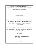
Fig. 1. 1H NMR spectrum interval of N-acetylated Ch samples, named as Ch60 (a); Ch45 (b); Ch35 (c); Ch25 (d); Ch15 (e) and Ch5 (f), assigned to H1’ and H1 signals,
used for determination of DA and PA.
depolymerized (Fig. S4) – ones prepared on heterogeneous medium.
This occurrence is due to the slightly higher probability to have a fre
quency of GlcNAc-GlcNAc residues and then increased chances to form a
block-wise distribution on heterogeneous conditions mainly at higher
acetylation levels (DA > 50 %) (Hirano et al., 1989; Vårum, Anthonsen,
Grasdalen, & Smidsrød, 1991). Nevertheless, Ch1x, Ch45 and Ch60
samples nearly accomplished the requirement for a random-like
distribution.
Thus, the independent Mw and DA values with acetamido groups
randomly distributed (A ~ 1) have been successfully achieved to eval
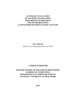
uate the morphological feature from XRD patterns (Fig. 2). As illustrated
in Fig. 2a, the diffractograms of chitins reveals the highly ordered
5
W.M. Facchinatto et al.
Carbohydrate Polymers 250 (2020) 116891
pattern of αCh, that preserves the orthorhombic P212121 symmetry with
antiparallel chains displacement (Minke & Blackwell, 1978), compared
to the typical profile of hydrated βCh allomorph that reveals a mono
clinic P21 symmetry with parallel displacement and lower intersheet
interaction across bc projection (Gardner & Blackwell, 1975). The two
diffraction peaks with the highest intensities comprising between 2θ ~
8◦ -11◦ and 19◦ -21◦ are mainly assigned to the hydrated crystalline
planes (020)h and (110)h, respectively, whereas secondary peaks are
predominantly evidenced on αCh. Such allomorph exhibits the peaks
centered at 12.8◦ and 22.8◦ related to the anhydrous planes (110)a and
(120)a reflections, respectively, while the peak at 26.5◦ which describes
the (013)a reflection are evidenced on both chitins diffractograms
(Fig. 2a) and seems to be related to DA, once its relative intensity de
creases from Ch60 to Ch5 (Fig. 2b).
All Ch samples and βCh (Fig. 2b,c) shows a broader peak at 19◦ -21◦ ,
hindering the (220)h reflection at 20.7◦ that only clearly appears on αCh
(Osorio-Madrazo et al., 2010). The absence of (110)a reflection on Ch
samples has been attributed to confirm the diffraction pattern of a hy
drated (tendom) crystalline form. In such case, the hydrated Ch samples
are stabilized by O3…O5 hydrogen bonds and water-bridges between
chains, which allows a twofold helical conformation to be preferentially
formed (Okuyama, Noguchi, Miyazawa, Yui, & Ogawa, 1997; Sikorski,
Hori, & Wada, 2009)
Although single crystals of Ch have been identified with ortho
rhombic P212121 unit cell, the same symmetry found on αCh allomorph
(Cartier, Domard, & Chanzy, 1990; Sikorski et al., 2009), an extensive
crystalline disruption is provided by the high penetration of water
molecules to produce Ch samples, which reduces the average crystallite
sizes and leads to a structure expansion across b axis, due to the fact
there are no intersheet hydrogen bonds between C(61)O…HOC(62) along
this axis (Cho et al., 2000). Nevertheless, the hydrated Ch preserves the
N2…O6 hydrogen bonds along b and then granting the intersheet par
allel arrangement on bc projection (Okuyama et al., 1997).
The crystallite dimensions L020 and L110 from Ch samples, obtained
by deconvolving the respective peaks (Fig. S5 and S6), converged the
values closer to those exhibited by βCh (Table 1), consequently losing
the structural compactness and then achieving a diffraction pattern
more similar to such allomorph (Saito, Putaux, Okano, Gaill, & Chanzy,
1997). All the procedures involved on the preparation of Ch samples
enabled this crystalline disruption and, consequently, shifted the peaks
at 8◦ -11◦ and 19◦ -21◦ to higher scattering angles. The first one contin
uously shifts and decreases its relative intensity suggesting that the
crystal structure was slightly distorted by decreasing the DA (Cho et al.,
2000; Zhang, Xue, Xue, Gao, & Zhang, 2005), while the variability on
19◦ -21◦ peak width are possibly ascribed to non-uniform deformations
of crystallites (Fig. 2b) (Garvey, Parker, & Simon, 2005). Indeed, by
lowering the peak intensity at 8◦ -11◦ , the hydrated (020)h reflection
should be closer to those exhibited by a completely amorphous pattern
(Osorio-Madrazo et al., 2010), thus decreasing the regularity provided
by interchain hydrogen bonds between C(73)=O…HNC(21) and C(73)=
O…HOC(61) across a axis.
As observed in Fig. 2c, there are no significative variations on the
diffraction patterns as function of Mw , especially regarding the molar
mass changes among samples with lower DA (Ch2x and Ch3x). This
result agrees to previous studies in which was found that the crystallinity
is influenced by lowering the molar mass from Ch sample with DA > 20
% (Ogawa & Yui, 1993; Savitri et al., 2014), similarly to the recorded for
Ch1x (DA ~ 30 %) that shows few profile changes in the diffraction
pattern compared to those from Ch1 x 3 h and 6 h.
The long-range ordering was estimated by means of crystallinity
Fig. 2. XRD patterns of α- and βCh (a); Ch5-60 and βCh (b); USAD (Ch1x, Ch2x and Ch3x) and depolymerized (3 h and 6 h) Ch samples (c).
6
W.M. Facchinatto et al.
Carbohydrate Polymers 250 (2020) 116891
confirm that the best coincidence between the 13C CPMAS and the
quantitative 13C DPMAS spectra is achieved at TC = 3000 μs, once the
signal integral ratio IC=O /ICH3 ~ 1 reveals equivalent amount of both
groups in the structure, as expected. Thus, 13C CPMAS with TC = 3000 μs
will be used here instead of the very time consuming 13C DPMAS spectra.
However, it is important to point out that the chain mobility in the
sample can change the optimal TC , so such approach would only be
possible if all samples have similar molecular mobility.
The specific mobility along the molecular segments can be formally
confirmed through the DIPSHIFT experiments. Such technique provides
the access to the molecular mobility by monitoring the strength of the
1 13
H- C dipolar interaction, which can be reduced by molecular motions.
This is probed by applying a pulse sequence that modulates each 13C
signal in the CPMAS spectrum by a factor that depend on the dipolar
coupling to its next neighbor 1H nuclei during an evolution time t1 . The
plot of the intensity of each 13C signal as a function of t1 provide the so
called DIPSHIFT curves, which have a “smile like” shape starting from a
maximum value at t1 = tr reaching a minimum at t1 = tr /2 and restoring
to a value that depend on the T2 relaxation time of that specific carbon
spin. The dependence of the DIPSHIFT curves on 1H-13C dipolar inter
action strength appears in the minimum intensity value reached at t1 =
tr /2 in such a way that higher is the dipolar interaction strength lower is
the minimum intensity. Because molecular motions with rates higher
and 10 kHz average out the dipolar interaction, this minimum value is
increased for mobile segments. Slower motion, i.e., with rates in the low
kHz frequency scales, reduces the T2 relaxation time and show up in the
DIPSHIFT curves as an intensity reduction at t1 = tr (DeAzevedo et al.,
2008; Munowitz et al., 1981). As showed in Fig. 4b, the minimum in
– O carbons is ~ 0.9, which is trivially
tensity at t1 = tr /2 achieved to C–
associated to the lack of directly bonded 1H. Still the minimum intensity
achieved by the CH3 carbon is ~ 0.7, which is closer to methyl carbons
of L-alanine, confirming that the decrease of dipolar interaction is
mainly consequence of the fast motion around its C3 symmetry axis. For
carbons C1 to C5 the minimum intensity is ~ 0.25. This is a typical value
obtained for CH carbons on glucose units of rigid carbohydrates (Sim
mons et al., 2016) pointing to a rigid backbone structure in the Ch
sample. For rigid CH2 carbons the minimum intensity of the DIPSHIFT
curves should reach ~ 0. This is not the case of the C6 signal, where the
minimum intensity is ~ 0.2. This is associated to local motion of the
CH2OH side chain, which also decrease the T2 values and leads to a
smaller final intensity at t1 = tr for the C6 carbons as compared to car
bons C1-C5.
All Ch samples showed similar DIPSHIFT profiles, showing that all
samples have similar chain mobility. This information is important
because it supports the use of the 13C CPMAS, instead of the
index applying two different methods (CrI1 and CrI2 ) of quantification
on XRD patterns. The corresponding CrI1 and CrI2 values are listed on
Table S1. Distinct results of crystallinity index have been achieved for a
given sample, being evident the considerable influence of the method
employed and achieving CrI1 > CrI2 almost to all samples. Nevertheless,
both values for each case tends to increase with DA mainly on samples
prepared in homogeneous conditions, except for Ch60, that revealed a
slightly decreases, and Ch35, that showed an unexpected increase on
CrI1 . The deacetylated and depolymerized Ch showed closer CrI1 values,
which means that the straightly relationship with DA and crystallinity is
not clearly observed on samples originally prepared in heterogeneous
conditions. Additionally, a slight decrease on CrI2 values is only
observed lowering the Mw of Ch2x and Ch3x samples. Such discrepancy
confirms the unsolved issue regarding the exact contribution of amor
phous phase on scattering profile, as already pointed out (Ioelovich,
2014; Osorio-Madrazo et al., 2010), despite the possibility to carry
similar tendencies through both methods mainly on products homoge
nously prepared. In this sense, the accurate and reproductive determi
nation of Ch crystallinity, even considering a wider structural
variability, is largely affected by the processing steps of Ch preparation
and XRD method, which is also not able to differentiate the molecular
origin of the amorphous components.
3.2. Part II: conformation and short-range molecular ordering
As it is well known the TC dependence of the 13C CPMAS spectral
profile arises from cross-polarization (CP) transfer rate, which depends
on the dipolar coupling between the 13C and the neighbor 1H nuclei
(Metz et al., 1996; Tanner et al., 1990). Thus, the 13C CPMAS spectra
were initially acquired varying TC to seek for an optimal condition that
minimizes the signal dependence on the polarization transfer (Kasaai,
2010). This procedure was applied on the Ch25 sample due to the in
termediate content of acetamido groups compared to the other samples.
A set of 13C CPMAS spectra were acquired with different TC and the
spectral profile was compared to that of a quantitative 13C DPMAS
spectrum as shown in Fig. 3. Therefore, with TC = 3000 μs, the 13C
CPMAS spectrum achieves a similar profile to the exhibited by the 13C
DPMAS spectrum in the whole spectral range. All DACP at 3000 μs are
listed on Table S1.
–O
Fig. 4a compares the 13C CPMAS signal intensity of CH3 and C–
groups as function of TC . As expected, the CH3 signal shows faster CP
build-up, due to three hydrogens direct bonded, and shorter decay time,
due to the fast rotation around the C3 axis leading to a shorter relaxation
ˆ et al., 1987). These results
time decay in rotating frame (T1ρ ) (Saito
Fig. 3. 13C CPMAS spectrum profiles of Ch25 at variable TC values (1000 to 4000 μs) compared to
interval of C4, C5-C3, C6 and C2 signals (b); C1 (c) and CH3 (d) signals.
7
13
C DPMAS profile at the whole spectral range (a) covering the
W.M. Facchinatto et al.
Carbohydrate Polymers 250 (2020) 116891
13
Fig. 4. CP build-ups for CH3 ad C–
–O groups of Ch25 (a); DIPSHIFT curves acquired from Ch25 (b); CPMAS spectrum of α-, βCh and Ch5-60 at TC = 3000 μs (c); and
comparation of N-acetylated Ch spectrum profiles at TC = 3000 μs, showing the conformational dependence with DA (d).
quantitative, but very time consuming, 13C DPMAS spectra for evalu
ating the DA and the NMR crystallinity of the samples.
The 13C CPMAS spectra of Ch and chitin allomorphs are shown in
Fig. 4c. Although βCh reveals overlapped C5 and C3 signals, those are
usually split on αCh leading to different chemical shifts, which indicates
the main influence of packing and geometrical effects on polymeric
chains (Focher et al., 1992; Heux et al., 2000). Additionally, the asym
– O from αCh are probably consequence of an inef
metrical shape of C–
ficient removal of the strong dipolar interaction between the direct
bonded quadrupolar 14N nucleus (Tanner et al., 1990). Indeed, such
behaviors suggest higher density and homogeneity, due to the antipar
allel arrangement of αCh chains, compared to the broad signals of βCh
and Ch samples that suggest lower homogeneity (Fig. 4c).
The profile of N-acetylated Ch samples follows the tendency assigned
– O and CH3 signals, in accordance with DA variation (Fig. 4c). A
to C–
closer overview of this current region of the spectra, detached on Fig. 4d,
shows that all signals show significant changes and notably C1, C4 and
C6 signals clearly increase with DA. The C4 signal in the 13C CPMAS
spectra has been widely used to estimate the fraction between the in
ternal (more ordered chain) and surface (more disordered) fibrils
structures, which is usually referred as crystalline index (Park et al.,
2010). Similarly, it is reasonable to consider that those signals propor
tionally increase with Ch crystallinity. The spectral line shape is sensi
tive to the molecular conformation and content of ordered segments,
being applicable a qualitative understanding based on γ-effect concept
(Born & Spiess, 1997; Tonelli & Schilling, 1981). For this approach, it
has to be firstly considered a molecular model building made by helical
symmetry with a period of 10.34 Å across a fiber axis. Such model was
properly used for explain the torsion angles of glycosidic linkages of Ch
ˆ et al. (1987),
on 13C NMR data as complementary means to XRD by Saito
and was formally detailed by Okuyama et al. (1997). This model in
cludes two dihedral angles (φ, ψ) in the main-chain conformation rep
resented by glycosidic C(1)-O1-C(41) linkage, and a third dihedral angle
(χ) at C(5)-C(6) that define the orientation o O6. Although φ and ψ are
average stable with low degree of freedom, which is ensured by
hydrogen bonds between O3…O5, χ can fell into three orientations at
-60◦ , +60◦ and 180◦ , satisfying the gauche-gauche, gauche-trans and
trans-gauche conformations, respectively, with respect to C(4) (Okuyama
et al., 1997). As already mentioned, it is well-accepted that O6 are not
comprising on C(61)O…HOC(62) but still participates on C(73)=O…HOC
(61) hydrogen bonds on β-forms, meaning that DA actually affects the
population of possible conformations of C(6)OH group. Therefore, we
suppose that a wider distribution of these conformations is proportion
ally achieved by decreasing the DA and so the population of this seg
ments packed in a regular way. Consequently, the electronic structure
around C(6) and C(4), located at two σ-bonds of distance, experiences
different dipolar interactions, reflecting on those CP signals.
Differently from XRD data, the conformational refinement achieved
by 13C CPMAS allows to observe slightly variations on depolymerized Ch
spectrum (Fig. 5). However, those are mainly assigned on C1 and C6
signals, showing no significant changes on C4. Considering that the
depolymerization extensively undergoes on glycosidic linkages, this
result confirms that C1 and C6 signals are quite sensitive to main-chain
conformation specially on first depolymerization step, while C4 signal
reveals great dependence with the DA but none significant changes with
molar masses. An exception regards to Ch1x that shows few changes on
these related signals, probably ascribed to some packing influence that
remains after heterogenous deacetylation of βCh. According to studies
ˆ et al., 1987), the chains
(Focher et al., 1990; Heux et al., 2000; Saito
length dependence of C4 were only found at higher (annealing) tem
peratures, however such behavior was not formally ruled by the authors.
A proof of concept concerning the ordered and disordered contri
bution on C4 and C6 signals was carried by means of TC ranged from 50
to 4500 μs on ChC sample, which actually presented split assignments
for both signals (Fig. 6a). The spectral interval ranged in 95− 50 ppm
shows that each assigned C4 peak responds to dipolar interaction at
differently CP rates. According to the C4 signal evolution profile, the
downfield shifted C4 peak quickly recoveries the magnetization even at
shorter TC (50 μs) compared to the upfield shifted peak, that requires
longer TC values to be totally polarized. Such behavior is typically
8
W.M. Facchinatto et al.
Carbohydrate Polymers 250 (2020) 116891
the correlations between nearby 1H nucleus from overlapped C4 signal
of Ch samples. Thus, the 2D HETCOR spectra was carried to provide the
heteronuclear correlation at distances higher than 1H-13C direct bonding
(Kono, 2004). The C4 signal of ChC (Fig. 6b) revealed distinguished 13C
chemical shifts (F2), each one referred to the same broad signal of
aliphatic 1H nucleus (F1). Although all Ch samples have shown over
lapped C4 signals, these have also achieved different 13C correlations
with similar protons, as clearly observed on Ch15 (Fig. 6c) and Ch45
(Fig. 6d), which can be related to different populations of possible
conformations. Heteronuclear correlations with protons from different
chemical groups are also observed on chitin allomorphs and N-acety
lated Ch (Fig. S7), as a consequence of a longer mixing time.
Given the dependency of C4 and C6 signals on conformational order,
the peak deconvolution method was used to estimate the fraction be
tween ordered (crystalline) and disordered (amorphous) content in the
sample. The C4 and C6 signals were decomposed into Lorentzian and
Gaussian functions for crystalline and amorphous contributions,
respectively, according to non-linear quantification of individual states
of order proposed by Larsson, Wickholm, and Iversen (1997) for cellu
lose. The resulted peak deconvolution from the spectral region of in
terest of N-acetylated Ch and chitin allomorphs are shown in Fig. 7. For
more reliable quantification, it was set an equal number of curves at the
same chemical shift and FWHM to all samples, including for depoly
merized Ch (Fig. S8). The estimative of crystallinity index of C4 and C6
signals (CrICP ) is listed on Table 1 and, as observed, the content of or
dered structures increases with DA, being nearly constant by changing
the molar masses.
A comparative analysis regarding the average crystallinity index
obtained from C4 and C6 (CrICP ) and the corresponding values calcu
lated from XRD patterns with DACP are shown in Fig. 8. The intrinsic
dependence from structural and morphological features are consider
ably more evident through the proposing method employed on 13C
CPMAS spectra, compared to the current methods from XRD. SSNMR
should provide consistent results also avoiding problems with baseline
Fig. 5. 13C CPMAS spectrum of USAD Ch1x (a); Ch2x (b) and Ch3x (c), with
respect to depolymerized (3 h and 6 h) Ch samples.
ascribed to changes on molecular packing, once the spin diffusion is
longer on amorphous phases, which have naturally lesser packed
arrangement than the crystalline one (Ando & Asakura, 1998). Each C4
peak can be properly described by such physical behavior, leading to
distinguished chemical shifts for crystalline and amorphous domains, as
expected by the γ-effect. In fact, and considering an wide distribution of
χ dihedral angles, the trans isomerism provides higher regularity and it is
commonly downfield shifted, while gauche is associated to lesser regu
larity and it is upfield shifted (Born & Spiess, 1997), as confirmed by C4
signal of ChC. However, this behavior was not clearly evidenced on C6
split peaks, despite the influence χ dihedral angles on C(6)OH
conformation.
Taking into account the whole set of results, it is reasonable to verify
Fig. 6. 13C CPMAS spectra of ChC sample showing the conformational dependence of carbon signals at variable TC values (50 to 4500 μs) (a); 2D HETCOR spectrum
of ChC (b); Ch15 (c) and Ch45 (d), proving that even without C4 signal splitting, distinguished correlations can be taken regarding the kind of protons.
9
W.M. Facchinatto et al.
Carbohydrate Polymers 250 (2020) 116891
Fig. 7. Peak deconvolution method applied on 13C CPMAS spectra (TC =3000 μs) of αCh (a), βCh (b) and N-acetylated Ch5-60 (c) samples, to estimate the shortrange molecular ordering from C4 and C6 signals, allowing the quantification of CrICP .
determination, as commonly found on XRD methods. For instance,
however, it is important to highlight that the physical origin remains
different from each technique and the following results of short-range
behavior (as probed in SSNMR) may not replace the long-range
behavior (as probed in XRD) that attains the bulk for every case.
The multivariate SVD analysis (Forato et al., 1998) was also applied
to the CPMAS spectra of acetylated Ch using its predicted values of CrICP
and DACP , in the same spectral range used for peak deconvolution
(Fig. 7). The concentration of the components CrI * and DA * and its
correlations with the predicted values were calculated from distinct
intervals, as indicated in Table S2. Since the SVD method aims to esti
mate the concentration of the components based on spectra profile
changes, it was not possible to obtain a satisfactory correlation including
the chitin allomorphs spectra in the set of samples due to the additional
influence of intersheet packing. It can be noted that all assigned regions
are governed by both components, indicating that the concentration
matrix is able to estimate CrI * and DA * independently from CH3 and C
= O signals, with an exception of DA * from 90− 67 ppm region. In this
10
W.M. Facchinatto et al.
Carbohydrate Polymers 250 (2020) 116891
Fig. 8. Crystallinity index calculated according to peak height (CrI1 ) and
amorphous subtraction (CrI2 ) method from XRD patterns; average contribution
from ordered domains from C4 and C6 signals of 13C CPMAS spectra (CrI CP ),
with respect to the DACP of αCh, βCh, N-acetylated Ch5-60 (a); USAD and
depolymerized Ch (3 h and 6 h) Ch (b) samples.
sense, higher CrI * correlation compared to DA * in 90− 67 ppm interval
highlights the major influence of short-range ordering, which essentially
confirms the fundamental relevance of using C4 signal for structural
analysis.
The correlation of the components generated by the calibration
matrix (Fig. 9a) indicate that both ones coexist proportionally, as ex
pected. Higher number of intersection points of these curves at 85− 75
ppm further indicates that the local geometry between C4 and C6 is
mediated by DA *. In addition, the theoretical spectrum profile gener
ated for pure components (Fig. 9b) suggests that the C4 signal tends to fit
the exhibited by βCh profile, evidencing the contribution regarding the
chemical shift separation at distinguish C4 signal portions between the
ordered and disordered structures. However, the clear distinction
observed between βCh and a fully crystalline profile indicates that the
crystallinity of such allomorph is also dependent on how the chains are
packaged, as already mentioned. This finding extends to αCh that even
showing closer DACP to βCh (Table S1), the chains arrangement affects
the CrICP and, consequently, the spectral profile.
Fig. 9. Profile of the components DA * and CrI * generated from calibration
matrix, X (a); 13CPMAS spectra profile relationship of βCh, Ch60 and Ch5 with
the theoretical profiles of crystalline and amorphous Ch. These spectra were
normalized by C1 signal area.
hydrogen bonds that participates on the stabilization of twofold helical
conformation by decreasing the DA. Consequently, the amount of
hydrogen bonds between C(73)=O…HNC(21) and C(73)=O…HOC(61)
decreases, leading to typical diffraction pattern with lower crystallinity.
Although the crystallinity indexes CrI1 and CrI2 proportionally increases
with DA, no significant changes were recorded varying the molar
masses.
The 13C CPMAS spectra fitted closely the profile exhibited by DPMAS
at TC = 3000 μs. In fact, it was found that the C4 signal splitting strongly
evidenced the CP rate variability of ordered and disordered conforma
tions, which was confirmed by HETCOR experiments. The non-linear
deconvolution of C4 and C6 signals showed a growing contribution of
the crystalline content downfield shifted (Lozentzian curves), and some
loss of magnetization upfield shifted (Gaussian curves) assigned to the
amorphous content by increasing the DA. Once the C(73)=O…HOC(61)
hydrogen bonds increases with DA, lesser mobility of C(6)OH is allowed,
probably leading the C(6)OH population to an growing contribution of
trans conformation with respect do C(4).
The CrICP values proportionally increases with DA but no significant
changes were found as function of molar mass. High correlation with
crystallinity was found using the peaks from 90− 67 ppm and applying
SVD analysis. Finally, according to the SVD multivariate analysis the
spectra of pure crystalline and amorphous clearly illustrated that C4
signal is strongly related to crystallinity. Therefore, this work provided a
4. Conclusion
Chitosans (Ch) with variable degrees of N-acetylation and molar
masses were successfully prepared on homogeneous conditions, all
exhibiting random pattern of acetylation (DA ~ 1). While acetylated Ch
(DA ranging as 5–60 %) showed just slight variations of Mw , the DA
values were mostly preserved after depolymerization of Ch (Mw ranging
as 0.15–1.2 × 10⁶ g mol− 1).
The XRD pattern of Ch samples exhibited crystallite lattice di
mensions L020 and L110 , related to 2θ ~ 8◦ -11◦ and 19◦ -21◦ , respectively,
closer to those presented by βCh. For all β-forms, the absence of anhy
drous (110)a plane on XRD pattern is straightly related to O3…O5
11
W.M. Facchinatto et al.
Carbohydrate Polymers 250 (2020) 116891
novel approach of crystallinity index quantification of chitosans,
extending the knowledge regarding to the origin of short-range molec
ular ordering, without requiring to external amorphous standards or
exhibiting meaningful impact from molecular weight on C4 signal
shape.
Cartier, N., Domard, A., & Chanzy, H. (1990). Single crystals of chitosan. International
Journal of Biological Macromolecules, 12(5), 289–294. />Chatelet, C., Damour, O., & Domard, A. (2001). Influence of the degree of acetylation on
some biological properties of chitosan films. Biomaterials, 22(3), 261–268. https://
doi.org/10.1016/S0142-9612(00)00183-6.
Cho, Y.-W., Jang, J., Park, C. R., & Ko, S.-W. (2000). Preparation and solubility in acid
and water of partially deacetylated chitins. Biomacromolecules, 1(4), 609–614.
/>DeAzevedo, E. R., Saalwachter, K., Pascui, O., De Souza, A. A., Bonagamba, T. J., &
Reichert, D. (2008). Intermediate motions as studied by solid-state separated local
field NMR experiments. The Journal of Chemical Physics, 128(10). />10.1063/1.2831798, 104505-1-104505–104512.
Facchinatto, W. M., Fiamingo, A., dos Santos, D. M., & Campana-Filho, S. P. (2019).
Characterization and physical-chemistry of methoxypoly(ethylene glycol)-gchitosan. International Journal of Biological Macromolecules, 124, 828–837. https://
doi.org/10.1016/j.ijbiomac.2018.11.246.
Fan, Y., Saito, T., & Isogai, A. (2008). Chitin nanocrystals prepared by TEMPO-mediated
oxidation of α-chitin. Biomacromolecules, 9(1), 192–198. />bm700966g.
Fan, Y., Saito, T., & Isogai, A. (2009). TEMPO-mediated oxidation of β-chitin to prepare
individual nanofibrils. Carbohydrate Polymers, 77(4), 832–838. />10.1016/j.carbpol.2009.03.008.
Fiamingo, A., Delezuk, J. A. M., Trombotto, S., David, L., & Campana-Filho, S. P. (2016).
Extensively deacetylated high molecular weight chitosan from the multistep
ultrasound-assisted deacetylation of beta-chitin. Ultrasonics Sonochemistry, 32,
79–85. />Focher, B., Beltrame, P. L., Naggi, A., & Torri, G. (1990). Alkaline N-deacetylation of
chitin enhanced by flash treatments. Reaction kinetics and structure modifications.
Carbohydrate Polymers, 12(4), 405–418. />90090-F.
Focher, B., Naggi, A., Torri, G., Cosani, A., & Terbojevich, M. (1992). Chitosans from
Euphausia superba. 2: Characterization of solid state structure. Carbohydrate
Polymers, 18(1), 43–49. />Forato, L. A., Bernardes-Filho, R., & Colnago, L. A. (1998). Protein structure in KBr
pellets by infrared spectroscopy. Analytical Biochemistry, 259, 136–141. https://doi.
org/10.1006/abio.1998.2599.
Francis Suh, J.-K., & Matthew, H. W. (2000). Application of chitosan-based
polysaccharide biomaterials in cartilage tissue engineering: A review. Biomaterials,
21(24), 2589–2598. />Gardner, K. H., & Blackwell, J. (1975). Refinement of the structure of β-Chitin.
Biopolymers, 14, 1581–1595. />Garvey, C. J., Parker, I. H., & Simon, G. P. (2005). On the interpretation of X-ray
diffraction powder patterns in terms of the nanostructure of cellulose I fibres.
Macromolecular Chemistry and Physics, 206(15), 1568–1575. />10.1002/macp.200500008.
Gonil, P., & Sajomsang, W. (2012). Applications of magnetic resonance spectroscopy to
chitin from insect cuticles. International Journal of Biological Macromolecules, 51(4),
514–522. />Goodrich, J. D., & Winter, W. T. (2007). Alpha-Chitin nanocrystals prepared from shrimp
shells and their specific surface area measurement. Biomacromolecules, 8(1),
252–257. />Grząbka-Zasadzi´
nska, A., Amietszajew, T., & Borysiak, S. (2017). Thermal and
mechanical properties of chitosan nanocomposites with cellulose modified in ionic
liquids. Journal of Thermal Analysis and Calorimetry, 130(1), 143–154. https://doi.
org/10.1007/s10973-017-6295-3.
Guibal, E. (2004). Interactions of metal ions with chitosan-based sorbents: A review.
Separation and Purification Technology, 38(1), 43–74. />seppur.2003.10.004.
Gupta, K. C., & Jabrail, F. H. (2006). Effects of degree of deacetylation and cross-linking
on physical characteristics, swelling and release behavior of chitosan microspheres.
Carbohydrate Polymers, 66(1), 43–54. />carbpol.2006.02.019.
Harish Prashanth, K. V., Kittur, F. S., & Tharanathan, R. N. (2002). Solid state structure of
chitosan prepared under different N-deacetylating conditions. Carbohydrate
Polymers, 50(1), 27–33. />Heux, L., Brugnerotto, J., Desbri`eres, J., Versali, M. F., & Rinaudo, M. (2000). Solid state
NMR for determination of degree of acetylation of chitin and chitosan.
Biomacromolecules, 1(4), 746–751. />Hirano, S., Tsuchida, H., & Nagao, N. (1989). N-acetylation in chitosan and the rate of its
enzymic hydrolysis. Biomaterials, 10(8), 574–576. />Huang, M., Khor, E., & Lim, L. Y. (2004). Uptake and cytotoxicity of chitosan molecules
and nanoparticles: Effects of molecular weight and degree of deacetylation.
Pharmaceutical Research, 21(2), 344–353. />PHAM.0000016249.52831.a5.
Ioelovich, M. (2014). Crystallinity and hydrophility of chitin and chitosan. Research and
Reviews - Journal of Chemistry, 3(3), 7–14.
Islam, M. M., Shahruzzaman, M., Biswas, S., Nurus Sakib, M., & Rashid, T. U. (2020).
Chitosan based bioactive materials in tissue engineering applications-A review.
Bioactive Materials, 5(1), 164–183. />bioactmat.2020.01.012.
Kang, X., Kirui, A., Muszy´
nski, A., Widanage, M. C. D., Chen, A., Azadi, P., … Wang, T.
(2018). Molecular architecture of fungal cell walls revealed by solid-state NMR.
Nature Communications, 9(1), 1–12. />
Conflicts of interest
The authors declare no conflict of interest.
CRediT authorship contribution statement
William Marcondes Facchinatto: Conceptualization, Writing original draft, Writing - review & editing, Methodology, Formal analysis.
Danilo Martins dos Santos: Conceptualization, Writing - original draft,
Writing - review & editing, Methodology, Formal analysis. Anderson
Fiamingo: Resources, Writing - original draft, Writing - review & edit
ing. Rubens Bernardes-Filho: Writing - review & editing, Methodol
´rgio Paulo Campana-Filho: Resources, Writing
ogy, Formal analysis. Se
- review & editing. Eduardo Ribeiro de Azevedo: Resources, Writing review & editing. Luiz Alberto Colnago: Supervision, Conceptualiza
tion, Resources, Writing - original draft, Writing - review & editing.
Acknowledgments
The authors are grateful for the financial support from the National
Council for Scientific and Technological Development, CNPq(141353/
2016-3, 303753/2018-8), and the S˜
ao Paulo Research Foundation,
FAPESP (2016/20970-2;2016/09720-4;2017/20973-4;2017/24465-3;
2019/13656-8). This study was financed in part by National Council for
the Improvement of Higher Education, CAPES – Finance Code 001.
Appendix A. Supplementary data
Supplementary material related to this article can be found, in the
online version, at doi: />References
Ahmed, S., & Ikram, S. (2016). Chitosan based scaffolds and their applications in wound
healing. Achievements in the Life Sciences, 10(1), 27–37. />als.2016.04.001.
Ahmed, S., Annu, Ali, A., & Sheikh, J. (2018). A review on chitosan centred scaffolds and
their applications in tissue engineering. International Journal of Biological
Macromolecules, 116, 849–862. />Aiba, S. (1992). Studies on chitosan: 4. Lysozymic hydrolysis of partially N-acetylated
chitosans. International Journal of Biological Macromolecules, 14(4), 225–228. https://
doi.org/10.1016/S0141-8130(05)80032-7.
Åkerholm, M., Hinterstoisser, B., & Salm´en, L. (2004). Characterization of the crystalline
structure of cellulose using static and dynamic FT-IR spectroscopy. Carbohydrate
Research, 339(3), 569–578. />Ando, I., & Asakura, T. (1998). Solid state NMR of polymers: Studies in physical and
theoretical chemistry. Retrieved from. Tokyo: Elsevier
m/bookseries/studies-in-physical-and-theoretical-chemistry/vol/84/suppl/C.
Baranwal, A., Kumar, A., Priyadharshini, A., Oggu, G. S., Bhatnagar, I., Srivastava, A., …
Chandra, P. (2018). Chitosan: An undisputed bio-fabrication material for tissue
engineering and bio-sensing applications. International Journal of Biological
Macromolecules, 110, 110–123. />Bernardinelli, O. D., Lima, M. A., Rezende, C. A., Polikarpov, I., & DeAzevedo, E. R.
(2015). Quantitative 13C MultiCP solid-state NMR as a tool for evaluation of
cellulose crystallinity index measured directly inside sugarcane biomass.
Biotechnology for Biofuels, 8(1), 110. />Bielecki, A., Kolbert, A. C., De Groot, H. J. M., Griffin, R. G., & Levitt, M. H. (1990).
Frequency-switched Lee—Goldburg sequences in solids. Advances in Magnetic and
Optical Resonance, 14(C), 111–124. />Born, R., & Spiess, H. W. (1997). Ab initio calculations of conformational effects on 13C
NMR spectra of amorphous polymers. In J. Seeling (Ed.), NMR basic principles and
progress (1st ed., pp. 1–127). New York: Springer.
Cardozo, F. A., Facchinatto, W. M., Colnago, L. A., Campana-Filho, S. P., & Pessoa, A.
(2019). Bioproduction of N-acetyl-glucosamine from colloidal α-chitin using an
enzyme cocktail produced by Aeromonas caviae CHZ306. World Journal of
Microbiology & Biotechnology, 35(8), 114. />
12
W.M. Facchinatto et al.
Carbohydrate Polymers 250 (2020) 116891
Kasaai, M. R. (2010). Determination of the degree of N-acetylation for chitin and
chitosan by various NMR spectroscopy techniques: A review. Carbohydrate Polymers,
79(4), 801–810. />Kaya, M., Mujtaba, M., Ehrlich, H., Salaberria, A. M., Baran, T., Amemiya, C. T., …
Labidi, J. (2017). On chemistry of γ-chitin. Carbohydrate Polymers, 176, 177–186.
/>Knaul, J. Z., Kasaai, M. R., Bui, V. T., & Creber, K. A. M. (1998). Characterization of
deacetylated chitosan and chitosan molecular weight review. Canadian Journal of
Chemistry, 76(11), 1699–1706. />Kono, H. (2004). Two-dimensional magic angle spinning NMR investigation of naturally
occurring chitins: Precise 1H and 13C resonance assignment of α- and β-chitin.
Biopolymers, 75(3), 255–263. />Kubota, N., & Eguchi, Y. (2005). Facile preparation of water-soluble N-Acetylated
chitosan and molecular weight dependence of its water-solubility. Polymer Journal,
29(2), 123–127. />Kumirska, J., Weinhold, M. X., Sauvageau, J. C. M., Thă
oming, J., Kaczy
nski, Z., &
Stepnowski, P. (2009). Determination of the pattern of acetylation of low-molecularweight chitosan used in biomedical applications. Journal of Pharmaceutical and
Biomedical Analysis, 50(4), 587–590. />Kurita, K., Ishii, S., Tomita, K., Nishimura, S.-I., & Shimoda, K. (1994). Reactivity
characteristics of squid β-chitin as compared with those of shrimp chitin: High
potentials of squid chitin as a starting material for facile chemical modifications.
Journal of Polymer Science Part A: Polymer Chemistry, 32(6), 1027–1032. https://doi.
org/10.1002/pola.1994.080320603.
Kurita, K., Kamiya, M., & Nishimura, S. I. (1991). Solubilization of a rigid
polysaccharide: Controlled partial N-acetylation of chitosan to develop solubility.
Carbohydrate Polymers, 16(1), 83–92. />90072-K.
Lamarque, G., Viton, C., & Domard, A. (2004). Comparative study of the second and third
heterogeneous deacetylations of α- and β-chitins in a multistep process.
Biomacromolecules, 5(5), 1899–1907. />Larsson, P. T., Wickholm, K., & Iversen, T. (1997). A CP/MAS carbon-13 NMR
investigation of molecular ordering in celluloses. Carbohydrate Research, 302(1–2),
19–25. />Lavall, R. L., Assis, O. B. G., & Campana-Filho, S. P. (2007). Beta-chitin from the pens of
Loligo sp.: Extraction and characterization. Bioresource Technology, 98(13),
2465–2472. />Lavertu, M., Darras, V., & Buschmann, M. D. (2012). Kinetics and efficiency of chitosan
reacetylation. Carbohydrate Polymers, 87(2), 1192–1198. />carbpol.2011.08.096.
Lavertu, M., Xia, Z., Serreqi, A. N., Berrada, M., Rodrigues, A., Wang, D., … Gupta, A.
(2003). A validated 1H NMR method for the determination of the degree of
deacetylation of chitosan. Journal of Pharmaceutical and Biomedical Analysis, 32(6),
1149–1158. />Mao, S., Shuai, X., Unger, F., Simon, M., Bi, D., & Kissel, T. (2004). The depolymerization
of chitosan: Effects on physicochemical and biological properties. International
Journal of Pharmaceutics, 281(1–2), 45–54. />ijpharm.2004.05.019.
Metz, G., Ziliox, M., & Smith, S. O. (1996). Towards quantitative CP-MAS NMR. Solid
State Nuclear Magnetic Resonance, 7(3), 155–160. />Miguel, S. P., Moreira, A. F., & Correia, I. J. (2019). Chitosan based-asymmetric
membranes for wound healing: A review. International Journal of Biological
Macromolecules, 127, 460–475. />Milot, C., McBrien, J., Allen, S., & Guibal, E. (1998). Influence of physicochemical and
structural characteristics of chitosan flakes on molybdate sorption. Journal of Applied
Polymer Science, 68(4), 571–580. />(19980425)68:4<571::AID-APP8>3.3.CO;2-1.
Minke, R., & Blackwell, J. (1978). The structure of α-Chitin. Journal of Molecular Biology,
120(2), 167–181. />Munowitz, M. G., Griffin, R. G., Bodenhausen, G., & Huang, T. H. (1981). Twodimensional rotational spin-echo nuclear magnetic resonance in solids: Correlation
of chemical shift and dipolar interactions. Journal of the American Chemical Society,
103(10), 2529–2533. />Mutungi, C., Passauer, L., Onyango, C., Jaros, D., & Rohm, H. (2012). Debranched
cassava starch crystallinity determination by Raman spectroscopy: Correlation of
features in Raman spectra with X-ray diffraction and 13C CP/MAS NMR
spectroscopy. Carbohydrate Polymers, 87(1), 598–606. />carbpol.2011.08.032.
Ogawa, K., & Yui, T. (1993). Crystallinity of partially N-acetylated chitosans. Bioscience,
Biotechnology, and Biochemistry, 57(9), 1466–1469. />bbb.57.1466.
Okuyama, K., Noguchi, K., Miyazawa, T., Yui, T., & Ogawa, K. (1997). Molecular and
crystal structure of hydrated chitosan. Macromolecules, 30(19), 5849–5855. https://
doi.org/10.1021/ma970509n.
Osorio-Madrazo, A., David, L., Trombotto, S., Lucas, J. M., Peniche-Covas, C., &
Domard, A. (2010). Kinetics study of the solid-state acid hydrolysis of chitosan:
Evolution of the crystallinity and macromolecular structure. Biomacromolecules, 11
(5), 1376–1386. />Ottøy, M. H., Vårum, K. M., & Smidsrød, O. (1996). Compositional heterogeneity of
heterogeneously deacetylated chitosans. Carbohydrate Polymers, 29(1), 17–24.
/>Park, S., Baker, J. O., Himmel, M. E., Parilla, P. A., & Johnson, D. K. (2010). Cellulose
crystallinity index: Measurement techniques and their impact on interpreting
cellulase performance. Biotechnology for Biofuels, 3(1), 1–10. />10.1186/1754-6834-3-10.
Pavinatto, A., Fiamingo, A., Bukzem, A. D. L., Silva, D. S., Santos, D. M., Senra, T. A. D.,
… Campana Filho, S. P. (2017). Chemically modified chitosan derivatives. In
G. L. Dotto, S. P. Campana-Filho, & L. A. de Almeida (Eds.), Chitosan based materials
and its applications (Vol. 3, pp. 111–137). Sharjah: Bentham Science Publishers.
Pires, C. T. G. V. M. T., Vilela, J. A. P., & Airoldi, C. (2014). The effect of chitin alkaline
deacetylation at different condition on particle properties. Procedia Chemistry, 9,
220–225. />Piron, E., & Domard, A. (1998). Interaction between chitosan and uranyl ions. Part 2.
Mechanism of interaction. International Journal of Biological Macromolecules, 22(1),
33–40. />Reddy, D. H. K., & Lee, S.-M. (2013). Application of magnetic chitosan composites for the
removal of toxic metal and dyes from aqueous solutions. Advances in Colloid and
Interface Science, 201–202, 68–93. />Richardson, S. C. W., Kolbe, H. V. J., & Duncan, R. (1999). Potential of low molecular
mass chitosan as a DNA delivery system: Biocompatibility, body distribution and
ability to complex and protect DNA. International Journal of Pharmaceutics, 178(2),
231–243. />Saitˆ
o, H., Tabeta, R., & Ogawa, K. (1987). High-resolution solid-state 13 C NMR study of
chitosan and its salts with acids: Conformational characterization of polymorphs and
helical structures as viewed from the conformation-dependent 13 C chemical shifts.
Macromolecules, 20(10), 2424–2430. />Saito, Y., Putaux, J., Okano, T., Gaill, F., & Chanzy, H. (1997). Structural aspects of the
swelling of β chitin in HCl and its conversion into α chitin. Macromolecules, 30,
3867–3873. />Saito, Y., Okano, T., Gaill, F., Chanzy, H., & Putaux, J.-L. (2000). Structural data on the
intra-crystalline swelling of β-chitin. International Journal of Biological
Macromolecules, 28(1), 81–88. />Santos, D. M., Bukzem, A. D. L., & Campana-Filho, S. P. (2016). Response surface
methodology applied to the study of the microwave-assisted synthesis of quaternized
chitosan. Carbohydrate Polymers, 138, 317–326. />carbpol.2015.11.056.
Sarode, S., Upadhyay, P., Khosa, M. A., Mak, T., Shakir, A., Song, S., … Ullah, A. (2019).
Overview of wastewater treatment methods with special focus on biopolymer chitinchitosan. International Journal of Biological Macromolecules, 121, 1086–1100. https://
doi.org/10.1016/j.ijbiomac.2018.10.089.
Savitri, E., Juliastuti, S. R., Handaratri, A., Sumarno, & Roesyadi, A. (2014). Degradation
of chitosan by sonication in very-low-concentration acetic acid. Polymer Degradation
and Stability, 110, 344–352. />polymdegradstab.2014.09.010.
Schenzel, K., Fischer, S., & Brendler, E. (2005). New method for determining the degree
of cellulose I crystallinity by means of FT Raman spectroscopy. Cellulose, 12(3),
223–231. />Schipper, N. G. M., Vårum, K. M., & Artursson, P. (1996). Chitosans as absorption
enhancers for poorly absobable drugs. 1: Influence of molecular weight and degree
of acetylation on drug transport across human intestinal epithelial (Caco-2) cells.
Pharmaceutical Research, 13(11), 1686–1692. />1016444808000.
Sikorski, P., Hori, R., & Wada, M. (2009). Revisit of α-chitin crystal structure using high
resolution X-ray diffraction data. Biomacromolecules, 10(5), 1100–1105. https://doi.
org/10.1021/bm801251e.
Silva, D. S., Almeida, A., Prezotti, F. G., Facchinatto, W. M., Colnago, L. A., CampanaFilho, S. P., … Sarmento, B. (2017). Self-aggregates of 3,6- O,O’ -dimyristoylchitosan
derivative are effective in enhancing the solubility and intestinal permeability of
camptothecin. Carbohydrate Polymers, 177, 178–186. />carbpol.2017.08.114.
Simmons, T. J., Mortimer, J. C., Bernardinelli, O. D., Pă
oppler, A. C., Brown, S. P.,
DeAzevedo, E. R., … Dupree, P. (2016). Folding of xylan onto cellulose fibrils in
plant cell walls revealed by solid-state NMR. Nature Communications, 7, 1–9. https://
doi.org/10.1038/ncomms13902.
Sinha, N., Grant, C. V., Wu, C. H., De Angelis, A. A., Howell, S. C., & Opella, S. J. (2005).
SPINAL modulated decoupling in high field double- and triple-resonance solid-state
NMR experiments on stationary samples. Journal of Magnetic Resonance, 177(2),
197–202. />Sogias, I. A., Khutoryanskiy, V. V., & Williams, A. C. (2010). Exploring the factors
affecting the solubility of chitosan in water. Macromolecular Chemistry and Physics,
211(4), 426–433. />Sorlier, P., Denuzi`
ere, A., Viton, C., & Domard, A. (2001). Relation between the degree of
acetylation and the electrostatic properties of chitin and chitosan.
Biomacromolecules, 2(3), 765–772. />Struszczyk, H. (1987). Microcrystalline chitosan. I. Preparation and properties of
microcrystalline chitosan. Journal of Applied Polymer Science, 33(1), 177–189.
/>Tanner, S. F., Chanzy, H., Vincendon, M., Claude Roux, J., & Gaill, F. (1990). Highresolution solid-state Carbon-13 nuclear magnetic resonance study of chitin.
Macromolecules, 23(15), 3576–3583. />Tonelli, A. E., & Schilling, F. C. (1981). Carbon-13 NMR chemical shifts and the
microstructure of polymers. Accounts of Chemical Research, 14(8), 233–238. https://
doi.org/10.1021/ar00068a002.
Vachoud, L., Zydowicz, N., & Domard, A. (1997). Formation and characterisation of a
physical chitin gel. Carbohydrate Research, 302(3–4), 169–177. />10.1016/S0008-6215(97)00126-2.
Van Rossum, B. J., Fă
orster, H., & De Groot, H. J. M. (1997). High-field and high-speed
CP-MAS 13 C NMR heteronuclear dipolar-correlation spectroscopy of solids with
frequency-switched lee-goldburg homonuclear decoupling. Journal of Magnetic
Resonance, 124(2), 516–519. />
13
W.M. Facchinatto et al.
Carbohydrate Polymers 250 (2020) 116891
Wei, S., Ching, Y. C., & Chuah, C. H. (2020). Synthesis of chitosan aerogels as promising
carriers for drug delivery: A review. Carbohydrate Polymers, 231(December 2019),
115744. />Weinhold, M. X., Sauvageau, J. C. M., Kumirska, J., & Thă
oming, J. (2009). Studies on
acetylation patterns of different chitosan preparations. Carbohydrate Polymers, 78(4),
678–684. />Yuan, Y., Chesnutt, B. M., Haggard, W. O., & Bumgardner, J. D. (2011). Deacetylation of
chitosan: Material characterization and in vitro evaluation via albumin adsorption
and pre-osteoblastic cell cultures. Materials, 4(8), 1399–1416. />10.3390/ma4081399.
Zhang, Y., Xue, C., Xue, Y., Gao, R., & Zhang, X. (2005). Determination of the degree of
deacetylation of chitin and chitosan by X-ray powder diffraction. Carbohydrate
Research, 340(11), 1914–1917. />Zhou, Y., Shi, H., Zhao, Y., Men, Y., Jiang, S., Rottstegge, J., … Wang, D. (2011).
Confined crystallization and phase transition in semi-rigid chitosan containing long
chain alkyl groups. CrystEngComm, 13(2), 561–567. />c0ce00165a.
Vårum, K. M., Anthonsen, M. W., Grasdalen, H., & Smidsrød, O. (1991). Determination of
the degree of N-acetylation and the distribution of N-acetyl groups in partially Ndeacetylated chitins (chitosans) by high-field n.m.r. Spectroscopy. Carbohydrate
Research, 211(1), 1723. />Viă
etor, R. J., Newman, R. H., Ha, M. A., Apperley, D. C., & Jarvis, M. C. (2002).
Conformational features of crystal-surface cellulose from higher plants. The Plant
Journal, 30(6), 721–731. />Villas-Boas, F., Facchinatto, W. M., Colnago, L. A., Volanti, D. P., & Franco, C. M. L.
(2020). Effect of amylolysis on the formation, the molecular, crystalline and thermal
characteristics and the digestibility of retrograded starches. International Journal of
Biological Macromolecules, 163, 1333–1343. />ijbiomac.2020.07.181.
Wang, T., & Hong, M. (2016). Solid-state NMR investigations of cellulose structure and
interactions with matrix polysaccharides in plant primary cell walls. Journal of
Experimental Botany, 67(2), 503–514. />Webster, A., Osifo, P. O., Neomagus, H. W. J. P., & Grant, D. M. (2006). A comparison of
glycans and polyglycans using solid-state NMR and X-ray powder diffraction. Solid
State Nuclear Magnetic Resonance, 30(3–4), 150–161. />ssnmr.2006.07.001.
14