Low methyl-esterified pectin protects pancreatic β-cells against diabetesinduced oxidative and inflammatory stress via galectin-3
Bạn đang xem bản rút gọn của tài liệu. Xem và tải ngay bản đầy đủ của tài liệu tại đây (2.95 MB, 11 trang )
Carbohydrate Polymers 249 (2020) 116863
Contents lists available at ScienceDirect
Carbohydrate Polymers
journal homepage: www.elsevier.com/locate/carbpol
Low methyl-esterified pectin protects pancreatic β-cells against diabetesinduced oxidative and inflammatory stress via galectin-3
T
Shuxian Hua,*, Rei Kuwabaraa, Martin Beukemaa, Michela Ferrarib, Bart J. de Haana,
Marthe T.C. Walvoortb, Paul de Vosa,1, Alexandra M. Sminka,1
a
Immunoendocrinology, Division of Medical Biology, Department of Pathology and Medical Biology, University of Groningen, University Medical Center Groningen,
Hanzeplein 1, EA 11, 9713 GZ, Groningen, The Netherlands
b
Stratingh Institute for Chemistry, University of Groningen, Nijenborgh 7, 9747 AG, Groningen, The Netherlands
A R T I C LE I N FO
A B S T R A C T
Keywords:
Dietary pectin
Streptozotocin
Inflammatory cytokine
Islet β-cell
Type 1 Diabetes
Galectin-3
Insufficient intake of dietary fibers in Western societies is considered a major contributing factor in the high
incidence rates of diabetes. The dietary fiber pectin has been suggested to be beneficial for management of both
Diabetes Type 1 and Type 2, but mechanisms and effects of pectin on insulin producing pancreatic β-cells are
unknown. Our study aimed to determine the effects of lemon pectins with different degree of methyl-esterification (DM) on β-cells under oxidative (streptozotocin) and inflammatory (cytokine) stress and to elucidate
the underlying rescuing mechanisms, including effects on galectin-3. We found that specific pectins had rescuing
effects on toxin and cytokine induced stress on β-cells but effects depended on the pectin concentration and DMvalue. Protection was more pronounced with low DM5 pectin and was enhanced with higher pectin-concentrations. Our findings show that specific pectins might prevent diabetes by making insulin producing β-cells
less susceptible for stress.
1. Introduction
Pancreatic islet inflammation is the main pathophysiological features of Type 1 Diabetes and late-period Type 2 Diabetes (Sudhahar
et al., 2018). β-cells possess an active oxidative metabolism and a low
antioxidant enzyme content (Gerber & Rutter, 2017). Therefore, they
are susceptible to damage by oxidative and nitrosative stress (Gerber &
Rutter, 2017). This stress is caused by overproduction of free radical
species such as reactive oxygen species (ROS) and nitric oxide (NO) and
is involved in induction of β-cell apoptosis (Dabhi & Mistry, 2015;
Fujimaki et al., 2015). Also, during progression of the autoimmunity
causing Type 1 Diabetes, invading immune cells and secretion of cytokines by those cells also provoke islet-inflammation and apoptosis by
ROS and NO overproduction (Merriman & Fu, 2019).
Recently, a high pectin diet has been suggested to be effective for
diabetes management (J. Wu et al., 2017). Most of these beneficial
effects are attributed to altering glucose tolerance (García-Carrizo, Picó,
Rodríguez, & Palou, 2019; Samout et al., 2016). However, it has not
been investigated whether pectins can also directly impact β-cells.
Pectin is a heteropolysaccharide dietary fiber that is isolated from cell
walls of terrestrial plants (Dranca & Oroian, 2018) and can be taken up
in blood (De Leoz et al., 2013; Eiwegger et al., 2010; Hong et al., 2004;
Porporatto, Bianco, & Correa, 2005). Pectins derived from lemon are
mainly composed of a backbone of α-1,4-linked-D-galacturonic acid
residues that are partly methyl-esterified (Moreira et al., 2014). The
percentage of methyl-esterification, known as degree of methyl-esterification (DM), impacts function of several biological processes (Eliaz &
Raz, 2019; Samout et al., 2016). However, the role of DM and the exact
molecular mechanism behind the effects on islet survival have not been
investigated.
Pectin is a natural and specific inhibitor of galectin-3 (Gal-3) (Zhang
et al., 2016). Gal-3, a β-galactoside-binding lectin, is involved in cellular communication, inflammation, and apoptosis (Sehrawat & Kaur,
2020). Gal-3 is widely expressed in different cell types and found both
intracellularly and extracellularly (Sehrawat & Kaur, 2020). Recent
evidence suggests that Gal-3 is essential in development of diabetes and
shows high expression in diabetic individuals (Yilmaz, Cakmak, Inan,
Darcin, & Akcay, 2015). Gal-3-deficiency has shown to prevent diabetogenesis (Mensah-Brown et al., 2009; Yilmaz et al., 2015) and is highly
expressed in pancreatic tissue (Sparre et al., 2002). Dietary fiber pectin
may prevent pancreatic β-cell damage during oxidative and inflammatory stress depending on DM via Gal-3. To gain more insight in
⁎
Corresponding author.
E-mail address: (S. Hu).
1
Shared last authorship.
/>Received 2 April 2020; Received in revised form 28 July 2020; Accepted 30 July 2020
Available online 06 August 2020
0144-8617/ © 2020 The Author(s). Published by Elsevier Ltd. This is an open access article under the CC BY license
( />
Carbohydrate Polymers 249 (2020) 116863
S. Hu, et al.
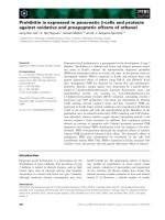
Fig. 1. Treatment schedule. Cadaveric human islets and MIN6 cells were incubated according to this treatment scheme. When adding the components (pectin,
streptozotocin (STZ), a-Lactose, or cytokines) to the culture system, this was done without medium replacement. The former component was present during the
following incubation.
how pectin may influence islet function, we studied the impact of pectin
on human islets and a mouse β-cell line under oxidative and inflammatory stress, induced by streptozotocin (STZ) or the proinflammatory cytokines Interferon-γ (IFN-γ), Tumor necrosis factor-α
(TNF-α), and Interleukin 1-β (IL-1β). We investigated if the effects of
pectin are dependent on the degree of methyl-esterification. To gain
insight in a possible role of Gal-3, the Gal-3 antagonist α-lactose was
applied in this study to block Gal-3 during challenge of β-cells. Our data
demonstrates that pectins protect human and mouse β-cells from oxidative and inflammatory processes in a DM-dependent fashion.
2018). The molecular weight of pectin was measured using high pressure size exclusion chromatography. The DM was determined with an
Ultimate 3000 high-performance liquid chromatography (HPLC)
system (Thermo Scientific). The constituent monosaccharide content
and composition was determined by gas chromatography as previously
described (Sahasrabudhe et al., 2018). The DM value of pectin was
confirmed by analyzing the release of methanol by high-performance
liquid chromatography (Voragen, Schols, & Pilnik, 1986). The DM was
calculated as the total mass of released methanol (mol) from per
100 mol of galacturonic acid.
2. Materials and methods
2.4. Nuclear magnetic resonance spectroscopy (NMR)
2.1. Cell culture
The mouse insulinoma MIN6 cell line (ATCC, Manassas, VA, USA)
was cultured in DMEM (Lonza, Basal, Switzerland) supplemented with
15 % fetal bovine serum (FBS, Lonza), 50 μmol/L β-mercaptoethanol,
2 mmol/L L-glutamine, 50 U/mL penicillin, and 50 mg/L streptomycin
(all from Sigma-Aldrich, St. Louis, MO, USA) in 5% CO2 (CO2: O2:
N2 = 5 : 20 : 75) at 37 ℃.
DM5, DM18, and DM69 pectins (17 mg) were suspended in 0.75 mL
of D2O. All of the samples had a pH in the range 4−4.7. 1H-, 13C-NMR
and Heteronuclear Multiple Quantum Coherence (HMQC) spectra were
recorded on a Bruker Spectrometer (600, 150.9 MHz; Mannheim,
Germany) in a 5-mm tube at 80 °C using D2O as solvent. 1H and 13C
chemical shifts were reported with 3-(trimethylsilyl) propionic-2,2,3,3d4 acid (δ 0.00 for 1H and for 13C) and acetone (δ 2.22 for 1H and δ
30.89, δ 215.94 for 13C) as internal reference.
2.2. Human islet isolation and culture
2.5. Human islet and MIN6 cell treatments
Human islets were isolated from cadaveric pancreata: three batches
were isolated at the Leiden University Medical Center (Leiden, The
Netherlands) (Ichii et al., 2005) and two batches were provided through
the JDRF award 31-2008-416 (ECIT Islet for Basic Research program,
Milan, Italy). Procedures were performed in accordance with the Code
of Proper Secondary Use of Human Tissue in The Netherlands as formulated by the Dutch Federation of Medical Scientific Societies. After
shipment, islets were handpicked and cultured in CMRL-1066 (GIBCO,
Bleiswijk, the Netherlands), containing 10 % FBS, 50 U/mL penicillin,
and 50 mg/L streptomycin, as previously described (Smink et al.,
2016). Islets were cultured in 5% CO2 at 37 ℃.
To investigate the effect of pectins on healthy β-cells, MIN6 cells
and human islets were incubated with lemon pectin (DM5, DM18,
DM69) dissolved in culture medium at a final concentration of 0.5, 1,
and 2 g/L for 24 h (Fig. 1a). To determine the influence of pectins under
stress, the cells and islets were incubated with the pectins for 1 h and
then exposed for 24 h to either the apoptosis-inducer streptozotocin
(STZ, Sigma-Aldrich) or the proinflammatory cytokines IFN-γ, TNF-α,
and IL-1β (all from ImmunoTools, Friesoythe, Germany) (Fig. 1b). All
experiments with human islets and cells were performed at 37 ℃ during
this study. For these experiments, the cells were treated with mouse or
human proinflammatory cytokines, IFN-γ (2000 U/mL), TNF-α (2000
U/mL), and IL-1β (150U/mL). Cell viability, apoptosis, ROS, NO, and
oxygen consumption rate (OCR) were measured after exposure to the
above-described conditions. Furthermore, to test whether effects are
Gal-3 dependent, MIN6 cell respiratory capacity was quantified in the
presence of the Gal-3 inhibitor α-lactose (Fig. 1c). For this, cells were
incubated with or without α-lactose (20 mM) (Sigma-Aldrich) 1 h before pectin incubation. For all of the above treatments, components
were added into the culture system without medium replacement.
2.3. Pectin samples
Lemon pectins with DM values of DM5 and DM18 were purchased
from CP Kelco (Lille Skensved, Denmark). The DM 69 pectin was purchased from Andre Pectin (Yantai, China). Endotoxin levels in pectin
samples were quantified with a Limulus amebocyte lysate assay and
showed to be below the detection level of 0.1 μg/L (Sahasrabudhe et al.,
2
Carbohydrate Polymers 249 (2020) 116863
S. Hu, et al.
1
H-NMR of DM18 showed the following α-GalA peaks: H-1 at 5.10, H-2
at 3.78, H-3 at 3.99, H-4 at 4.45, and H-5 at 4.82 ppm. The peak at
3.82 ppm can be assigned to the OCH3 group. The integration of the
OCH3 group corresponds to ∼20 % methylation. The 13C-NMR of
DM18 showed peaks of α-GalA: C-1 at 100.03, C-2 at 69.03, C-3 at
69.51, C-4 at 71.66, and C-5 at 78.89 ppm. The peak at 53.52 ppm
belongs to the OCH3 group. The 1H-NMR of DM69 that can be assigned
to α-GalA: H-1 and H-5 appear as a multiplet at 5.05 ppm, H-2 at 3.69,
H-3 at at 3.98, and H-4 at 4.46 ppm. The OCH3 peak resides at 3.82 ppm
and its integration suggests ∼61 % methylation. However, significant
overlap of the methyl peak in the 1H spectrum prevents accurate determination of methylation. The poor solubility and high gelling capacity of DM69 prevented the acquisition of a detailed 13C NMR
spectrum, so the 13C chemical shifts were revealed by HMQC.
2.6. Cell viability assays
To investigate the effect of pectins on β-cell viability, WST-1 was
applied (Roche, Indianapolis, IN, USA). To investigate the effect of
pectins on β-cell apoptosis, the cells and islets were stained by Alexa
Fluor® 488 annexin V (Biolegend, San Diego, CA, USA) and propidium
iodide (PI, Thermo Scientific, Eugene, OA, USA). For staining, islets
were fixed for 15 min with 4% (w/v) paraformaldehyde (Merck,
Darmstadt, Germany). Immunofluorescence staining of insulin was
performed as described previously (Liu et al., 2019). See supplementary
methods for details.
2.7. Oxidative stress assays
Intracellular ROS was detected according to the manufacturer’s instruction of a Cellular ROS Assay Kit (Abcam, Cambridge, UK). The NO
concentration in the supernatants was measured with a Nitric Oxide
Assay Kit (Invitrogen, Vienna, Austria) according to manufacturer’s
instructions. See supplementary methods for details.
3.2. Protective effect under STZ-induced stress
We first investigated the effects of pectins on human islets without
any stressor. Pectins did not significantly influence cell viability with
any of the tested DM-value (DM5, DM18 and DM69) or at any concentration (0.5, 1, and 2 g/L) (Fig. 2a, b). To investigate effects of pectin
on islets under stress we first tested the impact of pectins on islet-cells
exposed to STZ, i.e. a well-known β-cell apoptosis inducer (Biswas,
Gupta, Verma, & Singh, 2017). Human islets were incubated with
pectins with DM5, DM18, and DM69 and at concentrations of 0.5, 1,
and 2 g/L for 1 h. Subsequently 5 mM STZ was added and incubated for
24 h.
STZ significantly reduced viability by 53.8 ± 8.6 % (p < 0.001;
Fig. 2c). This reduction was less when islets were pre-incubated with
pectins. DM5 pectin showed the most pronounced protection. It prevented the STZ-induced viability decrease by 38.7 ± 10.5 %
(p < 0.01) at a concentration of 1 g/L but not at 0.5 g/L. DM5 pectin
exposure at a concentration of 2 g/L showed even a stronger protective
effect and prevented the decrease in viability after STZ exposure by
46.3 ± 13.7 % (p < 0.01). Effects were still present but less pronounced with DM18 pectin which only at 2 g/L significantly prevented
STZ-induced reduction in viability by 41.8 ± 9.7 % (p < 0.01). Pectin
of higher DM, i.e., DM 69, did not show any significant protective effects. The effect of pectins on islet apoptosis was also investigated. STZ
at a concentration of 5 mM significantly increased islet apoptosis with
54.8 ± 7.5 % (p < 0.001) compared to the untreated islets (Fig. 2d,
e). DM5 pectin at a concentration of 2 g/L significantly prevented STZinduced apoptosis with 46.0 ± 8.5 % (p < 0.001). This effect was not
observed when islets were pre-incubated with pectin of higher DM or
lower concentrations.
Islets are clusters of several different cell types (Cabrera et al.,
2006). To confirm relevance of our findings for β-cells we also performed the above-described experiments with a pure β-cell line. Since
there is no human β-cell line available that responds to glucose stimulation like in healthy individuals, we used the mouse MIN6 cell line
that does respond to glucose changes (Hastoy et al., 2018; Ishihara
et al., 1993). As in islets, pectin alone did not show any effects on β-cell
viability under homeostatic culture conditions (Fig. 2f). However, STZ
significantly decreased cell viability by 53.0 ± 1.1 % (p < 0.001)
(Fig. 2g), which was significantly prevented by DM5 pectin at 1 g/L by
33.8 ± 6.1 % (p < 0.05). DM5 pectin exposure at 2 g/L showed a
stronger protective effect and prevented the decline by 40.5 ± 2.0 %
(p < 0.001). DM18 pectin at 2 g/L significantly inhibited the decrease
by 27.5 ± 2.3 % (p < 0.05). DM69 pectin did not have a significant
effect here. STZ-induced apoptosis in 61.2 ± 6.8 % of the MIN6 cells
(p < 0.001; Fig. 2h). DM5 pectin at a concentration of 1 g/L and 2 g/L
significantly prevented STZ-induced cell apoptosis by respectively
46.8 ± 8.5 % (p < 0.01) and 53.4 ± 7.3 % (p < 0.001). DM18
pectin at 2 g/L also significantly inhibited apoptosis by 45.5 ± 7.6 %
(p < 0.01). There was no rescuing effect of DM69 pectin.
2.8. Oxygen consumption analysis
The effect of pectins on mitochondrial function was measured by the
Agilent Seahorse XF24 Extracellular Flux Assay Kit (Seahorse
Bioscience, North Billerica, MA, USA). MIN6 cells (4 × 10⁴ cells/well)
were seeded in XF24 cell culture microplates (Seahorse Bioscience) and
treated with pectin, STZ, or cytokines in the presence and absence of αlactose. See supplementary methods for details.
2.9. Statistical analysis
Parametric distribution of data was confirmed using KolmogorovSmirnov tests. Data are expressed as mean ± standard error of mean
(SEM). Statistical differences of parametric data were analyzed using
one-way ANOVA, while nonparametric data were analyzed with a
Kruskal-Wallis test. P-values < 0.05 were considered to be statistically
significant (*p < 0.05, **p < 0.01, and ***P < 0.001). The data
were analyzed using GraphPad Prism (version7.00; GraphPad Software
Inc, La Jolla, CA, USA).
3. Results
3.1. Structural characterization of pectins
The structural characterization of pectin DM5, DM18, and DM69
reported in Table 1 was confirmed by NMR analysis (Figs. S1–S8). The
major carbohydrate residue α-linked galacturonic acid (α-GalA) and
the degree of methylation was determined. The 1H-NMR spectrum of
DM5 showed the following α-GalA peaks: H-1 at 5.09, H-2 at 3.76, H-3
at 3.98, H-4 at 4.44, and H-5 at 4.77 ppm. 13C-NMR of DM5 presented
the following α -GalA peaks: C-1 at 99.94, C-2 at 69.06, C-3 at 69.64, C4 at 71.81, and C-5 at 78.91 ppm. The peak at 174.71 ppm is assigned to
the carbonyl group (C6). No significant OCH3 signal was observed. The
Table 1
Structural characteristics of the pectins. Degree of methyl-esterification
(DM) is defined as the amount of methanol (mole) per 100 mol of the total
galacturonic acid in the sample. Molecular weight = Mw. Rhamnose = Rha,
arabinose = Ara, galactose = Gal, glucose = Glc and Uronic acid = UA.
Pectin
DM5
DM18
DM69
Mw (KDa)
36
53
81
Monosaccharide content (mol%)
Carbohydrate
content (%)
Rha
Ara
Gal
Glc
UA
0
0
1
0
0
2
3
3
8
0
1
1
95
95
87
68
73
83
3
Carbohydrate Polymers 249 (2020) 116863
S. Hu, et al.
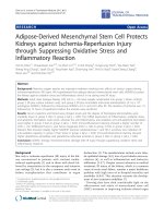
Fig. 2. Effects of pectins with different DM (DM5, DM18, and DM69) on cell survival in human islets and MIN6 cells exposure to STZ. Human islets (a and b)
or MIN6 cells (f) were incubated with pectin followed by measuring cell viability (a and f) using a WST-1 assay and insulin staining (b). To investigate the effect of
pectins on islet β-cell under streptozotocin (STZ)-induced stress, islets (c-e) or MIN6 cells (f-h) were incubated with pectins for 1 h followed by co-incubation for 24 h
with 5 mM STZ and pectin. After incubation cell viability was determined by a WST-1 assay (c and g). Islet cell apoptosis was determined by co-staining with Annexin
V and PI, and analyzed under a fluorescence microscope (d). Islet Annexin PI staining results were analyzed by using Image J gradation analysis (e). MIN6 cell
apoptosis was detected using a flow cytometric assay with Annexin V and PI staining (h). Results are plotted as mean ± SEM (n = 5). The statistical differences were
quantified using one-way ANOVA analysis with Newman-Keuls multiple comparisons test (*p < 0.5, **p < 0.01, ***p < 0.001). Scale bar denotes 100 μm.
cells (Fig. 3b, c). However, the apoptosis was significantly reduced with
43.9 ± 5.8 % (p < 0.001) when islets were pre-incubated with DM5
pectin at 2 g/L. Pectins of DM18 and DM69 did not show an effect on
apoptosis at any concentrations.
We also studied the impact of cytokines and pectins on MIN6 cells.
Cytokine-treated cells showed a decrease in cell viability of 57.9 ± 2.6
% (p < 0.001) compared to controls. This decrease was prevented by
low-DM pectins (Fig. 3d). DM5 pectin at 1 g/L significantly prevented
the viability decrease with 44.0 ± 5.4 % (p < 0.01). When further
increasing the dosages of DM5 pectin to 2 g/L, the pectin almost completely prevented the negative effect on viability (p < 0.001). DM18
pectin at 2 g/L demonstrated a protective effect but less effective as
DM5 pectin. It prevented decrease in cell viability with 27.8 ± 4.0 %
(p < 0.05). DM69 pectin-pretreated cells did not show significant
differences. To also determine the influence of pectin on cytokine-induced β-cell apoptosis, we measured apoptosis of MIN6 cells incubated
with cytokines in presence and absence of pectin. Cytokines increased
apoptosis by 77.2 ± 7.6 % (p < 0.001; Fig. 3e). DM5 pectin at a
concentration of 1 g/L and 2 g/L significantly prevented this increase
3.3. Protective effect under inflammatory stress
To determine the role of pectins on β-cell survival under cytokine
stress we tested the effects after exposure to the cocktail of IL-1β + IFNγ + TNF-α, which has been identified as essential effector molecules in
the initiation of Type 1 Diabetes (van der Torren et al., 2016). The
cytokines significantly decreased human islet viability with 63.2 ± 8.3
% (p < 0.001; Fig. 3a). However, a pre-incubation with pectin prevented this viability decrease. DM5 at a concentration of 1 g/L significantly prevented the viability decline with 45.7 ± 8.0 %
(p < 0.001) and at 2 g/L almost completely prevented the negative
effects on viability. Effects were less at higher DM as DM18 pectin only
prevented cytokine damage at 2 g/L significantly with 43.8 ± 10.9 %
(p < 0.01) and not at lower concentrations. DM69 pectin did not influence islet viability after cytokine exposure. Inflammatory cytokineinduced islet apoptosis is the main cause of islet loss (Arroyo-Jousse,
Garcia-Diaz, Codner, & Pérez-Bravo, 2016). Therefore, apoptosis was
investigated following incubation with pectins and cytokines. Cytokines
induced apoptosis in 53.1 ± 5.5 % (p < 0.001) of the human islet-
4
Carbohydrate Polymers 249 (2020) 116863
S. Hu, et al.
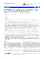
Fig. 3. Effects of DM5, DM18, or DM69 pectin on cell survival of human islets and MIN6 cells exposure to proinflammatory IFN-γ, TNF-α, and IL-1β
cytokine cocktail. Human islets (a-c) and MIN6 cells (d and e) were incubated with pectins for 1 h followed by co-incubated of pectins and cytokines (IFN-γ, TNF-α,
and IL-1β) for an additional 24 h. After incubation, cell viability was determined by a WST-1 assay (a and d). Cell apoptosis was determined by co-staining with
Annexin V and PI. Human islets were imaged with a fluorescence microscope (b) and analyzed by using Image J gradation analysis (c). MIN6 cell apoptosis was
detected using a flow cytometric assay (e). Results are plotted as mean ± SEM (n = 5). The statistical differences were analyzed using one-way ANOVA analysis with
Newman-Keuls multiple comparisons test. (*p < 0.5, **p < 0.01, ***p < 0.001). Scale bar denotes 100 μm.
3.4. Pectin attenuates generation of free radicals
with 43.4 ± 8.3 % (p < 0.05) and 63.7 ± 8.6 % (p < 0.01). DM18
pectin only showed a rescuing effect at 2 g/L, the apoptosis was reduced
with 51.9 ± 7.4 % (p < 0.01) compared to cells treated with cytokines alone. DM69 pectin did not prevent cytokine-induced apoptosis.
As early stages of STZ-induced β-cell damage are characterized by
free radical generation, we studied whether pectins impact the release
5
Carbohydrate Polymers 249 (2020) 116863
S. Hu, et al.
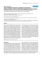
Fig. 4. Effects of DM5, DM18, or DM69 pectin on STZ- or cytokine-induced free radical generation in human islets and MIN6 cells. Human islets (a, b, e and
f) or MIN6 cells (c, d, g and h) were incubated with 2 g/L pectin for 1 h followed by 24 h co-incubation with pectin and 5 mM STZ (a-d) or a cytokine cocktail (e-h).
Intracellular ROS was measured with a DCFDA Cellular ROS Detection Assay Kit (a, c, e and g). Nitric oxide (NO) was detected using a Nitric Oxide Assay Kit (b, d, f
and h). Results represent are expressed as mean ± SEM (n = 5). The statistical differences were analyzed using one-way ANOVA analysis with Newman-Keuls
multiple comparisons test. (*p < 0.5, **p < 0.01, ***p < 0.001).
cytokines, trigger β-cell death via apoptosis (Kim, Lee, Gao, & Jung,
2005). Therefore, we investigated production of ROS and NO in both
islets and MIN6 cells after treatment with our cytokine cocktail and 2 g/
L of the different pectins. Treatment with cytokines significantly increased ROS production in islets with 70.3 ± 12.7 % (p < 0.001;
Fig. 4e). Pretreatment with DM5 pectin significantly prevented the
cytokine-induced ROS generation, which was similar to untreated
controls (p < 0.001). A preincubation with DM18 did also significantly
reduce ROS production with 40.0 ± 14.9 % (p < 0.05). However,
DM69 pectin exposure did not prevent the increase. NO was elevated
after cytokine incubation with 52.5 ± 14.6 % (p < 0.01; Fig. 4f). This
increase was prevented by DM5 pectin with 38.4 ± 16.4 %
(p < 0.05), but not by DM18 and DM69 pectin.
In MIN6 cells we observed similar results as in the human islets.
Cytokines provoked a significant increase of ROS with 74.5 ± 6.9 %
(p < 0.001) as compared with untreated controls (Fig. 4g). DM5 pectin
suppressed this overproduction with 58.9 ± 7.7 % (p < 0.001).
DM18 pectin has less effect but still significantly suppressed the increase in ROS production with 26.1 ± 9.7 % (p < 0.05). DM69 pectin
did not show a significant effect. Furthermore, cytokines enhanced NO
release in MIN6 cells with 35.2 ± 6.2 % (p < 0.001; Fig. 4h). DM5
pectin prevented this NO increase with 31.4 ± 6.8 % (p < 0.01),
which made the NO level similar to the untreated control. DM18 and
DM69 pectin did not prevent the NO increase.
of ROS and NO (Wu et al., 2016). Since 2 g/L pectins had the most
pronounced effect, these experiments were only done with this concentration. ROS generation was significantly increased with
41.4 ± 8.4 % (p < 0.05) after human islets were treated with STZ
(Fig. 4a). This increase was prevented by DM5 pectin with 43.2 ± 6.8
% (p < 0.05), but not by DM18 and DM69 pectin. STZ also strongly
impacted NO synthesis of islets, which was enhanced with 73.7 ± 5.3
% (p < 0.001; Fig. 4b) compared to untreated controls. DM5 and
DM18 pectin prevented this enhancement with respectively 76.2 ± 5.7
% (p < 0.001) and 56.3 ± 7.7 % (p < 0.01). DM69 pectin was unable to prevent the NO overproduction.
We also studied ROS and NO generation in MIN6 cells showing that
STZ provoked an increased production of ROS with 66.9 ± 4.0 %
(p < 0.001) as compared with untreated controls (Fig. 4c). DM5 pectin
significantly prevented this increase with 50.1 ± 4.5 % (p < 0.01)
compared to cells incubated with only STZ. DM18 pectin also significantly suppressed the increase in ROS production with 26.1 ± 4.4
% (p < 0.05), but DM69 pectin did not show significant prevention.
Consistent with the human islet results, STZ enhanced NO release from
MIN6 cells with 38.4 ± 1.9 % (p < 0.01; Fig. 4d). DM5 pectin prevented this STZ-induced NO increase with 34.9 ± 1.9 % (p < 0.01)
resulting in similar levels of NO compared to the untreated control.
DM18 pectin also significantly prevented STZ-induced NO increase with
24.8 ± 1.9 % (p < 0.05), whereas DM69 pectin did not impact NO
release.
Elevated intracellular free radicals, induced by proinflammatory
6
Carbohydrate Polymers 249 (2020) 116863
S. Hu, et al.
Fig. 5. Effect of DM5, DM18, or DM69 pectin on oxygen consumption rate of MIN6 cells after STZ or cytokine-induced stress. (a) A schematic overview of the
mitochondrial stress test. Arrows indicate the subsequent addition of the ATPase inhibitor oligomycin, the uncoupling reagent FCCP, and the inhibitors of the
electron transport chain rotenone/antimycin A. (b) MIN6 cell were seeded in a Seahorse cell culture plate. After incubation with pectins at 2 g/L for 1 h, STZ at a final
concentration of 5 mM (b-e) or a cytokine cocktail (f-i) was added to each well and co-incubated with pectin for 24 h. OCR of treated cells was investigated using
Seahorse Bioscience XF24 extracellular flux analyzer. Fig. c-e and g-i respectively represent individual parameters for basal respiration, spare respiration, and ATPlinked respiration. Results are plotted as mean ± SEM (n = 5). The statistical differences were analyzed using one-way ANOVA analysis with Newman-Keuls
multiple comparisons test. (*p < 0.05, **p < 0.01, ***p < 0.001).
exposed to DM18 pectin prevented the STZ-induced basal respiration
decrease with 16.9 ± 2.8 % (p < 0.05) and ATP-linked respiration
with 22.4 ± 4.0 % (p < 0.05). The spare respiration was still affected
by STZ. DM69 pectin did not protect the cells from STZ-induced respiratory damage.
To determine the influence of pectin on cytokine-induced mitochondrial dysfunction, we treated MIN6 cells with the cytokines in
the presence and absence of pectins at 2 g/L and measured OCR after
24 h. The culture of β-cells with cytokines significantly decreased OCR,
it reduced the basal respiration with 52.8 ± 2.0 % (p < 0.001), spare
respiration with 53.9 ± 8.0 % (p < 0.001), and ATP-linked respiration with 64.6 ± 1.8 % (p < 0.001; Fig. 5f–i). However, cells preincubated with pectins were protected from these effects. DM5 pectin
significantly prevented the decrease in basal respiration with
48.1 ± 3.5 % (p < 0.001), spare respiration with 33.6 ± 3.2 %
(p < 0.05), and ATP-linked respiration with 69.7 ± 5.2 %
(p < 0.001) as compared with cells incubated with only cytokines.
DM18 pectin was less efficient but still had significant protective effects. It prevented a basal respiration decrease of 25.8 ± 4.9 %
(p < 0.05), reduced spare respiration with 35.3 ± 3.8 % (p < 0.05),
and prevented ATP-linked respiration decline with 37.7 ± 7.0 %
(p < 0.01). DM69 pectin did not show any significant effects here.
3.5. Pectin prevents damage in energy metabolism
The release of free radicals is often accompanied by mitochondrial
dysfunction, which leads to disorders in the cellular energy metabolism
(Banerjee et al., 2020). To investigate whether the tested pectins contribute to maintenance of β-cell energy metabolism after STZ exposure,
we determined the effect of pectin on the OCR of MIN6 cells, which is
an indicator of mitochondrial respiration in islet-cells (Fig. 5a) (Llacua,
de Haan, & de Vos, 2018). As these assays require high cell amounts, it
could only be performed with MIN6 cells and not with the rarely
available human islets.
STZ has a strong negative impact on the OCR of β-cells, which was
prevented by pectins in a DM-dependent fashion (Fig. 5b–e). MIN6 cells
incubated with STZ showed a decreased respiration rate, including a
significantly decreased basal respiration, which was 46.8 ± 2.4 %
(p < 0.001) lower than controls, a spare respiration reduction of
23.2 ± 3.6 % (p < 0.05), and a reduced ATP production of
53.5 ± 3.6 % (p < 0.001). A pre-incubation with DM5 pectin almost
completely prevented these negative effects. The mitochondrial basal
respiration rate, spare respiration, and ATP-linked respiration values
were identical to the untreated controls when cells were exposed to
DM5 pectin and STZ. DM18 pectin had a partial rescue effect. Cells
7
Carbohydrate Polymers 249 (2020) 116863
S. Hu, et al.
Fig. 6. DM5 pectin influences islet cell respiration through Galectin-3 (Gal-3). To verify if pectin prevents the STZ- or cytokine-induced OCR reduction through
interaction with Gal-3, OCR was measured in the presence and absence of the Gal-3 ligand α-lactose (Lac). MIN6 cells were seeded in a Seahorse cell culture plate.
Lactose at 20 mM was incubated with MIN6 cells for 1 h, followed by DM5 pectin and STZ (a-d) or a cytokine cocktail (e-h) for an additional 24 h. OCR of treated cells
was investigated using the Seahorse Bioscience XF24 extracellular flux analyzer. Fig. b-d and f-h respectively represent individual parameters for basal respiration,
spare respiration, and ATP-linked respiration. Results are plotted as mean ± SEM (n = 5). The statistical differences were analyzed using one-way ANOVA analysis
with Newman-Keuls multiple comparisons test. (*p < 0.05, **p < 0.01, ***p < 0.001).
3.6. Pectins rescue damaged cells by binding to Gal-3
4. Discussion
Pectin influences adipocyte metabolism and cancer cell apoptosis as
a natural ligand of Gal-3 (Fang et al., 2018; Zhang et al., 2016). To
verify if pectin prevents STZ-induced OCR reduction through interaction with Gal-3, the OCR was measured in the presence and absence of
the Gal3 antagonist α-lactose (Fang et al., 2018). Since DM5 pectin
showed the most pronounced effect, this experiment was only done
with DM5 pectin. An incubation with α-lactose alone did not influence
β-cell respiration in normal culture conditions (Fig. 6a–d). The mitochondrial basal respiration rate, spare respiration, and ATP-linked
respiration of MIN6 cells exposed to α-lactose were identical to untreated controls. However, in the presence of α-lactose, the preventive
effects of DM5 pectin were compromised. The preincubation with αlactose before pectin and STZ resulted in reduced basal respiration,
spare respiration, and ATP-linked respiration, which was respectively
24.4 ± 4.4 % (p < 0.01), 19.5 ± 8.2 % (p < 0.05), and 31.6 ± 5.7
% (p < 0.01) lower than cells treated with pectin and STZ.
We also treated MIN6 cells with DM5 pectin and the cytokines with
and without preincubation of α-lactose. A preincubation of α-lactose
prevented the protective effect of DM5 pectin on cytokine-damaged βcells (Fig. 6e). The protective effect of DM5 on basal respiration was no
longer present, inclusion of the α-lactose decreased the basal respiration with 56.8 ± 2.7 % (p < 0.001) compared with cells treated with
DM5 pectin and cytokines (Fig. 6f). The preincubation of lactose did not
show considerable influence on spare respiration (Fig. 6g). The rescue
effect of DM5 pectin on ATP-linked respiration was fully counteracted
by preincubation of α-lactose (Fig. 6h). This indicates that DM5 pectin
binds with Gal-3 and regulates β-cell metabolism.
Type 1 and Type 2 Diabetes are frequently associated with oxidative
and inflammatory stress-induced β-cell loss (Danobeitia et al., 2017;
Hu, Kuwabara, de Haan, Smink, & de Vos, 2020; Usmani-Brown et al.,
2019). Many report beneficial effects of pectin on glucose management,
but not much is known about the direct impact on β-cells under stress
(García-Carrizo et al., 2019; Wu et al., 2017). Previous studies have
shown that pectin may regulate the function of Gal-3, which have been
shown to participate in cytokine-induced apoptosis (Nishikawa et al.,
2018). Here, we showed that pectins can rescue β-cell viability and
their respiratory metabolism under STZ- or cytokine-induced stress.
Furthermore, pectins reduced oxidative and nitrosative stress in both
human islets and β-cells. This protective effect against STZ or inflammatory stress was dependent on the DM and the concentration of
the pectins.
Previous mouse studies demonstrate a potential role for Gal-3 in
anti-diabetic effect of pectins (Li et al., 2016; Mensah-Brown et al.,
2009). These studies did not investigate the direct protective effects of
pectin on islets. Islets are believed to be micro-organs containing several cell types (Cabrera et al., 2006; Saksida et al., 2013). By using a
combination of human islets and a β-cell line, we proved the direct
protective effects of pectin on β-cells. We show pectins rescue from
oxidative or inflammatory stress through Gal-3. Gal-3 is synthesized by
free ribosomes in the cytosol but can easily cross the plasma membrane
and the endomembrane system to translocate into the nucleus, mitochondria, and extracellular matrix (Sehrawat & Kaur, 2020). Since
pectin is a macromolecule and cannot access cytoplasm, pectin likely
binds with Gal-3 at the surface of the cellular membrane and in the
8
Carbohydrate Polymers 249 (2020) 116863
S. Hu, et al.
Fig. 7. Schematic illustration of the rescuing effects of low-DM pectin on β-cells. Cellular membrane and extracellular Gal-3 binding with low-DM pectin
inhibits mitochondrial dysfunction, ROS and NO overproduction, and apoptosis in β-cells under STZ- or proinflammatory cytokine-induced stress.
et al., 2016). The procedure applied for pectin de-esterification results
in shorter chains and a decrease in the molecular weight of the pectin
(Hotchkiss et al., 2002; Sahasrabudhe et al., 2018). This procedure may
also liberate binding regions from complicated long-chain pectins and
allow them to freely bind with Gal-3. Therefore, the lowest DM pectin
has a maximum amount of freely binding regions per unit mass that
leads to a more prominent effect (Eliaz, 2019).
The composition of sidechain monosaccharides might influence the
biological characteristics of pectins (Torkova et al., 2018). Among the
tested pectins in this study, similar sugar composition patterns were
found in DM5 and DM18 pectin. However, DM5 pectin showed significantly stronger protection against β-cell damage. This indicates the
DM values of pectins do influence the protective ability of pectin. Additionally, it was suggested that the pectin structure present a high
diversity according to their different origin (Muller-Maatsch et al.,
2016). Recent studies of pectins extracted from other sources, e.g. okra,
sour cherry pomace, and papayas, suggest the essential roles of pectin’s
molecular weight and the degree of methyl-esterification on its chemical and biological characteristics (Mao et al., 2020; Prado et al.,
2020). The comparisons of pectin extract from different sources may
also contribute to a deeper understanding of structure-related biological
function.
Our study suggests that low-DM lemon pectins, could potentially be
applied in the prevention and management of diabetes by protecting βcells against inflammatory and oxidative stress. This is, to the best of
our knowledge, a new explanation as to why increased dietary fiber
intake is associated with a lower frequency of hyperglycemia.
Treatment of intestinal inflammation and tumors with dietary fibers
showed that these polysaccharides are taken up by gastrointestinal
macrophages, transported to the bone marrow, and subsequently secreted into the peripheral circulation (De Leoz et al., 2013; Eiwegger
et al., 2010; Hong et al., 2004; Porporatto et al., 2005). This indicates
extracellular matrix (Sahasrabudhe et al., 2018).
The mechanism behind the anti-apoptotic effects may be via three
ways (Fig. 7). First, pectin is directly inhibiting pro-apoptotic Gal-3
located on the cell membrane (Fukumori et al., 2003). This binding
suppresses related mitochondrial apoptotic pathways and rescues mitochondrial respiratory function (Fukumori et al., 2003). Second, pectin
is hampering intracellular danger signal delivery. Gal-3 mediates the
ligation of cell surface glycoproteins and increases the affinity of cell
binding, which in turn facilitates intercellular signaling (Colin Hughes,
2001). The severely impaired β-cell can generate danger signals that
induce nearby cell apoptosis (Paredes-Juarez et al., 2015). The binding
of pectin to Gal-3 potentially suppresses danger signal delivery, subsequently improving cell survival under stress. Third, another plausible
explanation could be that pectin induces translocation of Gal-3 to the
perinuclear membranes (Yu, Finley, Raz, & Kim, 2002). This translocation protects mitochondrial integrity and inhibits apoptosis (Yu et al.,
2002). However, the exact mechanism by which Gal-3 translocates
remains subject of debate (Funasaka, Raz, & Nangia-Makker, 2014).
Further research will enable us to exactely pinpoint which of the three
mechanism described above is applicable to Gal-3 in β-cell apoptotic
processes by pectins.
The protective effect against STZ or cytokines is dependent on
pectin concentration and DM value of the pectin. The low-DM pectin at
high concentration showed the highest efficiency in protection against
oxidative and inflammatory damage. The DM-dependent effect may be
related to the molecular weight of pectin, which can be explained by
differences in the valid binding-domain densities of pectins with different DM values. Considering that pectin is a long-chain polymer, each
single chain has multiple binding regions (Eliaz & Raz, 2019). Although
longer chain polymers could contain more binding regions, limited by
complicated structure and distance between regions, partial regions in
long-chain pectin may not be able to freely bind with Gal-3 (Zhang
9
Carbohydrate Polymers 249 (2020) 116863
S. Hu, et al.
pectins can be blood-born, and influence tissue metabolism. The insufficient intake of dietary fibers in Western societies has been believed
to be one of the major causes of the high incidence rates of both Type 1
Diabetes and Type 2 Diabetes (Mishra et al., 2019). Previous studies
report beneficial effects of dietary fiber on glucose metabolism, but
none of these effects is attributed to directly impacting metabolism or
viability of β-cells under stress (Wu et al., 2016, 2017). Our study demonstrates that low-DM pectin plays an essential role in maintaining βcell metabolism and promoting survival under stressful conditions. Islet
transplantation, a promising treatment for Type 1 Diabetes, which is
challenged by oxidative stress-induced islet graft loss, could also benefit
from these results (Bottino et al., 2004; Hu & de Vos, 2019). As a natural polymer with excellent biocompatibility (Singha et al., 2017), lowDM pectin could be applied as a preincubation or coating of the islet
graft to improve graft survival. Based on its biocompatibility and biodegradability, we believe that the highly abundant and low-cost natural
polymer pectin might has a great potential for reducing the expense and
cytotoxicity of diabetes treatment (Singha et al., 2017). In conclusion,
this study provides new insights in how pectin can contribute to
maintenance of health. Our data reveals an unrecognized influence of
pectin on β-cell apoptosis in the oxidative and inflammatory context,
showing to improve β-cell survival through binding with Gal-3.
(2004). Response of human islets to isolation stress and the effect of antioxidant
treatment. Diabetes, 53(10), 2559.
Cabrera, O., Berman, D. M., Kenyon, N. S., Ricordi, C., Berggren, P.-O., & Caicedo, A.
(2006). The unique cytoarchitecture of human pancreatic islets has implications for
islet cell function. Proceedings of the National Academy of Sciences of the United States of
America, 103(7), 2334–2339.
Colin Hughes, R. (2001). Galectins as modulators of cell adhesion. Biochimie, 83(7),
667–676.
Dabhi, B., & Mistry, K. N. (2015). Oxidative stress and its association with TNF-α-308 G/C
and IL-1α-889 C/T gene polymorphisms in patients with diabetes and diabetic nephropathy. Gene, 562(2), 197–202.
Danobeitia, J. S., Chlebeck, P. J., Shokolenko, I., Ma, X., Wilson, G., & Fernandez, L. A.
(2017). Novel fusion protein targeting mitochondrial DNA improves pancreatic islet
functional potency and islet transplantation outcomes. Cell Transplantation, 26(11),
1742–1754.
De Leoz, M. L. A., Wu, S., Strum, J. S., Niñonuevo, M. R., Gaerlan, S. C., Mirmiran, M.,
et al. (2013). A quantitative and comprehensive method to analyze human milk
oligosaccharide structures in the urine and feces of infants. Analytical and
Bioanalytical Chemistry, 405(12), 4089–4105.
Dranca, F., & Oroian, M. (2018). Extraction, purification and characterization of pectin
from alternative sources with potential technological applications. Food Research
International, 113, 327–350.
Eiwegger, T., Stahl, B., Haidl, P., Schmitt, J., Boehm, G., Dehlink, E., et al. (2010).
Prebiotic oligosaccharides: In vitro evidence for gastrointestinal epithelial transfer
and immunomodulatory properties. Pediatric Allergy and Immunology, 21(8),
1179–1188.
Eliaz, I. (2019). Letter to the Editor: Not all modified citrus pectins are the same: Size does
matter. American Journal of Physiology Heart and Circulatory Physiology, 316(5),
H1232–H1233.
Eliaz, I., & Raz, A. (2019). Pleiotropic effects of modified Citrus pectin. Nutrients, 11(11),
2619.
Fang, T., Liu, D.-d., Ning, H.-m., Dan, L., Sun, J.-y., Huang, X.-j., et al. (2018). Modified
citrus pectin inhibited bladder tumor growth through downregulation of galectin-3.
Acta Pharmacologica Sinica, 39(12), 1885–1893.
Fujimaki, K., Ogihara, T., Morris, D. L., Oda, H., Iida, H., Fujitani, Y., et al. (2015). SET7/
9 enzyme regulates cytokine-induced expression of inducible nitric-oxide synthase
through methylation of lysine 4 at histone 3 in the islet β cell. The Journal of Biological
Chemistry, 290(27), 16607–16618.
Fukumori, T., Takenaka, Y., Yoshii, T., Kim, H.-R. C., Hogan, V., Inohara, H., et al. (2003).
CD29 and CD7 mediate Galectin-3-Induced type II T-Cell apoptosis. Cancer Research,
63(23), 8302–8311.
Funasaka, T., Raz, A., & Nangia-Makker, P. (2014). Nuclear transport of galectin-3 and its
therapeutic implications. Seminars in Cancer Biology, 27, 30–38.
García-Carrizo, F., Picó, C., Rodríguez, A. M., & Palou, A. (2019). High-esterified pectin
reverses metabolic malprogramming, improving sensitivity to Adipostatic/Adipokine
hormones. Journal of Agricultural and Food Chemistry, 67(13), 3633–3642.
Gerber, P. A., & Rutter, G. A. (2017). The role of oxidative stress and hypoxia in pancreatic beta-cell dysfunction in diabetes mellitus. Antioxidants & Redox Signaling,
26(10), 501–518.
Hastoy, B., Godazgar, M., Clark, A., Nylander, V., Spiliotis, I., van de Bunt, M., et al.
(2018). Electrophysiological properties of human beta-cell lines EndoC-betaH1 and
-betaH2 conform with human beta-cells. Scientific Reports, 8(1), 16994.
Hong, F., Yan, J., Baran, J. T., Allendorf, D. J., Hansen, R. D., Ostroff, G. R., et al. (2004).
Mechanism by which orally administered β-1,3-Glucans enhance the tumoricidal
activity of antitumor monoclonal antibodies in murine tumor models. Journal of
Immunology (Baltimore, Md : 1950), 173(2), 797–806.
Hotchkiss, A. T., Savary, B. J., Cameron, R. G., Chau, H. K., Brouillette, J., Luzio, G. A.,
et al. (2002). Enzymatic modification of pectin to increase its calcium sensitivity
while preserving its molecular weight. Journal of Agricultural and Food Chemistry,
50(10), 2931–2937.
Hu, S., & de Vos, P. (2019). Polymeric approaches to reduce tissue responses against
devices applied for islet-cell encapsulation. Frontiers in Bioengineering and
Biotechnology, 7(134).
Hu, S., Kuwabara, R., de Haan, B. J., Smink, A. M., & de Vos, P. (2020). Acetate and
butyrate improve β-cell metabolism and mitochondrial respiration under oxidative
stress. International Journal of Molecular Sciences, 21(4), 1542.
Ichii, H., Pileggi, A., Molano, R. D., Baidal, D. A., Khan, A., Kuroda, Y., et al. (2005).
Rescue purification maximizes the use of human islet preparations for transplantation. American Journal of Transplantation: Official Journal of the American Society of
Transplantation and the American Society of Transplant Surgeons, 5(1), 21–30.
Ishihara, H., Asano, T., Tsukuda, K., Katagiri, H., Inukai, K., Anai, M., et al. (1993).
Pancreatic beta cell line MIN6 exhibits characteristics of glucose metabolism and
glucose-stimulated insulin secretion similar to those of normal islets. Diabetologia,
36(11), 1139–1145.
Kim, W. H., Lee, J. W., Gao, B., & Jung, M. H. (2005). Synergistic activation of JNK/SAPK
induced by TNF-α and IFN-γ: Apoptosis of pancreatic β-cells via the p53 and ROS
pathway. Cellular Signalling, 17(12), 1516–1532.
Li, P., Liu, S., Lu, M., Bandyopadhyay, G., Oh, D., Imamura, T., et al. (2016).
Hematopoietic-derived Galectin-3 causes cellular and systemic insulin resistance.
Cell, 167(4), 973–984 e912.
Liu, Q., Chiu, A., Wang, L.-H., An, D., Zhong, M., Smink, A. M., et al. (2019).
Zwitterionically modified alginates mitigate cellular overgrowth for cell encapsulation. Nature Communications, 10(1), 5262.
Llacua, L. A., de Haan, B. J., & de Vos, P. (2018). Laminin and collagen IV inclusion in
immunoisolating microcapsules reduces cytokine-mediated cell death in human
pancreatic islets. Journal of Tissue Engineering and Regenerative Medicine, 12(2),
Funding
This research was funded by Juvenile Diabetes Research Foundation
(JDRF) grant, grant number 2-RSA-2018-523-S-B and Manpei Suzuki
Diabetes Foundation.
CRediT authorship contribution statement
Shuxian Hu: Conceptualization, Methodology, Investigation,
Formal analysis, Software, Writing - original draft. Rei Kuwabara:
Conceptualization, Methodology, Writing - review & editing. Martin
Beukema: Conceptualization, Methodology, Writing - review & editing.
Michela Ferrari: Conceptualization, Methodology, Investigation,
Formal analysis, Writing - review & editing. Bart J. de Haan:
Conceptualization, Methodology, Writing - review & editing. Marthe
T.C. Walvoort: Conceptualization, Methodology, Writing - review &
editing, Supervision. Paul de Vos: Conceptualization, Methodology,
Writing - review & editing, Supervision, Project administration.
Alexandra M. Smink: Conceptualization, Methodology, Writing - review & editing, Supervision.
Acknowledgments
The authors acknowledge the support of China Scholarship Council.
Human islets were provided through the JDRF award 31-2008-416
(ECIT Islet for Basic Research program).
Appendix A. Supplementary data
Supplementary material related to this article can be found, in the
online version, at doi: />References
Arroyo-Jousse, V., Garcia-Diaz, D. F., Codner, E., & Pérez-Bravo, F. (2016). Epigenetics in
type 1 diabetes: TNFa gene promoter methylation status in Chilean patients with type
1 diabetes mellitus. The British Journal of Nutrition, 116(11), 1861–1868.
Banerjee, A., Singh, S., Prasad, S. K., Kumar, S., Banerjee, O., Seal, T., et al. (2020).
Protective efficacy of Tinospora sinensis against streptozotocin induced pancreatic
islet cell injuries of diabetic rats and its correlation to its phytochemical profiles.
Journal of Ethnopharmacology, 248, Article 112356.
Biswas, J., Gupta, S., Verma, D. K., & Singh, S. (2017). Streptozotocin alters glucose
transport, connexin expression and endoplasmic reticulum functions in neurons and
astrocytes. Neuroscience, 356, 151–166.
Bottino, R., Balamurugan, A. N., Tse, H., Thirunavukkarasu, C., Ge, X., Profozich, J., et al.
10
Carbohydrate Polymers 249 (2020) 116863
S. Hu, et al.
Singha, N. R., Karmakar, M., Mahapatra, M., Mondal, H., Dutta, A., Roy, C., et al. (2017).
Systematic synthesis of pectin-g-(sodium acrylate-co-N-isopropylacrylamide) interpenetrating polymer network for superadsorption of dyes/M (II): Determination of
physicochemical changes in loaded hydrogels. Polymer Chemistry, 8(20), 3211–3237.
Smink, A. M., de Haan, B. J., Paredes-Juarez, G. A., Wolters, A. H., Kuipers, J., Giepmans,
B. N., et al. (2016). Selection of polymers for application in scaffolds applicable for
human pancreatic islet transplantation. Biomedical Materials (Bristol, England), 11(3),
Article 035006.
Sparre, T., Christensen, U. B., Mose Larsen, P., Fey, S. J., Wrzesinski, K., Roepstorff, P.,
et al. (2002). IL-1beta induced protein changes in diabetes prone BB rat islets of
Langerhans identified by proteome analysis. Diabetologia, 45(11), 1550–1561.
Sudhahar, V., Okur, M. N., Bagi, Z., O’Bryan, J. P., Hay, N., Makino, A., et al. (2018). Akt2
(Protein kinase B Beta) Stabilizes ATP7A, a copper transporter for extracellular superoxide dismutase, in vascular smooth muscle: Novel mechanism to limit endothelial dysfunction in type 2 diabetes mellitus. Arteriosclerosis, Thrombosis, and
Vascular Biology, 38(3), 529–541.
Torkova, A. A., Lisitskaya, K. V., Filimonov, I. S., Glazunova, O. A., Kachalova, G. S.,
Golubev, V. N., et al. (2018). Physicochemical and functional properties of Cucurbita
maxima pumpkin pectin and commercial citrus and apple pectins: A comparative
evaluation. PloS One, 13(9), Article e0204261.
Usmani-Brown, S., Perdigoto, A. L., Lavoie, N., Clark, P., Korah, M., Rui, J., et al. (2019).
Β cell responses to inflammation. Molecular Metabolism, 27, S104–S113.
van der Torren, C. R., Verrijn Stuart, A. A., Lee, D., Meerding, J., van de Velde, U.,
Pipeleers, D., et al. (2016). Serum cytokines as biomarkers in islet cell transplantation
for type 1 diabetes. PloS One, 11(1), Article e0146649.
Voragen, A. G. J., Schols, H. A., & Pilnik, W. (1986). Determination of the degree of
methylation and acetylation of pectins by h.p.l.c. Food Hydrocolloids, 1(1), 65–70.
Wu, H., Li, G.-N., Xie, J., Li, R., Chen, Q.-H., Chen, J.-Z., et al. (2016). Resveratrol
ameliorates myocardial fibrosis by inhibiting ROS/ERK/TGF-β/periostin pathway in
STZ-induced diabetic mice. BMC Cardiovascular Disorders, 16, 5.
Wu, J., Chen, M., Shi, S., Wang, H., Li, N., Su, J., et al. (2017). Hypoglycemic effect and
mechanism of a pectic polysaccharide with hexenuronic acid from the fruits of Ficus
pumila L. in C57BL/KsJ db/db mice. Carbohydrate Polymers, 178, 209–220.
Yilmaz, H., Cakmak, M., Inan, O., Darcin, T., & Akcay, A. (2015). Increased levels of
galectin-3 were associated with prediabetes and diabetes: new risk factor? Journal of
Endocrinological Investigation, 38(5), 527–533.
Yu, F., Finley, R. L., Jr., Raz, A., & Kim, H. R. (2002). Galectin-3 translocates to the
perinuclear membranes and inhibits cytochrome c release from the mitochondria. A
role for synexin in galectin-3 translocation. The Journal of Biological Chemistry,
277(18), 15819–15827.
Zhang, T., Zheng, Y., Zhao, D., Yan, J., Sun, C., Zhou, Y., et al. (2016). Multiple approaches to assess pectin binding to galectin-3. International Journal of Biological
Macromolecules, 91, 994–1001.
460–467.
Mao, Y., Millett, R., Lee, C. S., Yakubov, G., Harding, S. E., & Binner, E. (2020).
Investigating the influence of pectin content and structure on its functionality in bioflocculant extracted from okra. Carbohydrate Polymers, 241, Article 116414.
Mensah-Brown, E. P., Al Rabesi, Z., Shahin, A., Al Shamsi, M., Arsenijevic, N., Hsu, D. K.,
et al. (2009). Targeted disruption of the galectin-3 gene results in decreased susceptibility to multiple low dose streptozotocin-induced diabetes in mice. Clinical
Immunology (Orlando, Fla), 130(1), 83–88.
Merriman, C., & Fu, D. (2019). Down-regulation of the islet-specific zinc transporter-8
(ZnT8) protects human insulinoma cells against inflammatory stress. The Journal of
Biological Chemistry, 294(45), 16992–17006.
Mishra, S. P., Wang, S., Nagpal, R., Miller, B., Singh, R., Taraphder, S., et al. (2019).
Probiotics and prebiotics for the amelioration of type 1 diabetes: Present and future
perspectives. Microorganisms, 7(3), 67.
Moreira, H. R., Munarin, F., Gentilini, R., Visai, L., Granja, P. L., Tanzi, M. C., et al.
(2014). Injectable pectin hydrogels produced by internal gelation: pH dependence of
gelling and rheological properties. Carbohydrate Polymers, 103, 339–347.
Muller-Maatsch, J., Bencivenni, M., Caligiani, A., Tedeschi, T., Bruggeman, G., Bosch, M.,
et al. (2016). Pectin content and composition from different food waste streams. Food
Chemistry, 201, 37–45.
Nishikawa, H., Liu, L., Nakano, F., Kawakita, F., Kanamaru, H., Nakatsuka, Y., et al.
(2018). Modified Citrus pectin prevents blood-brain barrier disruption in mouse
subarachnoid hemorrhage by inhibiting Galectin-3. Stroke, 49(11), 2743–2751.
Paredes-Juarez, G. A., Sahasrabudhe, N. M., Tjoelker, R. S., de Haan, B. J., Engelse, M. A.,
de Koning, E. J. P., et al. (2015). DAMP production by human islets under low oxygen
and nutrients in the presence or absence of an immunoisolating-capsule and necrostatin-1. Scientific Reports, 5(1), 14623.
Porporatto, C., Bianco, I. D., & Correa, S. G. (2005). Local and systemic activity of the
polysaccharide chitosan at lymphoid tissues after oral administration. Journal of
Leukocyte Biology, 78(1), 62–69.
Prado, S. B. R., Beukema, M., Jermendi, E., Schols, H. A., de Vos, P., & Fabi, J. P. (2020).
Pectin interaction with immune receptors is modulated by ripening process in papayas. Scientific Reports, 10(1), 1690.
Sahasrabudhe, N. M., Beukema, M., Tian, L., Troost, B., Scholte, J., Bruininx, E., et al.
(2018). Dietary Fiber pectin directly blocks toll-like receptor 2–1 and prevents doxorubicin-induced ileitis. Frontiers in Immunology, 9(383).
Saksida, T., Nikolic, I., Vujicic, M., Nilsson, U. J., Leffler, H., Lukic, M. L., et al. (2013).
Galectin-3 deficiency protects pancreatic islet cells from cytokine-triggered apoptosis
in vitro. Journal of Cellular Physiology, 228(7), 1568–1576.
Samout, N., Bouzenna, H., Dhibi, S., Ncib, S., ElFeki, A., & Hfaiedh, N. (2016).
Therapeutic effect of apple pectin in obese rats. Biomedecine & Pharmacotherapy, 83,
1233–1238.
Sehrawat, S., & Kaur, M. (2020). Galectin-3 as a modifier of anti-microbial immunity:
Unravelling the unknowns. Glycobiology.
11