Xylan microparticles for controlled release of mesalamine: Production and physicochemical characterization
Bạn đang xem bản rút gọn của tài liệu. Xem và tải ngay bản đầy đủ của tài liệu tại đây (2.36 MB, 9 trang )
Carbohydrate Polymers 250 (2020) 116929
Contents lists available at ScienceDirect
Carbohydrate Polymers
journal homepage: www.elsevier.com/locate/carbpol
Xylan microparticles for controlled release of mesalamine: Production and
physicochemical characterization
´ria Maria Oliveira Alves b, Camila de Oliveira Melo c,
Silvana Cartaxo da Costa Urtiga a, Vito
b
Marini Nascimento de Lima , Ernane Souza d, Arcelina Pacheco Cunha e,
´gila Maria Pontes Silva Ricardo e, Elquio Eleamen Oliveira b, Eryvaldo So
´crates Tabosa
Na
do Egito a, *
a
Graduate Program in Health Sciences, Federal University of Rio Grande do Norte, Gen. Gustavo Cordeiro de Faria, 59010-180, Natal, Rio Grande do Norte, Brazil
Department of Biology, State University of Paraíba, Hor´
acio Trajano, 58070-450, Jo˜
ao Pessoa, Paraíba, Brazil
c
Federal University of Paraíba, Conjunto Presidente Castelo Branco III, 58033-455, Jo˜
ao Pessoa, Paraíba, Brazil
d
University of Michigan, College of Pharmacy, 428 Church St., Ann Arbor, Michigan, 48109, USA
e
Laboratory of Polymers and Materials Innovation, Department of Organic and Inorganic Chemistry, Sciences Center, Federal University of Cear´
a, Campus of Pici,
60455-760, Fortaleza, Cear´
a, Brazil
b
A R T I C L E I N F O
A B S T R A C T
Keywords:
Mesalazine
Biopolymer
Hemicellulose
Drug delivery systems
DDsolver
Xylan extracted from corn cobs was used to produce mesalamine-loaded xylan microparticles (XMP5-ASA) by
cross-linking polymerization using a non-hazardous cross-linking agent. The microparticles were characterized
by thermal analysis (DSC/TG), X-ray diffraction (XRD), Infrared spectroscopy (FTIR-ATR) and scanning electron
microscopy (SEM). A comparative study of the in vitro drug release from XMP5-ASA and from gastro-resistant
capsules filled with XMP5-ASA (XMPCAP5-ASA) or 5-ASA was also performed. NMR, FTIR-ATR, XRD and
DSC/TG studies indicated molecularly dispersed drug in the microparticles with increment on drug stability. The
release studies showed that XMPCAP5-ASA allowed more efficient drug retention in the simulated gastric fluid
and a prolonged drug release lasting up to 24 h. XMPCAP5-ASA retained approximately 48 % of its drug content
after 6 h on the drug release assay. Thus, the encapsulation of 5-ASA into xylan microparticles together with
gastro-resistant capsules allowed a better release control of the drug during different simulated gastrointestinal
medium.
Chemical compounds studied in this article:
Sodium trimetaphosphate (PubChem CID
24579)
Mesalamine (PubChem CID 4075)
1. Introduction
Over the last years, biopolymers extracted from agricultural wastes
have received a great attention in several research fields, of which xylan
has substantial importance (Lucena, Costa, Eleamen, Mendonỗa, &
Oliveira, 2017; Oliveira et al., 2010; Samanta et al., 2012, 2015). Xylan,
the most common hemicellulose and the second most abundant
biopolymer in the plant kingdom, can be extracted from many different
agricultural products including wheat straw, corn stalks and cob, sor
ghum and sugar cane, hulls and husks from starch production, as well as
from forest and pulping waste products from hardwoods and softwoods
´ & Heinze, 2000; Kayseriliog
˘lu, Bakir, Yilmaz, & Akkas¸,
(Ebringerova
2003).
Several beneficial properties related to xylans have been reported in
the literature, such as antiphlogistic effects, immune function, anti
mutagenic activity, inhibitory action on the growth rate of tumors and
´ & Heinze, 2000; Ebringerov
antimicrobial activity (Ebringerova
a,
, Alfo
ădi, & Hr balova
, 1998; Melo-Silveira et al., 2012,
Hrom´
adkova
2019). Additionally, studies reported that xylan has the ability to remain
intact in the physiological stomach environment and small intestine,
once its complete degradation requires the activity of several enzymes
specifically produced by human colonic microflora (Rubinstein, 1995).
Such characteristic would allow the use of this polymer as a suitable raw
material for the development of a colon-specific mesalamine (5-ASA)
drug (Oliveira et al., 2010).
5-ASA is an anti-inflammatory drug commonly used on the treatment
of Crohn’s disease and ulcerative colitis (Mladenovska et al., 2007).
However, the conventional oral administration of 5-ASA is associated to
* Corresponding author at: Department of Pharmacy, Federal University of Rio Grande do Norte, Rua Gen. Gustavo Cordeiro de Faria, SN, CEP 59010-180, Natal,
Rio Grande do Norte, Brazil.
E-mail address: (E.S.T. Egito).
/>Received 3 March 2020; Received in revised form 6 August 2020; Accepted 6 August 2020
Available online 17 August 2020
0144-8617/© 2020 Elsevier Ltd. This article is made available under the Elsevier license ( />
S.C.C. Urtiga et al.
Carbohydrate Polymers 250 (2020) 116929
its absorption through the upper gastrointestinal mucosa, which is
responsible for the low bioavailability that compromises its pharmaco
logical effect in the colon region and causes several side-effects
including nephrotic syndrome, hepatitis and pancreatitis (Palma et al.,
2019; Sardo et al., 2019). Thus, 5-ASA delivery systems have been
developed to overcome these limitations, thereby, delivering maximal
amount of drug in the colon (Günter et al., 2018; Palma et al., 2019).
In this context, limited studies aiming the 5-ASA encapsulation into
xylan microparticles have been performed. Nagashima-Jr. and coworkers produced 5-ASA-loaded xylan microcapsules by interfacial
cross-linking polymerization using terephthaloyl chloride as the crosslinking agent (Nagashima-Jr et al., 2008). Despite its development
success, this system presented high toxicity, indicating that it does not
exhibit biological safety (Marcelino et al., 2015), probably due to re
sidual cross-linkers presence. It has been know that cross-linkers such as
epichlorohydrin, glutaraldehyde and terephthaloyl chloride are toxic
and the presence of residues could lead to major side effects, like
DNA-damage and cytotoxicity (Li, Wang, Li, Bhandari et al., 2009, Li,
Wang, Li, Chiu et al., 2009; Marcelino et al., 2015). To overcome this
drawback, 5-ASA-loaded xylan microcapsules were produced by
spray-drying process, an alternative that avoids the use of cross-linking
agents (Silva et al., 2013). However, the kinetic release of the drug from
the microparticles showed that spray-dried formulations completely
released the drug once in contact with the buffer medium due to the
formation of pores on the microparticles surface related to intrinsic
characteristic of the polymer (Nagashima-Jr et al., 2008; Silva et al.,
2013).
The aim of the present work was to prepare 5-ASA-loaded xylan
microparticles (XMP5-ASA) intended to 5-ASA controlled release and
colon specific delivery. Therefore, a cross-linking polymerization
method using a safe cross-linking agent was used in order to obtain the
microparticles. Physicochemical characterization, including micropar
ticles size, morphology, drug content and drug–polymer interaction, was
performed. A comparative study of the in vitro drug release from XMP5ASA and from gastro-resistant capsules filled with XMP5-ASA
(XMPCAP5-ASA) or 5-ASA alone was also performed. In addition, the
use of mathematical models of drug release was used to explain the drug
release from such delivery systems.
First, the corn cobs were dried at 50 ◦ C for 72 h and grinded. Subse
quently, the corn cobs powder was washed with water for 24 h at 25 ◦ C.
The residue (Residue I) was recovered by filtration and dried at 50 ◦ C for
24 h. To remove impurities, the Residue I was pre-purified with 1.3 %
(v/v) sodium hypochlorite for 1 h at 25 ◦ C. The same procedure of
filtration and drying was carried out and the Residue II was obtained.
The Residue II was treated with 4 % (v/v) sodium hydroxide for 4 h, at
25 ◦ C, to obtain the Extract I. This extract was neutralized and xylan was
separated by settling down after methanol addition. The xylan (precip
itate) was separated by filtration, washed several times with methanol
and isopropyl alcohol and dried at 50 ◦ C for 4 h.
2.3. Characterization of xylan
2.3.1. Quantification of free reducing sugars, total phenolic compounds and
protein
Free reducing sugars quantification was performed by Gas Chroma
tography (GC) (Model GC-2010 Plus, Shimadzu, Japan) coupled to a
Flame Ionization Detector (FID) (FID-2010 Plus, Shimadzu, Japan). The
hydrolysis of hemicellulose and derivatization of monosaccharides were
obtained according to the literature with some modifications (Lakhera &
´ndez-
Kumar, 2017; Pazur, Miskiel, & Liu, 1987; Ruiz-Matute, Herna
´ndez, Rodríguez-Sa
´nchez, Sanz, & Martínez-Castro, 2011; Sca
Herna
larone, Chiantore, & Riedo, 2008). Individual neutral sugars of the xylan
preparations were analyzed, after hydrolysis, by GC, in the form of
alditol. For acid hydrolysis of 10 mg of sample, 4 mol L− 1 of trifluoro
acetic acid was used for 6 h at 100 ◦ C. Residues of free monosaccharides
were converted to alditols by reduction with 22 mg of sodium borohy
dride (NaBH4). Then, samples and standards reduced to alditols were
acetylated using acetic anhydride and pyridine (2:1 v/v). The alditol
acetates were dissolved in chloroform and analyzed with a VF-5 ms inert
5 % phenylmethyl polysiloxane column (60 m × 0.25 mm ×0.25 μm). A
gun ACC-5000 model for self-injection of samples (1 μL) and standards
were used. The analyses were made with a split ratio of 1:20 for a better
resolution of the peaks under analysis by GC-FID. The carrier gas was
nitrogen (0.838 mL.min− 1) and the injector temperature was settled at
280 ◦ C and the detector (FID) at 300 ◦ C. The injections were performed
with a heating ramp programmed initially at 190 ◦ C for 4 min, in the
sequence, varying from 190 ◦ C to 230 ◦ C, with a rate of 4 ◦ C/min, and at
the end, remained at 280 ◦ C, for 8 min.
The protein content of the samples was determined based on the total
Nitrogen (N × 6.25), measured by the Kjeldahl method according to the
American Association of Cereal Chemists American Association of
Cereal Chemists (AACC) (1995), and the total phenolic content, by the
Folin-Ciocaulteau assay using gallic acid as calibration standard
´s, 1999).
(Singleton, Orthofer, & Lamuela-Ravento
2. Materials and methods
2.1. Materials
Sodium hydroxide, liquid paraffin, and acetic acid were purchased
from Vetec®, Chemical (Duque de Caxias, Rio de Janeiro, Brazil). Span®
80, 5-ASA, sodium trimetaphosphate (STMP), trifluoroacetic acid, acetic
anhydride, pyridine, chloroform, gallic acid, N,N-dimethylformamide,
N,N-dimethylacetamide, lithium chloride, dimethyl sulfoxide-d6 anhy
drous (DMSO-d6) and sodium phosphate monobasic and dibasic (com
ponents of sodium phosphate buffer) were purchased from Sigma˜o Paulo, Sa
˜o Paulo, Brazil). Tween® 80 and methanol
Aldrich Co. (Sa
˜o Paulo, Sa
˜o Paulo, Brazil). Acetone
were purchased from Sol-Tech (Sa
˜o Paulo, Brazil).
and ethanol were purchased from Cin´etica (Jandira, Sa
Petroleum ether was purchased from Panreac (Barcelona, Spain). Iso
propyl alcohol was purchased from Isofar (Duque de Caxias, Rio de
Janeiro, Brazil). Water was obtained from deionization, followed by a
reverse osmosis process using a Deionizer system, Model Osmose10 LX
˜o Paulo, S˜
GEHAKA (Sa
ao Paulo, Brazil). The corn cobs were kindly
˜o” (Joa
˜o Pes
provided by the commercial establishment “Casa do Serta
soa, Paraíba, Brazil) on March 2015. All chemicals were of analytical
grade and used as received without any further purification.
2.3.2. Gel permeation chromatography
The identification of xylan molecular weight was performed by Gel
Permeation Chromatography (GPC). The GPC analysis of xylan was
performed on two Shodex SB-803 M HQ (8 mm × 300 mm) and SB806 M HQ (8 mm × 300 mm) columns protected by a Shodex SB-G
(6 mm × 50 mm) pre-column using a chromatography system (SHI
MADZU LC-10AD, Kioto, Japan) with refractive index detectors RID10A and UV–vis SPD-20A. The columns, guard column and injection
system were maintained at 80 ◦ C. An adapted method was used for GPC
analysis (Shatalov, Evtuguin, & Pascoal-Neto, 1999). The eluent (N,
N-dimethylformamide 100 %) was pumped at a flow rate of 0.9 mL.
min− 1 and injection volume of 20 μL. Xylan was dissolved in N,
N-dimethylacetamide (DMAc) containing 0.5 % lithium chloride (LiCl)
(w/v) and filtered in a 0.45 μm Millipore Millex-FH (Polytetrafluoro
ethylene (PTFE)) filter before analysis. The average molecular weight of
the xylan was obtained using polystyrene calibration standards, based
on the studies of Fundador, Enomoto-Rogers, Takemura, & Iwata
(2012). The standard polystyrenes were obtained from Allcrom’s
PSS-PSKITL brand (Sao Paulo, Brazil) with molar masses in a magnitude
2.2. Extraction of xylan from corn cobs
The extraction and purification of xylan from corn cobs were per
formed following the methodology described by Oliveira et al. (2010).
2
S.C.C. Urtiga et al.
Carbohydrate Polymers 250 (2020) 116929
range of 102 to 106 Da. The experiments were performed in triplicate.
pH 7.4 and kept at 25 ◦ C ± 1 under magnetic stirring for 24 h. After
wards, the suspension was sonicated (amplitude of 40 % and 20 KHz) for
1 min. The suspension was filtered using nylon filter (0.45 μm). The
amount of 5-ASA encapsulated was determined through UV/Vis Spec
trophotometry (SP2000UV, Spectrum, Brazil) at λ =330 nm using a
previously validated spectrophotometric method, and the following
parameters: y = 0.0203x + 0.0406, R2 = 1. The entrapment efficiency
(EE%) was calculated by the following equation:
2.3.3. Nuclear magnetic resonance
1
H and 13C Nuclear Magnetic Resonance (NMR) spectra of xylan
were recorded on a spectrometer (Model Varian Unity Plus 400 MHz,
Quebec, Canada) at 400 MHz for 1H and 100 MHz for 13C, in DMSO-d6 at
25 ± 0.1 ◦ C.
2.4. Preparation of 5-ASA-loaded xylan microparticles
EE% = (Quantified drug content ÷ Initial drug content added) × 100
The 5-ASA-loaded xylan microparticles (XMP5-ASA) were prepared
according to the method described by Urtiga et al. (2017) with modi
fications using the cross-linking polymerization method, following four
main steps: (i) the aqueous phase was prepared by dissolving xylan,
5-ASA and STMP, the cross-linking agent, in 5 mL of NaOH 0.6 M so
lution under magnetic stirring for 10 min at 50 ◦ C; (ii) the oil phase was
prepared by dissolving 0.75 g of a mixture of Span® 80 and Tween® 80
(9.7:1 w/w) in 15 mL of liquid paraffin under mechanical stirring (IKA,
˜o Paulo, Brazil) at 50 ◦ C for 5 min; (iii) 1.5 mL
Model RW 20 DIGITAL, Sa
of aqueous phase was added into oil phase dropwise using a 5 mL glass
pipet with an internal ending tip diameter of 1.5 mm and kept under
high stirring at 50 ◦ C for 6 h, until microparticles formation; (iv) the
microparticles were settled by centrifugation and the supernatant was
discarded. Then, they were washed with acetone, petroleum ether and
ethanol. Afterwards, the microparticles were dried at 25 ◦ C for 24 h and
kept in sealed vials.
(1)
2.5.6. In vitro Drug release
5-ASA-loaded microparticles were placed into dialysis bags (MW cut˜o Paulo, Brazil), sealed and dropped
off 12,000 Da, Sigma-Aldrich®, Sa
into the release medium. An experiment was also performed using mi
croparticles placed into gastro-resistant Capsules (ReleaseCaps™,
˜o Paulo, Brazil) (XMPCAP5-ASA), added into dialysis
FagronCaps™, Sa
bags, sealed and dropped into the release medium, following sink con
dition. The system was kept at 37 ± 2 ◦ C with continuous magnetic
stirring at 100 rpm. Considering the transit time and the pH values
prevailing at different segments of the gastrointestinal tract, the in vitro
drug release study was performed following a gradient of pH. The
dialysis bag containing the formulation was first immersed in 0.1 M HCl
(pH 1.2) for 2 h to simulate the gastric medium. Thereafter, to simulate
mid jejunum, the system was transferred to phosphate buffer (pH 6.0)
and the drug release study was continued for 4 h. Finally, the dialysis
bags containing the formulation were immersed in phosphate buffer (pH
7.4) to simulate the colon region until complete 24 h of experiment.
Aliquots were withdrawn at predetermined time points and immediately
replaced with the same volume of dissolution medium. The drug
quantification was determined by UV spectrophotometry (SP2000UV,
Spectrum, Brazil) at 302 and 330 nm for HCl 0.1 M and phosphate
buffer media, respectively.
In order to determine the mechanism of drug release from the for
mulations, the experimental data were fitted to different kinetic models
using Excel® add-in DDSolver (Zhang et al., 2010). The main mathe
matical models were, then, analyzed (Table 1). The model that best
described the release data was evaluated based on the adjusted coeffi
cient of determination (R2 adjusted), the standard deviation of the re
siduals (RMSE) and the model selection criterion (MSC). The most
appropriate method will be that with the biggest R2 adjusted, smaller
RMSE and largest MSC (Zhang et al., 2010).
2.5. Characterization of the microparticles
2.5.1. Particle size distribution and morphology
The microparticles size distribution was determined by laser
diffraction method (CILAS, Model 1090, Orl´eans, France) at range of
0.10–500 μm. The microparticles morphology was studied by scanning
electron microscopy (SEM) at 15 kV (Model ZEISS LEO 1430, Jena,
Germany). The samples for SEM studies were mounted on metal stubs
with double-side adhesive carbon tapes and coated with gold/palladium
under argon atmosphere.
2.5.2. Fourier transform infrared-attenuated total reflectance (FTIR-ATR)
spectroscopy analysis
The interaction between the components during the cross-linking
process was evaluated by FTIR-ATR spectroscopy (Spectrum 65, Wal
tham, Massachusetts, USA). The FTIR-ATR spectroscopy measurements
were performed using the samples on solid state. The samples were
placed on the crystal area and the pressure arm was positioned over the
crystal / sample area. Each sample was subjected to 4 scans at 1 cm− 1
resolution at room temperature using acetone to clean the crystal be
tween the samples. The runs were carried out from 4000 to 700 cm− 1.
3. Results and discussion
3.1. Characterization of xylan from corn cobs
Chemical analyses of xylan were summarized in Table 1. The major
sugar components were xylose, arabinose and glucose. Minor amounts
of galactose, mannose and rhamnose were also detected. It has been
know that the arabinose/xylose ratios reflect the degree of branching of
xylan chains by arabinosyl residues, allowing to predict the polymer
solubility. In fact, higher arabinose contents can be related to the
´ & Hroma
´dkova
´, 2010;
polymer hydrosolubility (Ebringerova
´, Kova
´ˇcikova
´, & Ebringerova
´, 1999). Thus, the arabino
Hrom´
adkova
se/xylose ratio of our xylan-type hemicellulose was 0.19, revealing that
the xylan extracted in this work has poor water solubility.
In addition, the analyses of protein and phenolic compounds showed
the presence of a small content of these components (Table 1). Ac
cording to the literature, xylan-type hemicellulose isolated from annual
plants is usually contaminated with phenolic acids, proteins, and pectin
´ et al., 1999). Therefore, its functional properties may be
(Kaˇcur´
akova
affected by the presence of minor amounts of phenolic compounds,
which, by coupling with polysaccharide chains through ferulic acid di
mers, are responsible, at least partially, for the insolubility of annual
´kov´
plant heteroxylans (Kaˇcura
a et al., 1999).
2.5.3. X-Ray diffraction (XRD)
XRD analyses were performed for all formulations and components.
Measurements of X-ray scattering angle were conducted with a copper
anode (CuKα radiation, λ = 0, 15418 nm, 40 kV, 20 mA) fixed to the
diffractometer (Bruker, Model D8Advance, Karlsruhe, Germany). A
scanning rate of 2◦ /min throughout the range of 5 - 60◦ 2θ was used to
determine each spectrum.
2.5.4. Thermogravimetry (TG) and differential scanning calorimetry (DSC)
TG and DSC analyses were performed with a NETZSCH STA, Model
449 F3- JUPITER, Selb, Germany). Approximately 10 mg of the samples
(microparticles, xylan and 5-ASA) were placed in alumina pan and
heated from 25 to 450 ◦ C at a rate of 10 ◦ C.min− 1 under a Nitrogen flow
of 100 mL.min− 1.
2.5.5. Entrapment efficiency (EE)
A total of 50 mg of microparticles was suspended in phosphate buffer
3
S.C.C. Urtiga et al.
Carbohydrate Polymers 250 (2020) 116929
Table 1
Chemical composition of xylan-type hemicellulose isolated from corn cobs.
Compound
Phenolic compounds (%)a
Proteins (%) a
Xylan
1.26
1.38
Molar ratio %
Xyl
Rha
Ara
Gal
Glc
Man
64.65
5.17
12.64
2.81
11.04
3.69
Xyl = Xylose; Rha = Rhamnose; Ara = Arabinose; Gal = Galactose; Glc = Glucose; Man = Mannose.
a
Expressed as % of dry matter.
Concerning the xylan molecular weight, the GPC results revealed a
value of 31,300 g. mol− 1 with a polydispersity index of 1.06. Similar
value was found by Ren and co-workers who isolated xylan-type hemi
cellulose from wheat straw with a molecular weight of 26,800 g.mol− 1
and polydispersity of 2.93 (Ren, Sun, Liu, Cao, & Luo, 2007). The same
results were also detected in xylan-type hemicellulose isolated from
woods and pups of Eucalyptus spp. and Betula pendula (molecular weight
between 24,000–31,000 g.mol− 1) and in wood pulp and brewer’s spent
grain (17,000–19,000 g.mol− 1) (Laine et al., 2015; Pinto, Evtuguin, &
Pascoal-Neto, 2005). On the other hand, xylan-type hemicelluloses
extracted from corn cobs with molecular weight between 130,000–880,
000 g.mol− 1 were also found in the literature (Dhami, Harding, Eliz
´ et al., 1998; Hrom´
´
abeth, & Ebringerov´
a, 1995; Ebringerova
adkova
et al., 1999; Melo-Silveira et al., 2012; Van Dongen, Van Eylen, & Kabel,
2011). The differences in molecular weight, even within the same raw
material, can be explained by the seasonality and by the different
´ & Heinze, 2000).
extraction methods (Ebringerova
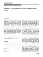
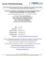
The 1H and 13C NMR spectra of xylan from NMR analyses (Fig. 1)
confirm that the powder obtained from corn cobs was mainly consti
tuted by xylan-type hemicellulose. Proton and carbon signals were
assigned by comparing the spectrum of xylan taken as a reference and
the chemical shifts detected in previous studies (Cordeiro, Almeida, &
Iacomini, 2015).
The 1H NMR spectrum depicted in Fig. 1A revealed that the β-(1→ 4)linked D-Xylpiranose units were characterized by the signals at δ 3.04,
3.21, 3.25, 3.49, 3.95 and 4.25 ppm, which correspond to H-2, H-5a, H3, H-4, H-5e and H-1, respectively. Furthermore, it is noted that the two
signals found downfield at δ 5.02 and 5.14 ppm were related to the
protons of the hydroxyl groups attached to the C-3 (–C–OH, δ 5.1 ppm)
and the C-2 (–C–OH, δ 5.2 ppm) positions of the D-xylpiranose units in
xylan (Fundador et al., 2012; Habibi, Mahrouz, & Vignon, 2005).
Additionally, a slight peak at δ 5.3 ppm was also observed, indicating the
presence of 4-O-methylglucuronic acid (Fundador et al., 2012).
The 13C NMR spectrum of xylan–type hemicellulose (Fig. 1B) pre
sented five signals at δ 102.21 (C-1), 75.88 (C-4), 74.39 (C-3), 73.08 (C2) and 63.69 (C-5) ppm, which were characteristic of D-xylopiranose
units presented in Xylan. Similar findings were observed with other
xylan sources (Cordeiro et al., 2015; Habibi & Vignon, 2005; Habibi
et al., 2005; Viana et al., 2011). No other additional signals were
observed related to neutral sugars and acetyl groups. It could be inferred
Fig. 1. Nuclear Magnetic Resonance spectra of corn cob xylan powder. 1H NMR spectrum (A) and
4
13
C NMR spectrum (B).
S.C.C. Urtiga et al.
Carbohydrate Polymers 250 (2020) 116929
that certain signals of the structural rally related to the monosaccharides
overlap with the signals observed for the xylose residues. Furthermore, it
can be inferred that, because the xylan–type hemicellulose analyzed
here was predominantly composed of xylose residues, and only minor
amounts of other monosaccharides, the signals of greater amplitude
depicted in the spectra correspond to xylose. In order to confirm the
spectroscopic results reported here, further characterization of the
chemical composition of the xylan was efficiently carried out by using
GC-FID (Table 1).
microparticles.
3.3. Fourier transform infrared-attenuated total reflectance (FTIR-ATR)
spectroscopy analysis
FTIR-ATR analyses were performed in order to investigate the
interaction between the components of the formulation. Therefore, the
analyses were carried out for the raw materials and microparticles. As
expected, the spectra of the xylan and STMP materials (Fig. 3) were
similar to those found in previous work published by our group (Oliveira
et al., 2010; Urtiga et al., 2017). However, the spectra of XMP and
XMP5-ASA were slightly different. Indeed, the presence of an intense
peak at 1110 cm− 1, related to the symmetrical stretching (P–O–P) in
pyrophosphates and other peaks from 750 cm− 1 to 775 cm− 1, attributed
to the vibrational stretching of the phosphorus bridges (O–P–O and/ or
P = O) and the symmetrical stretching (POP), respectively, were
observed. These peaks can be related to remaining STMP residues from
the cross-linking process (Parize, Stulzer, Laranjeira, Brighente, &
Souza, 2012; Suflet, Chitanu, & Popa, 2006; Urtiga et al., 2017). In
addition, the cross-linking process was confirmed by the presence of a
peak between 1200 and 1250 cm− 1, at 1217 cm-1 (Fig. 3), which is
attributed to the phosphate ester bond formation between the xylan and
the STMP during the cross-linking process (Urtiga et al., 2017).
Concerning 5-ASA, the spectrum of this drug presented absorption
bands at 2552, 1650, and 1580 cm− 1, which correspond to the vibra
tions of –NH2, –C = O and –C = C–, respectively (Tang et al., 2018). The
loss of intensity for all characteristic absorption bands of 5-ASA in the
microparticles spectrum was also observed, which can be attributed to
the encapsulation process, since it is characterized by the restriction in
the vibration.
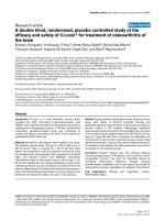
3.2. Characterization of xylan-based microparticles
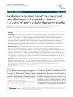
In order to obtain visual and morphological characterization of
xylan-based microparticles, the SEM (Fig. 2A-D) analysis was per
formed, wherein it was possible to observe the microparticles spherical
shape with the presence of residuals on their surface, which can be
related to the cross-linking agent that remained after washing process
(Fig. 2A-D). The particle size analysis revealed a mean diameter size of
12.66 ± 1.01 and 14.64 ± 0.5 μm for XMP and XMP5-ASA, respectively,
which confirmed the data from the SEM (Fig. 2). Thus, the addition of 5ASA to the aqueous phase containing the polysaccharide induced a slight
variation in the particle size after the cross-linking process.
The entrapment efficiency for XMP5-ASA was 65.41 ± 3.9 %.
Similar result was found by Palma et al. (2019) who produced 5-ASA-
loaded chitosan microcapsules by a spray-drying process with an
entrapment efficiency of 65–70%. On the other hand, a previous work
from our group showed a 5-ASA entrapment efficiency of 23.61 ± 0.15
and 24.98 ± 0.12 % for xylan-based microcapsules produced by
spray-drying and by interfacial cross-linking polymerization, respec
tively (Silva et al., 2013). Concerning the interfacial cross-linking pro
cess, in this work the authors attributed the low entrapment efficiency to
the several washing steps used to avoid any residual of organic solvent
and crosslinking agent (terephthaloyl chloride), which was also
responsible for the high microparticles toxicity (Marcelino et al., 2015;
Silva et al., 2013; Urtiga et al., 2017). In this work, similar washing steps
were used; however, it could be possible that the crosslinking process
using STMP as a cross-linking agent enhanced drug retention in the
3.4. X-Ray diffraction
Drug release kinetics from the microparticles can be affected by the
physical state of the drug in the polymeric matrix, which can vary from
˜ os, Peniche,
amorphous to well-defined crystalline state (Aranaz, Pan
Heras, & Acosta, 2017). Fig. 4 compares the XRD patterns of raw
Fig. 2. SEM images of xylan microparticles: XMP (A and B) and XMP5-ASA (C and D).
5
S.C.C. Urtiga et al.
Carbohydrate Polymers 250 (2020) 116929
between 10◦ and 50◦ (2θ) due to their crystalline nature (Cesar et al.,
2018; Li, Wang, Li, Bhandari et al., 2009, Li, Wang, Li, Chiu et al., 2009).
Concerning the microparticles XRD patterns (XMP and XMP5-ASA),
the results were similar to xylan alone. Indeed, they showed a broad
peak at the same angle as the xylan with the presence of some slight
crystallinity peaks between 15◦ and 35◦ (2θ), which could be related to
residuals of STMP from the cross-linking process, as confirmed by FTIRATR and SEM results. In addition, the characteristic diffraction peaks of
5-ASA did not appear on the XRD pattern of the XMP5-ASA, which could
be due to the perfected molecular dispersion of 5-ASA in the polymeric
matrix, corroborating to the SEM observation in which no 5-ASA crystals
were seen on the microparticles surface (Aranaz et al., 2017; Liu et al.,
2019).
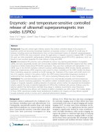
3.5. Thermogravimetry (TG) and differential scanning calorimetry (DSC)
Thermal analyses have been used to investigate interactions between
drug and polymers in several micro and nanoparticle formulations
(Oliveira et al., 2013). Fig. 5A illustrates the thermal behavior of xylan
expressed by the TG curve. The first degradation event was observed up
to 110 ◦ C with a mass loss of 8 %, which is suggestive of the water loss
presented in the xylan powder (Marcelino et al., 2015; Silva et al.,
2013). The second event occurred in the range of 193–410 ◦ C with a
mass loss of 62 %, which was related to the onset of the polymer
degradation processes. The thermal behavior of 5-ASA (Fig. 5A) showed
a single weight loss (97.8 %) between 269–394 ◦ C, attributed to
decomposition of the drug.
Regarding the thermal behavior of the microparticles (Fig. 5A), two
weight losses, similar to xylan events, were observed. The first degra
dation event (up to 116.4 ◦ C) showed a mass loss of 8–10 % and can be
attributed to the water loss present in the systems. The second thermal
event occurred between 165 and 320 ◦ C, with a mass loss of 36.5 % and
35.6 % for XMP and XMP5-ASA, respectively. As it can be seen, the
system showed smaller weight loss when compared to the xylan itself,
which may be attributed to the thermal stability of the phosphate ester
linkages formation between xylan and STMP during the crosslinking
process, highlighting that the cross-linking process was able to improve
˜ os, Pastrana,
the thermal stability of the microparticles (Brassesco, Fucin
´, 2019).
& Pico
The DSC curves of the samples were in agreement with the TGA
curves. The DSC curve for xylan and for microparticles (Fig. 5B) revealed
an endothermic event in the temperature range of 55–116 ◦ C, indicating
the water loss from xylan. An exothermic peak was also observed at
292 ◦ C, 287 ◦ C and 290 ◦ C for xylan, XMP and XMP5-ASA, respectively.
Concerning the 5-ASA, an endothermic peak was observed around
290 ◦ C, which matches the melting point of the drug (Cesar et al., 2018).
In addition, no thermal events related to 5-ASA were found in the
thermal curves of XMP5-ASA.
Fig. 3. FTIR-ATR spectrum of raw materials and xylan microparticles (XMP
and XMP5-ASA).
materials and the microparticles. The XRD pattern of xylan clearly
exhibited a typical feature of predominantly amorphous materials with
presence of slight crystallinity in the region of 10◦ to 30◦ (2θ). The
broader peaks at 19.6◦ and 29◦ (2θ) are characteristic of crystalline re
gions of semi crystalline xylan (Grodahl, Gatenholm, & Dekker, 2004).
On the other hand, STMP and 5-ASA themselves showed intense peaks
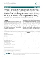
3.6. In vitro drug release
The in vitro release of 5-ASA from microparticles was studied in
simulated physiological dissolution media to mimic the passage of the
microparticles through the gastrointestinal tract. The results revealed
that approximately 52 % of the initial dose was released in less than 2 h
from XMP5-ASA into simulated gastric medium (pH = 1.2) (Fig. 6A).
This fast release of the drug can be explained by the formation of pores
on the surface of the microparticles, related to the intrinsic character
istics of the polymer (Nagashima-Jr et al., 2008; Silva et al., 2013).
However, in simulated gut medium (pH 6.0), 80 % of the drug was
released up to 6 h, which indicates that the formulation XMP5-ASA
might be able to reach the large intestine with approximately 20 % of
its initial loading of 5-ASA. Aiming to avoid the burst release effect
found on the gastric medium, gastro-resistant capsules were filled with
XMP5-ASA (XMPCAP5-ASA). The release profile of 5-ASA from the
XMPCAP5-ASA showed a Lag time up to 4 h. Additionally, in the
Fig. 4. X-ray powder diffraction patterns of raw materials and xylan micro
particles (XMP and XMP5-ASA).
6
S.C.C. Urtiga et al.
Carbohydrate Polymers 250 (2020) 116929
Fig. 5. TG (A) and DSC (B) curves of raw materials and xylan microparticles (XMP and XMP5-ASA).
predominant factor on the drug release of 5-ASA from the microparticles
(Table 2) (Peppas & Sahlin, 1989). Conversely, the release kinetics of
5-ASA from XMPCAP5-ASA were better fitted on the Korsmeyer-Peppas
model (Table 2) leading to release exponent (n) values of 0.26, indi
cating the presence of a Fickian diffusion transport (Jha, Chakraborty,
Chaudhuri, & Dey, 2016).
4. Conclusion
In this work, XMPCAP5-ASA was successfully produced as a new
formulation for the colonic release of 5-ASA. Additionally, no relevant
interactions among the components of the formulation, which could
interfere on the drug characteristics, were found, as demonstrated by the
FTIR-ATR spectroscopy results. The XRD and the thermal analyses
revealed that the 5-ASA was able to be molecularly dispersed in the
polymer matrix, inducing an increment on its thermal stability. In
addition, in vitro release studies of XMPCAP5-ASA showed the usefulness
of this new formulation for colonic delivery of 5-ASA from xylan mi
croparticles. In fact, approximately 50 % of the drug content was able to
be released at colonic pH (pH = 7.4), and the major mechanism of drug
release was the Fickian diffusion.
Fig. 6. (A) In vitro release profile of 5- ASA along the time as a function of the
pH, and (B) mathematical modeling of 5-ASA release profile according to
distinctive models. (5-ASA = free 5-ASA, CAP5-ASA = 5-ASA inside of gastroresistant
capsules;
XMP5-ASA = 5-ASA-loaded
xylan
microparticles;
XMPCAP5-ASA = 5-ASA-loaded xylan microparticles inside of gastroresistant capsules).
simulated gut medium (pH 6.0), only 49 % of 5-ASA was released, up to
6 h. Therefore, approximately 50 % of the initial loading of 5-ASA can
reach the large intestine by using such approach. In order to evaluate if
the gastro-resistant capsule was not the only factor able to promote the
drug release delay, samples containing free 5-ASA into the
gastro-resistant capsules (Fig. 6A) were also assayed. For this sample, it
was possible to evidence that the drug was totally released in 6 h of
experiment. The overall results concerning the release profile allow us to
infer the xylan microparticles importance on the 5-ASA release control,
providing an improvement on the drug availability in the colon.
The in vitro release data from the entire set of dissolution media (the
gradient of pH) were fitted together into mathematical kinetic equations
in order to describe the kinetic profile of 5-ASA from the systems. The
results obtained from the modeling (R2 adjusted, RMSE and MSC) of
each system, as well as their respective constant rates, were shown in
Table 2. The release kinetic profile presented by XMP5-ASA was better
fitted on the Peppas-Sahlin model (Fig. 6B), which explains that the drug
release occurred through two processes, the Fickian diffusion phenom
ena and the relaxation of the polymer chain. The application of this
model and the calculation of the k1 and k2 constants allows to evaluate
the impact of each mechanism in the drug release process. Indeed, once
k1 (44.83) > k2 (-5.61) it can be inferred that Fickian diffusion was the
CRediT authorship contribution statement
Silvana Cartaxo da Costa Urtiga: Conceptualization, Formal anal
´ ria Maria
ysis, Investigation, Methodology, Writing - original draft. Vito
Oliveira Alves: Conceptualization, Investigation. Camila de Oliveira
Melo: Conceptualization, Writing - review & editing. Marini Nasci
mento de Lima: Conceptualization, Investigation. Ernane Souza: Re
sources, Writing - review & editing. Arcelina Pacheco Cunha:
´gila Maria Pontes Silva Ricardo: Re
Methodology, Investigation. Na
sources, Writing - review & editing. Elquio Eleamen Oliveira:
Conceptualization, Supervision, Methodology, Writing - review & edit
´ crates Tabosa do Egito: Conceptualization, Method
ing. Eryvaldo So
ology, Supervision, Writing - review & editing, Project administration,
Funding acquisition.
7
S.C.C. Urtiga et al.
Carbohydrate Polymers 250 (2020) 116929
Table 2
Evaluation of different mathematical models for the in vitro release profile of the drug and the rate release constants of the data.
Formulation
Mathematical Model
Equation
F = 100 x (1- e
F = kH x t0.5
F = kKP x tn
–k1 x t
XMP5-ASAd
First-order
Higuchi
Korsmeyer-Peppas
)
Peppas-Sahlin
F = k1 x tm + k2 x t(2
x m)
F = 100 x [1 x e− k1 x
F = kH x (t-Tlag)0.5
F = kKP x (t-Tlag)n
(t – Tlag)
XMPCAP5-ASAe
First-order
Higuchi
Korsmeyer-Peppas
Peppas-Sahlin
F = k1 x (t-Tlag)m+k2 x (t-Tlag)(2
]
x m)
R2 Adjusteda
RMSEb
MSCc
Constants
0.94
0.69
0.92
7.22
17.76
8.97
2.88
1.02
2.33
kf1 = 0.39
kH g = 27.46;
kKP h = 45.68; n i = 0.28
kj1 = 44.83; k2 k = -5.61;
m l = 0.51
–
kH = 26.30
kKP = 48.48; n = 0.26
k1 = -132.24; k2 = 121.78;
m = 0.16
0.99
2.93
4.57
–
0.94
0.98
–
9.36
5.68
–
2.14
3.08
0.87
13.72
1.98
a
Adjusted coefficient of determination.
standard deviation of the residuals.
c
Model Selection Criterion.
d
5-ASA-loaded xylan microparticles.
e
5-ASA-loaded xylan microparticles inside of gastro-resistant capsules.
f
first order release constant.
g
Higuchi release constant.
h
release constant incorporating structural and geometric characteristics of the drug-dosage form.
i
diffusional exponent indicating the drug-release mechanism.
j
constant related to the Fickian kinetics.
k
the constant related to Case II relaxation kinetics.
l
diffusional exponent.
b
Declaration of Competing Interest
Grodahl, M., Gatenholm, P., & Dekker, M. (2004). Role of acetyl substitution in
hardwood xylan. Polysaccharides: Structural diversity and functional versatility (2nd
ed.) New York.
Günter, E. A., Markov, P. A., Melekhin, A. K., Belozerov, V. S., Martinson, E. A.,
Litvinets, S. G., et al. (2018). Preparation and release characteristics of mesalazine
loaded calcium pectin-silica gel beads based on callus cultures pectins for colontargeted drug delivery. International Journal of Biological Macromolecules, 120,
2225–2233.
Habibi, Y., & Vignon, M. R. (2005). Isolation and characterization of xylans from seed
pericarp of Argania spinosa fruit. Carbohydrate Research, 340(7), 1431–1436.
Habibi, Y., Mahrouz, M., & Vignon, M. R. (2005). D-Xylans from seed endosperm of
Opuntia ficus-indica prickly pear fruits. Comptes Rendus Chimie, 8(6–7), 1123–1128.
Hrom´
adkov´
a, Z., Kov´
aˇcikov´
a, J., & Ebringerov´
a, A. (1999). Study of the classical and
ultrasound-assisted extraction of the corn cob xylan. Industrial Crops and Products, 9
(2), 101–109.
Jha, J., Chakraborty, S., Chaudhuri, M. G., & Dey, R. (2016). In vitro release kinetics and
transferrin saturation study of intravenous iron sucrose entrapped in poly(ethylene
glycol)-assisted silica xerogel. Applied Biochemistry and Biotechnology, 178(7),
1351–1362.
Kaˇcur´
akov´
a, M., Wellner, N., Ebringerov´
a, A., Hrom´
adkov´
a, Z., Wilson, R. H., &
Belton, P. S. (1999). Characterisation of xylan-type polysaccharides and associated
cell wall components by FT-IR and FT-Raman spectroscopies. Food Hydrocolloids, 13
(1), 35–41.
Kayserilio˘
glu, B.S¸., Bakir, U., Yilmaz, L., & Akkas¸, N. (2003). Use of xylan, an agricultural
by-product, in wheat gluten based biodegradable films: Mechanical, solubility and
water vapor transfer rate properties. Bioresource Technology, 87(3), 239–246.
Laine, C., Kemppainen, K., Kuutti, L., Varhimo, A., Asikainen, S., Gră
onroos, A., et al.
(2015). Extraction of xylan from wood pulp and brewer’s spent grain. Industrial
Crops and Products, 70, 231–237.
Lakhera, A. K., & Kumar, V. (2017). Monosaccharide composition of acidic gum exudates
from Indian Acacia tortilis ssp. raddiana (Savi) Brenan. International Journal of
Biological Macromolecules, 94, 45–50.
Li, B.-Z., Wang, L.-J., Li, D., Bhandari, B., Li, S.-J., Lan, Y., et al. (2009). Fabrication of
starch-based microparticles by an emulsification-crosslinking method. Journal of
Food Engineering, 92(3), 250–254.
Li, B., Wang, L., Li, D., Chiu, Y. L., Zhang, Z., Shi, J., et al. (2009). Physical properties and
loading capacity of starch-based microparticles crosslinked with trisodium
trimetaphosphate. Journal of Food Engineering, 92(3), 255–260.
Liu, G., Hu, M., Zhao, Z., Lin, Q., Wei, D., & Jiang, Y. (2019). Enhancing the stability of
astaxanthin by encapsulation in poly (l-lactic acid) microspheres using a
supercritical anti-solvent process. Particuology, 44, 54–62.
Lucena, C. A. A., Costa, S. C., Eleamen, G. R. A., Mendonỗa, E. A. M., & Oliveira, E. E.
(2017). Desenvolvimento de biofilmes `
a base de xilana e xilana/gelatina para
produỗ
ao de embalagens biodegrad
aveis. Polớmeros, 27, 3541.
Marcelino, H. R., Silva, A. E., Gomes, M. C. S., Oliveira, E. E., Nagashima-Jr, T.,
Pinheiro, J. S., et al. (2015). Leads from physical, chemical, and thermal
characterization on cytotoxic effects of xylan-based microparticles. Polymers, 7(11),
1515.
Melo-Silveira, R. F., Fidelis, G. P., Costa, M. S., Telles, C. B., Dantas-Santos, N.,
Elias, S. O., et al. (2012). In vitro antioxidant, anticoagulant and antimicrobial
activity and in inhibition of cancer cell proliferation by xylan extracted from corn
cobs. International Journal of Molecular Sciences, 13(1), 409–426.
We wish to confirm that there are no known conflicts of interest
associated with this publication and there has been no significant
financial support for this work that could have influenced its outcome.
Acknowledgments
o de Aperfeiỗoaư
This study was financed in part by the Coordenaỗa
mento de Pessoal de Nớvel Superior - Brasil (CAPES) - Finance Code 001.
The authors would like to thank CETENE for XRD and thermal analyses
and “Casa do Sert˜
ao” for providing the corn cobs. The authors also
would like to thank Dr. L. Amaral-Machado and Dr. E. Alencar for the
deep revision and important scientific remarks of the final version of the
manuscript.
References
American Association of Cereal Chemists (AACC). (1995). Approved methods (8th ed.).
Saint Paul: American Association of Cereal Chemists (AACC).
´ & Acosta, N. (2017). Chitosan spray-dried
Aranaz, I., Pa˜
nos, I., Peniche, C., Heras, A.,
microparticles for controlled delivery of venlafaxine hydrochloride. Molecules, 22
(11), 1980.
Brassesco, M. E., Fuci˜
nos, P., Pastrana, L., & Pic´
o, G. (2019). Development of alginate
microparticles as efficient adsorption matrix for protein recovery. Process
Biochemistry, 80, 157–163.
Cesar, A. L. A., Abrantes, F. A., Farah, L., Castilho, R. O., Cardoso, V., Fernandes, S. O.,
et al. (2018). New mesalamine polymeric conjugate for controlled release:
Preparation, characterization and biodistribution study. European Journal of
Pharmaceutical Sciences, 111, 57–64.
Cordeiro, L. M. C., Almeida, C. P., & Iacomini, M. (2015). Unusual linear
polysaccharides: (1→5)-α-l-Arabinan, (1→3)-(1→4)-α-d-glucan and (1→4)-β-d-xylan
from pulp of buriti (Mauritia flexuosa), an edible palm fruit from the Amazon region.
Food Chemistry, 173, 141–146.
Dhami, R., Harding, S. E., Elizabeth, N. J., & Ebringerov´
a, A. (1995). Hydrodynamic
characterisation of the molar mass and gross conformation of corn cob heteroxylan
AGX. Carbohydrate Polymers, 28(2), 113–119.
Ebringerov´
a, A., & Heinze, T. (2000). Xylan and xylan derivatives – Biopolymers with
valuable properties, 1. Naturally occurring xylans structures, isolation procedures
and properties. Macromolecular Rapid Communications, 21(9), 542–556.
Ebringerov´
a, A., & Hrom´
adkov´
a, Z. (2010). An overview on the application of ultrasound
in extraction, separation and purification of plant polysaccharides. Central European
Journal of Chemistry, 8(2), 243–257.
Ebringerov´
a, A., Hrom
adkov
a, Z., Alfă
odi, J., & Hr balov
a, V. (1998). The
immunologically active xylan from ultrasound-treated corn cobs: Extractability,
structure and properties. Carbohydrate Polymers, 37(3), 231–239.
Fundador, N. G. V., Enomoto-Rogers, Y., Takemura, A., & Iwata, T. (2012). Syntheses and
characterization of xylan esters. Polymer, 53(18), 3885–3893.
8
S.C.C. Urtiga et al.
Carbohydrate Polymers 250 (2020) 116929
Samanta, A. K., Jayapal, N., Jayaram, C., Roy, S., Kolte, A. P., Senani, S., et al. (2015).
Xylooligosaccharides as prebiotics from agricultural by-products: Production and
applications. Bioactive Carbohydrates and Dietary Fibre, 5(1), 62–71.
Samanta, A. K., Senani, S., Kolte, A. P., Sridhar, M., Sampath, K. T., Jayapal, N., et al.
(2012). Production and in vitro evaluation of xylooligosaccharides generated from
corn cobs. Food and Bioproducts Processing, 90(3), 466–474.
Sardo, H. S., Saremnejad, F., Bagheri, S., Akhgari, A., Afrasiabi Garekani, H., &
Sadeghi, F. (2019). A review on 5-aminosalicylic acid colon-targeted oral drug
delivery systems. International Journal of Pharmaceutics, 558, 367–379.
Scalarone, D., Chiantore, O., & Riedo, C. (2008). Gas chromatographic/mass
spectrometric analysis of on-line pyrolysis–silylation products of monosaccharides.
Journal of Analytical and Applied Pyrolysis, 83(2), 157–164.
Shatalov, A. A., Evtuguin, D. V., & Pascoal-Neto, C. (1999). (2-O-α-D-Galactopyranosyl-4O-methyl-α-D-glucurono)-D-xylan from eucalyptus globulus Labill. Carbohydrate
Research, 320(1), 93–99.
Silva, A. E., Oliveira, E. E., Gomes, M. C. S., Marcelino, H. R., Silva, K. C. H., Souza, B. S.,
et al. (2013). Producing xylan/Eudragit(R) S100-based microparticles by chemical
and physico-mechanical approaches as carriers for 5-aminosalicylic acid. Journal of
Microencapsulation, 30(8), 787–795.
Singleton, V. L., Orthofer, R., & Lamuela-Ravent´
os, R. M. (1999). Analysis of total
phenols and other oxidation substrates and antioxidants by means of folin-ciocalteu
reagent. Methods in enzymology, 299, 152–178.
Suflet, D. M., Chitanu, G. C., & Popa, V. I. (2006). Phosphorylation of polysaccharides:
New results on synthesis and characterisation of phosphorylated cellulose. Reactive
and Functional Polymers, 66(11), 1240–1249.
Tang, P., Sun, Q., Zhao, L., Pu, H., Yang, H., Zhang, S., et al. (2018). Mesalazine/
hydroxypropyl-β-cyclodextrin/chitosan nanoparticles with sustained release and
enhanced anti-inflammation activity. Carbohydrate Polymers, 198, 418–425.
Urtiga, S. C. C., Gabi, C. A. A. L., Eleamen, G. R. A., Souza, B. S., Pessoa, H. L. F.,
Marcelino, H. R., et al. (2017). Preparation and characterization of safe
microparticles based on xylan. Drug Development and Industrial Pharmacy, 43(10),
1601–1609.
Van Dongen, F. E. M., Van Eylen, D., & Kabel, M. A. (2011). Characterization of
substituents in xylans from corn cobs and stover. Carbohydrate Polymers, 86(2),
722–731.
Viana, A. G., Noseda, M. D., Gonỗalves, A. G., Duarte, M. E. R., Yokoya, N.,
Matulewicz, M. C., et al. (2011). β-D-(1→4), β-D-(1→3) ‘mixed linkage’ xylans from
red seaweeds of the order nemaliales and palmariales. Carbohydrate Research, 346
(8), 1023–1028.
Zhang, Y., Huo, M., Zhou, J., Zou, A., Li, W., Yao, C., et al. (2010). DDSolver: An add-in
program for modeling and comparison of drug dissolution profiles. The AAPS
Journal, 12(3), 263–271.
Melo-Silveira, R. F., Viana, R. L. S., Sabry, D. A., Silva, R. A., Machado, D.,
Nascimento, A. K. L., et al. (2019). Antiproliferative xylan from corn cobs induces
apoptosis in tumor cells. Carbohydrate Polymers, 210, 245–253.
Mladenovska, K., Raicki, R. S., Janevik, E. I., Ristoski, T., Pavlova, M. J., Kavrakovski, Z.,
et al. (2007). Colon-specific delivery of 5-aminosalicylic acid from chitosan-Caalginate microparticles. International Journal of Pharmaceutics, 342(1), 124–136.
Nagashima-Jr, T., Oliveira, E. E., Silva, A. E., Marcelino, H. R., Gomes, M. C. S.,
Aguiar, L. M., et al. (2008). Influence of the lipophilic external phase composition on
the preparation and characterization of xylan microcapsules-a technical note. AAPS
PharmSciTech, 9(3), 814–817.
Oliveira, A. R., Molina, E. F., Mesquita, P. C., Fonseca, J. L. C., Rossanezi, G., FernandesPedrosa, M. F., et al. (2013). Structural and thermal properties of spray-dried
methotrexate-loaded biodegradable microparticles. Journal of Thermal Analysis and
Calorimetry, 112(2), 555–565.
Oliveira, E. E., Silva, A. E., Nagashima-Jr, T., Gomes, M. C. S., Aguiar, L. M.,
Marcelino, H. R., et al. (2010). Xylan from corn cobs, a promising polymer for drug
delivery: Production and characterization. Bioresource Technology, 101(14),
5402–5406.
Palma, E., Costa, N., Molinaro, R., Francardi, M., Paolino, D., Cosco, D., et al. (2019).
Improvement of the therapeutic treatment of inflammatory bowel diseases following
rectal administration of mesalazine-loaded chitosan microparticles vs Asamax®.
Carbohydrate Polymers, 212, 430–438.
Parize, A. L., Stulzer, H. K., Laranjeira, M. C. M., Brighente, I. M. C., & Souza, T. C. R.
(2012). Evaluation of chitosan microparticles containing curcumin and crosslinked
with sodium tripolyphosphate produced by spray drying. Quimica Nova, 35,
1127–1132.
Pazur, J. H., Miskiel, F. J., & Liu, B. (1987). Identification of furanose and pyranose ring
forms of carbohydrates by methylation, gas-liquid chromatography and mass
spectrometry. Journal of Chromatography A, 396, 139–147.
Peppas, N. A., & Sahlin, J. J. (1989). A simple equation for the description of solute
release. III. Coupling of diffusion and relaxation. International Journal of
Pharmaceutics, 57(2), 169–172.
Pinto, P. C., Evtuguin, D. V., & Pascoal-Neto, C. (2005). Structure of hardwood
glucuronoxylans: Modifications and impact on pulp retention during wood kraft
pulping. Carbohydrate Polymers, 60(4), 489–497.
Ren, J. L., Sun, R. C., Liu, C. F., Cao, Z. N., & Luo, W. (2007). Acetylation of wheat straw
hemicelluloses in ionic liquid using iodine as a catalyst. Carbohydrate Polymers, 70
(4), 406–414.
Rubinstein, A. (1995). Approaches and opportunities in colon-specific drug delivery.
Critical Reviews in Therapeutic Drug Carrier Systems, 12(2–3), 101–149.
Ruiz-Matute, A. I., Hern´
andez-Hern´
andez, O., Rodríguez-S´
anchez, S., Sanz, M. L., &
Martínez-Castro, I. (2011). Derivatization of carbohydrates for GC and GC–MS
analyses. Journal of Chromatography B, 879, 1226–1240.
9