Glucomannan and beta-glucan degradation by Mytilus edulis Cel45A: Crystal structure and activity comparison with GH45 subfamily A, B and C
Bạn đang xem bản rút gọn của tài liệu. Xem và tải ngay bản đầy đủ của tài liệu tại đây (7.86 MB, 13 trang )
Carbohydrate Polymers 277 (2022) 118771
Contents lists available at ScienceDirect
Carbohydrate Polymers
journal homepage: www.elsevier.com/locate/carbpol
Research Paper
Glucomannan and beta-glucan degradation by Mytilus edulis Cel45A:
Crystal structure and activity comparison with GH45 subfamily A, B and C☆
Laura Okmane a, Gustav Nestor a, Emma Jakobsson b, 1, Bingze Xu c, 2, Kiyohiko Igarashi d,
Mats Sandgren a, Gerard J. Kleywegt b, 3, Jerry Ståhlberg a, *
a
Department of Molecular Sciences, Swedish University of Agricultural Sciences, Uppsala, Sweden
Department of Cell and Molecular Biology, Uppsala University, Uppsala, Sweden
Center for Surface Biotechnology, Uppsala University, Uppsala, Sweden
d
Department of Biomaterial Sciences, Graduate School of Agricultural and Life Sciences, University of Tokyo, 1–1–1 Yayoi, Bunkyo-ku, Tokyo 113–8657, Japan
b
c
A R T I C L E I N F O
A B S T R A C T
Keywords:
Endoglucanase
Blue mussel
Cel45A
GH45
Beta-glucan
Glucomannan
The enzymatic hydrolysis of barley beta-glucan, konjac glucomannan and carboxymethyl cellulose by a β-1,4-Dendoglucanase MeCel45A from blue mussel, Mytilus edulis, which belongs to subfamily B of glycoside hydrolase
family 45 (GH45), was compared with GH45 members of subfamilies A (Humicola insolens HiCel45A), B (Tri
choderma reesei TrCel45A) and C (Phanerochaete chrysosporium PcCel45A). Furthermore, the crystal structure of
MeCel45A is reported.
Initial rates and hydrolysis yields were determined by reducing sugar assays and product formation was
characterized using NMR spectroscopy. The subfamily B and C enzymes exhibited mannanase activity, whereas
the subfamily A member was uniquely able to produce monomeric glucose. All enzymes were confirmed to be
inverting glycoside hydrolases. MeCel45A appears to be cold adapted by evolution, as it maintained 70% activity
on cellohexaose at 4 ◦ C relative to 30 ◦ C, compared to 35% for TrCel45A. Both enzymes produced cellobiose and
cellotetraose from cellohexaose, but TrCel45A additionally produced cellotriose.
1. Introduction
In aquatic ecosystems, cellulose is produced in large quantities by
algal plankton, which is an important energy source for filter feeding
organisms such as mussels (Newell et al., 1989). Not surprisingly,
cellulase activity has been demonstrated in the digestive tract of several
bivalves (Kaur, 1997; Onishi et al., 1985; Purchon, 1977). The highest
activity was found in so called crystalline styles, which most bivalves
and some gastropods (snails and slugs) use for digestion. The crystalline
style is a jelly-like translucent rod protruding into the stomach of the
mussel. It aids in digestion by being rotated and pushed against the
gastric shield, thus dragging the food from the gills into the stomach and
grinding the food like a pestle and mortar. The style dissolves gradually
and releases various enzymes that initiate extracellular digestion in the
stomach (Purchon, 1977).
In the blue mussel, Mytilus edulis, three enzymes with activity against
carboxymethyl cellulose (CMC) have been detected, the smallest of
which (around 20 kDa) has been purified from blue mussel collected off
the Swedish west coast. It has been characterized and the protein and
gene sequences have been determined (Xu et al., 2000). The mature
Abbreviations: AcCel45A, Ampullaria crossean Cel45A; BG, betaglucan; CBM, carbohydrate binding module; CMC, carboxymethyl cellulose; DPBB, double psi beta
barrel; GH, glycoside hydrolase; GH45, glycoside hydrolase family 45; GM, glucomannan; HiCel45A, Humicola insolens Cel45A; MeCel45A, Mytilus edulis Cel45A;
PASC, phosphoric acid swollen cellulose; PcCel45A, Phanerochaete chrysosporium Cel45A; PHBAH, p-Hydroxybenzoic acid hydrazide; RMSD, root mean square
deviation; TrCel45A, Trichoderma reesei Cel45A.
☆
Enzymes: EC3.2.1.4.
* Corresponding author at: Department of Molecular Sciences, Swedish University of Agricultural Sciences, POB 7015, SE-750 07 Uppsala, Sweden.
E-mail address: (J. Ståhlberg).
1
Present addresses: Emma Jakobsson, CIC biomaGUNE, Basque Research and Technology Alliance (BRTA), San Sebasti´
an, 20014 Guipúzcoa, Spain.
2
Present addresses: Bingze Xu, Medical Inflammation Research, Department of Medical Biochemistry and Biophysics, Karolinska Institutet, SE-171 77 Solna,
Sweden.
3
Present addresses: Gerard J. Kleywegt, EMBL-EBI, Wellcome Genome Campus, Hinxton, Cambridge CB10 1SD, UK.
/>Received 10 August 2021; Received in revised form 24 September 2021; Accepted 11 October 2021
Available online 21 October 2021
0144-8617/© 2021 The Authors. Published by Elsevier Ltd. This is an open access article under the CC BY license ( />
L. Okmane et al.
Carbohydrate Polymers 277 (2022) 118771
protein has 181 amino acid residues and consists of a single glycoside
hydrolase family 45 (GH45) catalytic domain without any carbohydrate
binding module or other accessory modules and was designated
MeCel45A. It has a broad temperature optimum between 30 and 50 ◦ C.
Interestingly, it retains over 50% of the maximum activity at 0 ◦ C. This
may be related to observations that in Sweden, mussels actively ingest
seston (suspended particles) at temperatures below 0 ◦ C, suggesting that
they can utilize spring phytoplankton blooms in boreal waters even at
low temperatures (Loo, 1992). Although the enzyme did not show any
activity at very high temperatures, it could withstand shorter periods
(10 min) at +100 ◦ C without irreversible loss of enzymatic activity (Xu
et al., 2000).
Today there are over 460 GH45 entries in the CAZy database (cazy.
org), which have been divided into three subfamilies, A, B and C
(Couturier et al., 2011; Igarashi et al., 2008; Nomura et al., 2019). All
three have been found in fungi. GH45 members of nematodes, insects,
springtail and bacteria belong to subfamily A (Kikuchi et al., 2004; Lee
et al., 2004; Mei et al., 2016; Pauchet et al., 2010, 2014; Song et al.,
2017; Valencia et al., 2013), while molluscs only have subfamily B
endoglucanases (Guo et al., 2008; Sakamoto & Toyohara, 2009; Tsuji
et al., 2013; Xu et al., 2000). Subfamily C is only found in basidiomycete
fungi, so far.
Three-dimensional (3D) structures of nine GH45 enzymes are
available in the PDB (rcsb.org), one of which is the structure of
MeCel45A described in this paper. They all share a conserved sixstranded double-ψ β-barrel (DPBB) also known as GH45-like domain
in their structure. The DPBB domain is evolutionarily and structurally
related to that of expansins and their homologues (loosenins, swollen
ins), where the same catalytic acid (aspartic acid) has been conserved in
the catalytic center motif (Payne et al., 2015). In GH45 subfamily A and
B, an additional aspartic acid at the catalytic center is proposed to act as
catalytic base in the inverting hydrolytic mechanism, but the corre
sponding residue is not conserved in subfamily C. In this regard, sub
family C appears to be more similar to expansin-like one-domain
proteins named loosenins, which also lack the putative catalytic base
residue. However, no hydrolytic activity of loosenins has been docu
mented yet, as opposed to subfamily C members which have shown β(1
→ 4)-endoglucanase activity (Igarashi et al., 2008).
Thus far, the most studied GH45 enzyme has been HiCel45A from the
ascomycete fungus Humicola insolens. As such, it often serves as a GH45
reference in structure and activity comparisons. HiCel45A is a subfamily
A member that is widely used in treating textiles, for example as part of
washing powders in the form of the product Carezyme from Novozymes.
In subfamily B, the first published structure was that of AcCel45A from
the snail Ampullaria crossean (Nomura et al., 2019), also known as
EG27II, which appears to be the most similar structure to MeCel45A. In
subfamily C there is only one enzyme with structures available, namely
PcCel45A from the white-rot basidiomycete fungus Phanerochaete
chrysosporium. Due to the exceptional nature of the catalytic center
among subfamily C members, a distinctive catalytic “Proton-relay”
mechanism has been proposed for PcCel45A (Nakamura et al., 2015).
GH45 enzymes hydrolyze β(1 → 4) linkages in soluble beta-glucans
via an inverting action mechanism, where one amino acid residue acts
as a general acid that protonates the glycosidic oxygen and another
residue acts as a general base that activates a water molecule to hy
drolyze the glycosidic bond. Such a catalytic mechanism leads to
inversion of position at the anomeric carbon, thus producing alphaanomers from β(1 → 4) linked glucans. The highest catalytic activities
of GH45 enzymes have been demonstrated on barley beta-glucan and
lichenan, lower activities on CMC, phosphoric-acid-swollen cellulose
(PASC) and hydroxyethyl cellulose (HEC), and minute activities on
crystalline cellulose substrates such as Avicel (Gilbert et al., 1990; Sal
oheimo et al., 1994; Schou et al., 1993). HiCel45A and other subfamily A
members are able to hydrolyze various cellulosic substrates (PASC,
CMC, Avicel and bacterial cellulose), as well as xylan and xyloglucan
among others (Vlasenko et al., 2010). Subfamily B members have been
shown to hydrolyze CMC (Karlsson et al., 2002; Liu et al., 2010; Nomura
et al., 2019), PASC, Avicel, glucomannan (Karlsson et al., 2002; Liu
et al., 2010), and xylan (Liu et al., 2010), whereas subfamily C member
PcCel45A is unable to hydrolyze xyloglucan and Avicel (Godoy et al.,
2018).
The roles of three residues proposed to be involved in the catalytic
mechanism of GH45 enzymes have been investigated experimentally by
mutations at those sites. Mutation of the catalytic acid leads to complete
inactivation of the enzyme in all GH45 subfamilies (D121N in HiCel45A,
D137A in AcCel45A, D117N in Fomitopsis palustris FpCel45, D114A and
D114N in PcCel45A) (Cha et al., 2018; Davies et al., 1995; Godoy et al.,
2018; Nakamura et al., 2015; Nomura et al., 2019). Mutation of the
catalytic base proposed for subfamily A and B, leads to inactivation in
subfamily A (HiCel45A D10N), but the activity is not completely lost in
subfamily B (AcCel45A D27A) (Davies et al., 1995; Nomura et al., 2019).
Subfamily C members lack an acidic residue at the corresponding posi
tion. Instead, an asparagine residue at another position, Asn92 in
PcCel45A, has been proposed to act as catalytic base. Mutation of this
residue, or the corresponding residue in other GH45 enzymes, drasti
cally reduced the enzymatic activity in members from all GH45 sub
families (D114N in HiCel45A, N112A in AcCel45A, N95D in FpCel45,
N92D in PcCel45A) (Cha et al., 2018; Davies et al., 1995; Nakamura
et al., 2015; Nomura et al., 2019).
There are very few studies where enzymes from different GH45
subfamilies have been compared side-by-side (Berto et al., 2019; Vla
senko et al., 2010). Here we describe the crystal structure of MeCel45A
and compare its enzymatic activity with representatives of GH45 sub
families A, B, and C. We hypothesise that i) the reaction mechanism is
inverting also in subfamily B and C, previously proven only for a sub
family A enzyme; ii) GH45 enzymes of subfamilies A, B and C show
differences in substrate specificity and bond cleavage preference on
beta-glucan and glucomannan; iii) MeCel45A is more similar in struc
ture and activity properties to the other GH45 subfamily B enzyme used
in this study, TrCel45A, than to the members of subfamily A and C; iv)
MeCel45A from blue mussel that lives in cold waters is more coldadapted and retains higher activity at low temperatures than the ho
mologous enzyme from a tropical fungus, TrCel45A.
2. Results
2.1. Isolation, purification and structure determination of MeCel45A
The MeCel45A enzyme was isolated from the digestive gland of the
common blue mussel, M. edulis, from waters off the Swedish west coast.
From 29 kg of blue mussel, 8 mg of pure enzyme was obtained, by using
a three-step purification procedure with immobilized metal affinity
(IMAC), size exclusion, and cation-exchange chromatography.
During the screening for crystallisation conditions it turned out that
microcrystals appeared already in the concentrated protein solution.
Crystals for X-ray analysis could be grown by equilibration against 0.6 M
sodium acetate, pH 5.5, and 0.1–0.5 M NaCl without addition of other
precipitants. The crystals were orthorhombic and the space group was
P212121 with one protein molecule per asymmetric unit. The structure
was solved by SIRAS (single isomorphous replacement with anomalous
scattering) using a heavy-atom derivative with Baker's dimercurial
(C10H16Hg2O6;
1,4-diacetoxymercuri-2,3-dimethoxybutane)
for
phasing, and was refined against a high-resolution dataset at 1.2 Å.
The refined structure model contains the complete polypeptide chain
(residues 1 to 181), 305 water molecules, one acetate molecule and two
polyethylene glycol (PEG) molecules. Proline residues 8 and 108 are
involved in cis-peptide bonds and all the 12 cysteine residues are
involved in disulfide bridges with the pairings 4/16, 30/69, 32/176, 65/
178, 72/157, and 103/113. Electron density maps indicated distinct
alternate conformations for the side chains of Thr20, Met55, Gln67,
Lys74, Gln85, Ser94, Asn105, His122, His130 and Asp132, which were
included in the refinement. Statistics relating to the quality of the X-ray
2
L. Okmane et al.
Carbohydrate Polymers 277 (2022) 118771
diffraction data and the refined protein model are summarized in
Tables S1 and S2.
lengthening of the loop where Asn109 is located by one residue
(Tyr107), as well as the substitution of two residues on either side of
subsite +2, where Arg24 and Lys89 in AcCel45A are replaced by Asn21
and Gln85, respectively, in MeCel45A.
2.2. Structure of MeCel45A
M. edulis Cel45A has a compact and globular structure with
approximate dimensions of 30 × 40 × 50 Å (Fig. 1A) built around a sixstranded β-barrel that has the characteristic DPBB fold (Fig. 1B). Loops
that connect the beta-strands combine to extend one of the faces of the
barrel into a shallow substrate-binding cleft (upwards in Fig. 1A). The
protein contains a few secondary structure elements in addition to the
canonical DPBB fold. At the N-terminus the first 14 residues form a very
short strand-turn-strand anti-parallel β-sheet directly before strand β1. A
short α-helix is present in the long loop between strands β1 and β2.
Finally, two α-helices are present in the stretch of 26 residues after
strand β6 at the C-terminus. The latter two helices form a protrusion
from the β-barrel on the side opposite to the substrate-binding surface
(downwards in Fig. 1A). On this protrusion three surface histidines are
located.
Among the GH45s with known structure, MeCel45A is most similar
to the other subfamily B enzyme from a mollusc, AcCel45A from
Ampullaria crossean. As expected from the high sequence similarity (48%
identity) the structures are very similar with a low root-mean-square
deviation (RMSD) of 1.7 Å over 168 aligned Cα atoms. In the overall
fold, MeCel45A has a three-residue deletion at the tip of a loop near the
N-terminus, and an eight-residue insertion near the C-terminus that
forms an extra alpha helix and extends the size of the protrusion from the
β-barrel. However, both these regions are distant from the active site and
not likely to influence the catalytic properties. The active site of
MeCel45A is nearly identical to that of AcCel45A, including the posi
tions of Asp24, Asn109 and Asp132 that correspond to the proposed
catalytic residues of AcCel45A (Asp27, Asn112, Asp137; Fig. 2), sug
gesting that these residues have the same function in MeCel45A. Asp132
at the bottom of the cleft is the catalytic acid that protonates the
glycosidic oxygen, Asp24 corresponds to the proposed catalytic base in
subfamily A enzymes (Asp10 in HiCel45A) and Asn109 is in the same
position as the proposed catalytic base of the subfamily C enzyme
PcCel45A (Asn92). Minor differences relative to AcCel45A include the
2.3. Structure comparison with other GH45 enzymes
The structure of MeCel45A was further compared with the other
GH45 enzymes used in the activity measurements, HiCel45A (subfamily
A), TrCel45A (subfamily B) and PcCel45A (subfamily C). For TrCel45A
no experimentally determined structure is yet available. Therefore, a
structure model of the catalytic domain of TrCel45A was built by ho
mology modelling using SWISS-MODEL and the structure of MeCel45A
as template. Percentage sequence identities and structural deviations
(RMSD) relative to MeCel45A are listed in Table 1, and a multiple
sequence alignment is shown in Fig. 3. The MeCel45A and PcCel45A
enzymes consist of a single catalytic domain alone, whereas HiCel45A
and TrCel45A are bimodular with a carbohydrate binding module
(CBM1) attached by a Ser/Thr-rich linker peptide to the catalytic
domain at the C-terminus.
The β-strands of the DPBB core superpose closely, but the surface
structures differ because of variations in the lengths of loop regions
flanking the β-barrel (Fig. 4). The subfamily B and C enzymes
(MeCel45A, TrCel45A, PcCel45A) are more similar to each other than to
HiCel45A of subfamily A. HiCel45A has longer loops surrounding the
catalytic center, forming a closed structure that resembles a tunnel,
while the others have an open cleft. In PcCel45A, loops extend the cleft
at both ends making the cellulose binding surface >5 Å longer (Fig. 5B).
The central part of the cleft is noticeably narrower in MeCel45A than in
TrCel45A and PcCel45A.
The location of catalytic residues is well conserved, except for the
catalytic base corresponding to Asp24 in MeCel45A, which is not present
in PcCel45A (Fig. 5). In PcCel45A a glycine residue occupies this posi
tion instead. Furthermore, HiCel45A has an aspartic acid (Asp114)
instead of asparagine at the location of the alternate base (Asn109 in
MeCel45A). Apart from the catalytic residues, a few additional amino
acids are conserved near the catalytic acid. These are Thr20, Tyr22 and
His130 in MeCel45A. His130 is on the same beta-strand as the catalytic
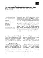
Fig. 1. Overall structure of M. edulis Cel45A. (A) Ribbon drawing showing the location of a shallow cleft on one face of the central six-stranded β-barrel with the
putative catalytic aspartate residues 24 and 132 sitting on either side of the cleft. On the other side, two α-helices at the C-terminus protrude from the β-barrel. (B)
Folding topology diagram with β-strands and α-helices numbered according to the generalized double-psi fold (Castillo et al., 1999). Cel45A contains an extra α-helix
at β1/β2, one short β-strand at the N-terminus and two C-terminal α-helices. The residue numbers of Cel45A at each end of the secondary structure elements
are indicated.
3
L. Okmane et al.
Carbohydrate Polymers 277 (2022) 118771
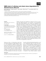
Fig. 2. M. edulis Cel45A (blue) structure superimposed on A. crossean Cel45A (gray). Assisting residues (Asn109 in MeCel45A; Asn112 in AcCel45A) and catalytic
center residues (Asp132, Asp24 in MeCel45A; Asp137, Asp27 in AcCel45A) are represented as sticks (from left to right respectively). (For interpretation of the
references to color in this figure legend, the reader is referred to the web version of this article.)
Table 1
GH45 endoglucanase structure and sequence similarities with MeCel45A.
Name
Organism
GenBank accession ID
PDB ID
UniProt accession ID
Percent sequence identitya
Structure similarity
RMSD (Å)
Cα
HiCel45A
TrCel45A
AcCel45A
PcCel45A
H. insolens
T. reesei
A. crossean
P. chrysosporium
CAB42307.1
CAA83846.1
ABR92638.1
BAG68300.1
2eng
N/A
5xbu
5kjo
P43316
P43317
A7KMF0
B3Y002
24
44
48
30
4.1
N/A
1.7
4.3
80
N/A
168
144
a
CBMs removed.
acid Asp132. The sidechain of Asp132 is positioned between Thr20 and
Tyr22 from the adjacent beta-strand, and conserved hydrogen bonds
connect the sidechains in the order Asp132-Thr20-His130.
In order to anticipate possible interactions with substrates, the
MeCel45A structure was superposed with available GH45 ligand com
plexes, and protein-ligand interactions were analyzed using LIGPLOT
(Figs. S1, S2). The structures chosen for comparison were: i) AcCel45A
with two cellobiose molecules bound in subsites − 3/− 2 and +1/+2,
respectively (PDB code 5XBX); ii) HiCel45A D10N mutant in complex
with cellohexaose where two cellotriosyl units are seen in subsites − 4/
− 3/− 2 and +1/+2/+3, respectively (PDB code 4ENG; Fig. 5A); and iii)
PcCel45A with two cellopentaose molecules bound in subsites − 5 to − 1
and +1 to +5, respectively (PDB code 3X2M; Fig. 5B). In the following,
residue numbers refer to MeCel45A unless indicated otherwise.
The position of sugar residues in subsites +1/+2 is very similar in all
the structures with several interactions in common. The glucose unit at
+1 is bound by hydrogen bonds between O4 and the catalytic acid
(Asp132) and between O6 and the alternate catalytic base (Asn109). At
subsite +2 the 6-hydroxyl is held in place by hydrogen bonds to the
backbone N and O atoms of Asn21 and to the sidechain of an asparagine
(Asn147), except in PcCel45A where the latter interaction is instead
with a backbone O atom (Gly131 in PcCel45A; Fig. 5). The subfamily B
enzymes also have a hydrogen bond between Trp112 NE1 and O3 that is
not present in HiCel45A or PcCel45A. There are several additional in
teractions in HiCel45A formed by the tunnel-enclosing loops that cover
the +1 subsite and partially subsite +2.
While the position of sugar units is similar at +1/+2, and presum
ably at − 1, cellulose binding deviates towards both ends of the active
site. At subsite +3 there is a small shift in the position of the glucose
residue between HiCel45A and PcCel45A. However, both positions
would clash with a protein loop in MeCel45A (at Gly84-Gln85) as well as
in AcCel45A and TrCel45A, suggesting that either the cellulose chain
takes on a different orientation in subfamily B enzymes from subsite +3
and onwards, or the loop assumes a different conformation when ac
commodating a substrate.
Towards the other end of the active site the − 1 subsite is only
occupied in PcCel45A but the mode of binding is likely similar in all
enzymes due to the high degree of conservation of the structures here.
The 6-hydroxymethyl arm of the sugar unit is deeply buried at the
bottom of the cleft and is used as a handle for positioning by hydrogen
bonding to the catalytic acid (Asp132) and by hydrophobic binding to
the tyrosine conserved at this site (Tyr22). On the other side of the sugar
ring, O3 is H-bonded to the alternate base (Asn109). At subsites − 2 and
− 3 the sugar positions are very similar in AcCel45A and PcCel45A. The
cellotrioside in HiCel45A is slightly shifted at subsite − 2 and displays
increasing deviation over subsites − 3 and − 4 relative to the cello
pentaose in PcCel45A, showing that the orientation of the cellulose
chain differs between these enzymes. The substrate binding in
MeCel45A at − 2/− 3 is likely similar to that seen in AcCel45A but may
deviate from PcCel45A at subsite − 4 due to the difference in position of
the tryptophan residue that forms a sugar-binding platform at this
subsite. All the enzymes have a tryptophan sidechain exposed at subsite
− 4, but this residue occupies different positions in the sequence in the
respective subfamilies and are oriented differently in the structures
(Fig. 5). The sidechain indole of Trp64 in MeCel45A (and Trp68 in
AcCel45A) is shifted around 4.5 Å and is tilted roughly 30 degrees
relative to Trp154 in PcCel45A, suggesting that a sugar residue at subsite
− 4 would likely be tilted to a similar extent. In HiCel45A it is Trp18 that
acts as the sugar-binding platform at this site.
2.4. Enzymatic activity
The hydrolytic activity of family GH45 endoglucanases HiCel45A,
MeCel45A, TrCel45A and PcCel45A were evaluated on soluble fractions
4
L. Okmane et al.
Carbohydrate Polymers 277 (2022) 118771
Fig. 3. Sequence alignment of M. edulis Cel45A,
H. insolens Cel45A, A. crossean Cel45A T. reesei
Cel45A and P. chrysosporium Cel45A catalytic mod
ules. Alignment visualized in ESPript 3.0. Secondary
structure elements of MeCel45A are represented as
springs (α-helices) and arrows (β-strands). Character
coloration according to ESPript 3.0: green numbers
indicate cysteine pairings; filled red box and a white
character indicate strict identity; red character –
similarity within a group; blue frame – similarity
across groups. (For interpretation of the references to
colour in this figure legend, the reader is referred to
the web version of this article.)
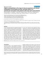
Fig. 4. Surface views of GH45 endoglucanases from subfamily A, B and C. Arrows point to substrate binding area in HiCel45A (PDB: 4ENG), PcCel45A (PDB: 5KJO),
MeCel45A (PDB: 1WC2), TrCel45A (GenBank: CAA83846.1, homology model). Catalytic acid and base are shown as sticks in the cartoon representation. Letters A, B,
C indicate subfamily membership.
of barley beta-glucan (BG), konjac glucomannan (GM) and carbox
ymethyl cellulose (CMC) (Fig. 6).
Activity was expressed as formation of reducing ends measured by
PHBAH assay. For all proteins the highest initial hydrolysis rates were
observed on beta-glucan, for which the initial production of reducing
ends was 2–7 times more rapid than on CMC (Table 2). On both CMC and
BG, MeCel45A showed the second highest initial rate, preceded by
HiCel45A and followed by PcCel45A and TrCel45A in that order.
With GM as a substrate the enzymes did not show any linear phase at
the start of the reaction, probably due to its heteropolymer nature, and
reliable initial rates could not be determined for GM. Therefore, in order
to gain a general understanding of the enzyme relative initial rates on
the different substrates we chose an initial product concentration that
was covered by all experiments (70 μM reducing ends), and then
compared the time needed to reach this concentration among the en
zymes (Table 2). For all enzymes GM was hydrolyzed faster than CMC,
but much more slowly than BG. The highest activity on GM was
exhibited by HiCel45A and TrCel45A, followed by MeCel45A and
PcCel45A in that order.
With BG as a substrate the reaction rapidly leveled off and appeared
to reach an end point within less than 1 h. Therefore, a 60 min time point
was used to calculate the yield of reducing ends from BG. The activity on
5
L. Okmane et al.
Carbohydrate Polymers 277 (2022) 118771
Fig. 5. Residues likely to interact with substrate in the active-site cleft of MeCel45A. (A) The active-site cleft of MeCel45A (PDB: 1WC2) aligned with the structure of
HiCel45A (PDB: 4ENG) with two cellotriose molecules bound in the substrate binding groove; (B) The active-site cleft of MeCel45A (PDB: 1WC2) aligned with the
structure of PcCel45A (PDB: 3X2M) with two cellopentaose molecules bound in the substrate binding groove. Substrate interacting residues are displayed as lines,
and residue labels are in italics for MeCel45A.
Fig. 6. General representations of the chemical structures of carboxymethyl cellulose (CMC), barley betaglucan (BG), and konjac glucomannan (GM).
CMC and GM did also level off, but no clear end point was reached, and
the yield was instead calculated from the amount of reducing ends after
24 h of hydrolysis. The yield is a measure of how many bonds in the
polymers the respective enzymes are able to hydrolyze.
The HiCel45A enzyme gave significantly higher yields on BG and on
CMC than the other enzymes, especially on BG with more than twice as
high concentration of reducing ends (Table 2). However, on GM the
pattern was quite different. The highest yield of reducing ends from GM
6
L. Okmane et al.
Carbohydrate Polymers 277 (2022) 118771
Table 2
Family GH45 endoglucanase substrate specificity. Initial rate of formation and yield of reducing ends on 0.1% carboxymethyl cellulose (CMC), barley beta-glucan (BG)
or konjac glucomannan (GM) as substrate. ±SD is standard deviation of triplicate determinations.
Name
Subfamily
Initial rate on 0.1% substrate
CMC
BG
(μM/μM)min
HiCel45A
MeCel45A
TrCel45A
PcCel45A
A
B
B
C
235 ±
183 ±
12 ±
56 ±
Yield on 0.1% substrate from 0.1 μM enzyme
9,9
9,7
4,1
5,0
− 1
± SD
− 1
(μM/μM)min
1139 ±
296 ±
90 ±
109 ±
58,5
43,5
37,3
58,6
± SD
Required time for 0.1 μM Cel45A
to produce 70 μM of reducing
sugar on 0.1% substrate
CMC
BG
GM
CMC
BG
GM
μM ± SD, 24 h (*22 h)
μM ± SD, 60 min
μM ± SD, 24 h
t, min
t, min
t, min
188 ± 4,8
128 ± 3,3
92 ± 4,0*
104 ± 14,7
547 ± 23,5
229 ± 6,6
225 ± 4,0
195 ± 20,2
192 ± 4,4
308 ± 42,0
1087 ± 43,2
240 ± 22,5
>60
>60
>100
>60
<1
<5
<30
<10
10
60
30
>60
was observed with TrCel45A and the lowest with HiCel45A. TrCel45A
deviated from the other enzymes in its activity on GM. The rate of hy
drolysis did not decline to the same extent over time as with the other
enzymes. The yield of reducing ends for TrCel45A on GM after 24 h was
nearly 10 times higher than it was on CMC after a similar period of time.
Enzymatic activities of MeCel45A and TrCel45A were additionally
evaluated on cellohexaose at +4 ◦ C and +30 ◦ C. Samples were analyzed
every 65 min for 6 h using high performance anion exchange chroma
tography (HPAEC). The main cleavage products of MeCel45A on cello
hexaose were cellobiose and cellotetraose, while the main products of
TrCel45A were cellobiose, cellotriose and cellotetraose (Fig. 7A–D). At
30 ◦ C MeCel45A and TrCel45A showed similar cellohexaose degrada
tion rates (1.38 ± 0.05 min− 1 and 1.26 ± 0.20 min− 1, respectively), but
at 4 ◦ C these enzymes performed significantly differently. For MeCel45A
the activity at 4 ◦ C was 72% of that at 30 ◦ C, whereas it was only 35% for
TrCel45A (Fig. 7E).
(Fig. S4) in contrast to the other enzymes.
Enzyme degradation of beta-glucan was also monitored by NMR with
a low enzyme concentration (0.3–0.5 μM). Formation of reducing-end
α-glucose was followed (Fig. S5) and corresponded well with the re
sults from PHBAH assays. The reducing-end residues formed were linked
at position 4 rather than position 3 (Fig. S6), showing a preference for
cleavage next to a β(1 → 4) linkage. This preference was the same for all
the four enzymes.
3. Discussion
All of the Cel45A enzymes tested in this study were able to produce
reducing ends on barley beta-glucan, konjac glucomannan and carbox
ymethyl cellulose. Barley beta-glucan was degraded most rapidly among
the three selected substrates. It is a linear glucose homopolymer with
β(1 → 3) and β(1 → 4) linkages, where three or four successive β(1 → 4)
linked glucose residues are followed by one pair of β(1 → 3) linked
residues (Fig. 6). During NMR experiments involving barley beta-glucan,
we found that the reducing-end residues were β(1 → 4) and not β(1 → 3)
linked. This shows that the Cel45A enzymes require a β(1 → 4) linkage
between subsites − 2 and − 1. However, further studies are required to
draw conclusions about restrictions for other subsites. The NMR ana
lyses confirm that Cel45A from M. edulis, T. reesei and P. chrysosporium
hydrolyze glycosidic bonds with an inverting action mechanism, thus
proving that this mechanism is indeed common among all GH45 sub
families known to date. An inverting catalytic mechanism has long been
proposed for GH45 enzymes, but to our knowledge had only been
experimentally proven for subfamily A member Cel45A from H. insolens
(Schou et al., 1993).
NMR spectroscopy on glucomannan revealed that HiCel45A can
cleave β(1 → 4) linkage between mannose and glucose (Man-Glc) as
pointed out by the cyan arrow in Fig. 8A, but the rate and preference for
such cleavage is low. However, HiCel45A was much faster at producing
non-reducing end glucose alongside reducing-end glucose, indicating
cleavage between two glucose residues (Glc-Glc) and a preference for
such linkages. Cleavage by HiCel45A also led to formation of monomeric
glucose, which was not observed from the other enzymes. Another major
difference was the absence of non-reducing end mannose in the
HiCel45A product profile, whereas in the case of TrCel45A, MeCel45A
and PcCel45A the formation of non-reducing end mannose and
reducing-end glucose was fast and simultaneous, suggesting a cleavage
between glucose and mannose (Glc-Man, magenta arrow in Fig. 8A) and
a preference for such linkages. Furthermore, a slow formation of
reducing-end mannose was accompanied by an increase of non-reducing
end mannose in TrCel45A, MeCel45A and PcCel45A, which indicates
cleavage between two mannose residues (Man-Man), thus mannanase
activity. While all of the Cel45A enzymes showed an activity against
glucomannan, Cel45A from T. reesei was outstanding in its ability to
continue glucomannan degradation even after 24-hour incubation. We
attribute this to the aforementioned mannanase activity, which was also
demonstrated in a previous study by Karlsson et al. (2002). An inter
esting observation was made regarding the levels of non-reducing end
2.5. NMR spectroscopy
The products from enzyme degradation of GM and BG were inves
tigated by NMR spectroscopy to observe differences in specificity on the
two substrates. Degradation of GM at 0.3 μM enzyme concentration was
observed by following the formation of reducing end α-Glc and yielded
initial build-up curves similar to those observed from PHBAH assays
(Fig. S3A). It confirmed that all four enzymes are acting through an
inverting mechanism, because α-Glc was formed rapidly before equi
librium with the β anomer was reached.
In order to obtain higher levels of degradation products from GM, the
enzyme concentration was increased to 35 μM. This caused reducing end
α-Glc to be formed very quickly before reducing end β-Glc was formed
by mutarotation (Fig. 8 and S3B), which further confirmed the inverting
mechanism of the enzymes. More interestingly, the formation of
mannose reducing ends was also observed (Fig. 8). Among the four
tested enzymes, TrCel45A was the most efficient in mannose cleavage,
whereas HiCel45A, MeCel45A and PcCel45A were similar in efficiency
after 23 h. However, HiCel45A and TrCel45A gave rise to the steepest
increase of Man reducing ends during the first 6 h, whereas MeCel45A
was the slowest.
Degradation of GM at high enzyme concentration (35 μM) also
allowed the detection of other degradation products. The 1H NMR
spectra after 23 h of degradation clearly shows a difference between
HiCel45A and the other enzymes (Fig. S4). Degradation by HiCel45A
produced non-reducing end glucose, whereas the other three enzymes
produced non-reducing end mannose (Fig. 8E and F). The non-reducing
end residues formed instantly, similar to the reducing-end glucose res
idues. Non-reducing end mannose was not detected from HiCel45A
degradation and only small amounts of non-reducing end glucose was
detected from TrCel45A, MeCel45A and PcCel45A degradation. In
addition, non-reducing end 2-acetylated mannose (2Ac-Man) was
formed from degradation by TrCel45A, MeCel45A and PcCel45A with
the highest amount from TrCel45A (Fig. S3C). Furthermore, a small
amount of monomeric glucose was observed from HiCel45A degradation
7
L. Okmane et al.
Carbohydrate Polymers 277 (2022) 118771
Fig. 7. Enzymatic activity of M. edulis Cel45A and T. reesei Cel45A on cellohexaose at +4 ◦ C and +30 ◦ C. HPAEC chromatogram time-lapse of product formation from
cellohexaose degradation by MeCel45A at (A) +4 ◦ C and (C) +30 ◦ C, by TrCel45A at (B) +4 ◦ C and (D) +30 ◦ C. Chromatogram legends represent timepoints of
degradation. (E) Hydrolytic activity is expressed as μM/min per 1 μM of enzyme of MeCel45A and TrCel45A on cellohexaose. The product formation rate is shown
with positive values and substrate degradation rate with negative values. Average enzymatic activity rates are depicted as bars and individual datapoints as filled
circles. Error bars represent standard deviation (n ≥ 3).
8
L. Okmane et al.
Carbohydrate Polymers 277 (2022) 118771
Fig. 8. (A) Simplified formula of a glucomannan chemical structure and exemplified degradation pathways. (B) 1H NMR spectra from a) GM control, b) GM after 12
min TrCel45A degradation, and c) GM after 24 h TrCel45A degradation. Signal assignments are 1) 2Ac-Man H2, 2) non-reducing end 2Ac-Man H2, 3) reducing end
Glc H1-α, and 4) reducing end Man H1-α. The asterisk highlights a starch-like impurity. NMR-derived progress curves are plotted to show the formation of (C)
reducing-end α-Man, (D) reducing-end α-Glc, (E) non-reducing end Man, and (F) non-reducing end Glc from degradation of glucomannan with 35 μM enzyme. Nonreducing end Man was not detected in samples with HiCel45A. Error bars correspond to ±1 standard deviation.
acetylated mannose residues. Cel45A from H. insolens did not facilitate
formation of products with an acetylated mannose residue at the nonreducing end. As expected, the cleavage product reducing-end glucose
residues were alpha-anomers for all enzymes, which once again con
firms the inverting nature of these endoglucanases.
All of the evaluated Cel45A enzymes were able to hydrolyze CMC, a
synthesized cellulose derivative with carboxymethyl group substitutions
(Fig. 6). Hydrolysis rates were comparatively slower and with lower
yields of reducing ends, presumably due to the presence of carbox
ymethyl substituents on some glucose residues. Interestingly, HiCel45A
gave a higher reducing end yield than the other enzymes, suggesting
more tolerance for substitutions despite having a more enclosed active
site. It would be reasonable to assume that such bulky substituents could
restrict the binding or cleavage in narrow active sites due to steric
hindrance.
Of the two subfamily B enzymes, MeCel45A retained over 70% of its
activity at +4 ◦ C relative to +30 ◦ C, compared to 35% for TrCel45A
(Fig. 7E). Considering the tropical habitat of T. reesei and the boreal
habitat of M. edulis, this could indicate GH45 enzyme evolution towards
cold adaptation in the mollusc. A broad optimum temperature range has
previously been demonstrated by Xu et al. (2000). Further investigation
of how the structure of this enzyme has evolved to retain high activity at
9
L. Okmane et al.
Carbohydrate Polymers 277 (2022) 118771
low temperatures could be an interesting study. The cellohexaose
cleavage product profile differed between the enzymes (Fig. 7A–D).
Cel45A from M. edulis produced predominantly cellobiose and cellote
traose, which implies a distinct organization of substrate in the active
site cleft. Cel45A from T. reesei produced cellotriose in addition to
cellobiose and cellotetraose, indicating a less definitive cleavage
preference.
Structure and sequence comparisons reveal several similarities and
differences between Cel45A from M. edulis and HiCel45A, AcCel45A,
TrCel45A and PcCel45A. While the DPBB core superposes closely, there
are variations in surface structures between the subfamilies due to dif
ferences in the lengths of flanking loop regions. In this regard, subfamily
B and C enzymes are more similar to each other than to the subfamily A
enzyme, where longer loops enclose the catalytic center, thus creating a
tunnel-like structure. In the subfamily C member PcCel45A, loops at
both ends of the substrate-binding cleft make the cleft longer than in the
other enzymes. MeCel45A appears to have the narrowest substrate
binding cleft among the subfamily B members. Substrate binding and the
catalytic mechanism of MeCel45A are most likely very similar to that in
AcCel45A, since their active sites are nearly identical. In all of the en
zymes a tryptophan residue is found at subsite − 4 (Trp64 in MeCel45A),
which most likely serves as a sugar binding platform. Interestingly, this
residue comes from different locations in the GH45 sequences, possibly
underlining the functional significance of this residue. Godoy et al.
(2018) mutated the corresponding tryptophan residue in PcCel45A
(W154A), which led to 50% decrease in catalytic activity on lichenan.
There is no GH45 structure available yet with substrate spanning
over the active site with an un-cleaved bond connecting the − 1 and +1
subsites. There are only a few structures where the − 1 subsite is occu
pied by the reducing-end residue of an oligosaccharide, but the con
formations differ between all of them. A distorted 4H5 half-chair is
refined in a cellotetraose complex with Thielavia terrestris Cel45A of
subfamily A (PDB code 5GLY) (Gao et al., 2017), whereas three variants
of PcCel45A of subfamily C in complex with cellopentaose display 3S1
skew, distorted 2H1 half chair, and 4C1 chair conformations, respec
tively, of the glucose residue at subsite − 1 (wildtype, N92D, N105D
variants; PDB codes 3X2M, 3X2H, 3X2K)(Nakamura et al., 2015).
In the absence of clear information from X-ray crystallography of
how a sugar residue will bind at the − 1 subsite prior to, during, and after
cleavage, the catalytic reaction has been examined by computational
methods. QM/MM calculations and transition path sampling (TPS)
molecular dynamics (MD) simulations of celloheptaose hydrolysis by
HiCel45A showed a conformational itinerary for the glucose unit at
subsite − 1, from 4C1 chair to 2SO skew in the Michaelis complex, over a
2,5
B boat transition state, via 5S1 skew to a 1C4 chair for the α-anomer
product (Bharadwaj et al., 2020). This type of catalytic itinerary, 2SO →
2,5 ‡
B → 5S1, has been proposed earlier for other GH families, including
inverting beta-glucoside active GH6 and GH8, and both retaining
(GH11, GH120) and inverting (GH43) beta-xylosidases (Ard`
evol &
Rovira, 2015).
While the role of the catalytic acid seems to be well established in all
three GH45 subfamilies, the assignment of catalytic base function is
more uncertain. The first assignment of catalytic residues was based on
structures of HiCel45A of subfamily A, where Asp121 was proposed to
act as general acid, implied by its hydrogen bonding to the glycosidic
oxygen (4OH) of a glucose residue at subsite +1, and Asp10 was pro
posed as the most likely general base. This was supported by complete
inactivation of HiCel45A upon mutations at these sites (D121N and
D10N mutants) (Davies et al., 1995). Consequently, these residues are
considered to be indispensable for hydrolysis by GH subfamily A
members. However, a role was also implicated for Asp114, since a
HiCel45A D114N mutant only retained less than 1% activity compared
to wildtype. It is interesting that in the subfamily A enzyme an Asp at
this location is much more active than Asn, whereas it seems to be the
opposite for subfamily C. PcCel45A has an Asn at this location and the
activity was drastically reduced upon mutation to Asp (N92D mutant)
(Nakamura et al., 2015).
Members of GH45 subfamily C lack the Asp residue corresponding to
the base in subfamily A. Instead, Nakamura et al. (2015) proposed
Asn92 as the catalytic base, on the basis of structure studies of PcCel45A
where high-resolution X-ray and neutron diffraction analyses revealed
amide/imidic acid tautomerization of Asn92 and a proton relay-network
that links Asn92 to the catalytic acid (Asp114 in PcCel45A) through a
chain of hydrogen bonds. Nearly all of the residues in the proton-relay
chain in PcCel45A (Asn92, Cys96, Phe95, Asn105, Ser14, His112,
Thr16, Asp114) are also present in MeCel45A (Asn109, Cys113, Trp112,
Asn123, Ser18, His130, Thr20, Asp132), but most are not conserved in
HiCel45A.
In the subfamily B enzymes, MeCel45A, AcCel45A and TrCel45A,
both of the candidate base residues are present. Mutation of either of
them in AcCel45A drastically reduced the activity but did not lead to
complete inactivation (Nomura et al., 2019). The activity reduction was
rather similar for the D27A and N112A mutants, about 30-fold and 60fold, respectively. Based on these results and the position of the residues
relative to a glucoside unit modelled at subsite − 1 of AcCel45A, Nomura
et al. (Nomura et al., 2019) proposed that Asn112 in its imidic acid form
acts as the catalytic base that activates a water molecule for nucleophilic
attack at the anomeric carbon, whereas Asp27 is of primary importance
for productive positioning of the glucose residue at subsite − 2. How
ever, the orientation of the modelled glucoside is quite different from
previous observations and models. The glucose residue modelled at
subsite − 1 of AcCel45A is flipped so that the 2OH and 3OH groups point
towards the bottom of the cleft, while the 6OH arm is pointing out from
the active site. The anomeric carbon is exposed on the side of the glucose
ring that is facing Asn112 and not Asp27 (Nomura et al., 2019). This is in
contrast to the crystal structures with sugar bound at subsite − 1 as well
as the QM/MM MD study by Bharadwaj et al. (2020). There, the glucose
residue is instead oriented with its 6OH arm bound at the bottom of the
cleft, while the 2OH and 3OH groups point out from the active site,
which exposes the anomeric carbon on the side of the ring that is facing
the Asp residue and not on the side where the Asn residue is located.
Considering the high structural similarity between AcCel45A and
MeCel45A, the corresponding residues in M. edulis Cel45A (Asp24,
Asn109 and Asp132) should be of similar importance. Asp132 is the
catalytic acid, but which residue is acting as the catalytic base, Asp24 or
Asn109, remains an open question. Further research is obviously needed
to fully elucidate the catalytic mechanism of GH45 and its subfamilies at
the molecular level.
4. Conclusions
Our results show that the GH45 enzymes studied here share several
common properties. All can hydrolyze barley beta-glucan, konjac glu
comannan and carboxymethylcellulose with an apparent preference for
barley beta-glucan. Hydrolysis by these enzymes leads to inversion of
configuration at the anomeric carbon, thus indicating an inverting ac
tion mechanism. We also demonstrated a few key differences such as
mannanase activity among subfamily B and C members, and the ability
of subfamily A member to produce monomeric glucose. We pointed out a
variation in product profile within subfamily B and a possible evolu
tionary cold adaptation of the enzyme in blue mussel.
5. Methods
5.1. Extraction and purification of MeCel45A
MeCel45A from Blue Mussel, Mytilus edulis, (UniProt entry P82186)
was prepared as described (Xu et al., 2000) with minor modifications –
only the digestive gland of the mussel was used and the acid precipita
tion and heat precipitation steps were omitted. The blue mussels were
collected from waters off the Swedish west coast, frozen and their
digestive glands excised (total 1.42 kg glands from 28.7 kg of whole
10
L. Okmane et al.
Carbohydrate Polymers 277 (2022) 118771
frozen mussels). The digestive glands were homogenized in a meat
blender (cooled with ice), stirred for 30 min and insoluble materials
were removed by centrifugation. The target protein was captured by
immobilized metal affinity chromatography (IMAC) on a STREAMLINE
Chelating gel (Amersham Biosciences) saturated with zinc ions and
elution with 20 mM sodium phosphate, pH 7.0, 1 M NaCl, 50 mM EDTA,
followed by size-exclusion chromatography on Superdex 75 pg (Amer
sham Biosciences) and finally cation exchange chromatography on a
Mono S column (Amersham Biosciences) eluted with a gradient of
0–300 mM NaCl in 20 mM sodium acetate, pH 5.5. MeCel45A eluted at
~200 mM NaCl. Activity against CMC during MeCel45A purification
was assayed using dinitrosalicylic acid as reducing sugar reagent ac
cording to Bhat and Wood (1998). The purified enzyme was concen
trated by ultra-filtration in 50 mM sodium acetate, 0.2 M NaCl, pH 5.5,
and was stored at +4 ◦ C prior to crystallisation screening, and at − 20 ◦ C
for further use. The final yield was 8 mg of pure MeCel45A enzyme
(extinction coefficient of 41,660 M− 1 cm− 1). Further details are pro
vided in the Supplementary Information file.
processed and scaled with the HKL program package (Otwinowski &
Minor, 1997). The structure was solved with SIRAS (single isomorphous
replacement with anomalous scattering) using a derivative with Baker's
dimercurial. The final structure model was refined with Refmac5
(Murshudov et al., 1997) at 1.2 Å resolution and final R and Rfree-values
of 0.147 and 0.162. Further details are provided in the Supplementary
Information file. Statistics of X-ray diffraction data sets and structure
refinement are summarized in Tables S1 and S2, respectively. Atomic
coordinates of the structure model and the structure factors have been
deposited with the Protein Data Bank (wwpdb.org) (wwPDB con
sortium, 2019) as PDB entry 1WC2.
5.4. Structure comparison
To include Cel45A from T. reesei in structure comparisons, a ho
mology model was created for TrCel45A, built in SWISS-MODEL
(Waterhouse et al., 2018) using MeCel45A (PDB code 1WC2; this
work) as a template. The PyMOL Molecular Graphics System, Version
ădinger, LLC) was used to align structures and investigate
1.8.6.0 (Schro
structural similarities. The structures were superposed using CEAlign
command and structure deviation was expressed as root mean square
deviation (RMSD, Å) over aligned Ca atoms, as generated by the CE
algorithm. LIGPLOT v.4.5.3 (Wallace et al., 1995) was used for plotting
protein-ligand interactions. The multiple sequence alignment was
created using MUSCLE (Madeira et al., 2019) online at the EMBL-EBI
server (ebi.ac.uk), later adjusted and visualized in ESPript (espript.
ibcp.fr) (Robert & Gouet, 2014). The percentage sequence-identity
matrix was created with Clustal 2.1 online using the EMBL-EBI server
(ebi.ac.uk) (Madeira et al., 2019).
5.2. Preparation of HiCel45A, PcCel45A, TrCel45A
All protein stock solutions were kept in 10 mM sodium acetate buffer
pH 5 with protein concentration of less than 1.0 mg/mL, stored at room
temperature directly before use, at +4 ◦ C the day before use and − 20 ◦ C
for further use.
Purified HiCel45A (GenBank: QCH00668, ext. coefficient 0.1% =
1.956) from Humicola insolens (including enzymatic deglycosylation
with endo H, followed by size exclusion chromatography on Superdex
75) was kindly provided by Kristian Bertel Rømer Mørkeberg Krogh at
Novozymes A/S, Denmark.
Methanol-induced heterologous expression of PcCel45A (GenBank:
BAG68300, ext. coefficient 0.1% = 1.270) was done in Komagataella
pastoris KM71H expression system according to Igarashi and colleagues
(Igarashi et al., 2008). Culture filtrate, supplemented with 1 M
(NH4)2SO4, was applied on a hydrophobic interaction chromatography
(HIC) column (Phenyl Sepharose; elution buffer 10 mM NaAc pH 5.0).
The collected protein solution was desalted on Biogel P6 column into a
20 mM TrisHCl pH 8.0 buffer. Desalting was followed by anion exchange
chromatography (DEAE Sepharose) with an elution buffer 0.5 M NaCl
20 mM TrisHCl pH 8.0 using the following elution gradient: 0–50% 400
mL; 50–100% 100 mL; 100% 200 mL. Size exclusion chromatography
(Superdex 75, buffer: 10 mM NaAc pH 5.0) was carried out as a final
purification step.
TrCel45A from Trichoderma reesei (GenBank: CAA83846, ext. coef
ficient 0.1% = 2.003) was kindly provided by Matti Siika-aho, VTT
Biotechnology, Finland. The full-length TrCel45A enzyme (with CDlinker-CBM1) was obtained from culture filtrate of a T. reesei strain
lacking the genes expressing the two major endoglucanases Cel7B and
Cel5A and was purified as described in Karlsson et al. (2002).
5.5. Reducing sugar assay
MeCel45A, TrCel45A, PcCel45A and HiCel45A were each incubated
at 30 ◦ C, 400 rpm, in 0.1 M sodium acetate buffer pH 5.0 on CMC (β-1,4
linkage, degree of carboxymethyl substitutions 0.60–0.95, dynamic
viscosity 0.7–1.5 Pa.s, purity >99%, BioChemika, Fluka), konjac glu
comannan (β-1,4 linkage, mannose: glucose = 60: 40, kinematic vis
cosity ~1.0 × 10⁻⁵ m2/s, purity >98%, Megazyme) or barley betaglucan
(mixed β-1,4 and β-1,3 linkages, kinematic viscosity 2.0–3.0 × 10⁻⁵ m2/
s, purity ~95%, Megazyme) dissolved in water. Reaction was stopped by
adding 1 M sodium hydroxide solution in a 1:1 ratio and cooling samples
on ice. Hydrolytic activity was determined colorimetrically by quanti
fying the reducing sugar formation with p-Hydroxybenzoic acid hy
drazide (PHBAH, Sigma) solution (Lever, 1972): 0.1 M PHBAH; 0.2 M
sodium potassium tartrate; 0.5 M sodium hydroxide solution. PHBAH
solution was added in 1:1 ratio to the samples, followed by incubation at
95 ◦ C for 15 min and cooling on ice for 10 min. Samples were kept at
room temperature for 5 min before absorption was read at 410 nm.
5.3. Crystallisation and structure determination of MeCel45A
5.6. Hydrolysis product analysis by high-performance anion-exchange
chromatography (HPAEC)
Crystallisation was done at room temperature by hanging drop
vapour diffusion (McPherson, 1982). Crystals were grown with 12 mg/
mL protein solution in 50 mM sodium acetate, pH 5.5, 0.2 M NaCl,
mixed with and equilibrated against 0.6 M sodium acetate, pH 5.5, and
0.125 or 0.375 M NaCl. The heavy-atom derivative was obtained by
adding a grain of solid of Baker's dimercurial (C10H16Hg2O6; 1,4-diace
toxymercuri-2,3-dimethoxybutane; Anatrace) to drops with crystals.
Crystals to be used for X-ray analysis were kept for 1–5 min in a cryoprotectant solution (30% monomethyl PEG 5000, 12.5% glycerol, 0.1
M sodium morpholine-ethane-sulfonic acid, pH 6.0, and 10 mM CoCl2)
and then flash-cooled using liquid nitrogen. X-ray diffraction data were
recorded at 100 K, in-house on a CuKα RIGAKU/R-AXIS IIC system
(native, 1.85 Å; dimercurial, 2.0 Å), and at beamline ID14-3, ESRF,
France (native, 1.2 Å resolution). Diffraction data were indexed,
0.075 μM MeCel45A and 0.075 μM TrCel45A each were incubated at
+30 ◦ C and +4 ◦ C with 85 μM cellohexaose (Seikagaku Corporation,
dissolved in water) in 0.1 M sodium acetate buffer pH 5.0.
Cellohexaose hydrolysis products were analyzed periodically (every
65 min for 6 h) using DIONEX ICS3000 equipped with Dionex CarboPac
PA200 column, HPAE-PAD detector. Column was equilibrated in 150
mM NaOH/50 mM Na acetate. Peak separation was achieved at a
flowrate 0.450 mL/min by three gradients: 1) from 150 mM NaOH/50
mM Na acetate to 150 mM NaOH/125 mM Na acetate in 20 min; 2) from
150 mM NaOH/125 mM Na acetate to 150 mM NaOH/250 mM Na ac
etate in 15 min; 3) 150 mM NaOH/250 mM Na acetate for 15 min.
Separation was followed by a cleaning step gradient from 150 mM
NaOH/250 mM Na acetate to 150 mM NaOH/50 mM Na acetate in 15
min. Cellobiose, cellotriose and cellotetraose (Seikagaku Corporation)
11
L. Okmane et al.
Carbohydrate Polymers 277 (2022) 118771
standards were used for peak identification and product quantification.
Database
5.7. NMR spectroscopy
Structural data have been deposited with the Protein Data Bank
under accession code 1WC2.
Degradation of beta-glucan and glucomannan with MeCel45A,
TrCel45A, PcCel45A, and HiCel45A was investigated by NMR spec
troscopy. The substrates were dissolved in D2O to yield 1% stock solu
tions. Substrates and enzymes were mixed in 3 mm NMR tubes to a total
volume of 160 μL, a final substrate concentration of 5–7 mg/mL, and 50
mM phosphate buffer (pH 5.0). The enzyme concentration was either
0.3–0.5 μM (for glucomannan and beta-glucan degradation) or 35 μM
(for glucomannan degradation).
NMR spectra were recorded on a Bruker Avance III 600 MHz spec
trometer using a 5 mm inverse detection cryoprobe equipped with z axis
gradient. The spectra were acquired at 30 ◦ C with the HDO signal as
internal reference (δ 4.71 ppm). After the enzyme was added, the tube
was quickly placed into the probe and hydrolysis was monitored as a
function of time. The first spectrum was acquired within 10 min after the
reaction started, and from then on spectra were acquired every 5–10
min. 1D spectra were obtained from the 1D NOESY experiment (noe
sygppr1d) for water suppression, with water presaturation during the
relaxation delay (1 s) and the mixing time (50 ms). A sweep width of
6000 Hz was used, and 32 scans of 64 K data points were acquired. The
enzyme and substrate concentrations were chosen to ensure fast enough
reaction to allow determination of the glucose anomers before muta
rotation occurred. Samples for glucomannan degradation with high
enzyme concentration (35 μM) were run in duplicate for each enzyme to
yield an error estimation.
Signal assignments were carried out with the help of various 2D NMR
experiments, including 1H,1H-TOCSY, 1H,1H-NOESY, and 1H,13C-HSQC,
as well as comparison with previous assignments of beta-glucan
(Petersen et al., 2013) and glucomannan (Mikkelson et al., 2013; Tele
man et al., 2003) and predicted chemical shifts from the CASPER tool
(Jansson et al., 2006; Lundborg & Widmalm, 2011).
Proton signals were integrated and normalized to yield ratios of
reducing-end or non-reducing end residues to interior residues. Since H1
of interior Man and Glc residues of glucomannan were hidden by the
water signal or affected by the water suppression, H2 of interior residues
was used for normalization. Reducing-end Man was calculated from the
ratio between reducing-end Man H1-α (5.17 ppm) and interior Man H2
(4.11 ppm). Reducing-end Glc was calculated from the ratio between
reducing-end Glc H1-α (5.22 ppm) and interior Glc H2 (3.35 ppm) and
from the ratio between reducing-end Glc H2-β (3.28 ppm) and interior
Glc H2 (3.35 ppm). For partly overlapping signals and signals with very
low abundance, peak heights were used rather than integrals. By this
means, non-reducing end 2Ac-Man was calculated from the ratio be
tween non-reducing end 2Ac-Man H2 (5.45 ppm) and interior 2Ac-Man
H2 (5.50 ppm). Similarly, non-reducing end Man was calculated from
the ratio between non-reducing end Man H2 (4.05 ppm) and interior
Man H2 (4.11 ppm), and non-reducing end Glc was calculated from the
ratio between non-reducing end Glc H2 (3.30 ppm) and interior Glc H2
(3.35 ppm). For beta-glucan, reducing-end Glc was calculated from the
ratio between the integrated signal of reducing-end Glc H1-α (5.23 ppm)
and β(1 → 4)-linked Glc H1 (4.54 ppm).
CRediT authorship contribution statement
Laura Okmane: Methodology, Validation, Formal analysis, Investi
gation, Data curation, Writing – original draft, Writing – review &
editing, Visualization. Gustav Nestor: Conceptualization, Methodol
ogy, Validation, Formal analysis, Investigation, Writing – original draft,
Writing – review & editing, Visualization. Emma Jakobsson: Valida
tion, Formal analysis, Investigation, Data curation, Writing – original
draft. Bingze Xu: Methodology, Formal analysis, Investigation, Re
sources, Writing – original draft. Kiyohiko Igarashi: Investigation,
Resources, Supervision. Mats Sandgren: Conceptualization, Resources,
Writing – review & editing, Supervision, Project administration, Fund
ing acquisition. Gerard J. Kleywegt: Conceptualization, Methodology,
Software, Validation, Formal analysis, Data curation, Writing – original
draft, Supervision, Funding acquisition. Jerry Ståhlberg: Conceptuali
zation, Methodology, Validation, Formal analysis, Investigation, Re
sources, Data curation, Writing – original draft, Writing – review &
editing, Visualization, Supervision, Project administration, Funding
acquisition.
Declaration of competing interest
No real or perceived conflicts.
Acknowledgements
We thank Jan-Christer Janson for initiation of the investigation of
enzymes from Blue mussel, Sabah Mahdi for help with protein crystal
lisation, Christina Divne for help with initial phasing, Matti Siika-aho
˜ oz for preparation and purification of TrCel45A
and In´es G. Mun
enzyme, Miao Wu for help with cultivation of K. pastoris, and Kristian
Bertel Rømer Mørkeberg Krogh at Novozymes A/S, Denmark, for kindly
providing the HiCel45A enzyme.
Appendix A. Supplementary data
Supplementary data to this article can be found online at https://doi.
org/10.1016/j.carbpol.2021.118771.
References
Ard`
evol, A., & Rovira, C. (2015). Reaction mechanisms in carbohydrate-active enzymes:
Glycoside hydrolases and glycosyltransferases. Insights from ab initio quantum
Mechanics/Molecular mechanics dynamic simulations. Journal of the American
Chemical Society, 137(24), 7528–7547. />Berto, G. L., Velasco, J., Tasso Cabos Ribeiro, C., Zanphorlin, L. M., Noronha
Domingues, M., Tyago Murakami, M., Polikarpov, I., de Oliveira, L. C., Ferraz, A., &
Segato, F. (2019). Functional characterization and comparative analysis of two
heterologous endoglucanases from diverging subfamilies of glycosyl hydrolase
family 45. Enzyme and Microbial Technology, 120, 23–35. />enzmictec.2018.09.005
Bharadwaj, V. S., Knott, B. C., Ståhlberg, J., Beckham, G. T., & Crowley, M. F. (2020).
The hydrolysis mechanism of a GH45 cellulase and its potential relation to lytic
transglycosylase and expansin function. Journal of Biological Chemistry. https://doi.
org/10.1074/jbc.RA119.011406. jbc.RA119.011406.
Bhat, K. M., & Wood, T. M. (1998). Methods for measuring cellulase activities. Methods
Enzymol., 160, 87–112.
Castillo, R. M., Mizuguchi, K., Dhanaraj, V., Albert, A., Blundell, T. L., & Murzin, A. G.
(1999). A six-stranded double-psi β barrel is shared by several protein superfamilies.
Structure, 7(2), 227–236. />Cha, J.-H., Yoon, J.-J., & Cha, C.-J. (2018). Functional characterization of a thermostable
endoglucanase belonging to glycoside hydrolase family 45 from Fomitopsis palustris.
Applied Microbiology and Biotechnology, 102(15), 6515–6523. />10.1007/s00253-018-9075-5
Couturier, M., Feliu, J., Haon, M., Navarro, D., Lesage-Meessen, L., Coutinho, P. M., &
Berrin, J.-G. (2011). A thermostable GH45 endoglucanase from yeast: Impact of its
Funding sources
This work was supported by the Swedish Research Council for
Environment, Agricultural Sciences and Spatial Planning (Formas),
Grant Number 2017-01130. GJK was supported by the Swedish Struc
tural Biology Network (SBNet), the Swedish Natural Science Research
Council (NFR) and the Royal Swedish Academy of Sciences (KVA). EJ
was supported by the Swedish Structural Biology Network (SBNet). KI
acknowledges Grant-in-Aid for Innovative Areas (No. 18H05494 to K.I.)
from the Japanese Ministry of Education, Culture, Sports and Technol
ogy (MEXT).
12
L. Okmane et al.
Carbohydrate Polymers 277 (2022) 118771
Igarashi, K. (2015). “Newton’s cradle” proton relay with amide–imidic acid
tautomerization in inverting cellulase visualized by neutron crystallography. Science
Advances, 1(7), Article e1500263. />Newell, C., Shumway, S., Cucci, T. L., & Selvin, R. (1989). The effects of natural seston
particle size and type on feeding rates, feeding selectivity and food resource
availability for the mussel Mytilus edulis linnaeus, 1758 at bottom culture sites in
Maine. Journal of Shellfish Research, 8, 187–196.
Nomura, T., Iwase, H., Saka, N., Takahashi, N., Mikami, B., & Mizutani, K. (2019). Highresolution crystal structures of the glycoside hydrolase family 45 endoglucanase
EG27II from the snail Ampullaria crossean. Acta Crystallographica Section D:
Structural Biology, 75(4), 426–436. />Onishi, T., Suzuki, M., & Kikuchi, R. (1985). In , 51. The distribution of polysaccharide
hydrolase activity in gastropods and bivalves (pp. 301–308) (2).
Otwinowski, Z., & Minor, W. (1997). Processing of X-ray diffraction data collected in
oscillation mode. Methods in Enzymology, 276, 307–326.
Pauchet, Y., Kirsch, R., Giraud, S., Vogel, H., & Heckel, D. G. (2014). Identification and
characterization of plant cell wall degrading enzymes from three glycoside hydrolase
families in the cerambycid beetle Apriona japonica. Insect Biochemistry and Molecular
Biology, 49, 1–13. />Pauchet, Y., Wilkinson, P., Chauhan, R., & Ffrench-Constant, R. H. (2010). Diversity of
beetle genes encoding novel plant cell wall degrading enzymes. PloS One, 5(12),
Article e15635. />Payne, C. M., Knott, B. C., Mayes, H. B., Hansson, H., Himmel, M. E., Sandgren, M.,
Ståhlberg, J., & Beckham, G. T. (2015). Fungal cellulases. Chemical Reviews, 115(3),
1308–1448. />Petersen, B. O., Olsen, O., Beeren, S. R., Hindsgaul, O., & Meier, S. (2013). Monitoring
pathways of β-glucan degradation by enzyme mixtures in situ. Carbohydrate
Research, 368, 47–51. />Purchon, R. D. (1977). The biology of the mollosca (2nd ed.). Pergamon Press.
Robert, X., & Gouet, P. (2014). Deciphering key features in protein structures with the
new ENDscript server. Nucleic Acids Research, 42(W1), W320–W324. https://doi.
org/10.1093/nar/gku316
Sakamoto, K., & Toyohara, H. (2009). Molecular cloning of glycoside hydrolase family 45
cellulase genes from brackish water clam Corbicula japonica. Comparative
Biochemistry and Physiology. Part B, Biochemistry & Molecular Biology, 152(4),
390–396. />Saloheimo, A., Henrissat, B., Hoffren, A. M., Teleman, O., & Penttilă
a, M. (1994). A novel,
small endoglucanase gene, egl5, from Trichoderma reesei isolated by expression in
yeast. Molecular Microbiology, 13(2), 219–228. />Schou, C., Rasmussen, G., Kaltoft, M.-B., Henrissat, B., & Schülein, M. (1993).
Stereochemistry, specificity and kinetics of the hydrolysis of reduced cellodextrins
by nine cellulases. European Journal of Biochemistry, 217(3), 947–953. https://doi.
org/10.1111/j.1432-1033.1993.tb18325.x
Song, J. M., Hong, S. K., An, Y. J., Kang, M. H., Hong, K. H., Lee, Y.-H., & Cha, S.-S.
(2017). Genetic and structural characterization of a thermo-tolerant, cold-active,
and acidic Endo-β-1,4-glucanase from Antarctic springtail, Cryptopygus antarcticus.
Journal of Agricultural and Food Chemistry, 65(8), 16301640. />10.1021/acs.jafc.6b05037
Teleman, A., Nordstră
om, M., Tenkanen, M., Jacobs, A., & Dahlman, O. (2003). Isolation
and characterization of O-acetylated glucomannans from aspen and birch wood.
Carbohydrate Research, 338(6), 525–534. />00491-3
Tsuji, A., Tominaga, K., Nishiyama, N., & Yuasa, K. (2013). Comprehensive enzymatic
analysis of the cellulolytic system in digestive fluid of the sea hare Aplysia kurodai.
Efficient glucose release from sea lettuce by synergistic action of 45 kDa
endoglucanase and 210 kDa ß-glucosidase. PLoS One, 8(6), Article e65418. https://
doi.org/10.1371/journal.pone.0065418
Valencia, A., Alves, A. P., & Siegfried, B. D. (2013). Molecular cloning and functional
characterization of an endogenous endoglucanase belonging to GHF45 from the
western corn rootworm,Diabrotica virgifera virgifera. 513(2), 260–267. https://doi.
org/10.1016/j.gene.2012.10.046
Vlasenko, E., Schülein, M., Cherry, J., & Xu, F. (2010). Substrate specificity of family 5, 6,
7, 9, 12, and 45 endoglucanases. Bioresource Technology, 101(7), 2405–2411.
/>Wallace, A. C., Laskowski, R. A., & Thornton, J. M. (1995). LIGPLOT: A program to
generate schematic diagrams of protein-ligand interactions. Protein Engineering, 8(2),
127–134. />Waterhouse, A., Bertoni, M., Bienert, S., Studer, G., Tauriello, G., Gumienny, R.,
Heer, F. T., de Beer, T. A. P., Rempfer, C., Bordoli, L., Lepore, R., & Schwede, T.
(2018). SWISS-MODEL: Homology modelling of protein structures and complexes.
Nucleic Acids Research, 46(W1), W296–W303. />wwPDB consortium. (2019). Protein Data Bank: The single global archive for 3D
macromolecular structure data. Nucleic Acids Research, 47(D1), D520–D528. https://
doi.org/10.1093/nar/gky949
Xu, B., Hellman, U., Ersson, B., & Janson, J.-C. (2000). Purification, characterization and
amino-acid sequence analysis of a thermostable, low molecular mass endo-β-1,4glucanase from blue mussel,Mytilus edulis. 267(16), 4970–4977. />10.1046/j.1432-1327.2000.01533.x
atypical multimodularity on activity. Microbial Cell Factories, 10, 103. https://doi.
org/10.1186/1475-2859-10-103
Davies, G. J., Tolley, S. P., Henrissat, B., Hjort, C., & Schülein, M. (1995). Structures of
oligosaccharide-bound forms of the endoglucanase V from Humicola insolens at 1.9
a resolution. Biochemistry, 34(49), 16210–16220. />bi00049a037
Gao, J., Huang, J.-W., Li, Q., Liu, W., Ko, T.-P., Zheng, Y., Xiao, X., Kuo, C.-J., Chen, C.C., & Guo, R.-T. (2017). Characterization and crystal structure of a thermostable
glycoside hydrolase family 45 1,4-β-endoglucanase from Thielavia terrestris. Enzyme
and Microbial Technology, 99, 32–37. />enzmictec.2017.01.005
Gilbert, H. J., Hall, J., Hazlewood, G. P., & Ferreira, L. M. A. (1990). The N-terminal
region of an endoglucanase from Pseudomonas fluorescens subspecies cellulosa
constitutes a cellulose-binding domain that is distinct from the catalytic centre.
Molecular Microbiology, 4(5), 759–767. />tb00646.x
Godoy, A. S., Pereira, C. S., Ramia, M. P., Silveira, R. L., Camilo, C. M., Kadowaki, M. A.,
Lange, L., Busk, P. K., Nascimento, A. S., Skaf, M. S., & Polikarpov, I. (2018).
Structure, computational and biochemical analysis of pc Cel45A endoglucanase from
phanerochaete chrysosporium and catalytic mechanisms of GH45 subfamily C
members. Scientific Reports, 8(1), 3678. />Guo, R., Ding, M., Zhang, S.-L., Xu, G., & Zhao, F. (2008). Molecular cloning and
characterization of two novel cellulase genes from the mollusc Ampullaria crossean.
Journal of Comparative Physiology B, 178(2), 209–215. />s00360-007-0214-z
Igarashi, K., Ishida, T., Hori, C., & Samejima, M. (2008). Characterization of an
endoglucanase belonging to a new subfamily of glycoside hydrolase family 45 of the
basidiomycete Phanerochaete chrysosporium. Applied and Environmental
Microbiology, 74(18), 5628–5634. />Jansson, P.-E., Stenutz, R., & Widmalm, G. (2006). Sequence determination of
oligosaccharides and regular polysaccharides using NMR spectroscopy and a novel
web-based version of the computer program Casper. Carbohydrate Research, 341(8),
1003–1010. />Karlsson, J., Siika-aho, M., Tenkanen, M., & Tjerneld, F. (2002). Enzymatic properties of
the low molecular mass endoglucanases Cel12A (EG III) and Cel45A (EG V) of
Trichoderma reesei. Journal of Biotechnology, 99(1), 63–78. />10.1016/S0168-1656(02)00156-6
Kaur, H. (1997). In , 1. Hydrolases in the crystalline style of a common fresh water mussel,
Lamellidens corrianus (Lea) (pp. 67–72) (2).
Kikuchi, T., Jones, J. T., Aikawa, T., Kosaka, H., & Ogura, N. (2004). A family of glycosyl
hydrolase family 45 cellulases from the pine wood nematode Bursaphelenchus
xylophilus. FEBS Letters, 572(1–3), 201–205. />febslet.2004.07.039
Lee, S. J., Kim, S. R., Yoon, H. J., Kim, I., Lee, K. S., Je, Y. H., Lee, S. M., Seo, S. J., Dae
Sohn, H., & Jin, B. R. (2004). CDNA cloning, expression, and enzymatic activity of a
cellulase from the mulberry Longicorn beetle, Apriona germari. Comparative
Biochemistry and Physiology Part B: Biochemistry and Molecular Biology, 139(1),
107–116. />Lever, M. (1972). A new reaction for colorimetric determination of carbohydrates.
Analytical Biochemistry, 47(1), 273–279. />90301-6
Liu, G., Wei, X., Qin, Y., & Qu, Y. (2010). Characterization of the endoglucanase and
glucomannanase activities of a glycoside hydrolase family 45 protein from
Penicillium decumbens 114–2. The Journal of General and Applied Microbiology, 56
(3), 223–229. />Loo, L. (1992). Filtration, assimilation, respiration and growth of Mytilus edulis L. at low
temperatures. Ophelia, 35(2), 123–131. />00785326.1992.10429974
Lundborg, M., & Widmalm, G. (2011). Structural analysis of glycans by NMR chemical
shift prediction. Analytical Chemistry, 83(5), 1514–1517. />ac1032534
Madeira, F., Park, Y. M., Lee, J., Buso, N., Gur, T., Madhusoodanan, N., … Lopez, R.
(2019). The EMBL-EBI search and sequence analysis tools APIs in 2019. Nucleic Acids
Research, 47(W1), W636–W641. />McPherson, A. (1982). Preparation and analysis of protein crystals. John Wiley & Sons.
Mei, H.-Z., Xia, D.-G., Zhao, Q.-L., Zhang, G.-Z., Qiu, Z.-Y., Qian, P., & Lu, C. (2016).
Molecular cloning, expression, purification and characterization of a novel cellulase
gene (Bh-EGaseI) in the beetle Batocera horsfieldi. Gene, 576(1, Part 1), 45–51.
/>Mikkelson, A., Maaheimo, H., & Hakala, T. K. (2013). Hydrolysis of konjac glucomannan
by Trichoderma reesei mannanase and endoglucanases Cel7B and Cel5A for the
production of glucomannooligosaccharides. Carbohydrate Research, 372, 60–68.
/>Murshudov, G. N., Vagin, A. A., & Dodson, E. J. (1997). Refinement of macromolecular
structures by the maximum-likelihood method. Acta Crystallographica. Section D,
Biological Crystallography, 53(Pt 3), 240–255. />S0907444996012255
Nakamura, A., Ishida, T., Kusaka, K., Yamada, T., Fushinobu, S., Tanaka, I., Kaneko, S.,
Ohta, K., Tanaka, H., Inaka, K., Higuchi, Y., Niimura, N., Samejima, M., &
13