Xylan adsorption on cellulose: Preferred alignment and local surface immobilizing effect
Bạn đang xem bản rút gọn của tài liệu. Xem và tải ngay bản đầy đủ của tài liệu tại đây (6.69 MB, 13 trang )
Carbohydrate Polymers 285 (2022) 119221
Contents lists available at ScienceDirect
Carbohydrate Polymers
journal homepage: www.elsevier.com/locate/carbpol
Xylan adsorption on cellulose: Preferred alignment and local surface
immobilizing effect
ăm c, Francisco Vilaplana a, b,
Emilia Heinonen a, b, Gunnar Henriksson a, c, Mikael E. Lindstro
a, c, *
Jakob Wohlert
a
b
c
Wallenberg Wood Science Center, KTH Royal Institute of Technology, Teknikringen 56-58, Stockholm 10044, Sweden
Division of Glycoscience, Department of Chemistry, KTH Royal Institute of Technology, AlbaNova University Centre, Roslagstullsbacken 21, Stcokholm 10691, Sweden
Department of Fibre and Polymer Technology, KTH Royal Institute of Technology, Teknikringen 56-58, Stockholm 10044, Sweden
A R T I C L E I N F O
A B S T R A C T
Keywords:
Xylan
Cellulose
Cell wall,
Molecular dynamics
Adsorption
Conformation
Interaction between xylan and cellulose microfibrils is required to maintain the integrity of secondary cell walls.
However, the mechanisms governing their assembly and the effects on cellulose surface polymers are not fully
clear. Here, molecular dynamics simulations are used to study xylan adsorption onto hydrated cellulose fibrils.
Based on multiple spontaneous adsorption simulations it is shown that an antiparallel orientation is thermo
dynamically preferred over a parallel one, and that adsorption is accompanied by the formation of regular but
orientation-dependent hydrogen bond patterns. Furthermore, xylan adsorption restricts the local dynamics of the
adjacent glucose residues in the surface layer to a level of the crystalline core, which is manifested as a three-fold
increase in their 13C NMR T1 relaxation time. These results suggest that xylan forms a rigid and ordered layer
around the cellulose fibril that functions as a transition phase to more flexible and disordered polysaccharide and
lignin domains.
1. Introduction
The interaction between xylan and cellulose is one of the most
important molecular scale phenomena in plants, both from a biological
perspective and in an industrial context. Together with glucomannan
and lignin, they constitute the major structural components of the wood
ănsson, & Mellerowicz, 2018). It is
secondary cell wall (Donev, Gandla, Jo
known that a certain amount of xylan is necessary to maintain the
integrity of the cell wall and to withstand the large gravitational forces
and turgor pressures associated with supporting the growing tree while
securing water and nutrient transport. Glucuronoxylan deficient Arabi
dopsis mutants result in plants with dwarfed stems and roots and reduced
cellulose content (Persson et al., 2007), highlighting the importance of
xylan-cellulose interactions from the viewpoint of the living organism.
From an industrial point of view, on the other hand, the tight asso
ciation between xylan and cellulose can become an obstacle for suc
cessful fractionation and modification of the individual lignocellulose
components, and for biotechnological transformation through sacchar
ification and fermentation processes. Adsorbed xylan has lower
accessibility for xylanolytic enzymes (Teleman, Larsson, & Iversen,
2001; Viikari, Kantelinen, Buchert, & Puls, 1994) and may also impede
the action of cellulolytic enzymes on cellulose surfaces (Meng &
Ragauskas, 2014; Zhang, Tang, & Viikari, 2012). It also interferes with
the conversion of dissolving pulp into further products (Sixta, 2006).
Nevertheless, the tendency of xylan and cellulose to interact can also be
beneficial as it results in reduced hornification (Yang, Berthold, & Ber
glund, 2018), higher strength of paper and higher pulping yield (Ribe,
Lindblad, Dahlman, & Theliander, 2010). An increased understanding of
cellulose-xylan interactions can thus be of importance for a better un
derstanding of the structure-function relationships of the plant cell
walls, for the development of more efficient processes for selective
extraction and purification of cell wall polysaccharides, and for the
design of high-performance materials.
On the molecular level, the association of xylan to cellulose is
accompanied by a change in conformation; from a twisted 3-fold screw
found in solution to a pseudo-flat 21-fold conformation (Berglund et al.,
2016; Busse-Wicher et al., 2014; Mazeau, Moine, Krausz, & Gloaguen,
2005), similar to that of the native cellulose polymers found in the
* Corresponding author at: Wallenberg Wood Science Center, KTH Royal Institute of Technology, Teknikringen 56-58, Stockholm 10044, Sweden.
E-mail addresses: (E. Heinonen), (G. Henriksson), (M.E. Lindstră
om), (F. Vilaplana),
(J. Wohlert).
/>Received 8 December 2021; Received in revised form 31 January 2022; Accepted 1 February 2022
Available online 17 February 2022
0144-8617/© 2022 The Authors. Published by Elsevier Ltd. This is an open access article under the CC BY license ( />
E. Heinonen et al.
Carbohydrate Polymers 285 (2022) 119221
elementary fibrils. Such conformational change is believed to extend the
cellulose crystal lattice. Thereby, it is also affecting the physical prop
erties of the cellulose, such as increasing the apparent crystallinity of the
surface cellulose determined from xylan-cellulose cross-peaks (Simmons
et al., 2016). On a related note, 13C NMR spin-lattice-relaxation times in
cellulose was observed to increase upon adsorption of xyloglucan,
´, 2015),
another hemicellulose (Terenzi, Prakobna, Berglund, & Furo
which indicates that the segmental dynamics of surface cellulose chains
may be affected as well.
Xylan is composed of a β-(1→4)-D-xylopyranosyl (Xylp) backbone,
which can be either acetylated or carry α-L-arabinopyranose sub
stitutions, and further substituted by 4-O-methylated β-(1→2)-D-glu
´, Hroma
´dkova
´, &
curonic acid units, depending on source (Ebringerova
Heinze, 2005). Both the amount of substitutions and the specific sub
stitution pattern is believed to affect xylan-cellulose association (Bos
mans et al., 2014; Busse-Wicher et al., 2016; Kabel, van den Borne,
Vincken, Voragen, & Schols, 2007; Martnez-Abad, Giummarella, Law
oko, & Vilaplana, 2018), as well as xylan degree of polymerization (DP)
(Crowe et al., 2021). Indeed, adsorption of xylan to cellulose has been
shown to increase with the removal of both O-acetyl (Kabel et al., 2007)
and arabinosyl (Andrewartha, Phillips, & Stone, 1979; Bosmans et al.,
2014; Kabel et al., 2007) side groups, as well as with decreasing amounts
of 4-O-Me-GlcA (Chimphango, Gă
orgens, & van Zyl, 2016; Linder,
Bergman, Bodin, & Gatenholm, 2003). However, low substituted xylan
has a strong tendency to self-associate already at low concentrations,
hence adsorbing to cellulose surfaces as aggregates rather than a
monolayer (Bosmans et al., 2014). Due to this partial insolubility, it is
challenging to experimentally determine the mode of adsorption of
single unsubstituted xylan chains, which is why molecular dynamics
(MD) simulations has become a convenient alternative to study xylancellulose interactions in detail (Busse-Wicher et al., 2014, 2016; Fal
coz-Vigne et al., 2017; Martnez-Abad et al., 2017; Mazeau et al., 2005).
MD simulations confirm both the strong interaction between xylan
and cellulose, and the benefits of a pseudo-flat conformation (BusseWicher et al., 2016; Falcoz-Vigne et al., 2017; Martnez-Abad et al., 2017;
Mazeau & Charlier, 2012; Pereira, Silveira, Dupree, & Skaf, 2017).
However, how xylan adsorption affected the underlying cellulose sub
strate has not been analyzed. Moreover, most simulations were started
by placing the xylan directly on top of the cellulose surface, parallel to
the glucan chains (Busse-Wicher et al., 2016; Martnez-Abad et al., 2017;
Pereira et al., 2017). One recent exception is the study by Gupta, Rawal,
Dupree, Smith, and Petridis (2021) where the xylan chains were placed a
small distance away from the surface and let to adsorb spontaneously,
although only the parallel orientation was examined. Thus, the extent to
which simulation results are affected by the initial conditions has been
poorly investigated. In the present work, cellulose-xylan complexes
were modeled without pre-adsorbing the xylans on the cellulose, and
using different relative starting orientations. From several microsecondlong MD simulations the spontaneous adsorption of unsubstituted xylan
to cellulose in water was analyzed with respect to xylan structure, its
relative orientation to cellulose, and also changes in both structure and
dynamics of the cellulose surface chains. Such detailed molecular level
understanding is helpful for developing more accurate cell wall models
at the supramolecular level and interpreting results from experimental
studies on lignocellulose.
also been proposed (Fernandes et al., 2011; Thomas, Martel, Grillo, &
Jarvis, 2020). The specific arrangement of chains into a fibril cross
section is still a matter of debate. However, a recent computational study
comparing different cross sections found that the hexagonal arrange
ment proposed by Newman (Newman et al., 2013) was the energetically
most favourable one (Yang & Kubicki, 2020), and is therefor used in the
present study. The number and arrangement of the cellulose chains
determines the ratio of hydrophilic to hydrophobic surface (Fig. 1),
which affects the possible interactions with hemicelluloses (Cosgrove,
2014).
The cellulose model used herein was generated using Cellulose
Builder (Gomes & Skaf, 2012) and consisted of 18 fully periodic chains
arranged as fibrils with a hexagonal cross-section in the cellulose I form
(Nishiyama, Langan, & Chanzy, 2002). Both cellulose and xylan topol
ogies were generated using the tLeap module in Amber tools (Case et al.,
2019) with the most recent release of GLYCAM06 force field (Kirschner
et al., 2008). Amber topologies were converted to GROMACS (Abraham
et al., 2015; Hess, Kutzner, van der Spoel, & Lindahl, 2008) using
ACPYPE (Bernardi, Faller, Reith, & Kirschner, 2019; da Silva & Vranken,
2012). Furthermore, the 1–4 interaction scale factors were set to 1.0. For
water, the TIP3P model was used (Jorgensen, Chandrasekhar, Madura,
Impey, & Klein, 1983).
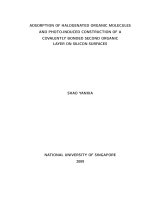
Four different configurations were prepared (Fig. 1). A cellulose
fibril of DP 12 was placed parallel to the z-axis. Next, a number of xylan
chains of DP 6 were placed in the vicinity of the fibril, on average 6.1 Å`
away from the cellulose surface. The first system (denoted S1) has six
xylan chains, where the top three ones are aligned parallel to the cel
lulose fibril and the bottom three are antiparallel. In the second system
(S2) every other chain is parallel. The third system (S3) has three chains
initially placed perpendicular to the fibril, and in the fourth system (S4)
has six perpendicular chains. An additional fifth system (S5) used a
similar arrangement as in S1, except that both cellulose and xylan were
elongated to DP 16 and DP 12, respectively. All systems were fully sol
vated using explicit water.
After energy minimization, all systems were subject to 1 μs of MD at
ambient temperature and pressure allowing the xylans to adsorb freely
to the cellulose surface. The time step was 0.002 ps and energies and
coordinates were saved every 5000 steps. Non-bonded interactions used
a 1.2 nm cutoff and long-range electrostatic interactions were included
using PME (Darden, York, & Pedersen, 1993; Essmann et al., 1995).
Temperature was controlled by the velocity-rescale thermostat (Bussi,
Donadio, & Parrinello, 2007), and pressure by semi-isotropic ParrinelloRahman pressure coupling (Parrinello & Rahman, 1981) with xy- and zaxis compressibilities set to 4.5 × 10− 5 and 4.5 × 10− 6 Pa− 1, respec
tively. Constraints were applied on all bonds using P-LINCS (Hess,
2008).
Simulations of S1 and S5 were extended by 100 ns, saving the co
ordinates every 5 ps to obtain sufficient data for calculating 13C NMR
spin-lattice (T1) relaxation times from
1
nH
= χ 2 (j(ωH − ωC ) + 3j(ωC ) + 6j(ωH + ωC ) )
T1 10 0
(1)
where j(ω) is the spectral density of the P2 rotational autocorrelation
function of the C–H bond vectors at the frequency ω. This is the
appropriate expression for an isotropic system, i.e., where all C–H
orientations are equally probable. Here, a carbon Larmor frequency of
100 MHz was used for comparing with previously published data (Chen,
´, Berglund, & Wohlert, 2019). The constant in front de
Terenzi, Furo
pends on the number of protons bonded to the carbon (nH, which is equal
to one in the case of C4) and on the dipole-dipole coupling constant . In
the present case the numerical value becomes nHχ 02/10=2.3 × 109 s− 2.
Calculations were performed separately for each C4-H4 bond in the
cellulose after which the values were grouped based on their location:
inner surface (meaning that the C6 hydroxymethyl is pointing inwards,
to the crystalline core) or outer surface (meaning that C6 points
2. Methods
The cellulose polymer consists of β-(1→4)-linked D-Glcp residues. In
nature the chains crystallize into fibrils of co-existing crystalline forms:
Iα and Iβ, where the latter dominates in higher plants (Nishiyama,
2009). Current understanding of the dimensions and the organisation of
the cellulose synthesis complex in wood gives an 18-chain fibril as the
most probable one (Cosgrove, 2014), which is also proposed by exper
imental measurements of wood fibril dimensions (Newman, Hill, &
Harris, 2013). However, fibril models containing 24 or 36 chains have
2
E. Heinonen et al.
Carbohydrate Polymers 285 (2022) 119221
A) Xylan DP12
C) Systems
B) Cellulose Iβ
1
2
X2
reducing end
X1
X3
X3
X1
ΦΨ
X6
X4
tg
X6
X4
X5
X5
gt
gg
X2
3
4
X2
110
360°
420°
2-fold
3-fold
X1
X2
200
1-10
Z
reducing end
hydrophilic
X1
X3
X6
X4
hydrophobic
X3
X5
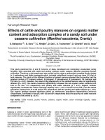
Fig. 1. A) β-(1→4)-xylan, 2-fold helix and 3-fold screw conformations. B) Cellulose I intrasheet hydrogen bonding pattern and dihedral angles of the glycosidic bond,
ϕ (O5’-C1’-O4-C4) and ψ (C1’-O4-C4-C3). Possible conformations of the hydroxymethyl group are trans-gauche (tg), gauche-gauche (gg) and gauche-trans (gt). Crosssection of 18 chain cellulose fibril used in this study, exposing the 110 and 110 (hydrophilic), and 200 (hydrophobic) crystallographic planes. C) Starting points of the
simulations: placement of the xylan chains around the cellulose either pre-aligned (S1 and S2) or perpendicular (S3 and S4). System S5 is similar to S1 except that
xylan chains are extended from DP 6 to DP 12, and the cellulose fibril from DP 12 to DP 16.
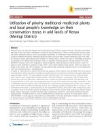
outwards). Reported values are logarithmic averages for each group.
xylan end-to-end distance and projecting it onto the fibril axis. Fig. 3
shows the alignment per xylan chain in each system as a function of
time, where a value of +1 indicates perfect alignment in a parallel
orientation, while − 1 means antiparallel. Generally, most xylan chains
are well aligned most of the time, which is shown by the histogram in
Fig. 3. In S5 fluctuations are small compared to those observed in S1-4.
This is an effect of the chains being twice as long, which reduces the
mobility of the xylans on the cellulose surface and the relative contri
bution from the more mobile chain ends. In S1 and S2 relatively large
fluctuations on the scale of several tens of nanoseconds occur. Xylan X4
in S2 rotated such that it interacted with both the fibril and its periodic
image and is therefore excluded from further analysis. However, it is
curious that this xylan-mediated fibril-fibril connection via xylan was
stable for more than 350 ns. In S3, xylans that were initially oriented
perpendicular to the fibril quickly aligned with the fibril axis whereas in
the more concentrated system (S4) it took longer time before they
settled in a partially aligned state, approximately 80–90% of full
alignment. An explanation for this is the crowding imposed by neigh
boring xylan chains, which is supported by the snapshots shown in
Fig. 2.
3. Results
MD simulations were run on systems with varying initial configu
ration, xylan concentration, and degree of polymerization. The resulting
trajectories were analyzed with respect to xylan orientation and
conformation, cellulose-xylan hydrogen bonding patterns, and finally
the effects on segmental mobility of surface cellulose chains.
3.1. Xylan aligns spontaneously on the cellulose fibril's surface
The systems were characterized with respect to the orientation and
alignment of the xylan polymers, and the effects of initial configuration,
chain length and concentration, respectively. Xylan orientation (paral
lel/antiparallel or perpendicular) and location (hydrophobic or hydro
philic surface) at the beginning and at the end of the simulations are
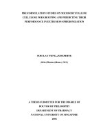
presented in Table 1. Snapshots of systems S1, S4 and S5 (Fig. 2), as well
as S2 and S3 (see Fig. A.1) are used to visualise the migration of xylans
once adsorbed.
The alignment of xylan oligomers was assessed by calculating the
Table 1
Alignment and location of the xylan chains with respect to the cellulose fibril.
3
E. Heinonen et al.
Carbohydrate Polymers 285 (2022) 119221
System 1 (S1)
System 4 (S4)
10 ns
500 ns
1000 ns
1000 ns
System 5 (S5)
10 ns
1000 ns
500 ns
1000 ns
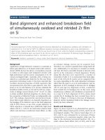
Fig. 2. Snapshots of the systems S1, S4 and S5 at 10, 500 and 1000 ns. The fibril surface is visualised with the VMD drawing method QuickSurf and the water
molecules are excluded for clarity. Blue-red end of xylan marks the non-reducing end. (For interpretation of the references to colour in this figure legend, the reader is
referred to the web version of this article.)
3.2. Xylan prefers antiparallel orientation to cellulose
in 30 cases, and unaligned in 2 cases. Here, as our xylan is unsubstituted
and thus differs from cellulose by just lacking the hydroxymethyl group,
a comparison can be made with the difference between the two cellulose
crystal allomorphs I and II. In cellulose I (native cellulose) all chains are
parallel, but upon alkali treatment it can be irreversibly converted to
cellulose II (Okano & Sarko, 1985; Simon, Glasser, Scheraga, & Manley,
1988), which has been shown to be thermodynamically the most stable
Curiously, all xylan chains in system S3 settled in an antiparallel
orientation. To investigate this behavior further, the simulation was
repeated 30 times (using different random seeds and for 150 ns) to ac
quire more statistics. Of the total 93 xylan chains (31 simulations using 3
chains each), the final orientation was antiparallel in 61 cases, parallel
4
E. Heinonen et al.
Carbohydrate Polymers 285 (2022) 119221
System 2 (S2)
System 3 (S3)
System 4 (S4)
System 5 (S5)
All xylans (S1-S5)
Relative frequency
Pre-aligned xylans
Perpendicular xylans
Pre-aligned xylans
System 1 (S1)
X1
X2
X3
X4
X5
X6
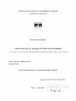
Fig. 3. Time evolution of xylan end-to-end-vector projections on the z-axis (cellulose fibril axis), where +1 means fully parallel and -1 means fully antiparallel.
Systems S1, S2 and S5 initially have xylans pre-aligned, while S3 and S4 initially have xylans perpendicular to the cellulose fibril axis. The xylan numbers are same as
in the Fig. 1. The bottom right panel displays a histogram of normalised absolute projections of all xylans.
allomorph (Goldberg et al., 2015). In this structure, every other chain is
antiparallel. Thus, xylan seems to be no different from cellulose in that
alternating chain polarity is slightly favoured. However, the native
orientation of xylan with respect to the cellulose microfibrils may still be
a consequence of biosynthetic mechanisms during cell wall formation,
rather than equilibrium thermodynamics.
The aspect of preferred orientation of has only seldom been
addressed in the simulation literature. Hanus and Mazeau (2006) found
that the differences in interaction energies in vacuum between parallel,
antiparallel and perpendicular xyloglucan on cellulose surface were
within the calculated error and hence all of them were well tolerated.
Later work on xylan adsorption to cellulose in vacuum indicated that the
parallel orientation would be preferred (Mazeau & Charlier, 2012). In
that work, a single simulation using three xylans in random starting
orientations was performed, and it was found that two out of three
finished parallel and one antiparallel. More recently Falcoz-Vigne et al.
5
E. Heinonen et al.
Carbohydrate Polymers 285 (2022) 119221
(2017) studied the preferred orientation by placing a xylo-oligomer at
either 30, 60 or 90 degree angle to the cellulose surface. Their 10 ns
simulation results show that xylan (DP 10) at 30 and 60 degrees tends to
rotate towards parallel orientation during the 10 ns simulation time,
while the xylan at 90 degrees did not, even after 30 ns. In the present
simulations, using xylans of DP 6, 30 ns is typically enough for achieving
alignment of the xylan chains. It could be that longer oligomers need
longer times to align. Moreover, since the present simulations show that
the statistical preference for antiparallel orientation after 60 trials re
mains fluctuating between 0.4 and 0.5:1 (parallel:antiparallel), any
outcome is probable if predictions are based based on too few obser
vations. Recent studies have concentrated on other aspects of xylancellulose interactions and usually assumed a parallel orientation
(Busse-Wicher et al., 2014; Martnez-Abad et al., 2017; Pereira et al.,
2017).
Martnez-Abad et al., 2017; Pereira et al., 2017), although recently Gupta
et al. (2021) observed a different kind of behavior. In their study an
unsubstituted xylan oligomer remained in 3-fold conformation and even
desorbed from the cellulose surface. This is possibly related to the choice
of interaction potentials, which will be addressed further down.
A)
X6
3.3. Xylan adopts a 2-fold conformation when adsorbed/aligned onto
cellulose surfaces
One of the most important structural features of polysaccharides is
the variation in how consecutive sugar residues are connected by the
glycosidic linkages. This largely determines the conformational space of
the polysaccharides (Varki, 2017). The β-(1→4)-linked backbone in
xylan in combination with the lack of an hydroxymethyl group on C5
makes an extended, twisted 3-fold screw the lowest energy conforma
tion in solution. However, a pseudo-flat 2-fold conformation is ener
getically allowed, and the barrier between the two conformations is
sufficiently low to allow inter-conversion between the two in equilib
rium (Berglund et al., 2016). In simulations, when xylan adsorbs to a flat
surface such as cellulose, the 2-fold conformation becomes the dominant
one, since it will maximize the specific interactions between the xylan
and the substrate (Busse-Wicher et al., 2016; Martnez-Abad et al., 2017).
This conformational change was also experimentally observed in solid
state CP/MAS 13C NMR (Simmons et al., 2016; Teleman et al., 2001).
It has been shown that the sum of the two dihedral angles ϕ and ψ is a
good indicator of the polysaccharide chain conformation (French &
Johnson, 2009). Thus, the conformational space can be reduced to just
one dimension. Here, ϕ and ψ were defined by the sequences O5’-C1’O4-C4 and C1’-O4-C4-C3, respectively. In this representation, a sum of
300◦ corresponds to a right-handed 3-fold helix, 360◦ to a 2-fold screw,
and 420◦ to a left-handed 3-fold helix (see Fig. 1 for the xylan confor
mations and Fig. 5 for examples of conformational plots, remaining
conformational plots are shown in Fig. A.2-A.6).
Histograms of ϕ + ψ collected from the simulations compared to a
histogram from a simulation of a free xylan in solution (Fig. 4) confirms
that unsubstituted xylan prefers the 2-fold conformation. This is in
agreement with previous MD simulations (Busse-Wicher et al., 2016;
B)
X4
Fig. 5. Example conformational plots of 3rd the glycosidic linkage of the xylan
xylan X6 and X4 (S4), where the diagonals indicate the sum of φ and ψ . 300◦
corresponds to a right-handed 3-fold helix, 360◦ to a 2-fold screw, and 420◦ to a
left-handed 3-fold helix.
Fig. 4. Left: Histograms of the sum of and from all simulations combined. Adsorbed xylan (yellow) is compared to a reference in water (magenta). Middle and right:
Xylan chains in S4 split in two groups based on the equilibrium projection length (see text Fig. 3). (For interpretation of the references to colour in this figure legend,
the reader is referred to the web version of this article.)
6
E. Heinonen et al.
Carbohydrate Polymers 285 (2022) 119221
A)
Antiparallel
Parallel
B)
C)
D)
Fig. 6. Short-range xylan-cellulose interactions in S3. Xylan chains are grouped based on (A) orientation (parallel/antiparallel) only, or (B–D) both orientation and
the closest cellulose surface. Xylan chains that could not unambiguously be assigned to either the hydrophilic or the hydrophopic surface were excluded. Panels (A)
and (B) show the total non-bonded energy while (C) displays Coulomb interactions, and (D) the Lennard-Jones (LJ) interactions.
Also histograms pertaining to adsorbed xylans displays a minor peak
around 440◦ that corresponds to a left-handed 3-fold conformation. To
investigate the origin of this peak, separate histograms of fully and
partially aligned xylan oligomers were constructed based on their
projections. For S4, full alignment correlates well with the 2-fold
conformation, and the 3-fold peak is suppressed when partially
aligned xylans are removed from the analysis. Instead, the distribution
of partially aligned xylans from the S4 overlaps perfectly with the
7
E. Heinonen et al.
Carbohydrate Polymers 285 (2022) 119221
H-bonds/10 xylose residues
A)
10
8
6
4
2
0
1
X2-G2
2
X2-G3
3
X2-G6
X3-G2
4
X3-G3
5
X3-G6
B)
H-bonds/ 10 residues
5
donor (OH)
acceptor (O)
4
3
2
1
0
X2-G2 X2-G3 X2-G6 X3-G2 X3-G3 X3-G6 X2-G2 X2-G3 X2-G6 X3-G2 X3-G3 X3-G6
parallel
antiparallel
C)
OH2
OH3
OH6
Xylan parallel
Glc
Xyl
Xylan antiparallel
Network A
Glc
Xyl
Network B
Glc
Xyl
Fig. 7. A) Average number of hydrogen bonds in each system per 10 xylose residues (Note: chain end residues excluded). Hydroxyl groups in xylose and glucose
residues are denoted with X and G, respectively, followed by the carbon number to which they are attached. B) Average number of hydrogen bonds in system S5 per
10 xylose residues located on hydrophilic surfaces, for parallel and antiparallel orientation separately (Note: chain end residues excluded). C) Snapshots of system S5
illustrating the hydrogen bonding patterns of parallel and antiparallel xylan.
reference. This indicates that xylans may indeed form stable complexes
with cellulose while retaining the 3-fold conformation, at least within
the time scale of these simulations.
In S5 the xylans were quite immobile and the 3-fold conformation
arises mainly from individual linkages that makes one of the chains (X4)
to bend on the cellulose surface (Fig. 2), which results in alternating
sections of 2- and 3-fold conformation. This is an curious observation
considering that recently Simmons et al. (2016) detected a minor
8
E. Heinonen et al.
Carbohydrate Polymers 285 (2022) 119221
fraction of 3-fold xylan in a wild type Arabidopsis, but only as a relatively
immobile component in CP-INADEQUATE spectra, not as a mobile
component in DP-INADEQUATE. Furthermore, their study showed that
the 3-fold xylan in the wild-type had higher dipolar order parameter
compared to the major 3-fold fraction in the cellulose deficient mutant.
Similarly, the 3-fold xylan of the wild type had 13C NMR T1-relaxation
time in same range with 2-fold xylan and cellulose. These observations
suggest that even 3-fold xylan in the secondary cell wall can be relatively
rigid, and perhaps close to cellulose. Thus, the presence of a minor 3-fold
fraction could indicate occasional bends in otherwise tightly bound
xylan, either in the middle of the polymer or at the chain ends.
network B reminds more that of the parallel xylan with G6 participating
in the H-bonding patterns: the two most common bonds being X2-G2
and X3-G6. These results show that a regular hydrogen bonding
pattern is observed between each xylan-cellulose pair, although the type
of the pattern may vary depending on the xylan orientation. It is inter
esting to note that the statistically more likely antiparallel orientation
also leads to the more extensive hydrogen bond pattern, possibly indi
cating a physical mechanism for this preference. However, the anti
parallel orientation is preferred also on the hydrophobic surfaces of the
cellulose microfibril where no hydrogen bonds are formed, pointing to a
more complex selection process.
To investigate this further, the short-range non-bonded energy be
tween xylan and cellulose in the multiple repeats of S3 were extracted
from the final 50 ns of the simulations and displayed as histograms based
on their respective orientation (Fig. 6). This quantity is a measure of the
proximity between xylan chains and the cellulose surface and should not
be interpreted as a binding energy. The distributions for antiparallel
chains are shifted towards lower energies compared to the parallel ones.
This indicates a more effective packing between the xylan and the cel
lulose in the former case. In both cases the energy is more negative for
chains adsorbed to hydrophilic surfaces compared to hydrophobic ones.
However, the fact that the binding has been shown to be consistently
stronger to the hydrophobic surfaces (Martnez-Abad et al., 2017) shows
the importance of other interaction terms, especially those mediated by
water. When the energy is further decomposed into contributions from
Coulomb and dispersion (Lennard-Jones) interactions, one finds, not
surprisingly, that the Coulomb energy dominates the short-range in
teractions at the hydrophilic surfaces, presumably a consequence of
forming hydrogen bonds, while dispersion dominates at the hydropho
bic surfaces where no hydrogen bonds are formed. The present analysis
shows that an antiparallel orientation leads to tighter interactions be
tween xylan and cellulose, which tentatively could contribute to the
statistical preference of that orientation over a parallel one. However,
since the difference arises in the already adsorbed state and no inter
conversion between orientations occurs (at least on MD timescales), the
actual selection process is likely governed by more long-ranged in
teractions, such as the molecular dipole moments of the individual
polymers.
3.4. Hydrogen bonding of xylan is orientation dependent
When xylan adsorbs to cellulose, hydrogen bonds (H-bonds) are
formed between the polymer and the substrate. However, H-bonding is
not required neither to drive the adsorption, nor for the xylan to adopt a
2-fold conformation, since both adsorption and a shift from 3-fold to 2fold occurs also on the hydrophobic surface. On the other hand, Hbonding does to some extent define the crystal lattice of cellulose, and
since it has been suggested that adsorbed xylan could extend the crys
talline structure, it is of interest to investigate the H-bonding charac
teristics of the xylan-cellulose systems. To that end, H-bonds between
the xylan and cellulose, for those chains adsorbed to hydrophilic surfaces,
were analyzed using standard geometric criteria: a cutoff angle of 30◦
and a cutoff distance of 3.5 Å. The first 100 ns of each simulation are
excluded from the analysis. Results are presented in Fig. 7. In the
following, specific hydroxyl groups are denoted by either X or G indi
cating whether they belong to xylan or cellulose residues, followed by
their respective carbon number.
The average number of interchain H-bonds varied between 7.5 and
10.4 per 10 xylosyl residues (Fig. 7A). This makes sense, since the xylans
adsorb on the cellulose surface primarily in 2-fold conformation, hence
every other residue has X2 and X3 directed towards the cellulose and
apparently at least one of them is participating in H-bonding. X2 is
somewhat more likely to participate in intermolecular H-bonding
compared to X3 in systems S1–3 and S5, while in S4, X2 and X3 both
participate in, on average, 4.6 and 4.2H-bonds, respectively. This dif
ference can be explained by the tendency of X3 to form an intrachain Hbond to O5 of the preceding xylose residue.
Next, the effect of xylan orientation on H-bonding pattern was
analyzed using data from S5 where chains are well aligned but not
forming aggregates, thus only forming H-bonds to cellulose and not to
neighboring xylans. There is a remarkable difference in the number of Hbonds depending on orientation: parallel xylan forms on average five Hbond to cellulose per 10 xylosyl residues while antiparallel forms nine.
This agrees with the visual observation that parallel xylans generally
form one bond per every other residue, whereas antiparallel xylans gives
a more complex pattern that often involves both of the free hydroxyls,
X2 and X3 (Fig. 7C).
Parallel xylan preferably forms a regular network of X2-G6 bonds
with a minor fraction of X3-G6 (Fig. 6B), which is in perfect agreement
with Busse-Wicher et al. (2014). In this pattern, X2 may be either donor
or acceptor, while X3 almost solely functions as an acceptor. This pattern
is reminiscent of that found in native cellulose (Nishiyama et al., 2002),
but not identical since xylan lacks the hydroxymethyl group.
Antiparallel xylan exhibit a more complex pattern. Of all possible Hbond combinations, only X3-G3 is rare. H-bonds are formed equally
likely through X2 and X3, often simultaneously. The most common Hbonds are X2-G3 and X3-G2 followed by X2-G6 and X3-G6. Again, X3
usually participates as an acceptor, which means that X2 in X2-G3, and
G2 in X3-G2, typically act as donors. On the other hand, X2-G2 and X2G6 may be formed either way. Visual observation reveals two kinds of Hbonding networks, here denoted A and B. Network A is characterized by
hydrogen bond pairs of X2-G3 and X3-G2, with G6 hardly participating
at all but rather pointing away in a gg conformation. On the contrary,
3.5. Adsorption of xylan as aggregates decreases the molecular mobility of
adjacent glucose residues
Previously Terenzi et al. (2015) have shown using 13C CP/MAS NMR
T1 relaxation times that the mobility of surface carbons in a hydrated
CNF/xyloglucan (XG) composite were significantly lower compared to
what would be expected from the weight averages of the individual
relaxation times of the same carbons in pure CNF and XG. They assigned
this to a decrease in XG dynamics due to the restrictions imposed by the
CNF interface, but also speculated whether the adsorbed XG could affect
the dynamics of cellulose surface chains. Here, such effects are investi
gated for the case of adsorbed xylan. T1 relaxation times can be calcu
lated from MD-simulations from the Fourier transform of the rotational
autocorrelation functions of specific C–H bond vectors, yielding com
parable results with experimental ones (Chen et al., 2019). The present
investigation concentrates on the dynamics of C4-H4 since the splitting
of the C4 peak in 13C NMR is commonly interpreted to arise from the
rigid core and more mobile surface/amorphous regions, respectively,
and to correlate with the hydroxymethyl conformation (Bardet, Emsley,
& Vincendon, 1997). To this end, systems S1 and S5 were simulated for
an additional 100 ns starting from the end of the 1 μs simulations. From
these simulations T1 relaxation times were calculated as described
above. The results were compared to reference simulations performed of
the same CNF models, but without xylan present. For the analysis, the
cellulose chains were divided into core chains, as well as hydrophobic
and hydrophilic surfaces. Individual residues of the hydrophilic surface
chains were further divided into hydrophilic (inner), having the
9
E. Heinonen et al.
Carbohydrate Polymers 285 (2022) 119221
hydroxymethyl group pointing into the crystal, and hydrophilic (outer),
having the hydroxymethyl group exposed to the xylan and/or water.
Average T1 values are presented in Table 2, where longer relaxation time
indicates more restricted molecular dynamics. The upper part of Table 2
shows averages of all glucose residues of both the xylan-cellulose ag
gregates and their respective references. In general, reference values
agrees with those previously reported using the same force field (Chen
et al., 2019): C4 relaxation times in hydrophilic (outer) are about half of
that in hydrophilic (inner), and within 35–42 s. Residues in hydrophobic
surfaces are more rigid compared to those in hydrophilic surfaces, and
overall relaxation times are 10–40 s higher in the xylan-cellulose sys
tems compared to the references.
In order to isolate possible effects from adsorbed xylan oligomers,
relaxation times in residues directly adjacent to adsorbed xylans (and
the corresponding residues of the reference) were calculated. This shows
that in S1 the T1 values of hydrophilic (outer) in contact with xylans in
crease from 30 to 121 s, compared to the same residues in the reference
system, clearly showing that adsorbed xylan has a direct effect on cel
lulose mobility. The corresponding increase in S5 is smaller; from 42 to
55 s. This was actually quite surprising since one would think that the
longer xylan chains in S5 would bind more stably to the cellulose and
therefor would confine the motions of adjacent glucoses to a larger
extent. However, while the longer xylans were indeed relatively
immobile on the cellulose surface, the shorter ones migrated to form
aggregates. This suggests that a certain amount of continuous surface
coverage is required to convert the mobile surface glucose residues into
more core-like.
Recently, it was proposed based on solid state NMR studies on native
Arabidopsis that xylan may induce a conformational change of the cel
lulose hydroxymethyl groups from the gt and gg conformations domi
nating in surfaces to tg, which is the dominant conformation in the
crystalline core, thus effectively make surface chains more crystal-like
(Dupree et al., 2015; Terrett et al., 2019). To investigate whether the
xylan adsorption affects the C6OH conformation, probability distribu
tions of the torsion angle ω of surface residues in S1 and S5 (and cor
responding references) were determined (Fig. B.1-B.4). As expected,
hydrophilic (inner) groups display very narrow distributions of a single
conformation, mainly tg, while distributions for hydrophilic (outer)
groups exposed to water are bi- and trimodal, meaning that they rotate
between states more often. For hydrophilic (outer) residues close to xylan,
there is no visible increase in tg, but rather a shift away from it to either
gt or gg. On the other hand, when compared to the reference, the peak
pertaining to the most probable conformation is amplified, indicating
that the time spent in that conformation increases, i.e., the motion
around the C5-C6 bond slows down. This is consistent with the forma
tion of regular H-bonding network, although that network is different
from that in native cellulose Iβ. However, from the perspective of cel
lulose mobility this appears to be of no consequence since the response
in T1 relaxation times clearly shows that these chains become crystallike regardless of H-bonding pattern. However, one cannot rule out
the possibility of the hydroxymethyls eventually converting to tg based
on these simulations alone. It is possible that significantly longer
simulations are required to observe it.
4. Discussion
Cellulose and hemicelluloses are the major structural components of
the plant cell wall and their molecular level association affects the
properties of the cell wall as whole. Cellulose has a straight and flat
structure due to the β-(1 → 4)-glycosidic bonds in two-fold conformation
and equatorially oriented hydroxyls at positions one and four in the
glucose β-pyranoside residue (Nishiyama, 2009). Xylans and gluco
mannans share these molecular features and can therefore adapt to the
cellulose structure, but also to more helical conformations (Berglund
et al., 2016; Busse-Wicher et al., 2014; Mazeau et al., 2005). This is
hardly a coincidence; one biological role of the non-cellulosic cell wall
polysaccharides is to bind the cellulose fibrils together through a less
crystalline and a more flexible phase, forming a composite material with
distinct biomechanical properties (Berglund et al., 2020). An ability to
adapt to different conformations seems to fulfill this function. Indeed,
the data in this work is in line with a view that xylans can form an
extension of the cellulose crystal structure that can function as a tran
sition phase both between rigid crystalline microfibrils, and to more
flexible polysaccharide phases and amorphous lignin. While MD simu
lations can indicate what type of molecular organizations are possible,
to this day, it is not known with certainty how the cellulose and hemi
celluloses are deposited in the cell wall of a growing tree.
Possible interactions between cellulose- and hemicellulose chains
include hydrogen bonding, van der Waals dispersion forces and hydro
phobic forces, which partially can be understood as the increase in
solvent entropy from excluding water molecules from the cellulose
surface (Chandler, 2005). The role of hydrogen bonding has often been
exaggerated in the field of cellulosics (Wohlert et al., 2021), but a recent
MD-study by Kishani, Benselfelt, Wågberg, and Wohlert (2021) on the
hemicellulose xyloglucan, suggested that the hydrophobic forces were
central for adsorption on cellulose. In addition, Nishiyama (2018)
showed that the cohesive energy of crystalline cellulose is dominated by
dispersion interactions. Yet, hydrogen bonds may still play an important
role in the formation of xylan-cellulose complexes: after the adsorption,
alignment and aggregation, a regular hydrogen bonding network can be
formed, and once formed, the interactions may be strong enough to
stabilize the chain conformation of xylan.
Based on polarized FTIR, Stevanic and Salm´
en (2009) and Olsson,
Bjurhager, Gerber, Sundberg, and Salm´en (2011) have suggested that
xylan is oriented parallel to the cellulose fibrils, while results presented
here show a preference for the antiparallel orientation. It is interesting if
both cellulose and xylan exhibit the same tendency for parallel organi
sation despite antiparallel being energetically more stable. The biosyn
thetic process seems to be the reason for the parallel oriented β-glucans
within a cellulose crystal: the individual chains are built in the cellulose
synthesizing complex (CSC) by sequentially adding new glucose units to
the reducing end of the chains, which then immediately coalesce
forming cellulose I (Cosgrove, 2014). However, antiparallel packing of
polysaccharides does occur in nature as the currently accepted crystal
unit cell of α-chitin consists of two antiparallel chains in a 2-fold
conformation (Ogawa, Lee, Nishiyama, & Kim, 2016; Sikorski, Hori, &
Wada, 2009). For xylan, more studies are needed on its biological as
sembly in the secondary cell wall to elucidate if there is a preferred
orientation, how it is controlled and whether it is similar throughout the
cell wall layers and the cell types.
One thing that seems increasingly certain, based on solid state NMRstudies (Dupree et al., 2015; Terrett et al., 2019) and the simulations
presented herein, is that the close association of 2-fold xylan to cellulose
microfibrils restricts the mobility of the surface polymers when quan
tified by 13C CP/MAS NMR T1 relaxation times. One possible reason for
this could be the exclusion of water from the cellulose surface upon
adsorption of a xylan aggregates, which causes a local decrease in the
mobility of cellulose, but the crowded environment imposed by the
Table 2
13
C NMR T1-relaxation times, logarithmic means in seconds.
S1
Reference CNF
S5
Reference CNF
All Glc
Hydrophobic surface
Hydrophilic (inner)
Hydrophilic (outer)
122
82
50
99
78
40
135
76
47
94
60
37
Glc next to Xylan
Hydrophobic surface
Hydrophilic (inner)
Hydrophilic (outer)
97
135
121
65
132
38
144
116
55
106
93
42
10
E. Heinonen et al.
Carbohydrate Polymers 285 (2022) 119221
adsorbed xylan chains themselves can also be expected to have an effect.
While it is difficult to determine how much of the fibril surfaces are
covered by xylan from NMR, here it was shown that in order have an
appreciable effect on the cellulose mobility, xylans should rather form a
continuous coverage than be sparsely distributed over the surface. In
this case, relaxation times becomes similar to those of the crystalline
core.
It is still a matter of debate how cellulose microfibrils are covered by
hemicelluloses. Among other factors, it depends on the cross-section of
the fibrils, which determines the type of surfaces that are exposed. In this
work an interesting tendency of the short xylan chains to migrate and
form aggregates close to the hydrophobic surfaces of the cellulose mi
crofibrils was observed. As a consequence, cellulose in the presence of
xylan might have its more hydrophobic parts preferentially covered,
which would affect cellulose in many aspects, such as water interaction
and aggregation, and in turn properties of the cell wall as such. The exact
structure of the cross-section of cellulose crystal in higher plants is not
known with certainty, but if hydrophobic surfaces are present, as in the
model used in this study, it seems likely that hemicelluloses can bind to
them in a stable way, and even with some preference over the hydro
philic surfaces.
How closely cellulose and xylan are associated possibly depends on
the level of the hydration of the cell wall. It is well known that the upon
drying, irreversible loss of swelling capacity occurs, and that the effect is
larger the more hemicelluloses are removed (Scallan & Laivin, 1993).
Recently Cresswell et al. (2021) showed by solid state NMR that some of
this loss seems to be due a removal of water found in between xylan and
cellulose, and subsequent adsorption of xylan to cellulose surface.
Thomas et al. (2020) and later Cresswell et al. (2021) have suggested
that some of the water would be accommodated in the void left by
missing C6OH group in xylan. In the present study, such confined water
molecules could indeed be found in the simulation trajectories, but only
rarely and for very short times (less than one nanosecond).
On a final note it is worth mentioning that results from MD simula
tions of cellulose/hemicellulose systems appear to be strongly affected
by the choice of potential parameters, i.e., the force field. In the present
work with the GLYCAM06 force field, all xylan oligomers eventually
bind to the cellulose fibril, and although some reorganization occurs on
the surface, especially for the shorter ones, they remain stably bound for
the remainder of the simulations with a major portion of the glycosidic
linkages in two-fold conformation. In a recent study by Gupta et al.
(2021) of xylan adsorption to a model cellulose surface using the
CHARMM36 force field it was found that unsubstituted xylan displayed
a smaller fraction of two-fold conformations compared to substituted
ones, indicating weaker interaction with cellulose, which, in some cases,
lead to the xylan chains desorbing. In yet another study by Falcoz-Vigne
et al. (2017) using the GROMOS56 carbohydrate force field, unsub
stituted xylan chains also adsorbed in a three-fold conformation, but still
remained stably bound. In cases where the xylan oligomer was initially
put in place in two-fold conformation, as opposed to spontaneous
adsorption, the two-fold screw has been observed to be stable even for
unsubstituted xylans with both GROMOS (Falcoz-Vigne et al., 2017) and
CHARMM (Cresswell et al., 2021), although in the latter case positional
restraints on the cellulose polymers were applied. Thus, the details of the
conformational behavior observed in these MD simulations appears to
be influenced by a delicate balance of interaction parameters. As one
example, the partial charge assigned to hydroxyl oxygen atoms is typi
cally − 0.1 e larger in GLYCAM06 compared to both CHARMM and
GROMOS, leading to a larger dipole moment of the hydroxyl groups.
Such differences could certainly influence the micro-scale structure and
presently it is not possible to say which parameter set represents the
xylan-cellulose system in the most realistic way.
different relative orientations show spontaneous adsorption, alignment
and conformational reorganization of xylan in the presence of cellulose
microfibrils. Regardless of the initial orientation, xylan adopts a
cellulose-like 2-fold conformation upon adsorption. Short oligomers
tend to migrate towards the hydrophobic surfaces and form stable ag
gregates, while longer ones are more immobile. Based on multiple in
dependent simulations starting from an initial perpendicular orientation
of the xylan chains with respect to the fibril axis, the results show that
the antiparallel orientation is thermodynamically more stable than the
parallel one, similar to cellulose II compared to the cellulose I. On the
hydrophilic surface, xylan forms regular H-bonding networks to cellu
lose that depends on its orientation, with an antiparallel orientation
leading to a more complex H-bonding pattern compared to a parallel
orientation. Finally, surface glucose residues that are covered by xylan
aggregates become less mobile and their 13C CP/MAS NMR T1 relaxation
times increase compared to the values typical of the crystalline core,
although no conversion of the hydroxymethyl group conformation from
gt/gg to tg is observed.
The data in this work agrees with a model where xylan forms a rigid
layer functioning as a transition phase from the crystalline cellulose fi
brils to more flexible domains containing disordered polysaccharides
and lignin. This study adds to the detailed molecular level understand
ing on how xylan interacts with cellulose fibrils, which is of large
importance for the construction of accurate plant secondary cell wall
models, considering that xylan is the main hemicellulose in hardwoods
and grasses and forms a significant part of softwoods. These structural
models contribute not only to a better understanding of the biological
function of xylan as a fundamental structural component in the plant cell
wall, but also to the implementation of efficient technologies for over
coming biomass recalcitrance, towards the development of a new gen
eration of bio based chemicals and materials from lignocellulose.
Funding
This work was supported by the Knut and Alice Wallenberg Foun
dation through the Wallenberg Wood Science Centre; The Swedish
Research Council (VR) [grant number 2020-04720] (F.V.).
CRediT authorship contribution statement
Emilia Heinonen: Methodology, Formal analysis, Investigation,
Writing – original draft. Gunnar Henriksson: Conceptualization,
ă m: Conceptualization,
Writing review & editing. Mikael E. Lindstro
Writing – review & editing. Francisco Vilaplana: Conceptualization,
Methodology, Writing – review & editing. Jakob Wohlert: Conceptu
alization, Methodology, Writing – review & editing.
Declaration of competing interest
The authors declare that they have no known competing financial
interests or personal relationships that could have appeared to influence
the work reported in this paper.
Appendix A. Supplementary data
Supplementary data to this article can be found online at https://doi.
org/10.1016/j.carbpol.2022.119221.
References
Abraham, M. J., Murtola, T., Schulz, R., P´
all, S., Smith, J. C., Hess, B., & Lindahl, E.
(2015). GROMACS: High performance molecular simulations through multi-level
parallelism from laptops to supercomputers. SoftwareX, 1–2, 19–25. />10.1016/j.softx.2015.06.001
Andrewartha, K. A., Phillips, D. R., & Stone, B. A. (1979). Solution properties of wheatflour arabinoxylans and enzymically modified arabinoxylans. Carbohydrate Research,
77, 191–204. />
5. Conclusions
Molecular dynamics simulations of xylan and cellulose starting from
11
E. Heinonen et al.
Carbohydrate Polymers 285 (2022) 119221
Bardet, M., Emsley, L., & Vincendon, M. (1997). Two-dimensional spin-exchange solidstate NMR studies of 13c-enriched wood. Solid State Nuclear Magnetic Resonance, 8,
25–32. />Berglund, J., dOrtoli, T. A., Vilaplana, F., Widmalm, G., Bergenstråhle-Wohlert, M.,
Lawoko, M., Henriksson, G., Lindstră
om, M., & Wohlert, J. (2016). A molecular
dynamics study of the effect of glycosidic linkage type in the hemicellulose backbone
on the molecular chain flexibility. The Plant Journal, 88, 56–70. />10.1111/tpj.13259
Berglund, J., Mikkelsen, D., Flanagan, B. M., Dhital, S., Gaunitz, S., Henriksson, G.,
Lindstră
om, M. E., Yakubov, G. E., Gidley, M. J., & Vilaplana, F. (2020). Wood
hemicelluloses exert distinct biomechanical contributions to cellulose fibrillar
networks. Nature Communications, 11. />Bernardi, A., Faller, R., Reith, D., & Kirschner, K. N. (2019). ACPYPE update for
nonuniform 1–4 scale factors: Conversion of the GLYCAM06 force field from AMBER
to GROMACS. SoftwareX, 10, Article 100241. />softx.2019.100241
Bosmans, T. J., St´ep´
an, A. M., Toriz, G., Renneckar, S., Karabulut, E., Wågberg, L., &
Gatenholm, P. (2014). Assembly of debranched xylan from solution and on
nanocellulosic surfaces. Biomacromolecules, 15, 924–930. />bm4017868
Busse-Wicher, M., Gomes, T. C. F., Tryfona, T., Nikolovski, N., Stott, K., Grantham, N. J.,
Bolam, D. N., Skaf, M. S., & Dupree, P. (2014). The pattern of xylan acetylation
suggests xylan may interact with cellulose microfibrils as a twofold helical screw in
the secondary plant cell wall of arabidopsis thaliana. The Plant Journal, 79, 492–506.
/>Busse-Wicher, M., Li, A., Silveira, R. L., Pereira, C. S., Tryfona, T., Gomes, T. C.,
Skaf, M. S., & Dupree, P. (2016). Evolution of xylan substitution patterns in
gymnosperms and angiosperms: Implications for xylan interaction with cellulose.
Plant Physiology, 171, 2418–2431. />Bussi, G., Donadio, D., & Parrinello, M. (2007). Canonical sampling through velocity
rescaling. The Journal of Chemical Physics, 126, Article 014101. />10.1063/1.2408420
Case, D. A., Ben-Shalom, I. Y., Brozell, S. R., Cerutti, D. S., Cheatham, T. E.,
Cruzeiro, V. W. D., Darden, T. A., Duke, R. E., Ghoreishi, D., Giambasu, G., Giese, T.,
Gilson, M. K., Gohlke, H., Goetz, A. W., Greene, D., Harris, R., Homeyer, N.,
Huang, Y., Izadi, S., … Kollman, P. A. (2019). Amber 2019. San Francisco: University
of California.
Chandler, D. (2005). Interfaces and the driving force of hydrophobic assembly. Nature,
437, 640–647. />Chen, P., Terenzi, C., Fur´
o, I., Berglund, L. A., & Wohlert, J. (2019). Quantifying localized
macromolecular dynamics within hydrated cellulose fibril aggregates.
Macromolecules, 52, 7278–7288. />Chimphango, A. F., Gă
orgens, J., & van Zyl, W. (2016). In situ enzyme aided adsorption of
soluble xylan biopolymers onto cellulosic material. Carbohydrate Polymers, 143,
172–178. />Cosgrove, D. J. (2014). Re-constructing our models of cellulose and primary cell wall
assembly. Current Opinion in Plant Biology, 22, 122–131. />pbi.2014.11.001
Cresswell, R., Dupree, R., Brown, S. P., Pereira, C. S., Skaf, M. S., Sorieul, M., Dupree, P.,
& Hill, S. (2021). Importance of water in maintaining softwood secondary cell wall
nanostructure. Biomacromolecules, 22, 4669–4680. />biomac.1c00937
Crowe, J. D., Hao, P., Pattathil, S., Pan, H., Ding, S.-Y., Hodge, D. B., & Jensen, J. K.
(2021). Xylan is critical for proper bundling and alignment of cellulose microfibrils
in plant secondary cell walls. Frontiers in Plant Science, 12. />fpls.2021.737690
da Silva, A. W. S., & Vranken, W. F. (2012). ACPYPE - AnteChamber PYthon parser
interfacE. BMC Research Notes, 5, 367. />Darden, T., York, D., & Pedersen, L. (1993). Particle mesh ewald: AnNlog(N) method for
Ewald sums in large systems. The Journal of Chemical Physics, 98, 10089–10092.
/>Donev, E., Gandla, M. L., Jă
onsson, L. J., & Mellerowicz, E. J. (2018). Engineering noncellulosic polysaccharides of wood for the biorefinery. Frontiers in Plant Science, 9.
/>Dupree, R., Simmons, T. J., Mortimer, J. C., Patel, D., Iuga, D., Brown, S. P., & Dupree, P.
(2015). Probing the molecular architecture of arabidopsis thaliana secondary cell
walls using two- and three-dimensional 13c solid state nuclear magnetic resonance
spectroscopy. Biochemistry, 54, 2335–2345. />Ebringerov´
a, A., Hrom´
adkov´
a, Z., & Heinze, T. (2005). In I. In Polysaccharides (Ed.),
Hemicellulose (pp. 1–67). Springer-Verlag. />Essmann, U., Perera, L., Berkowitz, M. L., Darden, T., Lee, H., & Pedersen, L. G. (1995).
A smooth particle mesh Ewald method. The Journal of Chemical Physics, 103,
8577–8593. />Falcoz-Vigne, L., Ogawa, Y., Molina-Boisseau, S., Nishiyama, Y., Meyer, V., PetitConil, M., Mazeau, K., & Heux, L. (2017). Quantification of a tightly adsorbed
monolayer of xylan on cellulose surface. Cellulose, 24, 3725–3739. />10.1007/s10570-017-1401-z
Fernandes, A. N., Thomas, L. H., Altaner, C. M., Callow, P., Forsyth, V. T.,
Apperley, D. C., Kennedy, C. J., & Jarvis, M. C. (2011). Nanostructure of cellulose
microfibrils in spruce wood. Proceedings of the National Academy of Sciences, 108,
E1195–E1203. />French, A. D., & Johnson, G. P. (2009). Cellulose and the twofold screw axis: Modeling
and experimental arguments. Cellulose, 16, 959–973. />s10570-009-9347-4
Goldberg, R. N., Schliesser, J., Mittal, A., Decker, S. R., Santos, A. F. L. O. M.,
Freitas, V. L. S., Urbas, A., Lang, B. E., Heiss, C., da Silva, M. D. M. C. R.,
Woodfield, B. F., Katahira, R., Wang, W., & Johnson, D. K. (2015). A thermodynamic
investigation of the cellulose allomorphs: Cellulose(am), cellulose iβ(cr), cellulose II
(cr), and cellulose III(cr). The Journal of Chemical Thermodynamics, 81, 184–226.
/>Gomes, T. C. F., & Skaf, M. S. (2012). Cellulose-builder: A toolkit for building crystalline
structures of cellulose. Journal of Computational Chemistry, 33, 1338–1346. https://
doi.org/10.1002/jcc.22959
Gupta, M., Rawal, T. B., Dupree, P., Smith, J. C., & Petridis, L. (2021). Spontaneous
rearrangement of acetylated xylan on hydrophilic cellulose surfaces. Cellulose, 28,
3327–3345. />Hanus, J., & Mazeau, K. (2006). The xyloglucan–cellulose assembly at the atomic scale.
Biopolymers, 82, 59–73. />Hess, B. (2008). P-LINCS: A parallel linear constraint solver for molecular simulation.
Journal of Chemical Theory and Computation, 4, 116–122. />ct700200b
Hess, B., Kutzner, C., van der Spoel, D., & Lindahl, E. (2008). GROMACS 4: Algorithms
for highly efficient, load-balanced, and scalable molecular simulation. Journal of
Chemical Theory and Computation, 4, 435–447. />Jorgensen, W. L., Chandrasekhar, J., Madura, J. D., Impey, R. W., & Klein, M. L. (1983).
Comparison of simple potential functions for simulating liquid water. The Journal of
Chemical Physics, 79, 926–935. />Kabel, M. A., van den Borne, H., Vincken, J.-P., Voragen, A. G., & Schols, H. A. (2007).
Structural differences of xylans affect their interaction with cellulose. Carbohydrate
Polymers, 69, 94–105. />Kirschner, K. N., Yongye, A. B., Tschampel, S. M., Gonz´
alez-Outeiri˜
no, J., Daniels, C. R.,
Foley, B. L., & Woods, R. J. (2008). GLYCAM06: A generalizable biomolecular force
field. Carbohydrates. Journal of Computational Chemistry, 29, 622–655. https://doi.
org/10.1002/jcc.20820
Kishani, S., Benselfelt, T., Wågberg, L., & Wohlert, J. (2021). Entropy drives the
adsorption of xyloglucan to cellulose surfaces – a molecular dynamics study. Journal
of Colloid and Interface Science, 588, 485–493. />jcis.2020.12.113
Linder, Å., Bergman, R., Bodin, A., & Gatenholm, P. (2003). Mechanism of assembly of
xylan onto cellulose surfaces. Langmuir, 19, 5072–5077. />la0341355
Martnez-Abad, A., Berglund, J., Toriz, G., Gatenholm, P., Henriksson, G., Lindstră
om, M.,
Wohlert, J., & Vilaplana, F. (2017). Regular motifs in xylan modulate molecular
flexibility and interactions with cellulose surfaces. Plant Physiology, 175, 1579–1592.
/>Martnez-Abad, A., Giummarella, N., Lawoko, M., & Vilaplana, F. (2018). Differences in
extractability under subcritical water reveal interconnected hemicellulose and lignin
recalcitrance in birch hardwoods. Green Chemistry, 20, 2534–2546. />10.1039/c8gc00385h
Mazeau, K., & Charlier, L. (2012). The molecular basis of the adsorption of xylans on
cellulose surface. Cellulose, 19, 337–349. />Mazeau, K., Moine, C., Krausz, P., & Gloaguen, V. (2005). Conformational analysis of
xylan chains. Carbohydrate Research, 340, 2752–2760. />carres.2005.09.023
Meng, X., & Ragauskas, A. J. (2014). Recent advances in understanding the role of
cellulose accessibility in enzymatic hydrolysis of lignocellulosic substrates. Current
Opinion in Biotechnology, 27, 150–158. />copbio.2014.01.014
Newman, R. H., Hill, S. J., & Harris, P. J. (2013). Wide-angle x-ray scattering and solidstate nuclear magnetic resonance data combined to test models for cellulose
microfibrils in mung bean cell walls. Plant Physiology, 163, 1558–1567. https://doi.
org/10.1104/pp.113.228262
Nishiyama, Y. (2009). Structure and properties of the cellulose microfibril. Journal of
Wood Science, 55, 241–249. />Nishiyama, Y. (2018). Molecular interactions in nanocellulose assembly. Philosophical
Transactions of the Royal Society A, 376. />Nishiyama, Y., Langan, P., & Chanzy, H. (2002). Crystal structure and hydrogen-bonding
system in cellulose iβ from synchrotron x-ray and neutron fiber diffraction. Journal of
the American Chemical Society, 124, 9074–9082. />Ogawa, Y., Lee, C. M., Nishiyama, Y., & Kim, S. H. (2016). Absence of sum frequency
generation in support of orthorhombic symmetry of α-chitin. Macromolecules, 49,
7025–7031. />Okano, T., & Sarko, A. (1985). Mercerization of cellulose. II. alkali–cellulose
intermediates and a possible mercerization mechanism. Journal of Applied Polymer
Science, 30, 325–332. />Olsson, A.-M., Bjurhager, I., Gerber, L., Sundberg, B., & Salm´
en, L. (2011). Ultrastructural organisation of cell wall polymers in normal and tension wood of aspen
revealed by polarisation FTIR microspectroscopy. Planta, 233, 1277–1286. https://
doi.org/10.1007/s00425-011-1384-1
Parrinello, M., & Rahman, A. (1981). Polymorphic transitions in single crystals: A new
molecular dynamics method. Journal of Applied Physics, 52, 7182–7190. https://doi.
org/10.1063/1.328693
Pereira, C. S., Silveira, R. L., Dupree, P., & Skaf, M. S. (2017). Effects of xylan side-chain
substitutions on xylan–cellulose interactions and implications for thermal
pretreatment of cellulosic biomass. Biomacromolecules, 18, 1311–1321. https://doi.
org/10.1021/acs.biomac.7b00067
Persson, S., Caffall, K. H., Freshour, G., Hilley, M. T., Bauer, S., Poindexter, P.,
Hahn, M. G., Mohnen, D., & Somerville, C. (2007). The arabidopsis irregular xylem8
mutant is deficient in glucuronoxylan and homogalacturonan, which are essential for
secondary cell wall integrity. The Plant Cell, 19, 237–255. />tpc.106.047720
12
E. Heinonen et al.
Carbohydrate Polymers 285 (2022) 119221
Ribe, E., Lindblad, M. S., Dahlman, O., & Theliander, H. (2010). Xylan sorption kinetics
at industrial conditions part 1. Experimental results. Nordic Pulp & Paper Research
Journal, 25, 138–149. />Scallan, A. M., & Laivin, G. V. (1993). The mechanism of hornification of wood pulps. In ,
2. Transactions of the 10th fundamental research symposium (pp. 1235–1260).
Sikorski, P., Hori, R., & Wada, M. (2009). Revisit of α-chitin crystal structure using high
resolution x-ray diffraction data. Biomacromolecules, 10, 1100–1105. https://doi.
org/10.1021/bm801251e
Simmons, T. J., Mortimer, J. C., Bernardinelli, O. D., Pă
oppler, A.-C., Brown, S. P.,
deAzevedo, E. R., Dupree, R., & Dupree, P. (2016). Folding of xylan onto cellulose
fibrils in plant cell walls revealed by solid-state NMR. Nature Communications, 7.
/>Simon, I., Glasser, L., Scheraga, H. A., & Manley, R. S. J. (1988). Structure of cellulose. 2.
Low-energy crystalline arrangements. Macromolecules, 21, 990–998. />10.1021/ma00182a025
Handbook of pulpSixta, H. (Ed.). Wiley. , (2006) />9783527619887
Stevanic, J. S., & Salm´en, L. (2009). Orientation of the wood polymers in the cell wall of
spruce wood fibres. Holzforschung, 63. />Teleman, A., Larsson, P. T., & Iversen, T. (2001). On the accessibility and structure of
xylan in birch Kraft pulp. Cellulose, 8, 209–215. />1013195030404
Terenzi, C., Prakobna, K., Berglund, L. A., & Fur´
o, I. (2015). Nanostructural effects on
polymer and water dynamics in cellulose biocomposites: 2h and 13c NMR
relaxometry. Biomacromolecules, 16, 1506–1515. />biomac.5b00330
Terrett, O. M., Lyczakowski, J. J., Yu, L., Iuga, D., Franks, W. T., Brown, S. P., Dupree, R.,
& Dupree, P. (2019). Molecular architecture of softwood revealed by solid-state
NMR. Nature Communications, 10. />Thomas, L. H., Martel, A., Grillo, I., & Jarvis, M. C. (2020). Hemicellulose binding and
the spacing of cellulose microfibrils in spruce wood. Cellulose, 27, 4249–4254.
/>Varki, A. (Ed.). (2017). Essentials of Glycobiology (3rd ed.). Cold Spring Harbor
Laboratory.
Viikari, L., Kantelinen, A., Buchert, J., & Puls, J. (1994). Enzymatic accessibility of xylans
in lignocellulosic materials. Applied Microbiology and Biotechnology, 41, 124–129.
/>Wohlert, M., Benselfelt, T., Wågberg, L., Fur´
o, I., Berglund, L. A., & Wohlert, J. (2021).
Cellulose and the role of hydrogen bonds: Not in chargeof everything. Cellulose.
/>Yang, H., & Kubicki, J. D. (2020). A density functional theory study on the shape of the
primary cellulose microfibril in plants. Effects of c6 exocyclic group conformation
and h-bonding. Cellulose, 27, 2389–2402. />Yang, X., Berthold, F., & Berglund, L. A. (2018). Preserving cellulose structure:
Delignified wood fibers for paper structures of high strength and transparency.
Biomacromolecules, 19, 3020–3029. />Zhang, J., Tang, M., & Viikari, L. (2012). Xylans inhibit enzymatic hydrolysis of
lignocellulosic materials by cellulases. Bioresource Technology, 121, 8–12. https://
doi.org/10.1016/j.biortech.2012.07.010
13