Alginate and tunicate nanocellulose composite microbeads – Preparation, characterization and cell encapsulation
Bạn đang xem bản rút gọn của tài liệu. Xem và tải ngay bản đầy đủ của tài liệu tại đây (4.79 MB, 10 trang )
Carbohydrate Polymers 286 (2022) 119284
Contents lists available at ScienceDirect
Carbohydrate Polymers
journal homepage: www.elsevier.com/locate/carbpol
Alginate and tunicate nanocellulose composite microbeads – Preparation,
characterization and cell encapsulation
Joachim S. Kjesbu a, Daria Zaytseva-Zotova a, Sanna Săamfors b, Paul Gatenholm b, c,
Christofer Troedsson d, Eric M. Thompson d, e, Berit Løkensgard Strand a, *
a
NOBIPOL, Department of Biotechnology and Food Science, Norwegian University of Science and Technology, N-7491 Trondheim, Norway
Department of Chemistry and Chemical Engineering, Biopolymer Technology, Wallenberg Wood Science Center, Chalmers University of Technology, Gothenburg,
Sweden
c
CELLHEAL AS, Sandvika, Norway
d
Ocean TuniCell AS, N-5258 Blomsterdalen, Norway
e
Department of Biological Sciences, University of Bergen, N-5006 Bergen, Norway
b
A R T I C L E I N F O
A B S T R A C T
Keywords:
Alginate
Cellulose nanofibrils
Tunicate
Microbeads
Cell encapsulation
Alginate has been used for decades for cell encapsulation. Cellulose nanofibrils (CNF) from tunicates are
desirable in biomedicine due to high molecular weight, purity, crystallinity, and sustainable production. We
prepared microbeads of 400–600 μm of alginate and tunicate CNF. Greater size, dispersity and aspect ratio were
observed in microbeads with higher fractions of CNF. CNF content in Ca-crosslinked alginate microbeads
decreased stability upon saline exposure, whereas crosslinking with calcium (50 mM) and barium (1 mM) yielded
stable microbeads. The Young's moduli of gel cylinders decreased when exchanging alginate with CNF, and
slightly increased permeability to dextran was observed in microbeads containing CNF. Encapsulation of MC3T3
cells revealed high cell viability after encapsulation (83.6 ± 0.4%) in beads of alginate and CNF. NHDFs showed
lower viability but optimizing mixing and production techniques of microbeads increased cell viability (from
66.2 ± 5.3% to 72.7 ± 7.5%).
1. Introduction
Alginates are commonly used for cell encapsulation due to hydrogel
formation in physiological conditions, resulting in high viability and
function of the encapsulated cells. Alginate hydrogel microbeads can be
produced by the extrusion of a viscous alginate solution through a
needle with an electrostatic potential between the needle and cross
linking solution (Strand et al., 2002). Alginates are naturally occurring
linear polysaccharides composed of 1,4-linked β-D-mannuronic acid (M)
and α-L-guluronic acid (G) residues which are arranged into blocks of
repeating M, G or MG. Alginates with a wide range of compositions can
be obtained from bacteria such as Azotobacter vinelandii and Pseudo
monas spp., or commercially from brown marine macroalgae (Phaeo
phyceae) (Draget et al., 2006; Gorin & Spencer, 1966; Govan et al.,
1981). The formation of hydrogels occurs with the ionic crosslinks of the
alginates, mainly facilitated by the G-blocks that are crosslinked by
multivalent cations and form stable crosslinks with Ca2+, Sr2+ and Ba2+
(Mørch et al., 2006a). Alginates are attractive in biomedical applications
due to their compatibility with high cell viability and low toxicity and
immunogenicity profile (Lee & Mooney, 2012).
Cellulose is another naturally occurring and ubiquitous polymer
consisting of 1–4 linked β-D-glucose residues. Various preparations of
cellulose such as fibers, fibrils and microcrystals can be isolated from
sources ranging from plant-based sources such as wood and agricultural
residues to algae. Cellulose can also be biosynthesized by bacteria or
produced by ocean dwelling animals known as tunicates such as Ciona
intestinalis (Klemm et al., 2011; Zhao & Li, 2014). These latter organisms
acquired the capacity to produce cellulose through lateral gene transfer
of a bacterial cellulose synthase gene at the base of the tunicate lineage
(Sagane et al., 2010). Nanocelluloses prepared by mechanical treat
ments, typically involving shearing, are termed cellulose nanofibrils
(CNF) (Dufresne, 2017). CNFs derived from tunicates have several
qualities which make them interesting in comparison with those derived
from plants and bacteria. Notably, they have high molecular weight, the
highest degree of crystallinity known in nature, the most robust fibrils
(highest aspect ratio and stiffness) and can be produced at very high
* Corresponding author.
E-mail address: (B.L. Strand).
/>Received 29 October 2021; Received in revised form 11 February 2022; Accepted 21 February 2022
Available online 25 February 2022
0144-8617/© 2022 The Authors. Published by Elsevier Ltd. This is an open access article under the CC BY license ( />
J.S. Kjesbu et al.
Carbohydrate Polymers 286 (2022) 119284
purity in the absence of contaminating lignins and hemicelluloses (Zhao
& Li, 2014).
The use of hydrogels incorporating CNF has garnered research in
terest within tissue engineering and regenerative medicine (Markstedt
et al., 2015; Nguyen et al., 2017). Hydrogels have several characteristics
that are similar to those of the extracellular matrix such as a high water
content, and rapid diffusion of nutrients, oxygen, and waste products
(Frampton et al., 2011). Within the field of 3D bioprinting, the rheo
logical and the mechanical properties of the biomaterials are key both to
printability, resolution and maintaining the desired shape of the con
structs. Although alginate can be crosslinked to a gel and solidify a
scaffold, it flows too quickly when printed alone, and thus yields low
print fidelity (Markstedt et al., 2015). For this reason, it is useful to
combine alginate with other materials, such as nanocellulose. Nano
cellulose dispersions are highly shear thinning: They exhibit very high
viscosities at close to zero shear rate, yet much lower viscosities at a high
shear rate (Markstedt et al., 2015). In other words, a combination of CNF
and alginate allows for a substrate that is readily extrudable, that retains
its shape following extrusion and that allows for crosslinking to maintain
a solid construct. Thus, it is a much used and commercially available
bioink for 3D printing (Athukoralalage et al., 2019; Wang et al., 2020).
Composite hydrogels of alginate and CNF have been used for tissue
engineering applications such as cartilage reconstruction (Martínez
´
Avila
et al., 2015) and in combination with conductive polymer for
energy storage (Franỗon et al., 2020). Although composite hydrogels of
alginate and CNF have recently been described, both regarding me
chanical properties (Aarstad et al., 2017; Heggset et al., 2019) as well as
in relevant applications (Markstedt et al., 2015), no studies have, to our
knowledge, investigated the production of alginate/tunicate CNF com
posite beads and subsequent encapsulation of cells that would be rele
vant for both cell therapy and tissue engineering applications.
We hypothesize that spherical and stable tunicate CNF-alginate
microbeads with a high number of viable cells can be produced by the
optimization of the parameters for production and gelling ions.
Trondheim, Norway) operated at 6.15 to 7 kV. Alginate was extruded
with a syringe pump (Graseby Medical Ltd., Watford, Hertfordshire, UK)
at 10–15 mL/h, using a 0.35 mm nozzle (Staedtler Mars GmbH & Co. KG,
Nuremburg, Germany). Microbeads were made from 1.8% (w/v) total
polysaccharide dry weight content, exclusively from alginate (A) or
different ratios of alginate and CNF (A/C). Stock solutions of 2.5% (w/v)
alginate and cellulose were mixed in different ratios corresponding to
their relative weight fractions in the final mixture (Table 2) and diluted
to a final concentration of 1.8% (w/v). The polysaccharides were
dispersed and diluted in 4.6% (w/v) mannitol (VWR International
BVBA, Leuven, Belgium) to provide physiological osmolarity. The gel
ling solution contained 50 mM CaCl2 (Sigma-Aldrich, St. Louis, MO,
USA) and 1 mM BaCl2 (Merck KGaA, Darmstadt, Germany), 1.64% (w/
v) mannitol, 10 mM HEPES (PanReac AppliChem GmbH, Darmstadt,
Germany) and was pH adjusted to 7.2–7.4. To measure the size stability
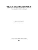
of calcium crosslinked microbeads, the microbeads in Fig. 3A were
produced without 1 mM BaCl2. Following gelation, microbeads were
rinsed to remove gelling solution and unreacted gelling ions using a
0.9% NaCl (VWR International BVBA, Leuven, Belgium), 2 mM CaCl2
and 10 mM HEPES solution at pH 7.2–7.4.
2.3. Visualization and size stability
Brightfield images and size determination of microbeads were ob
tained with a Nikon Eclipse TS100 microscope with a CFI Plan Fluor 4×/
0.13 Phl DL (Nikon, Tokyo, Japan, software NIS Elements v. 4.51, build
1145). To assess stability with regard to osmotic swelling, microbeads
were subjected to successive treatments in saline. Aliquots of 0.5 mL of
microbeads were exposed to 3 mL of 0.9% NaCl solution for 1 h on a tube
rotator. Images were captured and the saline solution was exchanged for
repeated saline treatments.
2.4. Confocal imaging of fluorescent microbeads
Confocal Laser Scanning Microscopy (CLSM) was performed on
microbeads produced from fluorescently labelled alginate (LF200S) and
CNF. The fluorescent labelling of alginate with fluoresceinamine using
carbodiimide chemistry has previously been described by Strand et al.
(2003) (Strand et al., 2003). Images of equatorial sections (30 μm) were
captured with an inverted confocal laser scanning microscope Zeiss
LSM800 (Carl Zeiss AG, Jena, Germany) with a motorized XY-stage, and
a C-apochromat 10× water-immersion objective (NA 0.45, WD 1.8 mm).
2. Materials and methods
2.1. Polysaccharides
Alginates, UP-LVG and LF200S were obtained from Novamatrix
(Sandvika, Norway) and FMC Biopolymer AS (Sandvika, Norway),
respectively. The composition and the molecular weight of the alginate
determined with 1H NMR (Grasdalen, 1983; Grasdalen et al., 1979) and
SEC-MALLS (Vold et al., 2006), respectively, are given in Table 1.
Alginate was labelled with fluoresceinamine for visualization in
confocal laser scanning microscopy (CLSM), as previously described
(Strand et al., 2003). CNF (TUNICELL ETC) derived from C. intestinalis
was obtained from Ocean TuniCell AS (Bergen, Norway), based on a
modified pulping procedure (Klemm et al., 2011; Zhao & Li, 2014) and
mechanical homogenization (Zhao et al., 2017). The CNF crystallinity
measured by X-ray diffraction (XRD) was 89.07 ± 1.60%. CNF average
fibril lengths and width were determined using atomic force microscopy
(AFM) at 2518 ± 827 nm, and 8.55 ± 3.37 nm, respectively.
2.5. Gel stiffness and syneresis
The same polysaccharide and gelling solutions described in the
“polysaccharides” and “production of microbeads” sections, were used
to produce gel cylinders of alginate, and alginate/CNF. Gel cylinders
were made by diffusion crosslinking (Skjåk-Bræk et al., 1989). Solutions
of alginate and alginate/CNF were extruded into cylindrical casts and
weighed. The cylindrical casts were enclosed in semipermeable mem
branes (Spectrum Laboratories, Inc., Rancho Dominguez, CA, USA). The
casts with alginate and alginate/CNF were placed in gelling baths for 24
h. Following gelation, the gels were weighed and compressed. A Stable
Micro Systems TA.XTplusC texture analyzer (Godalming, Surrey, UK), a
P/35 cylindrical probe and a 5 kg load cell were used for compression.
The compression was uniaxial and conducted at a probe speed of 0.1
mm/s with a trigger force of 1 g, at a temperature of 22 ◦ C. Exponent
2.2. Preparation of microbeads
Microbeads of alginate (A) and of alginate/CNF (A/C) were pro
duced with a custom-built electrostatic droplet generator (NTNU,
Table 1
Chemical composition of alginates given as fractions of G (FG) and M (FM), duplets (FGG, FMM, FMG/GM) and triplets (FGGM/MGG, FMGM, FGGG), estimates of G-block length
(NG>1) and weight average molecular weights (Mw). * LF200S was used exclusively as fluorescently labelled alginate for CLSM.
Alginate
FG
FM
FGG
FMM
FMG/GM
FGGM/MGG
FMGM
FGGG
NG>1
Mw (kDa)
UP-LVG
LF200S*
0.68
0.68
0.32
0.32
0.57
0.57
0.21
0.21
0.11
0.11
0.04
0.04
0.07
0.08
0.53
0.53
16
14
237
298
2
J.S. Kjesbu et al.
Carbohydrate Polymers 286 (2022) 119284
Table 2
Microbead nomenclature, alginate (A, UP-LVG) and cellulose nanofibril (C, TUNICELL ETC) constituents with corresponding percentages of the total polymer content,
and the corresponding alginate and cellulose nanofibril concentrations (w/v).
Microbead
Material(s), percentage of polymer in microbead
Concentration % (w/v)
A (100)
A/C (80/20)
A/C (50/50)
A/C (40/60)
A/C (30/70)
A/C (20/80)
UP-LVG
UP-LVG
UP-LVG
UP-LVG
UP-LVG
UP-LVG
1.80
1.44/0.36
0.90/0.90
0.72/1.08
0.54/1.26
0.36/1.44
(100%)
(80%)/TUNICELL
(50%)/TUNICELL
(40%)/TUNICELL
(30%)/TUNICELL
(20%)/TUNICELL
ETC (20%)
ETC (50%)
ETC (60%)
ETC (70%)
ETC (80%)
Connect software v. 7.0.3.0 (Hamilton, MA, USA) was used for data
collection and processing. Young's modulus (E) was calculated using the
initial slope of the force-deformation curves, with correction for syner
esis (Martinsen et al., 1989; Smidsrød et al., 1972) using the following
equations:
( )
L
E =S×
A
MC3T3 cells were cultured in ascorbic acid free α-MEM (ThermoFisher
Scientific, USA) supplemented with 1 μg/ml gentamicin, 2 mM gluta
mine and 10 % fetal calf serum (all supplements were from SigmaAldrich, St. Louis, MO, USA). Cells were sub-cultivated according to
the manufacturer's recommendations.
E
E Corr. = ( )2
Cells were mixed with alginate and alginate/CNF to a final concen
tration of 1 × 10⁶ cells/mL in 1.8% (w/v) polymer. Encapsulation of
cells was performed by electrostatic droplet production (EDP), as
described above (see section “Production of microbeads”). For cell
encapsulation, cell suspensions require mixing with polymer solutions
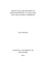
as well as extrusion (Fig. 1). Therefore, mixing and effects of extrusion
on cell viability were investigated. For alginate/CNF microbeads two
modes of mixing were tested: Stirring cell suspension into alginate and
CNF with a spatula (Fig. 1A), and by a cell mixing device (CellInk,
Boston, MA, USA) (Fig. 1B). Mixing was performed gently for approxi
mately 1 min in both approaches. To investigate the impact of
microbead production, some microbeads were gently extruded with a
pipette (Fig. 1C) for comparison with EDP (Fig. 1D). Following encap
sulation, microbeads and structures were gelled for 10 min before
rinsing off excess gelling solution in Dulbecco's Modified Eagle's Medium
(DMEM, Sigma-Aldrich, St. Louis, MO, USA). Encapsulated cells were
transferred into growth medium and cell viability was assessed as
described below.
2.9. Encapsulation
W0
W1
In which E is Young's modulus (Pa), L and A are the length (m) and
area (m2) of cylinders, and W0 and W1, respectively, are the masses (g) of
each sample prior to and following gelation.
2.6. Rheology
The shear viscosities of alginate, alginate/CNF and CNF were
analyzed using a TA Instruments Discovery HR-2 rheometer (God
alming, Surrey, UK). An aluminum plate-plate (20 mm, gap = 500 μm)
was used and a Peltier plate with a temperature of 25 ◦ C. The samples
were allowed to reach equilibrium temperature for 60 s prior to each
measurement. Shear viscosity was evaluated by increasing the shear rate
from 0.1 to 1000 s− 1.
2.7. Permeability
2.10. Cell viability
Diffusion of macromolecules was investigated with 40, 70 and 150
kDa FITC-conjugated dextrans (Sigma-Aldrich, St. Louis, MO, USA) and
absorbance spectrophotometry (VWR V1200, VWR International BVBA,
Leuven, Belgium) at 490 nm. Aliquots of 2 mL of microbeads were
incubated for 24 h at room temperature in 2 mL of 0.35% (w/v) dextran.
The dextran solution was removed and five to six samples of 250 mg of
microbeads were weighed. The microbeads were briefly rinsed in 1 mL
of PBS (Medicago, Uppsala, Sweden) and the absorbance was measured.
Three samples of microbeads were incubated in 1 mL PBS at room
temperature on a rotator. Absorbance in the solution was measured
immediately after incubation and at 15-minute intervals for 60 min. To
determine the initial concentration of FITC dextran following the rinse,
two to three samples of microbeads were dissolved in 1 mL of 0.15 M
EDTA (VWR International BVBA, Leuven, Belgium) and filtered to pro
vide a non-turbid solution. Identical microbeads were dissolved, filtered,
and used as the blank sample.
Encapsulated cells were transferred into 150 μL of serum-free growth
medium containing 1 μM DRAQ5 (Sigma-Aldrich, St. Louis, MO, USA)
and 4 μM Ethidium homodimer-1 (EthD-1, ThermoFisher Scientific,
USA) and incubated at room temperature for 30 min, to stain all (live
and dead) and only dead cells, respectively. Imaging was performed on a
Zeiss LSM800, as previously described (see section “Confocal imaging of
fluorescent microbeads”). To determine cell viability, image acquisition
was performed in triplicates with Z-projections of 50 stacks in 4.49 μm
intervals. Quantitative analysis of the images obtained was carried out
using ImageJ software (NIH). Differences between groups were
compared applying a two-tailed t-test (Microsoft Office Excel 365). The
significance level was set at 0.05. The results are expressed as mean ±
standard deviation (SD).
2.8. Cells
3.1. Shape and size of microbeads of alginate and CNF
In cell experiments, a pre-osteoblast cell line (MC3T3-E1 subclone 4,
ATCC® CRL-2593™) from Mus musculus, strain (C57BL/6) calvaria was
used. Additionally, Normal Human Dermal Fibroblasts (NHDFs), pri
mary cells derived from adult skin were used (Lonza, Basel,
Switzerland). NHDFs were cultured in FBM™ supplemented with
FGM™-2 Fibroblast SingleQuots™ Kit (Lonza, Basel, Switzerland).
Microbeads of alginate and of various alginate/CNF (A/C) ratios
were prepared using an electrostatic droplet generator, to produce
spherical beads of around 500 μm within a narrow size distribution,
compatible with high cell viablity upon encapsulation. The beads were
stabilized using barium in the gelling solution and characterised for
permeablity of dextrans with different molecular weight. Microbead
3. Results
3
J.S. Kjesbu et al.
Carbohydrate Polymers 286 (2022) 119284
Fig. 1. Encapsulation of cells in alginate and alginate/CNF microbeads. (A) Mixing CNF and alginate with cell suspension using a spatula. (B) Mixing of alginate and
CNF with cell suspension using a cell-mixing device. (C) Encapsulation by extrusion of microbeads using a pipette. (D) Encapsulation by electrostatic
droplet production.
preparation was initially evaluated with different solutions at the same
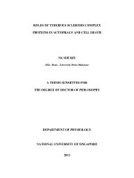
operating parameters (Fig. 2A), using a voltage of 7.00 kV, a flow rate of
10 mL/h and a nozzle diameter of 0.35 mm. During electrostatic
microbead production, solutions with greater fractions of CNF appeared
to increasingly elongate during extrusion. The maxima of elongation,
and the resulting microbeads are depicted in Fig. 2A. More spherical
composite microbeads with high CNF content (A/C (30/70) and A/C
(20/80)) were produced at a reduced voltage (from 7.00 kV to 6.15 kV)
and an increased flow rate (from 10 to 15 mL/h). Fig. 2B shows
microbead size, size distribution, and the degree of elongation denoted
as the aspect ratio for microbeads A/C (30/70) and A/C (20/80) pro
duced using the new parameters (6.15 kV and 15 mL/h). The lower CNF
content beads (60% CNF and below) where still produced with 7.00 kV
and 10 mL/h. Fig. 2D shows representative pictures of the produced
microbeads. Overall, the addition of CNF tended to increase size, size
distribution and elongated microbeads (Fig. 2B): Microbeads with 50%
CNF content or less had diameters of 449 ± 18 μm for A (100), 505 ± 13
μm for A/C (80/20) and 457 ± 37 μm for A/C (50/50) with aspect ratios
of 1.05 ± 0.03, 1.06 ± 0.06, and 1.11 ± 0.07, respectively. Above 50%
CNF, microbeads had greater diameters of 585 ± 78 μm for A/C (40/
60), 542 ± 86 μm for A/C (30/70) and 601 ± 131 μm for A/C (20/80)
with aspect ratios of 1.16 ± 0.18, 1.28 ± 0.17, and 1.23 ± 0.20,
respectively. The microbeads with high CNF content (80% CNF) had
both greater dispersity in size and aspect ratio (Fig. 2B), with some
batch-to-batch variability (Fig. S1). A reduced total polymer
concentration of 1.5% (w/v) produced results comparable to 1.8% (w/v)
while an increase to 2.0% (w/v) led to higher aspect ratios, and size and
dispersity (Fig. S1). To investigate the viscosity of alginate, CNF and
alginate/CNF dispersions at different shear rates, a frequency sweep was
performed (Fig. 2C), showing that the addition of CNF into alginate
solution results in non-Newtonian and high shear thinning flow char
acteristics compared with alginate alone. CNF alone (C (100)) consis
tently produced the highest viscosity at any shear rate. At low shear
rates, higher CNF content yielded substantially higher viscosity:
Approximately 500-fold greater for A/C (20/80) compared to A (100) at
0.1 S− 1. The difference in viscosity decreased at higher shear rates,
roughly overlapping (0.53–0.57 Pa⋅s) at 250 S− 1 for A (100), A/C (50/
50) and A/C (80/20).
3.2. Stability of alginate and alginate/CNF microbeads
To assess stability, A and A/C microbeads gelled with Ca2+ (50 mM)
or with Ca2+/Ba2+ (50 mM/1 mM) were subjected to successive saline
treatments (0.9% (w/v) NaCl). Calcium crosslinked microbeads (50 mM,
Fig. 3A) dissolved during the saline treatments. Increasing CNF content
reduced the stability of the beads where A/C (80/20) dissolved after four
treatments, A/C (50/50) after two treatments and A/C (20/80) after the
first treatment, in contrast to the Ca-alginate microbeads A (100) dis
solving after five treatments. Addition of 1 mM Barium ions to the gel
ling solution resulted in stable microbeads that did not dissolve during
4
J.S. Kjesbu et al.
Carbohydrate Polymers 286 (2022) 119284
Fig. 2. Alginate and alginate/CNF (A/C) composite materials (1.8% (w/v) total polymer concentration). (A) Electrostatic droplet production with equal operating
parameters (7 kV, 10 mL/h). The maxima of elongation of polymer solutions from the nozzle and resulting microbeads shown. Scale bar, 500 μm. (B) Diameters (left)
and aspect ratios (right) of microbeads produced at 7.00 kV and 10 mL/h for A (100), A/C (20/80), A/C (50/50) and A/C (40/60) and 6.15 kV, 15 mL/h for A/C (30/
70) and A/C (20/80). Diameters and aspect ratios are given as the mean ± SDEV, and scatter dots of individual values (n = 30). (C) Shear viscosity of 1.8% (w/v)
polymer solutions in 4.6% (w/v) mannitol. (D) Representative images of microbeads used for measurements of diameter and aspect ratio in (B). A (100): Alginate, A/
C (80/20): Alginate and CNF in 80/20 ratio, A/C (50/50): Alginate and CNF in 50/50 ratio, A/C (40/60): Alginate and CNF in 40/60 ratio, A/C (30/70): Alginate and
CNF in 30/70 ratio, A/C (20/80): Alginate and CNF in 20/80 ratio, C (100): CNF. Scale bar, 500 μm.
the saline treatments (Fig. 3B). Furthermore, calcium/barium cross
linked microbeads exhibited substantially greater size stability through
the saline treatments (Fig. 3B). CLSM images of equatorial sections of
alginate and alginate/CNF microbeads gelled in Ca2+/Ba2+ (50/1 mM),
produced with fluorescently labelled alginate are shown in Fig. 3B. A
slightly inhomogeneous distribution of alginate with greater signal from
the fluorescent alginate towards the rim of the microbeads was observed
for A (100) and A/C (80/20), but not for A/C (50/50). Due to limitations
in transmission of light through A/C (20/80) microbeads, these
microbeads were not visualized.
3.3. Stiffness of Ca/Ba-crosslinked alginate and alginate/CNF gels
To assess the stiffness of the Ca/Ba-crosslinked alginate/CNF com
posite gels, gel cylinders were chosen to reduce the complexity in
measuring Young's modulus on microbeads due to changes of contact
5
J.S. Kjesbu et al.
Carbohydrate Polymers 286 (2022) 119284
showing the lowest (27.6 ± 2.9 kPa) modulus. Syneresis (release of
water) was measured as the reduced weight of the material following
gelation. Increased content of CNF largely reduced the syneresis of the
hydrogels (Fig. 4B) with A (100) displaying the greatest syneresis (10.3
± 0.8%), followed by A/C (50/50) (6.4 ± 2.2%) and lastly A/C (20/80)
(1.3 ± 0.7%).
3.4. Permeability of Ca/Ba crosslinked alginate and alginate/CNF
microbeads
Initial uptake and subsequent release of dextrans from microbeads
was studied using FITC-conjugated dextrans and spectrophotometry.
Uptake of 40 kDa dextran was comparable for A (100) (0.089 ± 0.007%
(w/v)) and A/C (50/50) (0.088 ± 0.001% (w/v)). Uptake of 70 kDa
dextran was slightly higher in the composite beads (A/C (50/50): 0.084
± 0.001% (w/v)) as was 150 kDa dextran (0.070 ± 0.009% (w/v))
compared to microbeads with alginate alone (A (100): 70 kDa dextran =
0.070 ± 0.008%, and 150 kDa dextran = 0.061 ± 0.020% (w/v)). All
sizes of dextrans released rapidly from all microbeads with the greatest
fraction of release up to 15 min for both microbead types (Fig. 5D-F). A
slightly higher initial (t = 0) release of 40 kDa dextran was seen for A/C
(50/50) compared to A (100) (Fig. 5 D). The rate of release of 70 kDa
dextran was slightly higher overall for A/C (50/50) than for A (100)
(Fig. 5E), while release rates of 150 kDa were similar for both microbead
types (Fig. 5F).
3.5. Cell encapsulation in Ca/Ba-crosslinked composite microbeads
Cell viability in microbeads A (100) and A/C (50/50) was studied
using two cell types, the cell line MC3T3 and NHDF cells (Fig. 6A/C).
Shortly after encapsulation the viability of MC3T3 cells was slightly
greater (89.6 ± 2.6%) in A (100) than in A/C (50/50) microbeads (83.6
± 0.4%). The viability of NHDFs in the A (100) microbeads was signif
icantly (p < 0.05) higher (83.8 ± 5.0%.) than in the A/C (50/50)
microbeads (66.2 ± 5.3%). Production and handling throughout the
process of encapsulation may affect cell viability. Thus, the more sen
sitive NHDFs were chosen for evaluation of cell viability in A/C (50/50)
microbeads produced with different techniques of mixing: Either gentle
stirring with a spatula (Stir), or a cell-mixing device (Mixer), and
different approaches for encapsulation: either by extrusion with a
pipette (Pip.), or electrostatic droplet production (EDP) (Fig. 6B). In
summary, the mixing of polymers and cells had a greater impact on cell
viability than the production of microbeads. Mixing cells and polymers
Fig. 3. Size increase of alginate and alginate/CNF microbeads in saline treat
ments (0.9% NaCl). (A) Microbeads gelled in Ca2+ (50 mM) and (B) Ca2+/Ba2+
(50 mM/1 mM) and images of equatorial section (30 μm) by CLSM. Scale bar,
100 μm. Microbead diameters are given as the mean ± SDEV (n = 30). A (100):
Alginate, A/C (80/20): Alginate and CNF in 80/20 ratio, A/C (50/50): Alginate
and CNF in 50/50 ratio, A/C (80/20): Alginate and CNF in 80/20 ratio.
area upon uniaxial compression. Keeping the total polymer concentra
tion constant (1.8% (w/v)), Young's modulus decreased with increasing
content of CNF (Fig. 4A), with A (100) showing the highest (62.0 ± 9.6
kPa), A/C (50/50) slightly lower (55.4 ± 2.9 kPa) and A/C (20/80)
Fig. 4. Young's modulus and syneresis in gel cylinders of alginate (A 100), alginate/CNF (A/C 50/50) and A/C (20/80) gelled with Ca2+/Ba2+ (50 mM/1 mM). (A)
Young's modulus (E) corrected for syneresis, and (B) Syneresis. Measurements are given as the mean ± SDEV (N = 4). Statistically significant differences are
indicated by * (p < 0.05), ** (p < 0.01) and **** (p < 0.0001). Alginate, A/C (80/20): Alginate and CNF in 80/20 ratio, A/C (50/50): Alginate and CNF in 50/50
ratio, A/C (80/20): Alginate and CNF in 80/20 ratio.
6
J.S. Kjesbu et al.
Carbohydrate Polymers 286 (2022) 119284
Fig. 5. Uptake and release of FITC-dextrans (40, 70 and 150 kDa), from microbeads A (100) and A/C (50/50). (A–C) Initial concentration (% (w/v)) of dextrans in
microbeads (D–F) Percent release as a function of time where microbead A (100) is indicated by a solid line (—) and microbead A/C (50/50) is indicated by a dotted
line (—). Measurements are given as the mean ± SDEV (N = 3, 2 in A).
by stirring with a spatula (Stir) yielded lower viability than the purposebuilt cell-mixing device (Mixer). Production of microbeads with a
pipette (Pip.) yielded slightly lower viability than microbeads produced
by electrostatic droplet production (EDP) (Fig. 6B). NHDF viability in A/
C (50/50) microbeads was lowest (51.3 ± 3.4%) for microbeads pre
pared by mixing cells and polymers by stirring with a spatula followed
by pipette extrusion (Stir + Pip.). Viability was slightly higher (54.5 ±
3.7%) when microbeads were produced electrostatically (Stir + EDP).
Fig. 6. Viability of cells after encapsulation. (A) MC3T3 and NHDF cells in A (100), mixed directly in a syringe, and A/C (50/50) mixed with cell-mixer: mean ±
SDEV, statistically significant differences are indicated by * (p < 0.05, n = 2–4). (B) NHDF viability as a function of methods for cell-polymer mixing and extrusion.
Cells were mixed with an A/C (50/50) polymer solution by stirring with a spatula (Stir) or a cell-mixing unit (Mixer). Microbeads were prepared dropwise by a
pipetting (Pip.) or by electrostatic droplet production (EDP); mean ± SDEV (n = 2). (C) Z-projections of NHDFs and MC3T3s in microbeads. Scale bar, 200 μm. Dead
cells are shown in red (EthD-1), and both live and dead cells are green (DRAQ5). Viability was measured on the same day as encapsulation (A–C). (For interpretation
of the references to colour in this figure legend, the reader is referred to the web version of this article.)
7
J.S. Kjesbu et al.
Carbohydrate Polymers 286 (2022) 119284
Mixing cells and polymers with the cell mixer followed by pipette
extrusion (Mixer + Pip.) yielded slightly lower viability (67.2 ± 6.5%)
than electrostatically produced (Mixer + EDP) microbeads (72.7 ±
7.5%).
4. Discussion
(Mørch et al., 2012). A high concentration of barium used for cross
linking thus raises concerns about toxicity for in vivo applications.
However, a mixture of 50 mM Ca2+ with 1 mM Ba2+ lends itself to
producing high G alginate gels of sufficiently high strength and stability
to swelling, while minimizing exposure to barium (Mørch et al., 2006b;
Mørch et al., 2012).
4.1. Production of alginate/CNF microbeads
4.3. Elasticity of Ca/Ba-crosslinked alginate/CNF composite gels
Alginate together with nanofibrillated cellulose is a commonly used
bioink in 3D-bioprinting. However, to our knowledge, the application of
alginate/nanofibrillated cellulose for electrostatic production of
microbeads has not been reported. Furthermore, the high crystallinity
and purity of nanocellulose from tunicates represents a highly relevant
material for biomedical applications. Here, alginate and alginate/CNF
microbeads in the range of 400 to 600 μm were produced using an
electrostatic droplet generator. When starting from a fixed total polymer
concentration (1.8% (w/v)) known to be suitable for electrostatic pro
duction (Strand et al., 2002) of stable microbeads (Martinsen et al.,
1989; Mørch et al., 2006a), spherical microbeads of even size and size
distribution were produced with up to 50% CNF content. Increasing the
content of CNF in the polymer mixture resulted in elongation during
extrusion and generated microbeads with increased size, size dispersity,
and higher aspect ratios. Comparable results were obtained with
reduced total polymer concentration (1.5% (w/v)), with increasing
elongations at higher polymer concentrations (2.0% (w/v)). The shear
thinning effect of nanofibrillated cellulose is well known and was also
demonstrated here for alginate mixed with nanocellulose from tuni
cates. Alginate solutions have previously been shown to demonstrate
some shear thinning rheological properties, and have a greater loss
modulus compared to storage modulus over a wide range of frequencies
(Rezende et al., 2009). In general, nanocellulose dispersions demon
strate pronounced shear thinning effects, which are largely ascribed to
the alignment of cellulose fibrils when they are subjected to shear forces
(Hubbe et al., 2017). Greater elongation of the polymer solution upon
extrusion was observed for higher fractions of CNF, resulting in
microbeads exhibiting a greater aspect ratio, size, and size dispersity,
with some variation between batches. Considering the shear thinning
properties demonstrated by CNF, the production of microbeads might be
more sensitive to minor differences in the operational setup and thus the
flow. Hence, while the shear thinning properties of CNF are ideal for
printing as well as for production of microbeads up to a certain content
of CNF as shown here, this may be the limiting factor for proper droplet
production and shape recovery for microbead production using the
electrostatic droplet generator.
Although possible, investigating mechanical properties in microscale
beads entails considerable complexity in contrast to gel cylinders (Kim
et al., 2010). In the present study, gel cylinders were prepared by
diffusion of calcium (50 mM) and barium (1 mM) ions, to resemble the
gelation of microbeads. Decreasing alginate and increasing the CNF
concentration in the cylinders reduced both Young's modulus and syn
eresis. Previously, Aarstad et al. reported that the Young's moduli of
internally gelled, calcium saturated (50 mM) alginate/CNF gel cylinders
increased with increasing content (0.15–0.75% (w/v)) of cellulose
(Aarstad et al., 2017). In contrast, the present study kept the total
polymer concentration constant (1.8% (w/v)). Accordingly, the con
centration of alginate was lowered when CNF was incorporated. Ionic
crosslinking of alginate gels leads to a decrease in volume and weight
when compared to the solutions used to produce them (syneresis). This
effect is ascribed to the formation of junction zones, which are largely
responsible for generating the elastic properties in the ensuing alginate
hydrogel (Draget et al., 2001). Accordingly, the reduction in gel strength
of mixed alginate/CNF gels shown here is most likely caused by the
lower concentration of crosslinked material.
4.4. Permeability of Ca/Ba crosslinked alginate and alginate/CNF
microbeads
Permeability is an important variable in drug delivery systems, both
regarding loading and rates of release, and in constructs containing cells
that rely on diffusion of nutrients and cell products. De Vos et al. suggest
two main factors as relevant for quantification of permeability, namely
the rate of diffusion and the molecular weight cut-off (de Vos et al.,
2009). While both are linked to diffusion, molecular weight cut-off
(MWCO) alone does not predict diffusion since hydrogels are gener
ally non-uniform with respect to properties such as the size of pores and
their distribution, and material density (de Vos et al., 2009). In this
study, minor differences in the diffusion of dextrans (40–150 kDa) be
tween alginate and alginate/CNF microbeads were seen. Following in
cubation, all microbeads contained FITC-dextrans with slightly higher
initial concentrations for lower molecular weight dextrans. Alginate/
CNF microbeads held slightly more high molecular weight dextrans
following incubation and showed slightly faster release of dextrans
compared to alginate microbeads. This suggests that the rate of transfer
of nutrients and therapeutic products may be slightly increased by the
addition of CNF in microbeads. Cells entrapped within constructs such
as microbeads rely on diffusion of essential nutrients and oxygen
through the biomaterial. Previously, it has been reported that alginate/
CNF gels produce more porous structures than alginate alone (Siqueira
et al., 2019). The results herein showing slightly greater permeability of
alginate/CNF microbeads compared to alginate microbeads might be
linked to the higher porosity of these gel networks, as previously re
ported. What defines a desirable level of permeability is subject to
debate (Calafiore, 2018; Korsgren, 2017; Strand et al., 2017). Rokstad
et al. propose that what might be considered favorable permeability is
application dependent, whether the application be in vivo or in vitro
(Rokstad et al., 2014). In the context of immune isolation, some studies
have found simple alginate microbeads with limited permselectivity (i.
e., isolation against direct contact with immune cells) to be adequate for
sustained cell function in vivo (Duvivier-Kali et al., 2001; Omer et al.,
2003). On the other hand, some in vitro studies have shown improved
cell viability in hydrogels tailored for greater permselectivity against
4.2. Stability of alginate and alginate/CNF microbeads
Constructs made from nanocellulose and alginate within tissue en
gineering often use calcium ions for crosslinking (Krontiras et al., 2015;
Markstedt et al., 2015; Nguyen et al., 2017; Wu et al., 2018). However,
crosslinking alginate microbeads with barium or strontium ions has
previously been reported to produce highly stable microbeads compared
to crosslinking with calcium (Mørch et al., 2006b). Here, microbeads
containing increasing concentrations of CNF, crosslinked with calcium,
dissolved after fewer incubations with saline solutions. All of the cal
cium/barium crosslinked microbeads remained intact through saline
treatments and demonstrated greatly reduced swelling compared to
calcium crosslinked microbeads. Alginate gels are susceptible to ex
change with non-gelling ions or chelating compounds that lead to
swelling, compromised gel strength and dissolution (Rokstad et al.,
2014). Accordingly, stability is a concern for both in vitro and in vivo
applications where constructs are required to maintain their structure
over time. Although barium crosslinked alginate yields stable gels, it has
been shown in a mouse model that high G alginate barium crosslinked
(20 mM) microbeads (0.3 mL) exceed the tolerable intake of barium
8
J.S. Kjesbu et al.
Carbohydrate Polymers 286 (2022) 119284
inflammatory cytokines (Lin et al., 2010; Su et al., 2010).
content of CNF. Ionic crosslinking using calcium alone resulted in beads
with increasing content of CNF exhibiting reduced stability. However,
the addition of a low concentration of barium ions largely stabilized the
beads, even with a high CNF content. Compression of 1.8% (w/v) gel
cylinders revealed that Young's modulus decreased when adding CNF
into alginate, but syneresis was reduced. Spectrophotometry using FITCdextrans revealed that initial uptake and release rates were slightly
higher in microbeads with CNF compared to alginate alone, indicating a
slightly higher porosity. High (≈90%) viability was obtained for MC3T3
cells encapsulated in microbeads of alginate and alginate/CNF. The
viability following mixing and mode of extrusion was investigated in
alginate/CNF microbeads with NHDFs. Mixing was found to have
greater impact than extrusion and electrostatic bead generation, and
66% viability of NHFDs were obtained in alginate/CNF beads upon
optimizing the mixing protocol. The current study thus shows that
composite alginate and tunicate CNF microbeads can be produced with
an electrostatic bead generator. Such beads can be used for the encap
sulation of cells and hence have the potential for use in both cell therapy
and tissue engineering applications.
Supplementary data to this article can be found online at https://doi.
org/10.1016/j.carbpol.2022.119284.
4.5. Cell encapsulation
In tissue engineering, some applications require the entrapment of
cells such as in cell therapy. In the process of producing constructs for
cell immobilization, cell viability is a concern. Initial cell viability is
useful as an indicator of the tolerability of the selected approach.
Therefore, we encapsulated the mouse osteoblast precursor cell line
MC3T3 and primary normal human dermal fibroblasts (NHDFs) in
alginate and alginate/CNF microbeads to evaluate immediate viability
following production. Overall, viability was high for the cells after
encapsulation in the alginate/CNF microbeads, albeit higher viability
was seen for the MC3T3 cells than for the NHDF (90% vs 66%, respec
tively), and higher viability was seen in the alginate microbeads. Hence,
the effect on viability of mixing cells with polymers and extrusion was
also investigated for the NHDFs. With respect to the mixing of polymers
with cells and the mode of extrusion for microbead production the
greatest impact on viability (NHDFs) was observed in the mixing
process.
The decrease in viability of pre-osteoblast MC3T3 cells in the present
study is in agreement with previously published findings showing a
process-dependent decrease in the initial viability (down to 86–88%) of
MC3T3 cells (Ahn et al., 2012; Lee et al., 2015). However, in contrast to
the present study, previous studies have shown higher viability of
human dermal fibroblasts following bioprinting. Viability greater than
90% has been reported using different bioinks, based on type I collagen
(Lee et al., 2009), gelatin-poly(ethylene glycol)–tyramine (Hong et al.,
2019), ECM-like material (Rimann et al., 2016), or high viscosity bioink
based on 2% (w/w) of plant-derived nanofibrillated cellulose mixed
with 0.5% (w/w) alginate (Thayer et al., 2018). In the latter work, it was
also shown that viability of dermal fibroblasts was highly dependent on
the mixing procedure. While Thayer et al. reported human dermal
fibroblast viability over 90% at optimal mixing regimens (mixing unit or
mixing with a spatula for 30 and 60 s), viability dropped to 77.9 ± 14%
after mixing cells and bioink with a spatula for longer than 90 s. Simi
larly, shear-stress induced cell damage has been reported for mouse
L929 fibroblasts. The viability of these cells decreased in 3D-bioprinting
from 96 % to 76 % for 4 kPa and 18 kPa shear stresses, respectively
(Blaeser et al., 2016). These observations are in line with studies in 3D
bioprinting that found increased printing pressure and shear stress to
adversely affect cell viability (Koo & Kim, 2016; Nair et al., 2009; Shi
et al., 2018). A more prominent decrease in cell viability in A/C mixtures
as compared to A, found in our work, is in agreement with previous
observations for bovine chondrocytes. Viability of bovine chondrocytes
was found to be 81%, approximately 50%, and over 95% in 0.5% (w/w)
alginate/1.36% (w/w) nanocellulose mix, 1% (w/w) alginate sulfate/
1.36% (w/w) nanocellulose mix, and control cellulose-free alginates,
respectively (Müller et al., 2017). Therefore, it is likely that the viability
of the encapsulated cells in our study was dependent on the encapsu
lation material, the chosen method of production as well as the type of
cell encapsulated, as previously reported for other cell types (GungorOzkerim et al., 2018; Malda et al., 2013). We also showed a dependency
on material and cell type. However, cell viability was more strongly
affected by the mixing protocol than by extrusion and electrostatic
droplet generation.
CRediT authorship contribution statement
Joachim S. Kjesbu: Conceptualization, Methodology, Validation,
Investigation, Writing – original draft, Writing – review & editing,
Visualization. Daria Zaytseva-Zotova: Conceptualization, Methodol
ogy, Validation, Formal analysis, Investigation, Writing – original draft.
ămfors: Investigation, Writing original draft. Paul Gateư
Sanna Sa
nholm: Conceptualization, Writing – review & editing, Supervision,
Project administration. Christofer Troedsson: Conceptualization, Re
sources, Writing – review & editing. Eric M. Thompson: Conceptuali
zation, Resources, Writing – review & editing. Berit Løkensgard
Strand: Conceptualization, Methodology, Validation, Writing – review
& editing, Supervision, Project administration, Funding acquisition.
Declaration of competing interest
The authors declare the following financial interests/personal re
lationships which may be considered as potential competing interests:
Christofer Troedsson and Eric M. Thompson are both employed at Ocean
TuniCell, which provided the tunicate nanocellulose preparations used
in this study.
The remaining authors declare that the research was conducted in
the absence of any commercial or financial relationships that could be
construed as a potential conflict of interest.
Acknowledgements
Wenche I. Strand is acknowledged for performing 1H NMR. AnnSissel T. Ulset is acknowledged for analysis of alginates using SECMALLS. Their work was performed at the Department of Biotech
nology and Food Science at NTNU. MC3T3 cells were provided by Sarah
Lehnert and Kristin Grendstad, Department of Physics, NTNU, Trond
heim, Norway. The NHDF cells were kindly provided by SINTEF In
dustry, Department of Biotechnology and Nanomedicine, Trondheim,
Norway,
5. Conclusions
Funding
Here, we show that composite microbeads of alginate/tunicate CNF
can be produced with a narrow size range using an electrostatic bead
generator and the extrusion of composite material into a solution of
divalent cations. At a constant total polymer concentration of 1.8% (w/
v) a greater content of CNF in the microbeads was linked to elongation of
the polymers during extrusion and thus greater size, size distribution
and aspect ratio, making it difficult to produce spherical beads with 80%
The Research Council of Norway is acknowledged for funding of the
projects NFR-NANO 3D TUNINK and NFR-IPN TUNIGUIDE.
9
J.S. Kjesbu et al.
Carbohydrate Polymers 286 (2022) 119284
References
Markstedt, K., et al. (2015). 3D bioprinting human chondrocytes with nanocellulosealginate bioink for cartilage tissue engineering applications. Biomacromolecules, 16
(5), 1489–1496.
´
Martínez Avila,
H., et al. (2015). Novel bilayer bacterial nanocellulose scaffold supports
neocartilage formation in vitro and in vivo. Biomaterials, 44, 122–133.
Martinsen, A., Skjåk-Bræk, G., & Smidsrød, O. (1989). Alginate as immobilization
material: I. Correlation between chemical and physical properties of alginate gel
beads. Biotechnology and Bioengineering, 33(1), 79–89.
Mørch, Y. A., et al. (2006). Effect of Ca2+, Ba2+, and Sr2+ on alginate microbeads.
Biomacromolecules, 7(5), 1471–1480.
Mørch, Y. A., et al. (2006). Effect of Ca2+, Ba2+, and Sr2+ on alginate microbeads.
Biomacromolecules, 7(5), 1471–1480.
Mørch, Y. A., et al. (2012). Binding and leakage of barium in alginate microbeads.
Journal of Biomedical Materials Research. Part A, 100(11), 2939–2947.
Müller, M., et al. (2017). Alginate sulfate-nanocellulose bioinks for cartilage bioprinting
applications. Annals of Biomedical Engineering, 45(1), 210–223.
Nair, K., et al. (2009). Characterization of cell viability during bioprinting processes.
Biotechnology Journal, 4(8), 1168–1177.
Nguyen, D., et al. (2017). Cartilage tissue engineering by the 3D bioprinting of iPS cells
in a nanocellulose/alginate bioink. Scientific Reports, 7.
Omer, A., et al. (2003). Survival and maturation of microencapsulated porcine neonatal
pancreatic cell clusters transplanted into immunocompetent diabetic mice. Diabetes,
52(1), 69–75.
Rezende, R. A., et al. (2009). Rheological behavior of alginate solutions for
biomanufacturing. Journal of Applied Polymer Science, 113(6), 3866–3871.
Rimann, M., et al. (2016). Standardized 3D bioprinting of soft tissue models with human
primary cells. Journal of Laboratory Automation, 21(4), 496–509.
Rokstad, A. M. A., et al. (2014). Advances in biocompatibility and physico-chemical
characterization of microspheres for cell encapsulation. Advanced Drug Delivery
Reviews, 67–68, 111–130.
Sagane, Y., et al. (2010). Functional specialization of cellulose synthase genes of
prokaryotic origin in chordate larvaceans. Development, 137(9), 1483–1492.
Shi, J., et al. (2018). Shear stress analysis and its effects on cell viability and cell
proliferation in drop-on-demand bioprinting. Biomedical Physics & Engineering
Express, 4(4), Article 045028.
Siqueira, P., et al. (2019). Three-dimensional stable alginate-nanocellulose gels for
biomedical applications: Towards tunable mechanical properties and cell growing.
Nanomaterials (Basel), 9(1).
Skjåk-Bræk, G., Grasdalen, H., & Smidsrød, O. (1989). Inhomogeneous polysaccharide
ionic gels. Carbohydrate Polymers, 10(1), 31–54.
Smidsrød, O. H., Arne, & Lian, B. (1972). Properties of poly(1,4-hexuronates) in the gel
state. I. Evaluation of a method for the determination of stiffness. Acta Chemica
Scandinavia, 26, 79–88.
Strand, B. L., et al. (2002). Alginate-polylysine-alginate microcapsules: Effect of size
reduction on capsule properties. Journal of Microencapsulation, 19(5), 615–630.
Strand, B. L., et al. (2003). Visualization of alginate-poly-L-lysine-alginate microcapsules
by confocal laser scanning microscopy. Biotechnology and Bioengineering, 82(4),
386–394.
Strand, B. L., Coron, A. E., & Skjåk-Bræk, G. (2017). Current and future perspectives on
alginate encapsulated pancreatic islet. Stem Cells Translational Medicine, 6(4),
1053–1058.
Su, J., et al. (2010). Anti-inflammatory peptide-functionalized hydrogels for insulinsecreting cell encapsulation. Biomaterials, 31(2), 308–314.
Thayer, P. S., Orrhult, L. S., & Martínez, H. (2018). Bioprinting of cartilage and skin
tissue analogs utilizing a novel passive mixing unit technique for bioink
precellularization. Journal of Visualized Experiments, 131.
Vold, I. M. N., Kristiansen, K. A., & Christensen, B. E. (2006). A study of the chain
stiffness and extension of alginates, in vitro epimerized alginates, and periodateoxidized alginates using size-exclusion chromatography combined with light
scattering and viscosity detectors. Biomacromolecules, 7(7), 2136–2146.
de Vos, P., et al. (2009). Multiscale requirements for bioencapsulation in medicine and
biotechnology. Biomaterials, 30(13), 2559–2570.
Wang, X., Wang, Q., & Xu, C. (2020). Nanocellulose-based inks for 3D bioprinting: Key
aspects in research development and challenging perspectives in applications-a mini
review. Bioengineering (Basel, Switzerland), 7(2), 40.
Wu, Y., et al. (2018). 3D bioprinting of liver-mimetic construct with alginate/cellulose
nanocrystal hybrid bioink. Bioprinting, 9, 1–6.
Zhao, Y., & Li, J. (2014). Excellent chemical and material cellulose from tunicates:
Diversity in cellulose production yield and chemical and morphological structures
from different tunicate species. Cellulose, 21(5), 3427–3441.
Zhao, Y., et al. (2017). Cellulose nanofibers from softwood, hardwood, and tunicate:
Preparation–Structure–Film performance interrelation. ACS Applied Materials &
Interfaces, 9(15), 13508–13519.
Aarstad, O., et al. (2017). Mechanical properties of composite hydrogels of alginate and
cellulose nanofibrils. Polymers, 9(8), 378.
Ahn, S., et al. (2012). Cells (MC3T3-E1)-laden alginate scaffolds fabricated by a modified
solid-freeform fabrication process supplemented with an aerosol spraying.
Biomacromolecules, 13(9), 2997–3003.
Athukoralalage, S. S., et al. (2019). 3D bioprinted nanocellulose-based hydrogels for
tissue engineering applications: A brief review. Polymers, 11(5), 898.
Blaeser, A., et al. (2016). Controlling shear stress in 3D bioprinting is a key factor to
balance printing resolution and stem cell integrity. Advanced Healthcare Materials, 5
(3), 326–333.
Calafiore, R. (2018). Microencapsulation for cell therapy of type 1 diabetes mellitus: The
interplay between common beliefs, prejudices and real progress. Journal of diabetes
investigation, 9(2), 231–233.
Draget, K. I., et al. (2001). Effects of molecular weight and elastic segment flexibility on
syneresis in Ca-alginate gels. Food Hydrocolloids, 15(4), 485–490.
Draget, K. I., et al. (2006). Alginates. In Food polysaccharides and their applications (pp.
289–334). CRC Press.
Dufresne, A. (2017). Cellulose nanomaterial reinforced polymer nanocomposites. Current
Opinion in Colloid & Interface Science, 29, 1–8.
Duvivier-Kali, V. F., et al. (2001). Complete protection of islets against allorejection and
autoimmunity by a simple barium-alginate membrane. Diabetes, 50(8), 1698–1705.
Frampton, J. P., et al. (2011). Fabrication and optimization of alginate hydrogel
constructs for use in 3D neural cell culture. Biomedical Materials, 6(1), Article
015002.
Franỗon, H., et al. (2020). Ambient-dried, 3D-printable and electrically conducting
cellulose nanofiber aerogels by inclusion of functional polymers. Advanced Functional
Materials, 30(12), 1909383.
Gorin, P. A. J., & Spencer, J. F. T. (1966). Exocellular alginic acid from azotobacter
vinelandii. Canadian Journal of Chemistry, 44(9), 993–998.
Govan, J. R., Fyfe, J. A., & Jarman, T. R. (1981). Isolation of alginate-producing mutants
of Pseudomonas fluorescens, pseudomonas putida and Pseudomonas mendocina.
Journal of General Microbiology, 125(1), 217–220.
Grasdalen, H. (1983). High-field, 1H-n.m.r. spectroscopy of alginate: sequential structure
and linkage conformations. Carbohydrate Research, 118, 255–260.
Grasdalen, H., Larsen, B., & Smidsrød, O. (1979). A p.M.R. Study of the composition and
sequence of uronate residues in alginates. Carbohydrate Research, 68(1), 23–31.
Gungor-Ozkerim, P. S., et al. (2018). Bioinks for 3D bioprinting: An overview.
Biomaterials Science, 6(5), 915–946.
Heggset, E. B., et al. (2019). Viscoelastic properties of nanocellulose based inks for 3D
printing and mechanical properties of CNF/alginate biocomposite gels. Cellulose, 26
(1), 581–595.
Hong, S., et al. (2019). Coaxial bioprinting of cell-laden vascular constructs using a
gelatin-tyramine bioink. BiomaterialsScience, 7.
Hubbe, M., et al. (2017). Rheology of nanocellulose-rich aqueous suspensions: A review.
BioResources, 12, 9556–9661.
Kim, K., et al. (2010). Investigation of mechanical properties of soft hydrogel
microcapsules in relation to protein delivery using a MEMS force sensor. Journal of
Biomedical Materials Research Part A, 92A(1), 103–113.
Klemm, D., et al. (2011). Nanocelluloses: A new family of nature-based materials.
Angewandte Chemie International Edition, 50(24), 5438–5466.
Koo, Y., & Kim, G. (2016). New strategy for enhancing in situ cell viability of cell-printing
process via piezoelectric transducer-assisted three-dimensional printing.
Biofabrication, 8(2), Article 025010.
Korsgren, O. (2017). Islet encapsulation: Physiological possibilities and limitations.
Diabetes, 66(7), 17481754.
Krontiras, P., Gatenholm, P., & Hă
agg, D. A. (2015). Adipogenic differentiation of stem
cells in three-dimensional porous bacterial nanocellulose scaffolds. Journal of
Biomedical Materials Research Part B: Applied Biomaterials, 103(1), 195–203.
Lee, H. J., et al. (2015). A new approach for fabricating Collagen/ECM-based bioinks
using preosteoblasts and human adipose stem cells. Advanced Healthcare Materials, 4
(9), 1359–1368.
Lee, K. Y., & Mooney, D. J. (2012). Alginate: Properties and biomedical applications.
Progress in Polymer Science, 37(1), 106–126.
Lee, W., et al. (2009). Multi-layered culture of human skin fibroblasts and keratinocytes
through three-dimensional freeform fabrication. Biomaterials, 30(8), 1587–1595.
Lin, C.-C., et al. (2010). Regulating MCP-1 diffusion in affinity hydrogels for enhancing
immuno-isolation. Journal of controlled release : official journal of the Controlled
Release Society, 142(3), 384–391.
Malda, J., et al. (2013). 25th anniversary article: Engineering hydrogels for
biofabrication. Advanced Materials, 25(36), 5011–5028.
10