Two banana cultivars differ in composition of potentially immunomodulatory mannan and arabinogalactan
Bạn đang xem bản rút gọn của tài liệu. Xem và tải ngay bản đầy đủ của tài liệu tại đây (2.92 MB, 11 trang )
Carbohydrate Polymers 164 (2017) 31–41
Contents lists available at ScienceDirect
Carbohydrate Polymers
journal homepage: www.elsevier.com/locate/carbpol
Two banana cultivars differ in composition of potentially
immunomodulatory mannan and arabinogalactan
Tânia M. Shiga a , Nicholas C. Carpita c , Franco Maria Lajolo a,b ,
Beatriz Rosana Cordenunsi-Lysenko a,b,∗
a
Department of Food Science and Experimental Nutrition, University of São Paulo, Av. Prof. Lineu Prestes, 580, Bloco 14, São Paulo, SP, 05508-000, Brazil
Food Research Center (FoRC), São Paulo, SP, Brazil
c
Department of Botany & Plant Pathology, Purdue University, 915 West State Street, West Lafayette, IN 47907, USA
b
a r t i c l e
i n f o
Article history:
Received 30 August 2016
Received in revised form 10 January 2017
Accepted 21 January 2017
Available online 22 January 2017
Keywords:
Banana
Polysaccharides
Musa sp.
Mannan
Arabinogalactan
Cell wall
Polysaccharide structure
Bioactive polysaccharides
a b s t r a c t
Banana (Musa acuminata and M. acuminata x M. balbisiana) fruit cell walls are rich in mannans, homogalacturonans and xylogalacturonan, rhamnogalacturonan-I, and arabinogalactans, certain forms of which is
considered to have immunomodulatory activity. The cultivars Nanicão and Thap Maeo represent two
widely variants with respect to compositional differences in the forms of these polysaccharides. Nanicão has low amounts of mannan in the water-insoluble and water-soluble fraction. Both cultivars have
high amounts of water-soluble arabinogalactan. These commelinoid monocots lack the (1 → 3),(1 → 4)-d-glucans of grasses, but Thap Maeo has higher amounts of non-starch glucans associated with wild
species than does Nanicão. High amount of callose was found in both cultivars. As immunomodulatory
activity is associated with the fine structure and interaction of these polysaccharides, breeding programs
to introgress disease resistance from wild species must account for these special structural features in
retaining fruit quality and beneficial properties.
© 2017 Elsevier Ltd. All rights reserved.
1. Introduction
Fruits are rich in soluble and insoluble fiber, comprising complex
homo- and heteropolymers, such as pectins and hemicelluloses.
These polysaccharides are widely studied because their breakdown
and solubilization during fruit ripening yield softness and juiciness
to the pulp. Moreover, there is a growing interest in the biological
activity of its polysaccharides due to their potential beneficial effect
to the human health. Banana fruits contain various compounds,
such as lectin, polysaccharides, flavonoids, alkaloids, steroids and
glycosides that may produce physiological effects (Onyenekwe,
Okereke, & Owolewa, 2013; Scarminio, Fruet, Witaicenis, Rall, &
Di Stasi, 2012; Sansone, Sansone, Dias, Shiga, & Nascimento, 2016).
The commercially available bananas are diploids or triploids of
Musa acuminata (A genome) and Musa balbisiana (B genome), or
∗ Corresponding author at: Department of Food Science and Experimental Nutrition, University of São Paulo, Av. Prof. Lineu Prestes, 580, Bloco 14, São Paulo, SP,
05508-000, Brazil.
E-mail addresses: (T.M. Shiga),
(N.C. Carpita), (F.M. Lajolo),
(B.R. Cordenunsi-Lysenko).
/>0144-8617/© 2017 Elsevier Ltd. All rights reserved.
are hybrids of the two species produced with their crossbreeding, resulting in classification groups AAA, AA, AB, AAB and ABB.
Cultivar Thap Maeo is a variant of cultivar Mysore and belongs
to AAB genomic group, whilst Nanicão belongs to AAA genomic
group. Thap Maeo and Mysore cultivars are replacing traditional
commercial cultivars because to their higher resistance to Black
Sigatoka (black leaf-streak disease), a serious leaf spot disease of
bananas that results in yield losses of 33–50% (Mobambo et al.,
1993). Although resistance to Black Sigatoka is observed primarily
in leaves, changes in the cell-wall composition related to disease
resistance might be observed in the fruits, and these differences
might impact fruit quality. For instance, cultivar Mysore has anthocyanidin conjugates with cell-wall polymers (regarded to possess
strengthening and defense functions against pathogens) whilst,
Nanicão lacks of anthocyanidins linked to the cell wall and has
higher susceptibility to disease (Bennett et al., 2010).
Chemical composition and structure of the cell wall components
are the principal determinants of fruit texture, so characterization
of newly introduced cultivars for disease resistance is essential to
assure texture is not lost. Polysaccharides for the human health
are highly dependent on their structure (degree of branching,
molecule size and monosaccharide composition) that confers water
32
T.M. Shiga et al. / Carbohydrate Polymers 164 (2017) 31–41
holding properties, solubility and availability for fermentation
by colonic bacteria, and immunomodulatory effects (Blackwood,
Salter, Dettmar, & Chaplin, 2000).
In this work, we evaluated the composition and linkage structure of banana fruit wall polysaccharides to assess the impact of
introgression of a diverse germplasm to increase disease resistance
on the fine structure of wall polysaccharides that impart proper
pulp softening and beneficial properties for human health.
2. Material and methods
2.1. Material
Banana fruits Musa acuminata cv. Nanicão (genomic group AAA)
were purchased from CEAGESP (São Paulo State, Brazil) and M.
acuminata x M. balbisiana cv. Thap Maeo (genomic group AAB)
were harvested in the plantation located in Itapetininga (São Paulo
State, Brazil). Fruits were classified as ripe, based on ethylene and
respiration levels, and amounts of starch and soluble sugars. Ripe
banana fruits were peeled, frozen in liquid N2 and freeze-dried.
The freeze-dried materials were ground in mortar and boiled at
70–80 ◦ C in methanol: chloroform (1:1, v/v) for 30 min to remove
fat, pigments and to inactivate enzymes. The suspensions were filtered in sintered glass funnel, and the residues washed extensively
with acetone to remove pigments. The residues were dried at ambient temperature and kept in a desiccator until use. The defatted
material was used to cell wall polysaccharide extraction, enzymatic
assay and -glucan determination.
2.2. Extraction of cell wall polysaccharides
About 1 g of fruit pulp was soaked in 50 mL of 0.08 M sodium
phosphate, pH 6.0. Exactly 100 L of heat-stable ␣-amylase from
B. licheniformis (Megazyme International Ireland, EC 3.2.1.1) was
added, and the mixture was incubated at 40 ◦ C for 2 h. The pH
was then adjusted to 4.5 with HCl, and 200 L of amyloglucosidase
from A. niger (Megazyme International Ireland Limited, Ireland, EC
3.2.1.3) was added. The mixture was incubated at 35 ◦ C for 2 h,
then adjusted to pH 7.5 with NaOH, and 100 L of protease of B.
licheniformis (Megazyme International Ireland Limited, Ireland, EC
3.4.21.14) was added, and the mixture was incubated at 35 ◦ C for
an additional hour. The hydrolysate was centrifuged at 9000 g, and
the pellet was washed extensively with 3 vol. of 50 mL of deionized
water. About 100 mL of DMSO (90% in water, v/v) was added to the
residue, and the mixture was sonicated in ultra-sound bath for 1 h
(2x) and centrifuged at 10,000g for 10 min, and the supernatant was
discarded. The pellet was washed 3 times with 90% DMSO and then
washed exhaustively with water. The residue was re-suspended in
water, and freeze-dried and called water-insoluble polysaccharides
(WIP).
The aqueous supernatant (hydrolysis buffer) and the wash solutions were combined and brought to 80% EtOH (v/v). The ethanolic
mixture was heated to 70 ◦ C for 15 min and ice-cooled to precipitate the polysaccharides and other material, which were pelleted at
9000 g and washed extensively with chilled 80% EtOH. The ethanolinsoluble material was re-suspended in water, frozen in liquid N2
and freeze-dried. This fraction was named water-soluble polysaccharide (WSP).
2.3. Monosaccharide determination and linkage analysis
2.3.1. Carboxyl reduction of uronosyl residues
Duplicate samples of water-soluble and insoluble banana cell
wall materials were carboxyl-reduced with NaBD4 after activation
with a water-soluble carbodiimide, as described by Kim and Carpita
(1992) and modified by Carpita and McCann (1996). A colorimetric
assay for uronic acids in the presence of neutral sugars (FilisettiCozzi & Carpita, 1991) was used to confirm that the reduction of the
carboxyl groups was 95% or greater. For each of sets of materials,
two samples of each were used for monosaccharide and linkage
analysis.
2.3.2. Monosaccharide distribution
Monosaccharides were obtained from uronosyl-reduced cell
wall material (1–2 mg) by hydrolysis in 1 mL of 2 M trifluoroacetic
acid (TFA) at 120 ◦ C for 90 min. One-half millilitre of t-butyl alcohol was added, and the TFA-alcohol mixtures were evaporated in
a stream of nitrogen, reduced with NaBH4 , and alditol acetates
were prepared according to Gibeaut and Carpita (1991). Derivatives were separated by gas-liquid chromatography (GLC) on a
0.25 mm × 30 m column of SP-2330 (Supelco, Bellefonte, PA). Temperature was held at 80 ◦ C during injection then rapidly ramped to
170 ◦ C at 25 ◦ C min−1 , and then to 240 ◦ C at 5 ◦ C min−1 with a 10 min
hold at the upper temperature. Helium flow was 1 mL min−1 with
split-less injection. The electron impact mass spectrometry (EIMS)
was performed with a Hewlett-Packard MSD at 70 eV and a source
temperature of 250 ◦ C. The proportion of 6,6-dideuteriogalactosyl
was calculated using pairs of diagnostic fragments m/z 187/189,
217/219 and 289/291 according to the equation described in Kim
and Carpita (1992) that accounts for spillover of 13 C.
2.3.3. Linkage analyses
For linkage analysis, about 1 mg of polysaccharides dried in
a vacuum desiccator over P2 O5 were suspended in 2 mL of dry
DMSO (Pierce silylation grade) were per-O-methylated with Li+
methylsulfinylmethanide, prepared by adding 1.25 mL of 2.5 M nbutyllithium in hexanes (Aldrich) to the polysaccharides purged by
Argon. Upon evaporation of the hexanes and formation of Li+ alkoxide ions, methyl iodide (Aldrich) was added according to Gibeaut
and Carpita (1991). The per-O-methylated polymers were recovered after addition of water to the mixture and partitioning into
chloroform. The chloroform extracts were washed five times with
a three-fold excess of water each, and the chloroform was evaporated in a stream of nitrogen gas. The partly methylated polymers
were hydrolyzed in 2 M TFA for 90 min at 120 ◦ C. One-half millilitre
of t-butyl alcohol was added, the TFA-alcohol mixture was evaporated in a stream of nitrogen gas, and the sugars were reduced with
NaBD4 and acetylated. The partly methylated alditol acetates were
separated on the same column used to alditol acetates. After a hold
at 80 ◦ C for 1 min during injection, the derivatives were separated
in a temperature program of 160 ◦ C to 210 ◦ C at 2 ◦ C min−1 , then to
240 ◦ C at 5 ◦ C min−1 , with a hold of 5 min at the upper temperature.
All derivative structures were confirmed by electron-impact mass
spectrometry (Carpita & Shea, 1989).
2.4. Enzymatic digestion of cell wall polysaccharides
2.4.1. Cell wall polysaccharides composition
Suspensions containing 10 mg mL−1 of water-soluble and
water-insoluble polysaccharides in buffer were incubated with the
following glycosidases at 40 ◦ C for 2 h. A blank without enzyme
were also carried out in parallel.
2.4.1.1. Xyloglucan determination. Polysaccharide suspensions
were hydrolyzed using 10 U mL−1 of xyloglucanase from Paenibacillus sp. (E.C. 3.2.1.151, E-XEGP, Megazyme International
Ireland, Wicklow, Ireland) in 100 mM sodium acetate buffer, pH
5.5.
2.4.1.2. Mannan determination. Polysaccharide suspensions were
hydrolyzed using 3.9 U mL−1 of endo-(1 → 4)--d-mannanase from
T.M. Shiga et al. / Carbohydrate Polymers 164 (2017) 31–41
Bacillus sp. (E.C. 3.2.1.78, E-BMABS, Megazyme) in 100 mM glycine
buffer, pH 8.8.
2.4.1.3. Arabinan determination. Polysaccharide suspensions were
hydrolyzed using 15 U mL−1 of endo-/exo-arabinanase of Cellvibrio
japonicus (E.C. 3.2.1.99, E-ARBACJ, Megazyme) in 100 mM potassium phosphate buffer, pH 7.0.
2.4.1.4. Xylan determination. Polysaccharide suspensions were
hydrolyzed using 5 U mL−1 of -xylanase from Trichoderma viride
(E.C. 3.2.1.8, E-XYTR1, Megazyme) in 100 mM sodium acetate
buffer, pH 4.5.
2.4.1.5. Homogalacturonan determination. Polysaccharide suspensions were hydrolyzed using 5 U mL−1 of endopolygalacturonase
from Aspergillus niger (E-PGALS, E. C. 3.2.1.15, Megazyme) in
100 mM sodium acetate buffer, pH 4.0.
The reactions were terminated by boiling for 15 min and the
suspensions were centrifuged at 10,000g and the supernatants
filtered through 45-m pore filter and analyzed in a PA1 pellicular anion-exchange analytical column (Dionex Corp., Sunnyvale,
CA, USA, 250 × 4 mm) with its respective guard column, using a
DX500 HPAEC-PAD System (Dionex) equipped with a GP40 gradient pump an ED40 detector with a gold working electrode and
Ag/AgCl reference electrode and a AS50 autosampler to detect cell
wall polysaccharide oligomers.
The oligosaccharides were gradient eluted at 30 ◦ C at flow rate
of 1 mL min−1 , using 65 mM NaOH and NaOAc. Isocratic elution was
employed for 4 min, using 65 mM NaOH, followed by NaOAc gradient mM) ending at 40 min. A cleaning step of 10 min of NaOH
200 mM containing 200 mM NaOAc was added.
2.4.1.6. Monosaccharide determination. The hydrolysates were
brought to 80% EtOH by adding 4 vol. of absolute EtOH and centrifuged at 10,000g. The supernatant was transferred to a new
vial, dried under N2 stream, re-suspended in 2 M trifluoroacetic
acid (TFA) and hydrolyzed at 120 ◦ C for 90 min (Kim & Carpita,
1992). The hydrolysates were dried with 1 mL of t-butyl alcohol,
and suspended in water, and the monosaccharides were analyzed
on a CarboPac PA10 pellicular anion-exchange analytical column
(250 × 4 mm, with the corresponding guard column) using a DX500
HPAEC-PAD System (Dionex), according to Cordenunsi et al. (2008),
to detect xylose (Xyl), glucose (Glc), arabinose (Ara), mannose
(Man), rhamnose (Rha), fucose (Fuc), galactose (Gal), galacturonic
acid (GalA) and glucuronic acid (GlcA) released from cell wall
polysaccharides hydrolysis.
2.5. Determinations of ˇ-glucan
2.5.1. Pretreatment of water-insoluble polysaccharides
Water-insoluble polysaccharides were assayed with and without pre-treatment with protease from Bacillus licheniformis (EC
3.4.21.14, E-BSPRT from Megazyme).
Water-insoluble polysaccharides were submitted to protease
pre-treatment to remove resident enzymes. About 1 g of waterinsoluble polysaccharide was incubated with 35 U of protease for
1 h at 60 ◦ C with 50 mL of 0.08 M phosphate buffer, pH 7.5. The
reaction was terminated by boiling for 15 min to inactivate the
enzyme and centrifuged at 10,000g. The pellet was recovered and
exhaustively washed with 80% EtOH and freeze-dried.
2.5.1.1. Banana pulp preparation. For banana pulp -glucan assays,
the pulp was defatted as described above and washed exhaustively
with 80% EtOH in order to remove all free-sugar. The pulp samples
®
were washed with acetone and dried in a SpeedVac concentrator.
33
2.5.1.2. Sample preparation. Mixtures containing 3 mg mL−1 of
water-insoluble or water-soluble-polysaccharides and a suspension containing 30–50 mg mL−1 of defatted banana pulp were
prepared in 150 mM sodium phosphate buffer, pH 6.5.
2.5.1.3. Laminarinase assay. Banana cell wall and defatted banana
pulp suspensions were incubated for 2 h at 60 ◦ C with 5 U mL−1 of
laminarinase (EC 3.2.1.6, endo-1,3(4)--glucanase from Clostridium
thermocellum, E-LICACT, Megazyme).
2.5.1.4. Lichenase treatment. Banana cell wall and defatted banana
pulp suspensions were incubated for 2 h at 40 ◦ C with 1 U mL−1
of lichenase. (EC 3.2.1.73, endo-(1 → 3),(1 → 4)--d-glucanase from
Bacillus subtilis, Megazyme).
Banana isolated cell walls material, the incubation times were
up to 24 h.
The supernatants of laminarinase and lichenase and hydrolysis
were recovered, filtered through 45 m pore filter and analyzed
using a DX500 HPAEC-PAD System (Dionex) as mentioned in the
item 2.4.1 in order to detect -glucan oligomers. Samples of the
supernatant obtained from laminarinase and lichenase hydrolysis
®
were dried in a CentriVap , suspended in 2 M TFA and hydrolyzed
at 100 ◦ C for 1 h, 1 mL of t-butyl alcohol was added and the mixture
®
was dried in a CentriVap . The monosaccharides were suspended
in water and analyzed by HPAEC-PAD (Dionex) as earlier. Controls without laminarinase was also carried out as described earlier.
The oligomers and glucose released after cell wall polysaccharide
hydrolysis were separated on a CarboPac PA10 pellicular anionexchange analytical column in the same way mentioned above.
Reaction blanks without enzyme were carried out in parallel.
3. Results
3.1. Composition of cell wall polysaccharides
Owing to an enrichment of water-soluble polysaccharides, the
fruit pulps of Thap Maeo banana were denser than those of Nanicão,
with polysaccharide mass almost 2.5-fold higher (Fig. S1).
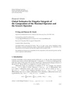
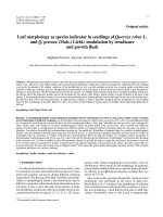
3.1.1. Monosaccharide composition
The compositions of water-soluble and insoluble polysaccharides in banana fruit pulp were vastly different, at least one-half of
the monosaccharide composition was Gal, with the remainder in
Man (Fig. 1A and C). Ara and Xyl were also present, but Glc was less
than 1 mol% of the water-soluble fraction. The uronosyl residues in
water-soluble and water-insoluble polysaccharides were carboxylreduced with NaBD4 to convert them to their respective neutral
sugars, distinguishing them as their 6,6-dideutero neutral sugars
by MS. GalA and GlcA were detected as smaller constituents of the
water-soluble fractions.
In stark contrast, glucans were the major polysaccharides of
water-insoluble fraction (Fig. 1A and C), when calculated in the
absence of Glc, GalA was more easily observed as a principal constituent, with modest amounts of Xyl, Ara, Gal and Man (Fig. 1A
and C). The great excess of GalA compared to Rha, indicated that
HG and its derivatives are the most abundant pectin.
Nanicão and Thap Maeo cultivars differ markedly in the
monosaccharide composition of their water-soluble polysaccharides (Fig. 1B and D). Thap Maeo had significantly more Man
(24 mol%) and slightly more GalA (2 mol%) than Nanicão, and consequently, significantly less Gal, Xyl, Ara and GalA (11, 6, 5 and
5 mol%, respectively) (Fig. 1B). Although glucans were the dominant polysaccharide of the water-insoluble fractions, Nanicão had
30 mol% larger proportions (Fig. 1D). Because of this difference, the
34
T.M. Shiga et al. / Carbohydrate Polymers 164 (2017) 31–41
Fig. 1. Thap Maeo (Genomic group AAB – grey bars) and Nanicão (Genomic group AAA – black bars) water-soluble and water-insoluble polysaccharide composition (A and
C). The values of Thap Maeo minus Nanicão values (B and D). Monosaccharide composition considering and excluding glucose content (at right). Rha, rhamnose; Fuc, fucose;
Ara, arabinose; Xyl, xylose; Man, mannose; Gal, galactose; Glc, glucose; GalA, galacturonic acid; GlcA, glucuronic acid. N = 4.
insoluble fractions of Thap Maeo had more GalA, Ara, Xyl and Gal
(14, 6, 6, and 4 mol%, respectively) (Fig. 1D).
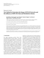
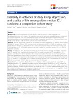
3.1.2. Linkage analysis of banana fruit pulp cell walls
Banana polysaccharides were characterized by high amounts of
water-soluble mannan and glucomannan, especially in Thap Maeo
that had 5-fold more 4-Man and 8-fold more 4,6-Man (Fig. 2A and
B).
Nanicão and Thap Maeo water-insoluble polysaccharides were
composed of medium amounts of mannans and glucomannans,
confirmed by 4-Man and 4,6-Man residues (both 4 mol%, 0.1 and
0.2 mol%, respectively) and 4-Glc (22 and 14 mol%) (Fig. 2C and D).
Both banana cultivars showed high amounts of water-insoluble
homogalacturonan and rhamnogalacturonan, especially Thap
Maeo. Thap Maeo had three-fold more 4-GalA, seven-fold more
t-Rha and 2,4-Rha and 6 fold more 2-Rha (Fig. 2C and D). In
contrast, Nanicão had more water-soluble homogalacturonan and
glucurono(arabino)xylan, containing about 2-times more 4-GalA,
2,4-Xyl, and 3,4-Xyl (Fig. 2A and B, Table 1).
T.M. Shiga et al. / Carbohydrate Polymers 164 (2017) 31–41
35
Fig. 2. Thap Maeo (Genomic group AAB – grey bars) and Nanicão (Genomic group AAA – black bars) water-soluble and water-insoluble polysaccharides linkage analysis (A
and C). The values of Thap Maeo minus Nanicão values (B and D). Monosaccharide composition considering glucose content. Rha, rhamnose; Fuc, fucose; Ara, arabinose;
Xyl, xylose; Man, mannose; Gal, galactose; Glc, glucose; GalA, galacturonic acid; GlcA, glucuronic acid; t, terminal; p, pyranose; f, furanose. N = 4.
Xyloglucan was the major hemicellulose, composed of 4-Glc,
4,6-Glc, t-Xyl, and 2-Gal, along with xylan (4-Xyl, 2,4-Xyl and 3,4Xyl)(Fig. 2A and C). Fucose was found in trace amounts. In Nanicão,
xyloglucan and xylan were more insoluble. Nanicão contained 2fold more 4-Glc and 4,6-Glc and 18 times more t-Xylp (Fig. 2C and
D).
Both cultivars showed high amounts of water-soluble arabinogalactans and galactans. However, in Thap Maeo, t-Araf and 4-Gal
were 3-fold higher in the water-insoluble polysaccharides, whilst in
water-soluble polysaccharides was 3-fold and 2-fold lower (Fig. 2C,
D, A and B, respectively).
3.2. Digestion of cell wall polysaccharides with hydrolytic
enzymes
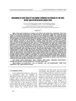
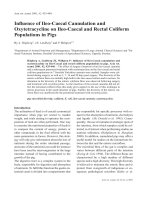
Enzymatic hydrolysis confirmed linkage analysis and revealed
that the major constituent of Nanicão and Thap Maeo banana pulp
cell wall were mannan, (Figs. 3, 4 and S2 ).
The oligomers obtained from mannanase, arabinanase and
xyloglucanase enzymatic digestion were hydrolyzed using TFA 2 M
to obtain the monosaccharide composition (Figs. S2 and 5 ). According to linkage analysis, both banana cultivar have high amounts
of homogalacturonans and small amounts of rhamnogalacturonan
I, mainly in water-insoluble polysaccharides. However, endopolygalacturonase hydrolysis produced a small peak containing high
36
T.M. Shiga et al. / Carbohydrate Polymers 164 (2017) 31–41
Table 1
Thap Maeo and Nanicão bananas cell wall polysaccharide composition. (For interpretation of the references to colour in this table, the reader is referred to the web version
of this article.)
molecular weight fragments (Figs. 3 and 4) and small amounts of
GalA (Figs. S2 and 5).
The (1 → 4)--d-mannanase-digestible mannans were found
mainly in the water-soluble polysaccharides of Thap Maeo banana
(Figs. 3, 4 and 5 and S2). The mannans in Nanicão were oligomeric,
soluble in 80% EtOH, and found even in samples that were not
treated with mannanase (Figs. S2 and 5). Only small amounts mannan from Thap Maeo pulps were solubilized by EtOH (Figs. 2 and
S2). While mannans were the predominate form of the polymer
of water-soluble polysaccharides, glucomannans predominated in
water-insoluble polysaccharides (Figs. S2 and 5). Enzymatic treatment with arabinanase released almost no oligomers (Figs. 3 and 4),
as evidenced by the low amounts of Ara released (Figs. S2 and 5).
Enzymatic treatment confirmed the results obtained by linkage
analysis. Only low amounts of 5-Araf residues were found in the
water-insoluble polysaccharides of either banana cultivars (Figs. 2
and S3), most probably released from short-chains of arabinans. On
the other hand, high amounts of 3-, 6- and 3,6-Gal were found, confirming the presence of righ amounts of arabinans. Probably, the
majority of t-Araf found belongs to arabinogalactans (Fig. 2). The
high amounts of t-Araf and 3,6-Gal in the water-soluble polysaccharide, suggests that arabinogalactans are water-soluble and has
a highly branched structure (Fig. 2A).
Xyloglucanase treatment released oligomers predominantly in
the water-insoluble fractions (Figs. 3 and 4). In both banana
cultivars, water-soluble polysaccharides had small amounts of
xyloglucan. On other hand, water-insoluble polysaccharides
showed higher amounts of xyloglucans, as can be seen by the presence of Glc, Gal and Xyl, (Figs. S2 and 5), as well as the 2-Xyl
and 4,6-Glc residues (Fig. 2). Xylanase did not produce significant amounts of oligomers despite the presence of 4- and 3,4-Xyl
(Figs. 3 and 4) and of t-Araf residues (Fig. 2).
3.2.1. Digestion of glucans with ˇ-glucanases
To confirm the chemical nature of glucose-rich polymer, the
water-insoluble polysaccharide fraction was assayed using laminarinase. The water-soluble polysaccharides do not have significant
amounts of glucose, however, almost all glucose are composed of
3,4-Glc and 4-Glc. We also tested the defatted banana pulp with
laminarinase.
Banana water-insoluble polysaccharide without pre-treatment
with protease were hydrolyzed and assayed for -glucan. Banana
pulp treated with laminarinase produced high amounts of glucose
in control samples (without laminarinase) and in samples containing enzyme (Fig. S4). Both samples produced high amounts of
glucose, showing -glucanase activity (Fig. S4).
Laminarinase hydrolyzes (1 → 3)--d- linkages of -glucans or
polysaccharide connected by -1,3 linkages containing a mixture of
(1 → 3)--d- linkages and either (1 → 4)--d- linkages or (1 → 6)-d- linkages. That means that laminarinase could be hydrolyzing
callose or there was endogenous -glucanase acting in banana
samples. To remove interference of endogenous enzymes, samples
were pre-treated with protease, and boiled for 15 min to inactivate
the protease added. Lichenase digests (1 → 4)--d-glucosyl units
in polymers that contain (1 → 3)--d-glucosyl at the non-reducing
end (Anderson & Stone, 1975), and not -d-glucans containing only
(1 → 3)--d- or (1 → 4)--d- bonds. Thus, the enzyme digests the
mixed-linkage (1 → 3)--d-, (1 → 4)--d-glucan but not cellulose
or callose.
Samples treated with protease were hydrolyzed with laminarinase and lichenase and resulted in high amounts of glucose, whilst
control produced only small amounts of glucose (Fig. S5). Both cultivars showed similar profiles (Fig. S5).
Water-soluble polysaccharides treated with laminarinase produced oligomers in a short period of time (15 min − 2 h) (Fig. 6).
Lichenase, in turn, required a very long period of hydrolysis to
T.M. Shiga et al. / Carbohydrate Polymers 164 (2017) 31–41
37
Fig. 3. Oligomer profile of Nanicão banana cell wall polysaccharides. The water-soluble and water-insoluble polysaccharides were hydrolyzed using mannanase, arabinanase,
xyloglucanase, xylanase and endopolygalacturonase. Red line, blank without enzyme; black line, samples containing enzyme. (For interpretation of the references to colour
in this figure legend, the reader is referred to the web version of this article.)
release oligomers (24 h) (Fig. 6). Hence, the digestion of both
banana cultivars using laminarinase and lichenase revealed no
(1 → 4)--d-glucosyl units containing (1 → 3)--d-glucosyl at the
non-reducing end (Fig. 6).
Oat, barley and laminarin were also hydrolyzed using laminarinase and lichenase to show the effectiveness of the enzymes used in
the -glucan assays in a well-known samples containing -glucans
(Fig. S6).
4. Discussion
4.1. Bananas polysaccharide composition
Banana
non-starch
polysaccharide
is
composed
of
(gluco)mannan and some mannan, made up of (1 → 4)--dlinked glucose, (1 → 4)--d-linked mannose and small amounts of
galacto(gluco)mannan. Homogalacturonans and rhamnogalacturonan I were the main acidic pectins in banana. Homogalacturonans
were not totally digested by endopolygalacturonase and produced
only small amounts of oligomers, the majority with high molecular
weight. The lack of digestion suggests that the enzyme could not
hydrolyze the galacturonans, possibly because galacturonans in
banana are acetylated or methylated.
The neutral and acidic pectin and hemicellulose ratios were different between cultivars revealing structural differences in the cell
wall composition. Differences in the genomic group may be correlated to those differences. The differences in the polysaccharide
composition and solubility may affect banana texture loss during
ripening, as shown in our previous work (Shiga et al., 2011).
Just small amounts of arabinan was detected in both banana
pulps. The majority of t-Araf were more likely released from arabinogalactan rather than arabinan. The large amounts of t-Araf
residues along with low amount of 5-Araf revealed that banana
pectin has short branches of arabinan. Corroborating the results of
linkage analysis, the enzymatic hydrolysis with arabinanase did not
produced high amounts of oligomers neither arabinose monomers
after TFA hydrolysis.
38
T.M. Shiga et al. / Carbohydrate Polymers 164 (2017) 31–41
Fig. 4. Oligomer profile of Thap Maeo banana cell wall polysaccharides. The water-soluble and water-insoluble polysaccharides were hydrolyzed using mannanase, arabinanase, xyloglucanase, xylanase and endopolygalacturonase. Red line, blank without enzyme; Black line, samples containing enzyme. (For interpretation of the references
to colour in this figure legend, the reader is referred to the web version of this article.)
There are two different types of arabinogalactan in banana pulp.
Type I arabinogalactan, composed of (1 → 4)--d-galactose with
t-Araf sidechain attached at the O-3 of the galactose units. Type
II arabinogalactan that has a short (1 → 3)--d- and (1 → 6)--dgalactan chains connected to each other by (1 → 3, 1 → 6)-linked
branch point residues. Type II arabinogalactan constitute the major
arabinogalactan in banana cell wall pulp, whilst type I arabinogalactan in average amounts.
Cultivars Nanicão and Thap Maeo showed similarities in their
polysaccharide compositions when water-soluble and waterinsoluble polysaccharides were combined (Figs. 2 and Fig. S3).
Basically, the main difference between both cultivars was correlated to the polysaccharides solubility and mannan content
(Table 1). In Thap Maeo, galacturonans, xyloglucan and xylans
showed to be more insoluble in water, while mannans were far
more soluble.
In terms of its nutritional characteristics, banana cell wall
has an interesting polysaccharide composition, since it is rich in
mannans, galactans and galacturonans. Studies conducted with
various plant materials showed that neutral polysaccharides such
as arabinogalactans, arabinoxylans and mannans have a potential immunomodulatory activity (Classen, Thude, Blaschek, Wack
& Bodinet, 2006; Im et al., 2010; Ramberg, Nelson, & Sinnott, 2010;
Liu, Willför, & Xu, 2015).
According to our previous works results, banana has high
amounts of mannose-rich polysaccharides and both, Thap Maeo
and Nanicão non-starch polysaccharides, are able to activate
macrophages (Cordenunsi et al., 2008; Shiga et al., 2011; Sansone,
Miranda Brito Sansone, Shiga, & Nascimento, 2016). However, the
effects depended of concentration and cultivar origin.
Polysaccharide immunomodulatory activity is associated with
the highly branched structure of arabinogalactans (Paulsen &
Barsett, 2005). According to ours results, banana has arabinogalactans containing high amounts of t-Araf side-chains. Banana
showed high amounts of 3-, 6- and 3,6- and 3,4-Gal, characteristic
of arabinogalactans. Hence, the majority of t-Araf residues seems
to belong to arabinogalactans rather than to arabinans and are
attached to galactose at O-3 position.
T.M. Shiga et al. / Carbohydrate Polymers 164 (2017) 31–41
39
Fig. 5. Hydrolysis profile of Thap Maeo banana cell wall oligomers using 2 M TFA. The water-soluble and water-insoluble polysaccharides were hydrolyzed using mannanase,
arabinanase, xyloglucanase, xylanase and endopolygalacturonase and the oligomers obtained hydrolyzed with 2 M TFA. Red line, standard; Black line, samples containing
monosaccharides hydrolyzed by endopolygalacturonase. (For interpretation of the references to colour in this figure legend, the reader is referred to the web version of this
article.)
40
T.M. Shiga et al. / Carbohydrate Polymers 164 (2017) 31–41
Fig. 6. Water-soluble polysaccharides from Nanicao and Thap Maeo bananas, digested with lichenase for 8 h, 16 h and 24 h and with laminarinase for 2 h. Red line, blank
without enzyme; Black line, samples containing enzyme. (For interpretation of the references to colour in this figure legend, the reader is referred to the web version of this
article.)
In our previous work, it was also observed high amounts of glucose in banana polysaccharides. Hence, in the present work we
decided to study the structure of banana glucans, since -glucans
is known to activate the immune system.
Samples hydrolyzed with laminarinase and control samples
(without laminarinase) released large amounts of glucose. Glucose
could be originated from hydrolysis of -glucan by laminarinase or
by endogenous -glucanase. When samples were pre-treated with
protease, and hydrolyzed by laminarinase, the amount of glucose
released by control (without laminarinase) did not reduce significantly. Samples pre-treated with protease were then hydrolyzed
with lichenase and released high amounts of glucose. Control samples (pre-treated with protease, without lichenase) released low
amounts of glucose, showing that banana has -glucanase activity
that was inactivated by protease treatment.
Presence of mixed-linkage glucans was not found, since
lichenase produced oligomers only after 24 h of hydrolysis.
Lichenase, hydrolyzes (1 → 4)--d-glucosidic linkages in polymers
containing (1 → 3) and (1 → 4) bonds, however, do not hydrolyzes
-d-glucans containing only (1 → 3) or (1 → 4) bonds. Hence,
lichenase cannot hydrolyze callose. Laminarinase is an endoglucanase that hydrolyzes (1–3) or (1–4) linkages only when the
glucose residue whose reducing group is involved in the linkage
to be hydrolyzed is itself substituted at C-3. The oligomers produced by laminarinase probably were derived from callose, which
was found in large amounts in banana cell wall.
5. Conclusions
Banana fruit cell wall is characterized by high amounts of
highly branched arabinans and mannans and small amounts of
short-chains of arabinans and galacto(gluco)mannan. Homogalacturonans and rhamnogalacturonan I were the main acidic pectins in
banana and were resistant to hydrolysis by endopolygalacturonase.
Thap Maeo and Nanicão differ with respect to its cell wall
polysaccharide composition ratio and solubility of polysaccharides
and, in proportion of each polysaccharide in the water-soluble and
water-insoluble fractions. Both cultivars also showed that banana
fruits may have a potential biological activity, mainly due to its high
arabinogalactan and mannan contents. Thap Maeo had more watersoluble galacturonans, galactans, arabinogalactans and -glucan as
callose, hence more -glucanase activity. However, in both cultivars studied mixed-linkage glucans were not present. This is an
important characteristic, since it makes Thap Maeo to have higher
probability to producing immunomodulatory activity.
T.M. Shiga et al. / Carbohydrate Polymers 164 (2017) 31–41
Acknowledgements
The authors acknowledge financial support of Núcleo de Apoio à
Pesquisa em Alimentos e Nutric¸ão – NAPAN and Coordenac¸ão para
a Pesquisa de Nível Superior – CAPES, for financial support (Process
BEX-10734/13-9).
We also thank the valuable help of Anna T. Olek, Matheus
Romanos Benatti and John F. Klimek from Purdue University for
their help with sugar and linkage analysis and Marcelo Sansone
from Department of Food Science and Experimental Nutrition from
University of São Paulo for his help in cell-wall material isolation.
Appendix A. Supplementary data
Supplementary data associated with this article can be found, in
the online version, at />079.
References
Anderson, M. A., & Stone, B. A. (1975). A new substrate for investigating the
specificity of -glucan hydrolases. FEBS Letters, 52, 202–207.
Bennett, R. N., Shiga, T. M., Hassimotto, N. M. A., Rosa, E. A. S., Lajolo, F. M., &
Cordenunsi, B. R. (2010). Phenolics and antioxidant properties of fruit pulp and
cell wall fractions of postharvest banana (Musa acuminata Juss.) cultivars.
Journal of Agricultural and Food Chemistry, 58, 7991–8003.
Blackwood, A. D., Salter, J., Dettmar, P. W., & Chaplin, M. F. (2000). Dietary fibre,
physicochemical properties and their relationship to health. Journal of the
Royal Society for the Promotion of Health, 120, 242–247.
Carpita, N. C., & McCann, M. C. (1996). Some new methods to study plant
polyuronic acids and their esters. In R. Townsend, & A. Hotchkiss (Eds.),
Progress in glycobiology (pp. 61–595). New York: Marcel Dekker.
Carpita, N. C., & Shea, E. M. (1989). Linkage structure of carbohydrates by gas
chromatography–mass spectrometry (CG–MS) of partially methylated alditol
acetates. In C. J. Biermann, & G. D. McGinnis (Eds.), Analysis of carbohydrates by
GLC and MS (pp. 157–216). Boca Raton: CRC Press.
Classen, B., Thude, S., Blaschek, W., Wack, M., & Bodinet, C. (2006).
Immunomodulatory effects of arabinogalactan-proteins from Baptisia and
Echinacea. Phytomedicine, 13, 688–694.
41
Cordenunsi, B. R., Shiga, T. M., & Lajolo, F. M. (2008). Non-starch polysaccharide
composition of two cultivars of banana (Musa acuminata L. cvs Mysore and
Nanicão). Carbohydrate Polymers, 71, 26–31.
Filisetti-Cozzi, T. M. C. C., & Carpita, N. C. (1991). Measurement of uronic acid
without interference from neutral sugars. Analytical Biochemistry, 197, 57–162.
Gibeaut, D. M., & Carpita, N. C. (1991). Clean-up procedure for partially methylated
alditol acetate derivatives of polysaccharides. Journal of Chromatography,
587(2), 284–287.
Im, S. A., Lee, Y. R., Lee, Y. H., Lee, M. K., Park, Y. I., Lee, S., et al. (2010). In vivo
evidence of the immunomodulatory activity of orally administered Aloe vera
gel. Archives of Pharmacal Research, 33(3), 451–456.
Kim, J. B., & Carpita, N. C. (1992). Changes in esterification of uronic acid groups of
cell wall polysaccharides during elongation of maize coleoptiles. Plant
Physiology, 98, 646–653.
Liu, J., Willför, S., & Xu, C. (2015). A review of bioactive plant polysaccharides:
Biological activities, functionalization, and biomedical applications. Bioactive
Carbohydrates and Dietary Fibre, 5, 31–61.
Mobambo, K. N., Gauhl, F., Vuylsteke, D., Ortiz, R., Pasberg-Gauhl, C., & Swennen, R.
(1993). Yield loss on plantain from Black Sigatoka leaf spot and field
performance of resistant hybrids. Field Crops Research, 35, 35–42.
Onyenekwe, P. C., Okereke, O. E., & Owolewa, S. O. (2013). Phytochemical screening
and effect of musa paradisiaca stem extrude on rat haematological parameters.
Current Research Journal of Biological Sciences, 5(1), 26–29.
Paulsen, B. S., & Barsett, H. (2005). Bioactive pectic polysaccharides. Advances in
Polymer Science, 186, 69–101.
Ramberg, J. E., Nelson, E. D., & Sinnott, R. A. (2010). Immunomodulatory dietary
polysaccharides: A systematic review of the literature. Nutrition Journal, 9,
54–75.
Sansone, M., Miranda Brito Sansone, A. C., Shiga, T. M., & Nascimento, J. R. O.
(2016). The water-soluble non-starch polysaccharides from bananas display
immunomodulatory properties on cultured macrophages. Food Research
International, 87, 125–133.
Sansone, A. C. M. B., Sansone, M., Santos Dias, C. T., & Nascimento, J. R. O. (2016).
Oral administration of banana lectin modulates cytokine profile and
abundance of T-cell populations in mice. International Journal of Biological
Macromolecules, 89, 19–24.
Scarminio, V., Fruet, A. C., Witaicenis, A., Rall, V. L., & Di Stasi, L. C. (2012). Dietary
intervention with green dwarf banana flour (Musa sp AAA) prevents intestinal
inflammation in a trinitrobenzenesulfonic acid model of rat colitis. Nutrition
Research, 32(3), 202–209.
Shiga, T. M., Soares, C. A., Nascimento, J. R. O., Purgatto, E., Lajolo, F. M., &
Cordenunsi, B. R. (2011). Ripening-associated changes in the amounts of starch
and non-starch polysaccharides and their contributions to fruit softening in
three banana cultivars. Journal of the Science of Food and Agriculture, 91,
1511–1516.