Fucogalactan from the giant mushroom Macrocybe titans inhibits melanoma cells migration
Bạn đang xem bản rút gọn của tài liệu. Xem và tải ngay bản đầy đủ của tài liệu tại đây (1.05 MB, 7 trang )
Carbohydrate Polymers 190 (2018) 50–56
Contents lists available at ScienceDirect
Carbohydrate Polymers
journal homepage: www.elsevier.com/locate/carbpol
Fucogalactan from the giant mushroom Macrocybe titans inhibits melanoma
cells migration
T
Shayane da Silva Milhorinia, Fhernanda Ribeiro Smiderlea, Stellee Marcela Petris Biscaiab,
⁎
Fabio Rogerio Rosadoc, Edvaldo S. Trindadeb, Marcello Iacominia,
a
Department of Biochemistry and Molecular Biology, Federal University of Parana, CEP 81531-980, Curitiba, PR, Brazil
Department of Cellular Biology, Federal University of Parana, CEP 81531-980, Curitiba, PR, Brazil
c
Department of Biosciences, Federal University of Parana, CEP 85950-000, Palotina, PR, Brazil
b
A R T I C L E I N F O
A B S T R A C T
Keywords:
Macrocybe titans
Fucogalactan
Chemical characterization
Melanoma B16-F10
An aqueous extract containing polysaccharides was obtained from the giant mushroom Macrocybe titans, and it
was purified by amylase treatment, freeze-thawing process and dialysis. The purified fraction (ESP) was analyzed by HPSEC and GC–MS which showed a homogenous polysaccharide with Mw 14.2 × 10³ g/mol composed
by galactose and fucose. NMR and methylation analysis of ESP confirmed the presence of a fucogalactan with a
(1 → 6)-linked α-D-Galp main chain partially substituted at O-2 by non reducing end units of α-L-Fucp residues in
the side chain. Its biological activity was evaluated against murine melanoma cells B16-F10. The fucogalactan
did not alter the viability, proliferative capacity and morphology of cells. However, this polysaccharide was able
to reduce the cell migration in vitro at 40% (100 μg/mL) and 33% (250 μg/mL). The results obtained showed that
Macrocybe titans fucogalactan is a promising agent capable of altering melanoma cell migration without decrease
the cell viability.
1. Introduction
For millennia, mushrooms have been used in culinary and folk
medicine by mankind, not only for their unique taste, but also because
of their nutritional value and bioactive compounds (Patel & Goyal,
2012; Ruthes, Smiderle, & Iacomini, 2016). A great variety of bioactive
molecules has been isolated from mushrooms, such as proteins (Zhang
et al., 2014), glycoproteins (Cui et al., 2013), secondary metabolites
(Baby, Johnson, & Govindan, 2015), lipopolysaccharides (Wasser,
2011) and polysaccharides (Moreno et al., 2016; Meng, Cheng, Han,
Chen, & Wang, 2017). The latter molecules are potent antitumor and
immunomodulating agents isolated from mushrooms, according to
studies dated from 1976 until now (Patel & Goyal, 2012; Smiderle,
Ruthes, & Iacomini, 2014). These medicinal molecules have been obtained from several mushrooms, such as Agaricus bisporus (Ruthes,
Rattmann, Carbonero, Gorin, & Iacomini, 2012), Agaricus brasiliensis
(Komura et al., 2010), Macrolepiota dolichaula (Samanta et al., 2015),
Lactarius rufus (Ruthes et al., 2012), Flammulina velutipes (Smiderle,
Carbonero, Sassaki, Gorin, & Iacomini, 2008), Lentinus edodes
(Carbonero et al., 2008), Pleurotus sajor-caju (Silveira et al., 2015),
among others. However, there are some mushroom species that are
unexplored and may have a medicinal potential that is still unknown,
⁎
such as the giant edible mushroom Macrocybe titans (H.E. Bigelow &
Kimbr.) Pegler, Lodge & Nakasone. This species is large and its fruit
body is white and firm and grows in large caespitose clusters which may
exceed 30 kg of fresh weight (Pegler, Lodge, & Nakasone, 1998). Despite of its peculiar morphology and popular consumption, M. titans has
never been studied about its polysaccharides and there are no reports
about its biological properties. Therefore, considering its large size, this
mushroom can be a promising source to obtain different bioactive
molecules in great yields, including the polysaccharides.
The biologically active mushroom polysaccharides that were mostly
isolated and studied from several Basidiomycetes are the β-D-glucans
(Smiderle, Sassaki, Griensven, & Iacomini, 2013). Besides that, there is
another class of mushroom polysaccharides that presents more than one
monosaccharide, and variable anomeric configuration, linkage and
branching type, including the presence of methyl groups in its structure
(Ruthes et al., 2016). Among these polysaccharides, there are the heterogalactans, which are mostly composed by a main chain of (1 → 6)linked α-D-Galp units, some of them partially methylated and substituted mainly by fucose or mannose (Rosado et al., 2003; Komura
et al., 2010; Ruthes et al., 2013).
Heterogactans have been largely explored about their biological
activities, such as immunomodulatory (Sun & Liu, 2009; Samanta et al.,
Corresponding author at: Department of Biochemistry and Molecular Biology, Federal University of Parana, CP 19046, Curitiba, PR, Brazil.
E-mail address: (M. Iacomini).
/>Received 21 November 2017; Accepted 21 February 2018
Available online 23 February 2018
0144-8617/ © 2018 Elsevier Ltd. All rights reserved.
Carbohydrate Polymers 190 (2018) 50–56
S.d.S. Milhorini et al.
pressure and precipitated with ethanol (3:1; v/v). The precipitated
polysaccharides were obtained by centrifugation (10.000 rpm, at 4 °C,
for 20 min) and dialyzed against distilled water (6–8 kDa). Dialysis was
stopped when the eluted material contained no sugar, which was verified by phenol-sulfuric acid method (Dubois, Gilles, Hamilton, Rebers,
& Smith, 1956). The polysaccharide fraction (P) was treated with αamylase (Sigma-Aldrich) and precipitated with ethanol (3:1; v/v). The
precipitate was dialyzed against tap water (6–8 kDa, for 24 h), concentrated under reduced pressure and submitted to freezing and slow
thawing (3×) until complete precipitation of cold-water insoluble
polysaccharides (Gorin & Iacomini, 1984). After centrifugation
(10.000 rpm, at 4 °C, for 20 min), the soluble material, which contained
the cold-water soluble polysaccharides (SP), was purified by dialysis
against distilled water through a membrane of 1000 kDa Mw cut-off,
giving rise to retained (RSP) and eluted (ESP) fractions, that were
concentrated under reduced pressure and freeze-dried. The yield of
each fraction was calculated in comparison to the initial weight of dried
mushroom.
2015), anti-inflammatory and antinociceptive (Komura et al., 2010;
Silveira et al., 2015) anti-sepsis (Ruthes et al., 2012), anti-oxidative
(Ding, Hou, & Hou, 2012; Samanta et al., 2015) and also antitumor
activities (Jeff et al., 2013; Wang, Sun, Wu, Yang, & Tan, 2014). The
latter one has showed great interest by the scientific and medical
community because tumors are one of the main cause of morbidity and
death worldwide (Bae, Jang, Yim, & Jin, 2005; Liu, Zeng, Li, & Shi,
2016, Tian, Zhao, Zeng, Zhang, & Zheng, 2016). According to the World
Health Organization (WHO/Cancer, 2018), cancer related-deaths were
totalized 8.8 millions in 2015, which implies that approximately 1 in 6
deaths in the world is due to cancer. Among the cancer types, the skin
cancer classified as melanoma is one of the most aggressive with the
highest rates of metastasis and mortality (Harries et al., 2016). It is the
eighth most common cancer that affects people from developed countries (Hamel et al., 2016). There are some studies that demonstrated
activity of mushroom polysaccharides against melanoma cells in vitro
(Bae et al., 2005; Han et al., 2006) and antitumor activity of such
molecules in vivo (Zhang, Yang, Chen, Hou, & Han, 2005).
Based on this, the aim of this study was to obtain polysaccharides
from Macrocybe titans fruiting bodies, that could exhibit some activity
against melanoma cells. Furthermore, the active polysaccharide was
purified and its chemical structure was carefully analyzed until complete elucidation.
2.3. Analysis of monosaccharide composition by GC–MS
The polysaccharide fractions (1 mg) were hydrolyzed with 2 M TFA
at 100 °C for 8 h, followed by evaporation to dryness. The dried carbohydrate samples were dissolved in distilled water (100 μL) and 1 mg
NaBH4 was added. The solution was held at room temperature overnight to reduce aldoses into alditols (Sassaki et al., 2008). The product
was dried and excess NaBH4 was neutralized by the addition of acetic
acid, and removed by the addition of methanol (×2) under a compressed air stream in a fume hood. Acetylation of the alditols was
performed in pyridine–Ac2O (200 μL; 1:1, v/v), heated for 30 min at
100 °C. The resulting alditol acetates were analyzed by GC–MS, and
identified by their typical retention times and electron impact profiles.
The relative percentage of correspondent monosaccharides was calculated by determination of each peak area with a Varian CP-3800 gas
chromatograph coupled to an Ion-Trap 4000 mass spectrometer, using a
VF5 column (30 m × 0.25 mm i.d.) programmed from 100 to 280 °C at
10 °C min−1, with He as carrier gas.
2. Material and methods
2.1. Biological material
The mushroom Macrocybe titans was kindly donated by Dr. Fábio
Rogério Rosado from the Department of Bioscience, Federal University
of Paraná – Palotina-PR, Brazil.
2.2. Extraction and purification procedures
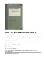
The dried and powdered fruiting bodies (300 g) were submitted to
several purification steps as shown in Fig. 1. Firstly, the apolar compounds were extracted with CHCl3:MeOH (2:1; v/v) at 60 °C during 3 h
(3×). Subsequently, the resulting residue was dried and extracted with
distilled water at room temperature under mechanical stirring during
6 h (3×). The aqueous extract was concentrated under reduced
2.4. Methylation analysis
Per-O-methylation of the purified polysaccharide (ESP; 5 mg) was
carried out using NaOH-Me2SO-MeI (Ciucanu & Kerek, 1984). After
isolation of the products by neutralization (HOAc), dialysis, and evaporation, the methylation process was repeated. The per-O-methylated
derivatives were hydrolyzed using 45% aqueous formic acid (1 mL) for
8 h at 100 °C followed by evaporation to dryness. The residue was
converted into partially O-methylated alditol acetates by reduction with
NaBD4 and acetylation with pyridine–Ac2O as describe above (Section
2.3), giving rise to a mixture of partially O-methylated alditol acetates,
which was analyzed by GC–MS using a VF5 capillary column as described above (Section 2.3). The derivatives were identified by their m/
z of positive ions and retention time, by comparison with standards, and
the results were expressed as relative percentage of each component
(Sassaki, Gorin, Souza, Czelusniak, & Iacomini, 2005).
2.5. Determination of homogeneity of polysaccharide fractions and
molecular weight (Mw) of fucogalactan
The homogeneity of polysaccharide fractions (P, SP and ESP) was
determined by high-performance size-exclusion chromatography
(HPSEC) coupled to refractive index detector. Four gel-permeation
Ultrahydrogel columns in series with exclusion sizes of 7 × 10⁶,
4 × 10⁵, 8 × 10⁴, and 5 × 10³ Da were used. The eluent was 0.1 M aq.
NaNO2 containing 200 ppm aq. NaN3 at 0.6 mL min−1. The sample,
previously filtered through a membrane (0.22 μm), was injected
(100 μL loop) at a concentration of 1 mg mL−1.
Fig. 1. Scheme of extraction and purification of M. titans fucogalactan.
51
Carbohydrate Polymers 190 (2018) 50–56
S.d.S. Milhorini et al.
2.6. Determination of relative molar mass
Table 1
Yields and monosaccharide composition of fractions obtained from M. titans.
The retention time of fucogalactan (ESP) was compared with a
curve of dextran patterns of different molecular masses (Sigma-Aldrich)
(5.00 × 10³; 9.40 × 10³; 1.72 × 10⁴; 4.02 × 10⁴; 7.22 × 10⁴;
1.24 × 10⁵; 2.66 × 10⁵; 4.87 × 10⁵; 2.00 × 10⁶), aiming to estimate its
relative molecular weight.
Fraction
P
SP
ESP
Yield (%)a
4.1
2.0
0.8
Monosaccharides (%)b
Fuc
Man
Gal
Glc
2.7
13.7
17
6.2
7.3
–
30.3
60.2
83
60.8
18.8
–
2.7. Nuclear magnetic resonance (NMR) spectroscopy
a
b
NMR spectra (coupled-HSQC; HSQC-DEPT; COSY; TOCSY) were
obtained using a 400 MHz Bruker model Avance III spectrometer with a
5 mm inverse probe. The analyses were performed at 70 °C and samples
(20 mg) were dissolved in D2O. Chemical shifts are expressed in ppm
(δ) relative to resonance of acetone at δ 30.2 and 2.22 corresponding to
13
C and 1H signals, respectively.
Yields relative to fungus dry weight.
Alditol acetates obtained after hydrolysis, NaBH4 reduction, and acetylation.
2.8. Cell culture
The cells used for cell culture were B16-F10 (BCRJ – Banco de
Células do Rio de Janeiro), maintained in Dulbecco’s Modified Eagle’s
Medium – DMEM (ThermoFisher, Waltham, MA, EUA, CAT 12800017), supplemented with 10% (v/v) fetal bovine serum (FBS)
(ThermoFisher, 12657), 10 mM Hepes (Sigma-Aldrich, H-4034),
0,25 μg/mL penicillin-streptomycin in 0,85% saline (ThermoFisher,
15140-148), 3.7 g/L sodium bicarbonate (ThermoFisher, 25080094) at
37 °C in 5% CO2 in humidified atmosphere.
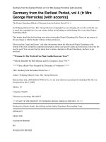
Fig. 2. Elution profile of P, SP and ESP fraction determined by HPSEC (refractive index
detector), eluted with 0.1 M NaNO2.
composition (Table 1) and HPSEC (Fig. 2) of the samples obtained after
precipitation with ethanol (P), freeze-thawing treatment (SP), and
elution through dialysis membrane (ESP). Crude polysaccharide extract
(P) contained fucose, mannose, galactose and glucose. This fraction (P)
showed a heterogeneous elution profile on HPSEC (Fig. 2) by the presence of two blunt peaks (at ∼43 min and ∼53 min), confirming the
necessity of further purification steps.
Fungi stock energy is glycogen and some species may contain high
amounts of this polysaccharide, which is easily removed by α-amylase
treatment (Synytsya & Novák, 2013). Therefore, this enzymatic hydrolysis was performed followed by freeze and thawing process, giving
rise to a soluble polysaccharide fraction (SP), after centrifugation. Its
elution profile on HPSEC still contained two peaks, although the one at
∼43 min was sharpened (Fig. 2) and the glucose content reduced
drastically (Table 1), indicating that a partial purification occurred.
Considering the difference of molecular weight observed on SP chromatogram (Fig. 2), a dialysis through a membrane of 1000 kDa Mw cutoff was performed as purification step. Only fucose and galactose were
detected on the eluted fraction (ESP), and its HPSEC profile showed
only one sharp peak at ∼53 min (Fig. 2). The molecular weight of the
purified fraction ESP was estimated at 14.2 × 10³ g/mol.
The methylation analysis (Table 2) was consistent with a polysaccharide composed of (1 → 6)-linked Galp main chain, indicated by
the presence of 2,3,4-Me3-Galp (56%) derivatives. This fraction also
presented 3,4-Me2-Galp (21.9%) and 2,3,4-Me3-Fucp (20.7%) derivates,
indicating that the main chain is branched at O-2 position by non reducing end units of Fucp in the proportion of each 2–3 units of Galp.
2.9. Viability and proliferation assays
The B16-F10 cells were cultured in 96-well plates (600 cells/well).
After 24 h, the cells were exposed to treatment (100 or 250 μg/mL) for
24, 48 or 72 h, with fucogalactan. Afterwards, the cell viability was
determined using Neutral Red (Sigma-Aldrich, N6634) as described in
Borenfreund & Puerner (1985), and proliferation assay was performed
using protocol by Gillies, Didier and Denton (1986), in the same plate
(Chiba, Kawakami, & Tohyama, 1998) with modifications. All experiments were compared to cells in the absence of treatment.
2.10. Morphology and migration assays
The morphology and cell migration were determined using a microscope EVOS FL Auto (ThermoFisher). Therefore cells (2.5 × 10⁴)
were cultured in 6-well plates, exposed or not (control) to fucogalactan
(100 and 250 μg/mL), for 72 h. After treatment, images were collected
using phase contrast filter for analysis of morphology. The scratch was
performed with a 10 μL tip, cells were washed 2 times with medium,
and finally added medium with 1% SFB. The images were collected
every 30 min, for 10 h. This assay was performed according to the
protocol describe by Liang, Park and Guan, (2007) with modifications.
The images were analyzed using a T-scratch program (free software by
CSELab, ETH, Zurich, Switzerland).
2.11. Statistical analysis
Table 2
Partially-O-methylalditol acetates formed on methylation analysis of fucogalactan isolated from M. titans.
Statistical analyses was performed using GraphPad Prism 5.0 software (GraphPad Software®, Inc.), being considered different p < 0.05
value, in different parametric tests, as indicated by each legend.
3. Results and discussion
3.1. Isolation and structural characterization of fucogalactan from M. titans
Partially O-methylated alditol acetatesa
Sample (mol%)b
Linkage typec
2,3,4-Me3-Fucp
2,3,4,6-Me4-Galp
2,3,4-Me3-Galp
3,4-Me2-Galp
20.7
1.4
56.0
21.9
Fucp-(1→
Galp-(1→
6→)-Galp-(1→
2,6→)-Galp-(1→
a
Analyzed by GC–MS, after methylation, total acid hydrolysis, reduction with NABD4
and acetylation.
b
% of peak area relative to total peak area.
c
Based on derived O-methylalditol acetates.
The dried and milled mushroom was used to prepare polysaccharide
extracts that were purified as described above (Section 2.2). The purification process was accompanied by analyses of monosaccharide
52
Carbohydrate Polymers 190 (2018) 50–56
S.d.S. Milhorini et al.
Fig. 3. HSQC-DEPT spectrum of fucogalactan (ESP), in D2O at 70 °C (chemical shifts are expressed in ppm). Correlations of CH2 groups are flipped to negative phase, and are shown in
grey color.
Fig. 4. Viability of B16-F10 cells determined by Neutral Red Assay.
C = control (untreated cells); Treated = 100 or 250 μg/mL of
Fucogalactan; Time 24, 48 and 72 h. The results are representative of three
independent experiments with technical quadruplicate. Data are shown as
mean ± SD, statistical analysis: One-way ANOVA, Tukey post-test, no
difference compared with control group.
Fig. 5. Proliferation of B16-F10 cells determined by Crystal Violet Assay.
C = control (untreated cells); Treated = 100 or 250 μg/mL of
Fucogalactan; Time: 24, 48 and 72 h. The results are representative of
three independent experiments with technical quadruplicate. Data are
shown as mean ± SD, statistical analysis: One-way ANOVA, Tukey posttest, no difference compared with control group.
53
Carbohydrate Polymers 190 (2018) 50–56
S.d.S. Milhorini et al.
Fig. 6. Morphology of B16-F10 cells after incubation with fucogalactan. Cells were treated with fucogalactan (100 μg/mL or 250 μg/mL), for 72 h and the treatment did not alter the cell
morphology. Control cells received no treatment.
which was cell migration.
It is known that cell migration is one of the major cellular events
and it is considered a hallmark of cancer, because it is through this
mechanism that tumor cell can achieve metastasis and thus generate
new secondary tumors (Hanahan & Weinberg, 2000, 2011).
After incubation of B16-F10 cells with fucogalactan, it was observed
that this polysaccharide was able to change the migration behavior
(Fig. 7). As shown in Fig. 7, it can be seen that after 10 h the control
cells migrated until closing the scratched area (0% open area). However, the cells treated with both concentrations of fucogalactan (100 or
250 μg/mL) still presented an average open area of 40% and 33%, respectively, after the same period. The inhibition of cell migration was
statistically different (**** p < 0.0001) when treated groups were
compared with control group.
By the results obtained, it was demonstrated that the fucogalactan
extracted from Macrocybe titans does not alter the viability, proliferation or morphology of melanoma cells. However, it was able to reduce
their migration, showing that this polysaccharide presented some antitumor effect in vitro even with no toxicity against the tested cells.
Many authors correlate antitumor capacity of some drugs with cytotoxicity or loss of cell viability (Ivanova, Krupodorova, Barshteyn,
Artamonova, & Shlyakhovenko, 2014; Shang et al., 2011; Srinivasahan
& Durairaj, 2015). However, in this case, there is a great possibility of
such drugs to damage or kill normal and healthy cells. Thus, recently
there is a search for compounds that do not alter the cellular viability,
but rather cause cellular modifications that lead to antitumor effects,
with minimum side effects (Biscaia et al., 2017; Meng, Liang, & Luo,
2016; Novaes, Valadares, Reis, Gonỗalves, & Menezes, 2011; Tong
et al., 2009). Based on this, the results presented on this study showed a
promising agent against melanoma cells, that is candidate of new studies to evaluate its antitumor effect in vivo.
The NMR analyses (coupled-HSQC; COSY; TOCSY, data not shown)
contributed to elucidate the chemical structure of the fucogalactan and
the signals were assigned according to the correlation among the NMR
spectra (coupled-HSQC; HSQC-DEPT; COSY; TOCSY) and to literature
values of similar fucogalactans isolated from other basidiomycetes
(Ruthes et al., 2012; Ruthes et al., 2013). The HSQC-DEPT (Fig. 3)
showed signals in the anomeric region (C-1/H-1) at δ 101.5/5.07, 98.2/
4.98 and 98.1/5.03 corresponding to Fucp units, 6-O and 2,6-di-Osubstituted Galp units, respectively. The α-configuration of Galp and
Fucp units was confirmed by the values of the coupling constants JC-1/H1
13
1 observed in H/ C coupled-HSQC spectrum (data not shown), which
was 171 Hz for Galp and 170,6 Hz for Fucp units (Perlin and Casu,
1969). The substitution at O-2 of 2,6 → )-D-Galp-(1→ was confirmed by
signals at δ 77.8/3.84 and 77.8/3.82. Furthermore, the O-6 substitution
of all Galp units was identified by the negative phase resonances at δ
66.8/3.71 and 66.8/3.91. The signals at δ 15.6/1.25 and 15.5/1.23 are
attributed to CH3 of Fucp units.
The data showed that the M. titans fucogalactan is composed of a
(1 → 6)-linked α-D-Galp main chain that is partially substituted at O-2
position by non reducing end units of α-L-Fucp residues.
3.2. Biological activity of fucogalactan from M. titans
The literature data has shown that several polysaccharides present
antitumor activities (Ale, Maruyama, Tamauchi, Mikkelsen, & Meyer,
2011; Hung, Hsu, Chang, & Chen, 2012; Srinivasahan & Durairaj, 2015;
Tong et al., 2009; Zong, Cao, & Wang, 2012), and fucogalactans of
mushrooms have already been evaluated for their biological effect,
presenting a reduction in late mortality rate caused by polymicrobial
sepsis (Ruthes et al., 2012); antinoceptive and anti-inflammatory activities in mice (Komura et al., 2010; Ruthes et al., 2013); and an increase of the expression of TNF-α gene and NO by macrophages
(Mizuno et al., 2000).
In the present work, the effect of the fucogalactan isolated from M.
titans on melanoma tumor cells was evaluated. The cells were exposed
to different concentrations of fucogalactan, for different periods to
determine their viability after incubation with the polysaccharide. No
difference was observed among the treatments and controls (Fig. 4).
Proliferation of tumor cells is an important target when it is desired
to develop antitumor drugs (Ale et al., 2011; Hung et al., 2012).
Therefore, the fucogalactan was evaluated on a cell proliferation assay,
however no alteration was observed after incubation of B16-F10 cells
with different concentrations of fucogalactan (Fig. 5). The next step was
to assess whether the treatment could affect the morphology of cells.
Tumor cell morphology is quite characteristic, as seen in Fig. 6
(control), showing adherent cells as well as rounded cells. The treated
groups (100 or 250 μg/mL) presented the same morphology of cancer
cells: as spindle-shaped and epithelial-like cells and stacked up on each
other (Fig. 6).
Thus, without changes in cell viability, proliferation and morphology, we then analyzed an important cell malignancy parameter,
4. Conclusion
A heteropolysaccharide was obtained from the giant edible mushroom Macrocybe titans by aqueous extraction. It was chemically characterized and its biological activity on melanoma cells was evaluated.
GC–MS, NMR and HPSEC analyzes showed that this polysaccharide is a
homogeneous fucogalactan with a (1 → 6)-linked α-D-Galp main chain
partially substituted at O-2 by non reducing end units of α-L-Fucp residues in the side chain. The activity of Macrocybe titans fucogalactan
against B16-F10 cells was firstly reported in this study. This polymer
showed a promising antitumor effect because it did not affect the cell
viability, proliferation and morphology; however it inhibited the migration of melanoma cells. Considering that M. titans is a mushroom
that reaches around 30 kg and therefore could provide high yields of
polysaccharides; and that the purified fucogalactan showed potential
effect against melanoma cells, the study of this mushroom showed to be
interesting and should be continued in vivo.
54
Carbohydrate Polymers 190 (2018) 50–56
S.d.S. Milhorini et al.
Fig. 7. Cell migration Assay. Cells were treated with fucogalactan (100 μg/mL or 250 μg/mL), for 72 h. Control cells received no treatment. On time 0 h, a scratch was produced on cells
and the migration process was recorded during 10 h. Fucogalactan altered cell migration in both concentrations. Data are shown as mean ± SD, statistical analysis: One-way ANOVA,
Dunnett post-test, **** p < 0.0001 compared with control group.
Acknowledgments
Phytochemistry, 114, 66–101. />Bae, J. S., Jang, K. H., Yim, H., & Jin, H. K. (2005). Polysaccharides isolated from Phellinus
gilvus inhibit melanoma growth in mice. Cancer Letters, 218, 43–52. .
org/10.1016/j.canlet.2004.08.002.
Biscaia, S. M. P., Carbonero, E. R., Bellan, D. L., Borges, B. S., Costa, C. R., Rossi, G. R., &
Trindade, E. S. (2017). Safe therapeutics of murine melanoma model using a novel
antineoplasic, the partially methylated mannogalactan from Pleurotus eryngii.
Carbohydrate Polymers, 178, 95–104. />117.
Borenfreund, E., & Puerner, J. A. (1985). A simple quantitative procedure using monolayer cultures for cytotoxicity assays (HTD/NR-90). Journal of Tissue Culture Methods,
9(1), 7–9. />Carbonero, E. R., Gracher, A. H. P., Freitas, C. S., Komura, D. L., Santos, A., Baggio, ...
Iacomini, M. (2008). Lentinus edodes heterogalactan: Antinociceptive and anti-inflamatory efects. Food Chemistry, 111, 531–537. />phytochem.2007.06.018.
Chiba, K., Kawakami, K., & Tohyama, K. (1998). Simultaneous evaluation of cell viability
The authors would like to thank the Brazilian funding agencies
Coordenaỗóo de Aperfeiỗoamento de Pessoal de Nớvel Superior (CAPES)
and Conselho Nacional de Desenvolvimento Científico e Tecnológico
(CNPq).
References
Ale, M. T., Maruyama, H., Tamauchi, H., Mikkelsen, J. D., & Meyer, A. S. (2011). Fucosecontaining sulfated polysaccharides from brown seaweeds inhibit proliferation of
melanoma cells and induce apoptosis by activation of caspase-3 in vitro. Marine
Drugs, 9(12), 2605–2621. />Baby, S., Johnson, A. J., & Govindan, B. (2015). Secondary metabolites from Ganoderma.
55
Carbohydrate Polymers 190 (2018) 50–56
S.d.S. Milhorini et al.
Rosado, F. R., Carbonero, E. R., Claudino, R. F., Tischer, C. A., Kemmelmeier, C., &
Iacomini, M. (2003). The presence of partially 3-O-methylated mannogalactan from
the fruit bodies of edible basidiomycetes Pleurotus ostreatus “florida” Berk. and
Pleurotus ostreatoroseus Sing. FEMS Microbiology Letters, 221, 119–124. .
org/10.1016/S0378-1097(03)00161-7.
Ruthes, A. C., Rattmann, Y. D., Carbonero, E. R., Gorin, P. A. J., & Iacomini, M. (2012).
Structural characterization and protective effect against murine sepsis of fucogalactans from Agaricus bisporus and Lactarius rufus. Carbohydrate Polymers, 87,
1620–1627. />Ruthes, A. C., Rattmann, Y. D., Malquevicz-Paiva, S. M., Carbonero, E. R., Córdova, M. M.,
Baggio, C. H., & Iacomini, M. (2013). Agaricus bisporus fucogalactan: Structural
characterization and pharmacological approaches. Carbohydrate Polymers, 92,
184–191. />Ruthes, A. C., Smiderle, F. R., & Iacomini, M. (2016). Mushroom heteropolysaccharides: A
review on their sources, structure and biological effects. Carbohydrate Polymers, 136,
358–375. />Samanta, S., Nandi, A. K., Sen, I. K., Maity, P., Pattanayak, M., & Islam, S. S. (2015).
Studies on antioxidative and immunostimulating fucogalactan of the edible mushroom Macrolepiota dolichaula. Carbohydrate Research, 413, 22–29. />10.1016/j.carres.2015.05.006.
Sassaki, G. L., Gorin, P. A. J., Souza, L. M., Czelusniak, P. A., & Iacomini, M. (2005). Rapid
synthesis of partially O-methylated alditol acetate standards for GC–MS: Some relative activities of hydroxyl groups of methyl glycopyranosides on Purdie methylation. Carbohydrate Research, 340, 731–739. />01.020.
Sassaki, G. L., Souza, L. M., Serrato, R. V., Cipriani, T. R., Gorin, P. A. J., & Iacomini, M.
(2008). Application of acetate derivaties for gas chromatography mass spectrometry:
Novel approaches on carbohydrates, lipids and amino acids analysis. Journal of
Chromatography A, 1208, 215–222. />083.
Shang, D., Li, Y., Wang, C., Wang, X., Yu, Z., & Fu, X. (2011). A novel polysaccharide from
Se-enriched Ganoderma lucidum induces apoptosis of human breast cancer cells.
Oncology Reports, 25, 267–272. />Silveira, M. L. L., Smiderle, F. R., Agostini, F., Pereira, E. M., Bonatti-Chaves, M., Wisbeck,
E., ... Iacomini, M. (2015). Exopolysaccharide produced by Pleurotus sajor-caju: Its
chemical structure and anti-inflammatory activity. International Journal of Biological
Macromolecules, 75, 90–96. />Smiderle, F. R., Carbonero, E. R., Sassaki, G. L., Gorin, P. A. J., & Iacomini, M. (2008).
Characterization of a heterogalactan and some nutricional values present in the edible mushroom Flammulina velutipes. Food Chemistry, 108, 329–333. .
org/10.1016/j.foodchem.2007.10.029.
Smiderle, F. R., Sassaki, G. L., Griensven, L. J. L. D. V., & Iacomini, M. (2013). Isolation
and chemical characterization of a glucogalactomannan of the medicinal mushroom
Cordyceps militaris. Carbohydrate Polymers, 97, 74–80. />carbpol.2013.04.049.
Smiderle, F. R., Ruthes, A. C., & Iacomini, M. (2014). Natural polysaccharides from
mushrooms: Anti-nociceptive and anti-inflammatory properties. In J.-M. Merillon, &
K. G. Ramawat (Eds.). Polysaccharides – bioactivity and biotechnology (pp. 1–25). Berlin
Heidelberg: Springer-Verlag. />Srinivasahan, V., & Durairaj, B. (2015). In vitro and apoptotic activity of polysaccharide
rich Morinda citrofolia fruit on MCF-7 cells. Asian Journal of Pharamceutical and
Clinical Research, 8(2), 190–193.
Sun, Y., & Liu, J. (2009). Purification, structure and immunobiological activity of a watersoluble polysaccharide from the fruiting body of Pleurotus ostreatus. Bioresource
Technology, 100, 983–986. />Synytsya, A., & Novák, M. (2013). Structural diversity of fungal glucans. Carbohydrate
Polymers, 92, 792–809. />Tian, Y., Zhao, Y., Zeng, H., Zhang, Y., & Zheng, B. (2016). Structural characterization of
a novel neutral polysaccharide from Lentinus giganteus and its antitumor activity
through inducing apoptosis. Carbohydrate Polymers, 154, 231–240. />10.1016/j.carbpol.2016.08.059.
Tong, H., Xia, F., Feng, K., Sun, G., Gao, X., Sun, L., & Sun, X. (2009). Structural characterization and in vitro antitumor activity of a novel polysaccharide isolated from
the fruiting bodies of Pleurotus ostreatus. Bioresource Technology, 100(4), 1682–1686.
/>Wang, D., Sun, S. Q., Wu, W. Z., Yang, S. L., & Tan, J. M. (2014). Characterization of a
water-soluble polysaccharide from Boletus edulis and its antitumor and immunomodulatory activities on renal cancer in mice. Carbohydrate Polymers, 105,
127–134. />Wasser, S. P. (2011). Current findings, future trends, and unsolved problems in studies of
medicinal mushrooms. Applied Microbiology and Biotechnology, 89, 1323–1332.
/>World Health Organization (2017). Cancer. [Retrieved from />Zhang, W., Yang, J., Chen, J., Hou, Y., & Han, X. (2005). Immunomodulatory and antitumour effects of na exopolysaccharide fraction from cultivated Cordyceps sinensis
(Chinese caterpillar fungus) on tumour-bearing mice. Biotechnology and Applied
Biochemistry, 42, 9–15. />Zhang, Y., Liu, Z., Ng, T. B., Chen, Z., Qiao, W., & Liu, F. (2014). Purification and
characterization of a novel antitumor protein with antioxidant and deoxyribonuclease activity from edible mushroom Pholiota nameko. Biochimie, 99, 28–37.
/>Zong, A., Cao, H., & Wang, F. (2012). Anticancer polysaccharides from natural resources:
A review of recent research. Carbohydrate Polymers, 90(4), 1395–1410. .
org/10.1016/j.carbpol.2012.07.026.
by neutral red, MTT and crystal violet staining assays of the same cells. Toxicology In
Vitro, 12(3), 251–258. />Ciucanu, I., & Kerek, F. (1984). A simple and rapid method for the permethylation of
carbohydrates. Carbohydrate Research, 131(2), 209–217. />0008-6215(84)85242-8.
Cui, F., Zan, X., Li, Y., Yang, Y., Sun, W., Zhou, Q., & Dong, Y. (2013). Purification and
partial characterization of a novel anti-tumor glycoprotein from cultured mycelia of
Grifola frondosa. International Journal of Biological Macromolecules, 62, 684–690.
/>Ding, X., Hou, Y. L., & Hou, W. R. (2012). Structure elucidation and antioxidant activity of
a novel polysaccharide isolated from Boletus speciosus Forst. International Journal of
Biological Macromolecules, 50(3), 613–618. />2012.01.021.
Dubois, M., Gilles, K. A., Hamilton, J. K., Rebers, P. A., & Smith, F. (1956). Colorimetric
method for determination of sugars and related substances. Analytical Chemistry, 28,
350–356. />Gillies, R. J., Didier, N., & Denton, M. (1986). Determination of cell number in monolayer
cultures. Analytical Biochemistry, 159(1), 109–113. />Gorin, P. A. J., & Iacomini, M. (1984). Polysaccharides of the lichens Cetraria islandica and
Ramalina usnea. Carbohydrate Research, 128(1), 119–132. />0008-6215(84)85090-9.
Hamel, J. F., Pe, M., Coens, C., Martinelli, F., Eggermont, A. M. M., Brandberg, Y., &
Bottomley, A. (2016). A systematic review examining factors influencing health related quality of life among melanoma cancer survivors. European Journal of Cancer,
69, 189–198. />Han, S. B., Lee, C. W., Kang, J. S., Yoon, Y. D., Lee, K. H., Lee, K., & Kim, H. M. (2006).
Acidic polysaccharide from Phellinus linteus inhibits melanoma cell metastasis by
blocking cell adhesion and invasion. International Immunopharmacology, 6, 697–702.
/>Hanahan, D., & Weinberg, R. A. (2000). The hallmarks of cancer. Cell, 100, 57–70. http://
dx.doi.org/10.1007/s00262-010-0968-0.
Hanahan, D., & Weinberg, R. A. (2011). Hallmarks of cancer: The next generation. Cell,
144(5), 646–674. />Harries, M., Malvehy, J., Lebbe, C., Heron, L., Amelio, J., Szabo, Z., & Schadendorf, D.
(2016). Treatment patterns of advanced malignant melanoma (stage III-IV) – a review
of current standards in Europe. European Journal of Cancer, 60, 179–189. http://dx.
doi.org/10.1016/j.ejca.2016.01.011.
Hung, C., Hsu, B., Chang, S., & Chen, B. (2012). Antiproliferation of melanoma cells by
polysaccharide isolated from Zizyphus jujuba. Nutrition, 28(1), 98–105. http://dx.
doi.org/10.1016/j.nut.2011.05.009.
Ivanova, T. S., Krupodorova, T. A., Barshteyn, V. Y., Artamonova, A. B., & Shlyakhovenko,
V. A. (2014). Anticancer substances of mushroom origin. Experimental Oncology,
36(2), 58–66.
Jeff, I. B., Li, S., Peng, X., Kassim, R. M. R., Liu, B., & Zhou, Y. (2013). Purification,
structural elucidation and antitumor activity of a novel mannogalactoglucan from the
fruiting bodies of Lentinus edodes. Fitoterapia, 84, 338–346. />1016/j.fitote.2012.12.008.
Komura, D. L., Carbonero, E. R., Gracher, A. H., Baggio, C. H., Freitas, C. S., Marcon, R., &
Iacomini, M. (2010). Structure of Agaricus spp. fucogalactans and their anti-inflammatory and antinociceptive properties. Bioresource Technology, 101, 6192–6199.
/>Liang, C., Park, A. Y., & Guan, J. (2007). In vitro scratch assay: A convenient and inexpensive method for analysis of cell migration in vitro. Nature Protocols, 2(2),
329–333. />Liu, M. M., Zeng, P., Li, X. T., & Shi, L. G. (2016). Antitumor and immunomodulation
activities of polysaccharide from Phellinus baumii. International Journal of Biological
Macromolecules, 91, 1199–1205. />Meng, X., Liang, H., & Luo, L. (2016). Antitumor polysaccharides from mushrooms: A
review on the structural characteristics, antitumor mechanisms and immunomodulating activities. Carbohydrate Research, 424, 30–41. />1016/j.carres.2016.02.008.
Meng, M., Cheng, D., Han, L., Chen, Y., & Wang, C. (2017). Isolation, purification,
structural analysis and immunostimulatory activity of water-soluble polysaccharides
from Grifola Frondosa fruiting body. Carbohydrates Polymers, 157, 1134–1143. http://
dx.doi.org/10.1016/j.carbpol.2016.10.082.
Mizuno, M., Shiomi, Y., Minato, K. I., Kawakami, S., Ashida, H., & Tsuchida, H. (2000).
Fucogalactan isolated from Sarcodon aspratus elicits release of tumor necrosis factor-a
and nitric oxide from murine macrophages. Immunopharmacology, 46, 113–121.
/>Moreno, R. B., Ruthes, A. C., Baggio, C. H., Vilaplana, F., Komura, D. L., & Iacomini, M.
(2016). Structure and antinociceptive effects of β-D-glucans from Cookeina tricholoma. Carbohydrates Polymers, 141, 220–228. />2016.01.001.
Novaes, M. R. C. G., Valadares, F., Reis, M. C., Gonỗalves, D. R., & Menezes, M. C. (2011).
The effects of dietary supplementation with Agaricales mushrooms and other medicinal fungi on breast cancer: Evidence-based medicine. Clinics, 66(12), 2133–2139.
/>Patel, S., & Goyal, A. (2012). Recent developments in mushrooms as anti-cancer therapeutics: A review. 3 Biotech, 2, 1–15. />Pegler, D. N., Lodge, D. J., & Nakasone, K. K. (1998). The pantropical genus Macrocybe
gen. nov. Mycologia, 90(3), 494–504. />Perlin, A. S., & Casu, B. (1969). Carbon-13 and proton magnetic resonance spectra of Dglucose-13C. Tetrahedron Letters, 34, 2919–2924. />
56