Effect of the molecular weight of chitosan on its antifungal activity against Candida spp. in planktonic cells and biofilm
Bạn đang xem bản rút gọn của tài liệu. Xem và tải ngay bản đầy đủ của tài liệu tại đây (1.6 MB, 8 trang )
Carbohydrate Polymers 195 (2018) 662–669
Contents lists available at ScienceDirect
Carbohydrate Polymers
journal homepage: www.elsevier.com/locate/carbpol
Effect of the molecular weight of chitosan on its antifungal activity against
Candida spp. in planktonic cells and biofilm
T
Lana Glerieide Silva Garciac, Glaucia Morgana de Melo Guedesa,
Maria Lucilene Queiroz da Silvaa, Débora Souza Collares Maia Castelo-Brancoa,
José Júlio Costa Sidrima, Rossana de Aguiar Cordeiroa, Marcos Fábio Gadelha Rochab,
Rodrigo Silveira Vieirac,⁎, Raimunda Sâmia Nogueira Brilhantea,⁎
a
Department of Pathology and Legal Medicine, School of Medicine, Specialized Medical Mycology Center, Postgraduate Program in Medical Microbiology, Federal
University of Ceará, Fortaleza-CE, Brazil, Brazil
b
School of Veterinary Medicine, Postgraduate Program in Veterinary Sciences, State University of Ceará, Fortaleza-CE, Brazil
c
Department of Chemical Engineering, Federal University of Ceará, Fortaleza-CE, Brazil
A R T I C LE I N FO
A B S T R A C T
Keywords:
Chitosan
Candida spp.
Inhibition
Biofilm
Molecular weight
Difficulties in the treatment of Candida spp. invasive infections are usually related to the formation of biofilms.
The aim of this study was to determine the effects of molecular weight (MW) of chitosan (using high (HMW),
medium (MMW) and low (LMW) molecular weight chitosan) on Candida albicans, Candida tropicalis and Candida
parapsilosis sensu stricto. The deacetylation degree (DD) and molecular weight M were measured by potentiometric titration and viscosimetry, respectively. The planktonic shape activity was quantified by broth microdilution, and the activity against biofilm was quantified by metabolic activity through XTT 2,3-bis(2-methoxy-4nitro-5-sulfophenyl)-5-[(phenylamino)carbonyl]- 2H-tetrazolium hydroxide and biomass formation (crystal
violet). The influence of chitosan MW on the planktonic form of Candida spp. was strain dependent. Fungal
growth decreased with increasing chitosan MW for C. tropicalis and C. parapsilosis, while chitosan MW did not
modulate the effect for C. albicans. With regard to the formation of biofilms, in both the adhesion and mature
phases, the biomass and metabolic activities of Candida spp. were reduced by about 70% and 80%, respectively
for each phase.
1. Introduction
Candida spp. are opportunistic pathogens, commonly isolated in a
hospital environment that are responsible for causing systemic infections, mainly in immune-compromised patients. These microorganisms
can colonize the surface of implant devices, producing a cellular aggregate embedded within a self-produced matrix of extracellular polymeric substances (EPSs), also known as biofilms. These biofilms are
largely associated with infections, limiting the lifetime of the device
and increasing the risk of infection (Leonhard & Schneider-Stickler,
2015).
The major Candida species associated with candidiasis infections is
C. albicans, a normal constituent of the human intestinal, oral cavity,
and vaginal microflora. C. albicans is one of the most important causes
of nosocomial fungemia (Lahkar et al., 2017). On the other hand, the
⁎
number of infections caused by non-C.albicans species, such as C. tropicalis and C. parapsilosis sensu stricto, has also increased, mainly due to
their high resistance to azole antifungal agents following biofilm production (Deorukhkar, Saini, & Mathew, 2014). As such, the development of new antifungal agents against Candida spp. biofilms has been
sought, such examples include essential oils (Souza et al., 2016), flavonoids (Seleem, Pardi, & Murata, 2017), and polysaccharides (SilvaDias et al., 2014).
Chitosan, a linear polysaccharide, obtained from the partial deacetylation of chitin, has been used against planktonic and biofilm cells for
different microorganisms (Costa et al., 2017; Sun, Shi, Wang, Fang, &
Huang, 2017). This biopolymer has been largely used as an antimicrobial agent, due to its chemical properties, biocompatibility, biodegradability and low toxicity (Muzzarelli et al., 2012). The antimicrobial activity of chitosan is influenced by a number of factors, one
Corresponding author at: Campus do Pici, s/n bloco 1010, Amadeu Furtado, CEP: 60440-900,Fortaleza, CE, Brazil.
E-mail addresses: (L.G.S. Garcia), (G.M.d.M. Guedes), (M.L.Q. da Silva),
(D.S.C.M. Castelo-Branco), (J.J.C. Sidrim), (R.d.A. Cordeiro), (M.F.G. Rocha),
(R.S. Vieira), (R.S.N. Brilhante).
/>Received 30 January 2018; Received in revised form 29 March 2018; Accepted 23 April 2018
Available online 25 April 2018
0144-8617/ © 2018 Elsevier Ltd. All rights reserved.
Carbohydrate Polymers 195 (2018) 662–669
L.G.S. Garcia et al.
(Rinaudo, Milas, & Le dung, 1993).
of the most important being the molecular weight. Differences in molecular weight can alter the properties of chitosan in two manners:
firstly, high molecular weight (HMW) chitosan presents increased adsorption on cell walls, leading to the coverage of cell walls, membrane
weakening, disruption and cell leakage; secondly low molecular weight
(LMW) chitosan can penetrate living cells, leading to the inhibition of
various enzymes and the disruption of protein synthesis, interfacing
with the synthesis of mRNA. Chitosan also inhibits microbial growth by
the chelation of nutrients and essential metals (Yuan, Lv, Tang, Zhang,
& Sun, 2016). Metal ions that combine with the cell wall molecules of
the microorganism are fundamental for cell wall stability. Therefore,
the chelation of these ions by chitosan has been proposed to represent a
possible mechanism of its action.
Kulikov et al. (2014) carried out a study to correlate the molecular
weight and antifungal activity of eight oligochitosan samples, with
molecular weights in the range 0.73–19.99 kDa, against planktonic cells
of Candida spp. Authors found that oligochitosans displayed activity
against yeast cell multiplication and caused severe cell wall modifications. The anti-biofilm activity of carboxymethyl chitosan was recently
demonstrated against non-C. albicans species by Tan, Leonhard, Ma,
Moser, and Schneider-Stickler (2018), who showed that 2.5 mgL−1 of
carboxymethyl chitosan inhibited 73.4% of multi-species biofilm formation.
While some studies have correlated the molecular weight of chitosan with its antifungal activity against planktonic cells or biofilm
formation, the effects of the molecular weight of chitosan, on antifungal
activity against the adhesion and development of Candida spp. biofilms
are still not well established. Due to the exopolymeric matrix produced
by biofilms and the defense mechanisms attributed to these communities, the behavior of chitosan against biofilms is different from that
against planktonic cells. The aim of this study was initially to chemically characterize chitosans with regard to their molecular weight and
deacetylation degree, and subsequently to investigate their antifungal
activity against planktonic cells and biofilms of C. albicans, C. tropicalis
and C. parapsilosis sensu stricto. The effect of chitosan on the morphology and structure of mature C. tropicalis biofilms was also observed
using confocal laser scanning microscopy (CLSM) and scanning electron
microscopy (SEM).
2.1.2. Deacetylation degree (DD) determination
Deacetylation degree was determined by potentiometric titration. A
known amount (25 mL–0.15 molL−1) of hydrochloric acid solution was
mixed with chitosan mass (∼ 0.20 g), soaked for 24 h for amino group
protonation, and then titrated with 0.1 molL−1 sodium hydroxide solution. At each known increase in NaOH volume, the potential in millivolts was measured, to produce a typical potentiometric titration
curve. Based on the first derivative of the titration curve, it was possible
to observe two inflexion points, which correspond to the volumes required to neutralize the HCl excess and the amino groups protonated in
chitosan samples. Using these two inflexion point values from the derivate curve, it was possible to determine the percentage of amino
groups on the chitosan chain by Eq. (4) (Vieira & Beppu, 2006).
M
× (V2 − V1) × 161 ⎤
%NH2 = ⎡ NaOH
⎥
⎢
W1
⎦
⎣
in which MNaOH is the molarity of the NaOH solution (mol L )), V1
and V2 are, respectively, the volume (L) of NaOH used to neutralize the
excess of HCl and the volume (L) of the protonated chitosan sample,
161 is the molecular weight of the monomeric unit of chitosan and W1
is the mass (g) of the sample in a dry state before titration.
2.2. Microorganisms
This study included 6 strains of C. albicans and 12 non-C. albicans (6
strains of C. tropicalis and 6 strains of C. parapsilosis sensu stricto from
the Fungal Culture Collection of the Specialized Medical Mycology
Center (CEMM, Federal University of Ceara). The procedures were
performed in a class II biological safety cabinet.
2.3. Preparation of chitosan and drugs
Chitosan solutions (10 mg/ml) were prepared in 1% (w/v) glacial
acetic acid 99% (Panreac, Barcelona, Spain) and stored under refrigeration. Control drugs, amphotericin B (AMB) and itraconazole
(ITC) (Sigma Chemical Corporation, USA), were prepared with DMSO
(Sigma-Aldrich) as solvent, according to the Clinical and Laboratory
Standards Institute (CLSI, 2008). Subsequently, AMB and ITC were
prepared in RPMI 1640 medium (Sigma, St. Louis) buffered at pH 7.0
with 0.165 M MOPS.
2. Materials and methods
2.1. Chitosan characterization
2.1.1. Molecular weight (MW) determination
This study used three kinds of chitosan obtained from Sigma-Aldrich
(Sigma Chemical Corporation, USA): high molecular weight (HMW –
419419), medium molecular weight (MMW – 448877) and low molecular weight (LMW – 448869) chitosan. The molecular weights of the
chitosans were determined by viscosimetry, as previously reported in
the literature (Huei & Hwa, 1996). Chitosan samples were prepared in
buffer solution (0.2 mol L−1 of sodium acetate and 0.3 mol L−1 of acetic
acid – pH ∼4.5). The relative viscosity, η, of chitosan samples was
measured using a Canon Fensk capillary viscometer at 30 ± 0.5 °C.
Specific viscosity was determined using Eq. (1)
ηsp = (ηsolution − ηsolvent)/ηsolvent)
2.4. Susceptibility testing
2.4.1. Planktonic form
The minimum inhibitory concentration (MIC) of HMW, MMW,
LMW, AMB and ITC against the Candida spp. planktonic cells was determined by a broth microdilution method (de Aguiar Cordeiro et al.,
2012; de Medeiros et al., 2017) as described in M27-A3 (CLSI, 2008;
CLSI, 2012). C. parapsilosis ATCC 22019 was included as a control for
each test (CLSI, 2008; CLSI, 2012). All strains were tested in duplicate.
Chitosan was used in concentrations of 2–512 μg/ml (Kulikov et al.,
2014). The antifungal concentrations, AMB and ITC, ranged from
0.03125–16 μg/ml. For chitosan samples, the MICs were defined as the
lowest concentrations able to inhibit 50% (MIC50%), 80% (MIC80%) and
100% (MIC100%) (Brilhante et al., 2014; Gadelha Rocha et al., 2011) of
fungal growth, compared to the drug-free control well. For AMB and
ITC, the MIC was defined as the lowest drug concentration that inhibited 100% and 50% of fungal growth, respectively (CLSI, 2008).
(1)
Intrinsic viscosity, [η], is defined as reduced viscosity, ηred, extrapolated to a chitosan concentration, C, of zero by Eq. (2):
[η] = (ηsp/C)c→0 = (ηred)c→0
(2)
Viscosity average molecular weight was calculated based on the
Mark–Houwink equation Eq. (3):
[η]
= KMVa
(4)
−1
2.4.2. Biofilm
2.4.2.1. Evaluation of the effect of chitosan on the initial adhesion of
Candida spp. biofilms. For biofilm experiment assays (biomass and
metabolic activity), the inoculum was prepared as described by
(3)
with K = 0.074 and a = 0.76
663
Carbohydrate Polymers 195 (2018) 662–669
L.G.S. Garcia et al.
scanning electron microscopy (SEM). Mature biofilms were formed as
previously described in the previous section on Thermanox™
Brilhante et al. (2016). Briefly, Candida spp. were cultivated on
Sabouraud dextrose agar for 48 h at 30 °C. Subsequently, a loop full
of cells was transferred to Sabouraud dextrose broth and incubated for
24 h at 30 °C in a rotary shaker at 150 rpm. The cells were collected by
centrifugation (3000 rpm, 10 min) and the pellet was washed twice
with PBS. Suspensions were adjusted to 1 × 10⁶ cells/ml in RPMI
medium. One-hundred μL of the inoculum was then transferred to flat
bottomed 96-well polystyrene plates with 100 μL of chitosan solution.
To determine the activity of chitosan against biofilms, the
microorganisms were exposed to four different growth conditions;
RPMI culture medium with the inoculum, without added chitosan
(considered as the positive growth control; Brilhante et al., 2015) and in
the presence of three different chitosan concentrations, which were
determined based on planktonic cell experiments, the MIC that was able
to inhibit 100% of growth (MIC100%), 4xMIC100% and 8xMIC100%. The
plates were incubated at 37° C for 90 min. The supernatants were then
removed and the wells were washed with PBS-Tween 20 (Brilhante
et al., 2016). Afterwards, the chitosan effect on biomass and metabolic
activity was determined as described previously (Brilhante et al., 2016).
All experiments were conducted in triplicate.
For biomass evaluation, the wells were washed three times with PBS
(pH = 7.4) and 0.05% (v v−1) Tween 20 to remove non-adhered cells.
Subsequently, wells were washed with 100 μL of 100% methanol and
the supernatant was aspirated. An aliquot of 100 μL of 0.3% crystal
violet (w v−1) was added to each well. After 20 min at 25 °C, the dye
solution was aspirated and the wells were washed twice with 200 μL of
sterile distilled water. Finally, 150 μL of 33% acetic acid (v v−1) were
added to stained wells and left for 30 s. After this time, the volume was
transferred to another plate and the optical density (OD) of the acetic
acid was immediately measured using a spectrophotometer at 590 nm
(Brilhante et al., 2016).
The metabolic activity of the biofilm was evaluated by metabolic
assay using (2-methoxy-4-nitro-5-sulfophenyl)-5-[(phenylamino) carbonyl]-2H-tetrazolium hydroxide (XTT; Sigma). The wells were washed
twice with PBS with 0.05% (v v−1) Tween 20 to remove non-adhered
cells. One-hundred-and-thirty-one μL of XTT solution (50 μL sterile PBS,
75 μL XTT and 6 μL menadione) (Sigma) were added to each well with
the aid of a multichannel pipette and the plates were incubated at 35 °C,
for 24 h in the dark, with stirring at 80 rpm. Afterwards, the contents of
each well were transferred to another 96-well plate, which was immediately submitted to spectrophotometric reading at 492 nm. RPMI
medium was included a negative control in all experiments (Brilhante
et al., 2016).
2.5.1. Confocal laser scanning microscopy (CLSM)
The C. tropicalis biofilms were analyzed by confocal microscopy to
correlate the reduction in XTT with visually evaluated effects on biofilm
metabolism and structure. The CLSM was analyzed according to
Brilhante et al. (2016). Mature biofilms were formed on Thermanox™,
as previously described. After incubation, biofilms were washed with
PBS and stained using LIVE/DEAD™ fluorescent dye (Invitrogen, USA).
The biofilms were analyzed using a Nikon C2 microscope at 488 nm for
the detection of SYTO 9 fluorescent dye, which identifies live cells, and
at 561 nm for the detection of propidium iodide, which identifies dead
or damaged cells. Several sections were obtained in the XY plane at
1 μm intervals along the Z axis. Three-dimensional reconstructions of
biofilms were obtained using the resident software and images were
processed with Photoshop software (Adobe Systems, San Jose, USA).
2.5.2. Scanning electron microscopy (SEM)
To visualize architectural differences between untreated and chitosan-treated C. tropicalis mature biofilms, the C. tropicalis biofilms were
also evaluated by scanning electron microscopy (SEM), according to the
methodology described by Brilhante et al. (2016), with minor modifications. Biofilms were formed directly on Thermanox™ coverslips
using 12-well tissue culture plates. After 24 h of growth, the coverslips
were washed with PBS and different LMW concentrations (MIC100%,
4xMIC100% and 8xMIC100%) were added to the samples.
2.5.3. Statistical analyses
Experimental results were expressed as means ± standard deviations (SD). Student’s t-Test and one-way analysis of variance (ANOVA)
were applied. Differences were considered to be statistically significant
at p < 0.05.
3. Results and discussion
3.1. Characterization of chitosan
The mean values of chitosan deacetylation degree (Table 1) were
81.8%, 84.2% and 79.0%, for HMW, MMW and LMW, respectively.
These are within the range reported in the literature (Kumar, 2000),
and within the range informed by Sigma-Aldrich (75–85%). This
parameter is important for correlation with antimicrobial activity, since
one of the mechanisms of action of chitosan is the interaction of negative cell walls with protonated amino groups from the chitosan chain
(Hosseinnejad & Jafari, 2016).
Another important property measured was molecular weight. The
values of MW found (Table 1) for HMW, MMW and LMW were
247,795.2 (g mol−1), 140,469.4 (g mol−1) and 75,774.77 (g mol−1),
respectively. These values were within the molecular weight range
accepted for chitosan, which is 104–106g mol−1 (Canella & Garcia,
2001). We defined low, medium and high molecular weight chitosan as
relating to one type of chitosan or another. This same classification has
been used in other studies described in the literature for different
2.4.2.2. Evaluation of the effect of chitosan effect on mature biofilms of
Candida spp.. Inocula were prepared as described in Section 2.4.2.1. To
allow biofilm formation, aliquots of 200 μL of the fungal suspension
were added to flat bottom 96-well plates and incubated for 48 h at
37 °C. The supernatants were then removed and the wells were washed
with PBS-Tween 20 (Brilhante et al., 2016). To determine the activity of
chitosan against mature biofilms, the biofilms were exposed to four
different growth conditions; RPMI culture medium with the inoculum,
without added chitosan (considered as the positive growth control;
Brilhante et al., 2015) and in the presence of three different chitosan
concentrations, which were determined based on planktonic cell
experiments, the MIC that was able to inhibit 100% of growth
(MIC100%), 4xMIC100% and 8xMIC100%. Plates were re-incubated at
37 °C and, after 48 h, the plates were washed with PBS-Tween 20 for the
evaluation of biomass and biofilm metabolic activity, as previously
described. All experiments were conducted in triplicate.
Table 1
Characterization of high, medium and low molecular weight chitosan (degree
of deacetylation and molecular weight).
Sample
Degree of deacetylation (%
mol)
2.5. Evaluation of the morphology and structure of Candida spp. biofilms
LMW
MMW
HMW
The effect of chitosan of low molecular weight (LMW) on the
morphology and structure of C. tropicalis mature biofilms was investigated using confocal laser scanning microscopy (CLSM) and
664
79.0 ± 1.0
84.2 ± 0.3
81.8 ± 0.1
Molecular weight
Intrinsic viscosity
(h)
Molecular weight
13.94
7.90
4.25
75,554.8 ± 0.01
140,469.4 ± 0.01
247,795.2 ± 0.03
Carbohydrate Polymers 195 (2018) 662–669
L.G.S. Garcia et al.
Table 2
Minimum Inhibitory Concentration (MIC) of high, medium and low molecular weight chitosan against Candida spp. in the planktonic form.
Chitosan
HMW (μg/ml)
MMW (μg/ml)
LMW (μg/ml)
Species
50%
Range
80%
Range
100%
Range
50%
Range
80%
Range
100%
Range
50%
Range
80%
Range
100%
Range
C.
C.
C.
C.
C.
C.
C.
C.
C.
C.
C.
C.
C.
C.
C.
C.
C.
C.
128
64
128
128
64
64
1
4
64
64
8
2
8
4
128
64
64
64
256
256
256
256
256
256
2
8
128
128
16
4
16
8
256
128
128
128
512
512
512
512
512
512
4
16
256
256
64
8
32
16
512
256
256
512
128
32
128
128
64
64
1
8
64
64
8
4
8
8
128
64
64
64
256
128
256
256
256
256
2
16
128
128
16
8
16
32
256
128
128
128
512
512
512
512
512
512
4
32
256
256
64
16
64
64
512
256
256
512
128
128
128
128
64
64
1
16
128
128
16
4
16
16
128
128
128
64
256
256
256
256
256
256
2
32
256
256
64
8
128
128
256
256
256
128
512
512
512
512
512
512
4
128
512
512
128
16
256
256
512
512
512
512
albicans CEMM-01-05-004
albicans CEMM 01-05-005
albicans CEMM 01-05-006
albicans CEMM 02-01-070
albicans CEMM 01-03-037
albicans ATCC 10231
parapsilosis CEMM-01-01-165
parapsilosis CEMM-01-01-193
parapsilosis CEMM-05-01- 054
parapsilosis CEMM-01-01-168
parapsilosis CEMM-01-01-166
parapsilosis ATCC 22019
tropicalis CEMM-03-06-076
tropicalis CEMM-03-06-068
tropicalis CEMM-03-06-072
tropicalis CEMM-03-06-069
tropicalis CEMM-03-06-077
tropicalis CEMM-03-06-079
Antifungals
Species
C. parapsilosis ATCC 22019
AMB (μg/ml)
ITC (μg/ml)
50%
80%
100%
50%
80%
100%
–
–
0.03
0.5
–
–
wild type of Neurospora crassa. These findings demonstrated association
with the amount of charges present in the membrane, since larger
amounts of fatty acids conferred greater negative charge on the membrane, facilitating the action of chitosan.
Based on our results and in accordance with the results of Palmeirade-Oliveira et al. (2011) and Palma-Guerrero et al. (2010), we suggest
that the strains that presented higher MIC values are chitosan-resistant
strains, due to a lower fatty acid content or fewer negative charges in
the cell membrane. A large variation in MIC values for the same species
were demonstrated by Alburquenque et al. (2010). They found MIC
values of low molecular weight chitosan ranging from 4.8–2500 mg/l
against strains of C. glabrata. This result agrees with the idea that
chitosan activity is strain-dependent. The influence of molecular weight
on the antifungal activity of chitosan was observed for C. tropicalis and
C. parapsilosis sensu stricto, with the highest activity for the HMW
sample. Numerous investigations have suggested that a variation in the
molecular weight of chitosan leads to two different mechanisms of
action. The mechanism of action high molecular weight chitosan occurs
via the deposition of chitosan on the cell wall; since the molecule
cannot pass through the membrane, the membrane becomes fragilized
and ruptures, resulting in cell leakage. On the other hand, low molecular weight chitosan penetrates the microbial cell and causes the inhibition of some enzymes and the disruption of protein synthesis (Kong,
Chen, Xing, & Park, 2010).
The relationship between molecular weight and the antifungal activity of chitosan is not well understood; thus, we aimed to determine
the association between the size of the polymer chain of the chitosan
and the type of microorganism. With regard to the activity of chitosan
against planktonic cells, an increase in molecular weight led to an increase in antifungal activity, although no correlation was observed
between molecular weight and the antifungal activity of chitosan
against Candida spp. Biofilms. Chien and Chou (2006) also demonstrated a relationship between molecular weight and antifungal activity. The authors demonstrated that chitosan of a high molecular
weight (MW = 357.3 kDa) and of low molecular weight
applications. Li et al. (2015) worked with chitosan of 2, 5 and 50 kDa,
and considered chitosan to be of low, medium and high molecular
weight, respectively. Additionally, Alakayleh et al. (2016) used chitosan of 8 and 88 kDa, and described these as of low and high molecular
weight, respectively.
3.2. Planktonic form
The influence of molecular weight on antifungal activity was evaluated and results are presented in Table 2. Data demonstrate that the
efficacy of chitosan depends not only on its molecular weight but also
on the microorganism studied. Within the same species, significant
differences were found between the MIC values for the different chitosans. LMW chitosan presented a MIC range of 4–512 μg/ml for
C.parapsilosis sensu strictu and HMW chitosan demonstrated a range of
16–512 μg/ml for C. tropicalis. Therefore, our study shows that the activity of chitosan is strain-dependent.
The differences found in the MIC values for the different Candida
species may be based on the composition and negative charge density
present in the cell wall. The major antimicrobial mechanism action of
chitosan is the electrostatic interaction between the positive charges of
the protonated amino groups of chitosan and the negative charges of
the cell wall, causing its disruption and the release of intracellular
components (Li, Yang, & Yang, 2015; Severino et al., 2015). Palmeirade-Oliveira et al. (2011) evaluated the surface charge density of Candida species and subsequently related a chitosan sensitivity profile.
They showed that C. albicans has lower a negative charge density at the
cell surface, followed by C. tropicalis and C. parapsilosis, presenting inversely proportional MIC values.
Another study carried out by Palma-Guerrero et al. (2010) demonstrated that chitosan-resistant fungi had lower amounts of unsaturated
fatty acids present in the cell membrane. This was demonstrated by
testing the antimicrobial activity of chitosan against a mutant of Neurospora crassa with a reduced amount of unsaturated fatty acids, leading
to a decrease in the antimicrobial activity of chitosan, compared to the
665
Carbohydrate Polymers 195 (2018) 662–669
L.G.S. Garcia et al.
(MW = 92.1 kDa), at the concentration of 20,000 μg/ml, was able to
inhibit the growth of Penicillium italicum by 90.5% and 78.6%, respectively. However, Qiu et al. (2014) observed the converse for the growth
of Fusarium concentricum, which was inhibited by 89% and 74% for low
molecular weight (viscosity of 20 mPa s−1) and high molecular weight
(viscosity of 92.5 mPa s−1), respectively. The antifungal activity of
chitosan against B. cinerea also decreased with increasing molecular
weight (Badawy & Rabea, 2009). As such, data show that numerous
factors such as fungal species, degree of deacetylation and the molecular weight of chitosan, as well as differences in the methods used to
obtain chitosan, can significantly influence the effects of chitosan on
antifungal activity.
3.3. Biofilm
One of the major virulence factors of Candida spp. is the ability to
form biofilms, which are the microbial communities linked to a matrix
of extracellular polymer substances that can form on biotic or abiotic
surfaces. The biofilm acts as a physical barrier and prevents the entry
and expression of the activity of drugs or toxic substances (Araújo,
Henriques, & Silva, 2017). Biofilms contribute to therapeutic failure
due to their resistance to antimicrobial agents and cause an increase in
mortality rates (De Vita et al., 2016). The biofilms of Candida spp.
present resistance to a broad spectrum of available antifungal drugs.
Azoles, including voriconazole, have no activity against pre-formed
Candida spp. biofilms (Uppuluri et al., 2011). For this reason, studies of
the agents that have activity against biofilm are necessary, as are investigations to determine which factors can influence their activity, in
order to improve the mode of application of these molecules. Although
the activity of chitosan against Candida spp. is known, it is important to
evaluate the parameters that can influence such activity. We worked
with different types of chitosan (high, medium and low molecular
weight), different microorganisms C. albicans and C. non-albicans, different stages of biofilm formation (adhesion and mature biofilm) and
used different activity parameters of evaluation (biomass and metabolic
activity).
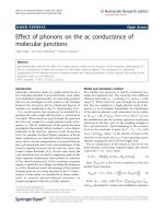
Fig. 1. Inhibitory effect of HMW, MMW and LMW chitosan on the biomass (A)
and the metabolic activity (B) of the biofilm adhesion phase of Candida spp.
Cells were co-incubated in 96-well plates with various concentrations (MIC100%,
4xMIC100%, 8xMIC100%) of HMW, MMW and LMW chitosan for 90 min and
biofilm production was compared to that of fungal cells incubated without
chitosan. *, P < 0.05, compared to the control groups. Values obtained are
given as the percentage of biofilm formation. Results are expressed as
mean ± SD.
3.3.1. Adhesion bioforms
Candida spp. biofilm formation begins with the adherence of round
yeast cells to a solid surface, which is crucial for all later stages of
biofilm development (Gulati & Nobile, 2016). Fig. 1 represents the effects of HMW, MMW and LMW on biomass (Fig. 1A) and metabolic
activity (Fig. 1B) during the adhesion phase of Candida spp. biofilm
formation (based on the percentage of reduction). Reductions in both
biomass and metabolic activity were observed, as compared to the
growth of the positive control, for all the concentrations used (MIC100%,
4xMIC100% and 8xMIC100%). By increasing the chitosan concentration,
progressive reductions in biomass and metabolic activity were observed. At 4 x MIC100%, the reductions were statistically significant (*
p < 0.05 in comparing the control group) for both biomass and metabolic activity. For HMW, MMW and LMW chitosan, when the concentration was increased to a maximum of 8xMIC100%, the biomass and
metabolic activities of the Candida spp. were inhibited by approximately 70%.
Several factors influence the adhesive capacity of yeasts, including
cellular hydrophobicity and electrostatic interactions (zeta potential)
between microbial cells and substrate surfaces. The phenomenon of
adhesion on inert surfaces (polystyrene) is commanded by the physicochemical properties of yeast cell surfaces (Rotrosen, Calderone, &
Edwards, 1986). It is probable that HMW, MMW and LMW chitosan
reduce the relative hydrophobicity of the cell surface. Panagoda,
Ellepola, and Samaranayake (2001) demonstrated that there is a relationship between the adhesion of microorganisms to buccal epithelial
cells and acrylic surfaces and the relative hydrophobicity of the microorganism cell surface. The material used in this study to evaluate
biofilm adhesion was polystyrene (PS), while Poly(methyl
methacrylate) (PMMA) was used in the study conducted by Panagoda
et al. (2001) and different materials may affect the interaction of the
yeast cell with the surface. The interactions that occur between the
PMMA surface and yeast cells may be of the dipole-dipole type or via
hydrogen bonds. Since polystyrene is more hydrophobic than PMMA or
cell membranes, it is possible that no charge or dipole interactions
occur between the surface and the yeast surface or chitosan. A study
carried out with different types of polymers (polytetrafluorethylene,
polyethyleneterephthalate and polystyrene) demonstrated that C. albicans cells present a greater adhesion on polystyrene surfaces due to its
higher hydrophobicity (Klotz, Drutz, & Zajic, 1985). Given that polystyrene favors the adhesion process, the efficacy of chitosan for preventing biofilm adhesion is further supported in this study.
Another important factor affecting the adhesion of Candida is its
expression of peripheral proteins called adhesins. Several Candida adhesins have been identified and play an important role in adhesion,
both on mammalian cells (HeLa cells) and on polystyrene surfaces (Li &
Palecek, 2003). Some of these adhesins are present on the surface of the
cell wall (Chaffin et al., 1998). Therefore, based on the mechanism of
action of chitosan, which consists of inducing cell wall damage, we
suggest that chitosan is capable of compromising the cell adhesion
process.
The present study revealed the ability of HMW, MMW and LMW
chitosan to affect an important virulence factor of Candida species, i.e.
surface colonization.
666
Carbohydrate Polymers 195 (2018) 662–669
L.G.S. Garcia et al.
nucleic acids (5%) (Nobile & Johnson, 2015; Zarnowski et al., 2014).
These components of the matrix have a predominantly anionic character, facilitating the action of chitosan in the biofilm matrix (Donlan &
Costerton, 2002). The use of substances capable of destroying the
physical integrity of the biofilm matrix is an attractive approach as the
consequent loss of the highly protective barrier, represented by the
exopolysaccharide matrix, exposes the sessile microbial cells to the
antifungal agents. It is believed that this mechanism of action occurs for
all the chitosan used in this study, due to the relatively high molecular
weight of the chitosans used. Chitosan penetration of the biofilm matrix
probably did not occur in this study and the activities of the types of
chitosan used were probably mediated by charge effects. Some studies
have reported that chitosan has an optimal antimicrobial activity in a
range of MW from 10k to 50k. Chitosans in this molecular weight range
have a better absorption profile and are able to penetrate the biofilm
matrix and reach the cell more efficiently. It is assumed that statistical
differences would be found if we worked with chitosans that presented
a broader range of MW, which would probably have different mechanisms of action.
With regards to DD, previous studies have shown that the antimicrobial activity of chitosan against planktonic cells increases in association with the increase in DD (Chien, Yen, & Mau, 2016; Chung &
Chen, 2008; Tsai, Su, Chen, & Pan, 2002). Increasing the deacetylation
degree leads to more available amino groups, increasing the electrostatic interaction with the fungal cell wall. Knowing that the exopolysaccharide matrix of biofilms contains components that give it a negative charge, it is believed that chitosan with higher DDs will also be
more effective against biofilms. The DDs of chitosan used in this study
were close and it was not possible to evaluate the influence of DD on
chitosan activity against biofilms. The close values of DD may have
contributed to the similar activities of the chitosans studied herein.
The efficacy of chitosan against biofilms of C. albicans, C. glabrata, C.
parapsilosis, and C. tropicalis was previously reported by Silva-Dias et al.
(2014). The authors showed that chitosan with a low molecular weight
(50 kDa), at a concentration of 1 × 10⁴ mg/l, was able to reduce biofilm
biomass and metabolic activity for all Candida species investigated up
to 90%. However, in their study, Silva-Dias et al. (2014) demonstrated
only the activity of low molecular weight chitosan. In this study, the
activities of medium and high molecular weight chitosan against biofilms of Candida spp. were also demonstrated.
Fig. 2. Inhibitory effects of HMW, MMW and LMW chitosan on the biomass (A)
and the metabolic activity (B) of the mature biofilm phase of Candida spp. Cells
were co-incubated in 96-well plates with various concentrations (MIC100%,
4xMIC100%, 8xMIC100%) of HMW, MMW and LMW chitosan for 48 h and their
biofilm production was compared to that of fungal cells incubated without
chitosan. *, P < 0.05, compared to the control groups Values obtained are given
as the percentage of biofilm formation. Results are expressed as mean ± SD.
3.3.2. Mature biofilms
As shown in Fig. 2, the activities of HMW, MMW and LMW chitosan
against mature Candida spp. biofilms were measured based on the
percentage reduction of biomass (Fig. 2A) and metabolic activity
(Fig. 2B). Significant reductions in growth control (compared to biofilms that were not exposed to chitosan) were observed when biofilms
were exposed to the concentration of 4xMIC100%. The percentage of
biomass and metabolic activity decreased in association with the increase in HMW, MMW and LMW chitosan concentrations. At the
highest concentration used (8xMIC100%), about 18.7% of the biomass
and 15.31% of the metabolic activity were observed, indicating a percentage reduction in these parameters of more than 80% for the three
kinds of chitosan studied. The results obtained in this study demonstrated no correlation between molecular weight and the antifungal
activity of chitosan against biofilms of Candida spp. The three different
chitosans showed statistically similar activities against mature biofilms
that were independent of molecular weight. These results contrast with
those obtained for planktonic cells, since the mature biofilms of Candida
spp. produce an exopolymeric matrix that hinders the penetration of
antimicrobial agents.
The mechanisms of action reported for chitosan activity against the
biofilms of Candida spp. are not as well described as they are for
planktonic cells. The action of chitosan on the extracellular biofilm
matrix can be attributed to the attraction of chitosan, due to its cationic
charges, to the exopolymeric components of the biofilm matrix that
consists of glycoproteins (55%), carbohydrates (25%), lipids) and
3.4. Morphology and structure of biofilms
3.4.1. Confocal laser scanning microscopy (CLSM)
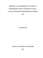
Confocal microscopy was used to correlate XTT reduction assays
with the visual effects on biofilm metabolism and structure (Fig. 3A–D).
The regions of green fluorescence correspond to metabolically-active
cells, while red fluorescence represents metabolically-inactive or nonviable cells. The biofilms of C. tropicalis cultivated in the absence of
chitosan showed regions of high metabolic activity (Fig. 3A), while
biofilms treated with LMW chitosan at concentrations of MIC100%,
4xMIC100% and 8xMIC100% showed a decrease in metabolic activity
(Fig. 3(B–D)). The decrease in metabolic activity reflects the stress
caused by LMW chitosan in the biofilm
3.4.2. Scanning electron microscopy (SEM)
SEM images were performed to show structural differences between
the biofilms of C.tropicalis treated with chitosan and those which were
untreated (Fig. 3E–H). In the absence of LMW, biofilms of C. tropicalis
showed blastoconidia, long and short hyphae that were organized in
dense structures and composed of multilayers of associated cells, as
observed in Fig. 3E. In the presence of chitosan, at the MIC100% obtained for planktonic cells, a reduced number of cells were observed
associated with the biofilms of C. tropicalis (Fig. 3F). Biofilms treated
with chitosan at concentrations of 4xMIC100% and 8xMIC100% presented
wrinkled and collapsed yeast cells (Fig. 3G and H), distinguishing them
667
Carbohydrate Polymers 195 (2018) 662–669
L.G.S. Garcia et al.
Fig. 3. Confocal microscopy (A–D) images and Scanning electron microscopy (E − H) of C. tropicalis biofilm exposed to different concentrations of LMW. Biofilms
developed without chitosan (A and E), and biofilms exposed to MIC100% (B and F), 4x MIC100% (C and G) and 8x MIC100% (D and H). The green color corresponds to
metabolically-active cells while the red areas represent metabolically-inactive or nonviable cells. Scalebars: 100 μm (Figure A–D), 50 μm (Figure E and F), 20 μm
(Fgures G e H). (For interpretation of the references to colour in this figure legend, the reader is referred to the web version of this article.)
from the regular and smooth surface observed in the yeasts of the
control biofilm.
213–219.
Clinical and Laboratory Standards Institute (2008). Reference method for broth dilution
antifungal susceptibility testing of yeasts; approved standard (3rd ed.). Wayne (PA): CLSI
Document M27-A3.
Clinical and Laboratory Standards Institute, & Laboratory Standards Institute (2012).
Reference method for broth dilution antifungal susceptibility testing of yeasts; fourth informational supplement. Wayne (PA): CLSI Document M27-A3.
Canella, K. M., & Garcia, R. B. (2001). Characterization of chitosan by gel permeation
chromatography-influence of preparation method and solvent. Química Nova, 24(1),
13–17.
Chaffin, W. L., López-Ribot, J. L., Casanova, M., Gozalbo, D., & Martínez, J. P. (1998). Cell
wall and secreted proteins of Candida albicans: Identification, function, and expression. Microbiology and Molecular Biology Reviews, 62(1), 130–180.
Chien, P. J., & Chou, C. C. (2006). Antifungal activity of chitosan and its application to
control post-harvest quality and fungal rotting of Tankan citrus fruit (Citrus tankan
Hayata). Journal of the Science of Food and Agriculture, 86(12), 1964–1969.
Chien, R. C., Yen, M. T., & Mau, J. L. (2016). Antimicrobial and antitumor activities of
chitosan from shiitake stipes: Compared to commercial chitosan from crab shells.
Carbohydrate Polymers, 138, 259–264.
Chung, Y. C., & Chen, C. Y. (2008). Antibacterial characteristics and activity of acidsoluble chitosan. Bioresource Technology, 99(8), 2806–2814.
Costa, E. M., Silva, S., Veiga, M., Vicente, S., Tavaria, F. K., & Pintado, M. E. (2017).
Investigation of chitosan’s antibacterial activity against vancomycin resistant microorganisms and their biofilms. Carbohydrate Polymers, 174, 369–376.
de Aguiar Cordeiro, R., Nogueira, G. C., Brilhante, R. S. N., Teixeira, C. E. C., Mourão, C.
I., Castelo, D. D. S. C. M., et al. (2012). Farnesol inhibits in vitro growth of the
Cryptococcus neoformans species complex with no significant changes in virulencerelated exoenzymes. Veterinary Microbiology, 159(3–4), 375–380.
de Medeiros, V. M., do Nascimento, Y. M., Souto, A. L., Madeiro, S. A. L., de Oliveira
Costa, V. C., Silva, S. M. P., et al. (2017). Chemical composition and modulation of
bacterial drug resistance of the essential oil from leaves of Croton grewioides.
Microbial Pathogenesis, 111, 468–471.
De Vita, D., Simonetti, G., Pandolfi, F., Costi, R., Di Santo, R., D’Auria, F. D., et al. (2016).
Exploring the anti-biofilm activity of cinnamic acid derivatives in Candida albicans.
Bioorganic & Medicinal Chemistry Letters, 26(24), 5931–5935.
Deorukhkar, S. C., Saini, S., & Mathew, S. (2014). Non-albicans Candida infection: An
emerging threat. Interdisciplinary perspectives on infectious diseases. 2014.
Donlan, R. M., & Costerton, J. W. (2002). Biofilms: survival mechanisms of clinically
relevant microorganisms. Clinical Microbiology Reviews, 15(2), 167–193.
Gadelha Rocha, M. F., Nascimento de Aguiar, F. L., Nogueira Brilhante, R. S., Aguiar
Cordeiro, R. D., Cordeiro Teixeira, C. E., Souza Collares Maia Castelo-Branco, D. D.,
et al. (2011). Extratos de Moringa oleifera e Vernonia sp. sobre Candida albicans e
Microsporum canis isolados de cães e gatos e análise da toxicidade em Artemia sp.
Ciência Rural, 41(10).
Gulati, M., & Nobile, C. J. (2016). Candida albicans biofilms: Development, regulation,
and molecular mechanisms. Microbes and Infection, 18(5), 310–321.
4. Conclusion
With regard to planktonic cells, the effect of MW was observed for
the strains of C. parapsilosis sensu stricto and C. tropicalis, with HMW
displaying the highest antimicrobial activity. With regard to biofilms,
HMW, MMW and LMW chitosan reduced biomass and metabolic activity both in the adhesion phase and in the mature biofilms. The MW
had no influence on the activity of chitosan against biofilms and the
three MWs displayed statistically similar effects. Therefore, it can be
concluded that chitosan showed promising results in the search for new
agents with antifungal activity against Candida spp. However, the
function of chitosan in biofilm control in vivo deserves further study.
References
Alakayleh, F., Rashid, I., Al-Omari, M. M., Al-Sou'od, K., Chowdhry, B. Z., & Badwan, A.
A. (2016). Compression profiles of different molecular weight chitosans. Powder
Technology, 299, 107–118.
Alburquenque, C., Bucarey, S. A., Neira-Carrillo, A., Urzúa, B., Hermosilla, G., & Tapia, C.
V. (2010). Antifungal activity of low molecular weight chitosan against clinical isolates of Candida spp. Medical Mycology, 48(8), 1018–1023.
Araújo, D., Henriques, M., & Silva, S. (2017). Portrait of Candida species biofilm regulatory network genes. Trends in Microbiology, 25(1), 62–75.
Badawy, M. E., & Rabea, E. I. (2009). Potential of the biopolymer chitosan with different
molecular weights to control postharvest gray mold of tomato fruit. Postharvest
Biology and Technology, 51(1), 110–117.
Brilhante, R. S. N., Malaquias, Â. D. M., Caetano, É. P., Castelo-Branco, D. D. S. C. M.,
Lima, R. A. C. D., Marques, F. J. D. F., et al. (2014). In vitro inhibitory effect of
miltefosine against strains of Histoplasma capsulatum var. capsulatum and
Sporothrix spp. Medical Mycology, 52(3), 320–325.
Brilhante, R. S. N., de Lima, R. A. C., de Farias Marques, F. J., Silva, N. F., Caetano É, P.,
Castelo, D. D. S. C. M., et al. (2015). Histoplasma capsulatum in planktonic and
biofilm forms: In vitro susceptibility to amphotericin B, itraconazole and farnesol.
Journal of Medical Microbiology, 64(4), 394–399.
Brilhante, R. S. N., de Oliveira, J. S., de Jesus Evangelista, A. J., Serpa, R., da Silva, A. L.,
de Aguiar, F. R. M., et al. (2016). Candida tropicalis from veterinary and human
sources shows similar in vitro hemolytic activity: Antifungal biofilm susceptibility
and pathogenesis against Caenorhabditiselegans. Veterinary Microbiology, 192,
668
Carbohydrate Polymers 195 (2018) 662–669
L.G.S. Garcia et al.
hydrophobicity. Mycoses, 44(1–2), 29–35.
Qiu, M., Wu, C., Ren, G., Liang, X., Wang, X., & Huang, J. (2014). Effect of chitosan and its
derivatives as antifungal and preservative agents on postharvest green asparagus.
Food Chemistry, 155, 105–111.
Rinaudo, M., Milas, M., & Le Dung, P. (1993). Characterization of chitosan. Influence of
ionic strength and degree of acetylation on chain expansion. International Journal of
Biological Macromolecules, 15(5), 281–285.
Rotrosen, D., Calderone, R. A., & Edwards, J. E., Jr. (1986). Adherence of Candida species
to host tissues and plastic surfaces. Reviews of Infectious Diseases, 8(1), 73–85.
Seleem, D., Pardi, V., & Murata, R. M. (2017). Review of flavonoids: A diverse group of
natural compounds with anti-Candida albicans activity in vitro. Archives of Oral
Biology, 76, 76–83.
Severino, R., Ferrari, G., Vu, K. D., Donsì, F., Salmieri, S., & Lacroix, M. (2015).
Antimicrobial effects of modified chitosan based coating containing nanoemulsion of
essential oils: Modified atmosphere packaging and gamma irradiation against
Escherichia coli O157: H7 and Salmonella typhimurium on green beans. Food Control,
50, 215–222.
Silva-Dias, A., Palmeira-de-Oliveira, A., Miranda, I. M., Branco, J., Cobrado, L., MonteiroSoares, M., et al. (2014). Anti-biofilm activity of low-molecular weight chitosan
hydrogel against Candida species. Medical Microbiology and Immunology, 203(1),
25–33.
Souza, C. M. C., Pereira Junior, S. A., Moraes, T. D. S., Damasceno, J. L., Amorim Mendes,
S., Dias, H. J., et al. (2016). Antifungal activity of plant-derived essential oils on
Candida tropicalis planktonic and biofilms cells. Sabouraudia, 54(5), 515–523.
Sun, Z., Shi, C., Wang, X., Fang, Q., & Huang, J. (2017). Synthesis, characterization, and
antimicrobial activities of sulfonated chitosan. Carbohydrate Polymers, 155, 321–328.
Tan, Y., Leonhard, M., Ma, S., Moser, D., & Schneider-Stickler, B. (2018). Efficacy of
carboxymethyl chitosan against Candida tropicalis and Staphylococcus epidermidis
monomicrobial and polymicrobial biofilms. International Journal of Biological
Macromolecules, 110, 150–156.
Tsai, G. J., Su, W. H., Chen, H. C., & Pan, C. L. (2002). Antimicrobial activity of shrimp
chitin and chitosan from different treatments and applications of fish preservation.
Fisheries Science, 68(1), 170–177.
Uppuluri, P., Srinivasan, A., Ramasubramanian, A., & Lopez-Ribot, J. L. (2011). Effects of
fluconazole, amphotericin B, and caspofungin on Candida albicans biofilms under
conditions of flow and on biofilm dispersion. Antimicrobial Agents and Chemotherapy,
55(7), 3591–3593.
Vieira, R. S., & Beppu, M. M. (2006). Interaction of natural and crosslinked chitosan
membranes with Hg (II) ions. Colloids and Surfaces A: Physicochemical and Engineering
Aspects, 279(1), 196–207.
Yuan, G., Lv, H., Tang, W., Zhang, X., & Sun, H. (2016). Effect of chitosan coating
combined with pomegranate peel extract on the quality of Pacific white shrimp
during iced storage. Food Control, 59, 818–823.
Zarnowski, R., Westler, W. M., Lacmbouh, G. A., Marita, J. M., Bothe, J. R., Bernhardt, J.,
et al. (2014). Novel entries in a fungal biofilm matrix encyclopedia. MBio, 5(4)
e01333-14.
Hosseinnejad, M., & Jafari, S. M. (2016). Evaluation of different factors affecting antimicrobial properties of chitosan. International Journal of Biological Macromolecules,
85, 467–475.
Huei, C. R., & Hwa, H. D. (1996). Effect of molecular weight of chitosan with the same
degree of deacetylation on the thermal, mechanical, and permeability properties of
the prepared membrane. Carbohydrate Polymers, 29(4), 353–358.
Klotz, S. A., Drutz, D. J., & Zajic, J. E. (1985). Factors governing adherence of Candida
species to plastic surfaces. Infection and Immunity, 50(1), 97–101.
Kong, M., Chen, X. G., Xing, K., & Park, H. J. (2010). Antimicrobial properties of chitosan
and mode of action: A state of the art review. International Journal of Food
Microbiology, 144(1), 51–63.
Kulikov, S. N., Lisovskaya, S. A., Zelenikhin, P. V., Bezrodnykh, E. A., Shakirova, D. R.,
Blagodatskikh, I. V., et al. (2014). Antifungal activity of oligochitosans (short chain
chitosans) against some Candida species and clinical isolates of Candida albicans:
Molecular weight–activity relationship. European Journal of Medicinal Chemistry, 74,
169–178.
Kumar, M. N. R. (2000). A review of chitin and chitosan applications. Reactive and
Functional Polymers, 46(1), 1–27.
Lahkar, V., Saikia, L., Patgiri, S. J., Nath, R., & Das, P. P. (2017). Estimation of biofilm,
proteinase & phospholipase production of the Candida species isolated from the oropharyngeal samples in HIV-infected patients. Indian Journal of Medical Research,
145(5), 635.
Leonhard, M., & Schneider-Stickler, B. (2015). Voice prostheses, microbial colonization and
biofilm formation. Biofilm-based healthcare-associated infections. Springer International
Publishing123–136.
Li, F., & Palecek, S. P. (2003). EAP1, a Candida albicans gene involved in binding human
epithelial cells. Eukaryotic Cell, 2(6), 1266–1273.
Li, Z., Yang, F., & Yang, R. (2015). Synthesis and characterization of chitosan derivatives
with dual-antibacterial functional groups. International Journal of Biological
Macromolecules, 75, 378–387.
Muzzarelli, R. A., Boudrant, J., Meyer, D., Manno, N., De Marchis, M., & Paoletti, M. G.
(2012). Current views on fungal chitin/chitosan, human chitinases, food preservation, glucans, pectins and inulin: A tribute to Henri Braconnot, precursor of the
carbohydrate polymers science, on the chitin bicentennial. Carbohydrate Polymers,
87(2), 995–1012.
Nobile, C. J., & Johnson, A. D. (2015). Candida albicans biofilms and human disease.
Annual Review of Microbiology, 69, 71–92.
Palma-Guerrero, J., Lopez-Jimenez, J. A., Pérez-Berná, A. J., Huang, I. C., Jansson, H. B.,
Salinas, J., et al. (2010). Membrane fluidity determines sensitivity of filamentous
fungi to chitosan. Molecular Microbiology, 75(4), 1021–1032.
Palmeira-de-Oliveira, A., Passarinha, L. A., Gaspar, C., Palmeira-de-Oliveira, R.,
Sarmento, B., Martinez-de-Oliveira, J., et al. (2011). The relationship between
Candida species charge density and chitosan activity evaluated by ion-exchange
chromatography. Journal of Chromatography B, 879(31), 3749–3751.
Panagoda, G. J., Ellepola, A. N. B., & Samaranayake, L. P. (2001). Adhesion of Candida
parapsilosis to epithelial and acrylic surfaces correlates with cell surface
669