Pharmacological prospection and structural characterization of two purified sulfated and pyruvylated homogalactans from green algae Codium isthmocladum
Bạn đang xem bản rút gọn của tài liệu. Xem và tải ngay bản đầy đủ của tài liệu tại đây (2.75 MB, 9 trang )
Carbohydrate Polymers 222 (2019) 115010
Contents lists available at ScienceDirect
Carbohydrate Polymers
journal homepage: www.elsevier.com/locate/carbpol
Pharmacological prospection and structural characterization of two purified
sulfated and pyruvylated homogalactans from green algae Codium
isthmocladum
T
Diego Araujo Sabrya, Sara Lima Cordeirob, Cynthia Haynara Ferreira Silvaa,
Eduardo Henrique Cunha Fariasc, Guilherme Lanzi Sassakid, Helena Bonciani Nadere,
⁎
Hugo Alexandre Oliveira Rochaa,
a
Department of Biochemistry, Universidade Federal do Rio Grande do Norte, Natal, Rio Grande do Norte, 59.078-970, Brazil
Instituto Federal de Educaỗóo, Ciờncia e Tecnologia do Rio Grande do Norte, Macau, Rio Grande do Norte, Brazil
Centro Universitário do Rio Grande do Norte, Natal, Rio Grande do Norte, Brazil
d
Department of Biochemistry and Molecular Biology, Universidade Federal do Paraná, Curitiba, Paraná, 81.531-980, Brazil
e
Discipline of Molecular Biology, Universidade Federal de São Paulo, São Paulo, São Paulo, 04.044-020, Brazil
b
c
ARTICLE INFO
ABSTRACT
Keywords:
NMR
HSQCed
Anticoagulant activity
Clexane®
aPTT
Two sulfated polysaccharides (SPs), F2 and F3, isolated from Codium isthmocladum were found to contain galactose, sulfate, and pyruvate. The apparent molecular weights of F2 and F3 were determined to be 62 and
61 kDa, respectively. NMR spectroscopy combined with chemical analysis showed that F2 and F3 have the same
structural features. However, F3 showed higher sulfate/sugar ratio (1/2.6) than F2 (1/4). F2 and F3 are essentially (1 → 3)-β-D-galactans with some branching at C6. Pyruvylation occurs at O3 and O4, forming 3,4-O-(1carboxyethylidene)-β-D-Galp residues; some of these pyruvylated residues contain sulfate groups at C6. Some
non-branching residues contain sulfate at C4. None of the SPs exhibited antioxidant activity. MTT results indicated that 1 mg/mL of both SPs about 40% of PANC-1 cell viability. At 10 μg/mL, F2 and F3 had 1.7-fold
longer clotting times compared to that of Clexane® at the same concentration. The higher sulfate content of F3 is
not a determining factor for pharmacological activities of galactans, considering that both F2 and F3 exerted the
effects.
1. Introduction
Green seaweed from the genus Codium are widely distributed
worldwide, except for the polar regions. Codium comprises 125 species
that are found in marine environments (Verbruggen et al., 2007). The
Brazilian coast is home to a diverse range of Codium species, including
Codium decorticatum, C. intertextum, C. profundum, C. repens, C. spongiosum, C. taylori, C. tomentosum, and C. isthmocladum (De OliveiraCarvalho, de, Pereira, & Pedroche, 2010; De Oliveira-Carvalho,
Oliveira, Pereira, & Verbruggen, 2012; Farias et al., 2008).
Several studies investigated the genus Codium and evaluated the
pharmacological/biological potential of the sulfated polysaccharides
(SPs) that are present in its cell wall (Ciancia et al., 2007; Estevez,
Fernández, Kasulin, Dupree, & Estevez, 2009; Fernández, Raffo,
Alberghina, & Ciancia, 2015). SPs obtained from different Codium
species
showed
diverse
pharmacological
applications
as
⁎
immunostimulants (Lee, Ohta, Hayashi, & Hayashi, 2010), anticoagulants (Li et al., 2015), anti-obesity agents, and protective agents for
liver-kidney functions (Kolsi et al., 2017). The diversity of pharmacological applications of Codium species could be attributed to the heterogeneity and complexity of their SPs.
Codium species have been described to produce different types of
SPs, such as sulfated mannans (Fernández, Estevez, Cerezo, & Ciancia,
2012), arabinans (Hayakawa et al., 2000), and galactans (Farias et al.,
2008). Of these, many structural studies have been conducted on sulfated galactans (SGs) in Codium species. Li et al. (2015) reported a SG
from C. divarticum whose main chain is formed by →3-β-D-galactopyranose→1 residues and substituted at the C4 position by branches of
1→-β-D-galactopyranose units or sulfate esters. Meanwhile, SGs from C.
decorticatum are composed of 3-, 6-, and 3,6-O-linked β-D-galactopyranose residues sulfated at C6 and predominantly at C4. In addition,
these SGs contain pyruvate ketals both linked to O-3 and O-4 as linked
Corresponding author.
E-mail address: (H.A. Oliveira Rocha).
/>Received 15 March 2019; Received in revised form 19 June 2019; Accepted 19 June 2019
Available online 20 June 2019
0144-8617/ © 2019 Elsevier Ltd. All rights reserved.
Carbohydrate Polymers 222 (2019) 115010
D.A. Sabry, et al.
to O-4 and O-6 (Fernández et al., 2015). Other studies reported highly
sulfated galactans from C. fragile and C. vermilara, composed of →3-β-Dgalactopyranose→1 residue, with sulfate esters at the C4 and C6 positions. These SGs are also pyruvylated at O-3 and O-4 (Ciancia et al.,
2007).
The structures of sulfated polysaccharides directly affect their biological activities because of the main structure, molecular weight, degree of sulfation, monosaccharide composition, and glycosidic linkages
(Costa et al., 2010). However, few studies investigated the relationship
between structure and biological activity. Meanwhile, the purification
process plays an important role in structural elucidation (Fernández
et al., 2015).
Farias et al. (2008) described a method to obtain five SP-rich fractions named F0.3v, F0.5v, F0.7v, F0.9v, and F1.2v, respectively, from
the green seaweed C. isthmocladum. Gel electrophoresis and monosaccharide composition analyses revealed that each fraction contained
different SPs. In addition, the authors purified and investigated the
chemical structure of two preponderantly 4-O-sulfated, 3-linked SGs
from F0.9v. Among the other fractions, F0.5v showed a potential
pharmacological use. However, electrophoretic analysis revealed that
F0.5v has heterogeneous profile and does require further purification
steps. Therefore, we purified SPs from the F0.5v fraction from C. isthmocladum, proposed their chemical features, and additionally evaluated
their pharmacological potential as antioxidant, antiproliferative, and
anticoagulant compounds.
F0.3v, F0.5v, F0.7v, F0.9v and F1.2v respectively. After the addition of
each acetone volume, the mixture was maintained at 4 °C for 24 h and
then collected by centrifugation (for 20 min at 10,000 x g), followed by
drying under vacuum atmosphere.
2.3. Anion exchange chromatography (AEC)
10 g of F0.5v was dissolved to a final concentration of 10 mg/mL
and added to 300 mL of MP500 Lewatit resin. The mixture was complexed for 18 h under gentle agitation at room temperature. The mixture was then applied to a glass column (25 x 5 cm i.d.). Firstly, the
mixture was washed with 300 mL of NaCl 0.3 M to remove unbound
compounds. Chromatography was then carried out by a stepwise gradient elution with seven different concentrations of NaCl (0.3 M, 0.5 M,
0.7 M, 1.0 M, 1.5 M, 2.0 M and 3.0 M). The column was washed three
times with each NaCl molar concentration, and the eluate was precipitated separately with 4 volumes of methanol at 4 °C and collected by
centrifugation (15 min at 10,000 x g) after 12 h, followed by drying
under vacuum atmosphere.
2.4. Agarose gel electrophoresis
All precipitate fractions obtained from AEC were analyzed by
agarose gel electrophoresis as described previously (Dietrich & Dietrich,
1976). Samples (50 μg) were applied to a 0.6% (w/v) agarose gel and
ran for 1 h at 110 V in 0.05 M 1,3 diaminopropane/acetate (PDA) buffer
at pH 9.0. After the run, the sulfated polysaccharides were fixed in the
gel with 0.1% CTV solution for 12 h. Following the fixation, the gel was
dried under airflow and stained with 0.1% (w/v) toluidine blue in a
solution of acetic acid:ethanol:water (0.1:5:4.9, v/v).
2. Material and methods
2.1. Chemicals and reagents
Toluidine blue, deuterium oxide (D2O), 1,3-diaminopropane, sodium pyruvate, cresol red, Sephadex G-100 medium, acetic anhydride
(Ac2O), Coomassie brilliant blue R-250, D-Galactose, D-Glucose, LArabinose, D-Mannose, L-Fucose, D-Xylose, and molecular weight
dextran standards were purchased from Sigma (St. Louis, MO, USA).
Methanol, ethanol, acetone, acetic acid, sulfuric acid, pyridine, N-cetylN,N,N-trimethylammonium bromide (CTV) were purchased from CRQ
(São Paulo, SP, Brazil). Maxatase, an alkaline protease from
Esporobacillus sp, was purchased from BioBrás (Montes Claros, MG,
Brazil). MP500 Lewatit resin was purchased from Bayer Chemicals (São
Paulo, SP, Brazil). Panc-1 cells were purchased from Rio de Janeiro Cell
Bank (Rio de Janeiro, RJ, Brazil). All other solvents and chemicals were
of analytical grade.
2.5. Physicochemical analysis of F2 and F3
Total sugar content measurement was performed by phenol-sulphuric acid method using galactose as a reference sugar (Dubois, Gilles,
Hamilton, Rebers, & Smith, 1956). Sulfate content was determined by
BaCl2-gelatin method (Dodgson & Price, 1962). Protein content was
measured as described by Bradford (1976). Pyruvate content was determined by high performance liquid chromatography (HPLC) measurement described by Ohta, Lee, Hayashi, and Hayashi (2009).
Molecular weight and homogeneity of F2 and F3 were determined
by gel permeation chromatography (GPC). Each fraction was dissolved
to a final concentration of 10 mg/mL and applied to a column containing Sephadex G-100 (130 x 1 cm i.d.). GPC was performed using an
isocratic elution mode. The molecular weight was estimated by reference to a calibration curve made by dextran sulfate standards (10,
40, 70, 147 and 500 kDa). And homogeneity of F2 and F3 was evaluated
by chromatographic profile.
Monosaccharide composition was determined by gas chromatography (GC) after derivatization. F2 and F3 were submitted to hydrolysis with 2 M HCl for 4 h at 100 °C. The hydrolyzed material was reduced with 3 mg NaBH4 and acetylated with Ac2O 50% (v/v) in
pyridine for 30 min at 100 °C (Sassaki et al., 2008), yielding alditol
acetates. The product was resuspended with chloroform and analyzed
by GC–MS in Varian Saturn 2000R in a DB225MS column (30 m
x0.25 mm i.d.), using a temperature programming at 40 °C/min rate
from 50 to 220 °C maintained for 40 min, with He as gas carrier at
1 mL/min flow rate (Sassaki, Gorin, Souza, Czelusniak, & Iacomini,
2005). Identification and quantitation of monosaccharides were performed by comparison with typical retention time of reference sugars:
D-Galactose, D-Glucose, L-Arabinose, D-Mannose, L-Fucose and D-Xylose.
2.2. Seaweed samples
Specimens of Codium isthmocladum (Vickers) were collected from
Pirambúzios beach, Nísia Floresta, Rio Grande do Norte, Brazil. The
seaweed was identified according to its morphology (Wynne, 1986) by
Dr. Valquíria Pereira Medeiros (Federal University of Juiz de Fora, MG,
Brazil) and a voucher specimen was deposited in the UFRN Herbarium
of the Institute of Biosciences, Federal University of Rio Grande do
Norte, under the registration code UFRN25933. The material collection
occurred under authorization of Brazilian National Management
System Genetic Heritage and Associated Traditional Knowledge (loose
translation) SISGEN n° A0D4240. The seaweed was washed with tap
water, air-dried in an aerated stove at 50 °C and crushed in a blender.
The dried powder was incubated with acetone to remove pigments and
lipids (Leite et al., 1998). Powdered alga was then submitted to proteolytic digestion by alkaline protease and maxatase (pH 8.0) to solubilize sulfated polysaccharides. The digestion was conducted by adding
three volumes of 250 mM NaCl to powdered alga, adjusting pH to 8.0
and finally adding maxatase. After incubating with protease for 24 h at
60 °C, the mixture was filtered and fractionated by precipitation with
different volumes of cold acetone: 1:0.3, 1:0.5, 1:0.7, 1:0.9 and 1:0.1.2
(v/v) as established by Farias et al. (2008), who named the fractions as
2.6. Desulfation
Desulfation of sulfated F2 and F3 was performed as described by
2
Carbohydrate Polymers 222 (2019) 115010
D.A. Sabry, et al.
Nagasawa, Inoue, and Tokuyasu (1979). Briefly, each sulfated polysaccharide (10 mg) was dissolved in distilled water and was added to
cation-exchange resin (H+ form). The mixture was filtered, the pH of
the supernatant was adjusted to 7.0 with pyridine and then freeze-dried
to give the pyridinium salt. The product was dissolved in 3 mL of dimethyl sulfoxide (DMSO) containing 10% (v/v) of methanol and then
the solution was heated for 4 h at 100 °C. After the reaction was completed, the desulfated polysaccharides were dialyzed (MWCO 6–8 kDa)
against distilled water and freeze-dried. The products were named DSF2
and DSF3, referring to desulfated F2 and F3, respectively.
and incubated at 95 °C for 90 min. Once the tubes were at room temperature 200 μL of each tube was collected and measured at an absorbance of 695 nm. The results were expressed as ascorbic acid equivalent
as of a standard curve.
2.11. Anticoagulant test
F2 and F3 anticoagulant test was performed in vitro by aPTT (activated partial thromboplastin time) test using a Labtest kit (Lagoa Santa,
MG, Brazil), in a Drake coagulometer (São Paulo, SP, Brazil) and a pool
of citrated human plasma. For the aPTT test, plasma (100 μL) was incubated at 37 °C with saline or SP (100 μL) for 1 min and then rabbit
cephalin (100 μL) was added. After 2.5 min, 25 mM CaCl2 (100 μL) was
added and the clotting time measured. Clexane was used as a positive
control on aPTT test. For PT test, plasma (100 μL) was incubated at
37 °C with saline or SP (100 μL) for 3 min. Thromboplastin (100 μL) was
then added and the clotting time was measured.
2.7. Methylation analysis
F2, F3 and their respective desulfated derivatives were per-O-methylated by the NaOH-DMSO-CH3I method described by Ciucanu and
Kerek (1984). Briefly, each sample was dissolved in DMSO to a final
concentration of 15 mg/mL and then 1 mL of methyl iodide was added
followed by powdered NaOH (30 mg). The mixture was submitted to
vigorous mechanical agitation for 30 min. The product was then dissolved in distilled water in an ice bath and the pH was adjusted to 7.0
with acetic acid. The material was dialyzed (MWCO 6–8 kDa), freezedried and the whole process was then repeated twice. Per-O-methylated
derivatives were then hydrolyzed by formic acid 45% (v/v) for 15 h at
100 °C. The product was reduced and acetylated as previously described
by Sassaki et al. (2008), resulting to partially O-methylated alditol
acetates and analyzed by GC–MS as previously described. The compounds were identified by their typical retention times and electron
impact fragmentation spectra (Sassaki et al., 2005).
2.12. Statistical analysis
The experiments performed were expressed as mean ± standard
deviation. To test differences between the tested concentrations as well
as treatments with different polysaccharides, one-way analysis of variance (ANOVA) and Newman-Keuls post-test was performed using
GraphPad Prism 5.01 2007 (GraphPad Software, Inc.), and results were
considered statistically significant when p ≤ 0.05.
3. Results and discussion
2.8. NMR spectroscopy
3.1. Isolation and physicochemical characterization of sulfated
polysaccharides isolated from C. isthmocladum
NMR 1D and 2D spectra were recorded at 30 °C using an Avance III
Bruker 400 MHz spectrometer equipped with a 5 mm inverse probe.
Polysaccharide (30 mg) was submitted to deuterium-exchange and
lyophilized three times with D2O 99% (v/v). The chemical shift of 1H
and 13C are expressed in δ (ppm) relative to TMSP (trimethylilsilylpropionate) as an internal standard (δ =0 ppm). Only 2D-NMR HMBC
was carried out at 65 °C, all the other experiments were performed at
30 °C. 1D-NMR 1H and 2D-NMR spectra (1H/1H COSY, 1H/1H TOCSY,
1
H/13C HSQC and 1H/13C HMBC) were analyzed using TopSpin Bruker
software version 3.2pl6.
Firstly, 1H chemical shifts assignments were determined by 2D
COSY and TOCSY spectra analysis. 1H/13C chemical shift correlations
were later determined by HSQC analysis and multiple bonding connections were obtained by HMBC experiment. 2D NMR experiments
were acquired using 24 scans per series of 4096 × 360 data points.
Five sulfated polysaccharide-rich fractions, named F0.3v, F0.5v,
F0.7v, F0.9v, and F1.2v, were obtained from the green seaweed C.
isthmocladum (Vickers), following the same starting material and precipitation method reported by Farias et al. (2008). In addition, these
five fractions yielded 17, 30, 27, 21, and 5% (w/w), respectively.
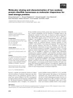
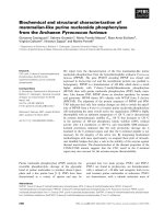
To confirm the presence of sulfated polysaccharides (SP), the fractions were subjected to agarose electrophoresis in PDA buffer, and their
migration profiles were analyzed (Fig. 1). After toluidine blue staining,
we observed bands that correspond to the predominant SP population
in each of the fractions. The F0.3v and F1.2v bands were weakly
stained, indicating low SP concentrations in these fractions, whereas
F0.5v, F0.7v, and F0.9v produced more intense bands, indicating higher
SP content. Due the PDA buffer, the SPs with the same structure have
similar interaction with the diamine and consequently show the same
electrophoretic mobility (Presa et al., 2018). The electrophoretic band
from F0.7v is composed of a mixture of F0.9v and the band from F0.5v
with more mobility. In addition, the SPs present in F0.9v and F1.2v
showed similar electrophoretic mobilities, indicating similarities in SP
composition, consistent with the results reported in previous characterization studies (Farias et al., 2008).
The SPs present in F0.7v, F0.9v, and F1.2v showed similar electrophoretic mobilities, indicating similarities in SP composition, consistent with the results reported in previous characterization studies
(Farias et al., 2008).
F0.5v produced two distinct electrophoretic bands, indicating the
presence of two distinct SP populations. In addition, the electrophoretic
mobilities of the bands were different from those observed in the F0.7v,
F0.9v, and F1.2v fractions, indicating the presence of unknown SPs in
F0.5v. Therefore, F0.5v was further subjected to anion-exchange
chromatography (AEC) to isolate these two SP families by stepwise
gradient elution.
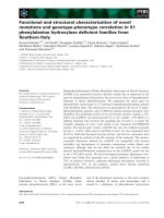
AEC of F0.5v produced seven subfractions named F0.3, F0.5, F0.7,
F1, F1.5, F2, and F3. The fractions were analyzed by agarose electrophoresis, and migration profile analysis showed that F2 and F3 migrate
2.9. 3-(4,5-Dimethylthiazol-2-yl)-2,5-diphenyltetrazolium bromide (MTT)
test assay
For the tests, 5 × 10³ cells/well were grown in 96-well plates with
DMEM medium containing the samples in concentrations of 0.05, 0.1,
0.25, 0.5 and 1 mg/mL for 24 and 48 h (each concentration in triplicate). After treatment, MTT (5 mg/mL) was added and cells were incubated for 4 h at 37 °C. The cell capacity to reduce MTT was determined by the colorimetric test of MTT as described earlier by
Mosmann (1983).
2.10. Determination of total antioxidant capacity
Total antioxidant capacity (TAC) was evaluated by colorimetric
assay based on the reduction of molybdenum (Mo) VI to V by sulfated
polysaccharides and subsequent formation of a green phosphate/Mo(V)
complex as used by Costa et al. (2010). The reagent solution containing
sulfuric acid (0.6 M), sodium phosphate (28 mM) and ammonium molybdate (4 mM) was added in tubes containing F2 and F3 (10 mg/mL),
3
Carbohydrate Polymers 222 (2019) 115010
D.A. Sabry, et al.
3.2. Structural characterization of F2 and DSF2 from C. isthmocladum by
GC–MS and NMR spectroscopy
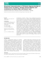
One-dimensional 1H-NMR analysis showed that F2 and F3 have the
same chemical shift fingerprint (Fig. 4), indicating that the two SGs
have the same structural features with slightly different sulfate contents, as shown in Table 1. Because of the high similarity in structure
and yield, all subsequent structural characterizations were conducted
using only F2.
To obtain information on linkage position and sulfation pattern of
F2, we performed a comparative GC–MS analysis between F2 and its
desulfated derivative (DSF2) per-O-methylated alditol acetates. The
identified alditol acetates and their respective quantities are listed in
Table 2.
F2 showed higher content of non-methylated residues even after
three rounds of methylation, which could be attributed to the highly
branched structure or conformation of F2 that blocks efficient methylation. In addition, the high sulfate/sugar ratio (1:4) and presence of
pyruvate groups in F2 can prevent methylation process. Nevertheless,
certain indications about the structure of F2 cannot be determined.
Only few residues were found to contain the 3-O-methylation, which
indicated that F2 predominantly consists of →3)-Galp-(1→ units
(Table 2).
Moreover, DSF2 showed higher contents of 2,4-di-O- and 2,6-di-Omethyl-galactitol, indicating the presence of sulfate groups at the C4
and C6 positions. In addition, DSF2 showed higher content of 2,4,6-triO-methyl-galactitol residues, which also indicated presence of sulfate
esters at C6 in F2. Desulfation can also eliminate pyruvylation, as evidenced by the reduction in 2-O-methyl-galactitol at the same proportion as the increase in 2,3,4-di-O-methyl-galactitol, indicating the presence of substituents at positions 3 and 4. Desulfation facilitated the
determination of sulfate groups because of variations in the number of
methylated residues between F2 and DSF2 units (Table 2).
Fig. 1. Electrophoretic migration of acetone fractions from Codium isthmocladum. The crude polysaccharides before acetone precipitation (CP), and
fractions precipitated with 0.3, 0.5, 0.7, 0.9, and 1.2 volumes of acetone (50 μg
each) were applied at agarose gel electrophoresis from the origin (Or) to the
positive pole (+).
3.3. Structural characterization of F2 and DSF2 from C. isthmocladum by
NMR spectroscopy
The 1H-NMR spectra (Fig. 4) of F2 and F3 showed no distinguishable anomeric signals, which is typical of SP spectra. However, an
anomeric region from δ 4.40 to 4.90 ppm, which represents the characteristic region of β-anomers, can be determined. However, the 1HNMR spectra show the presence of a strong peak at δ 1.63 ppm, indicating pyruvylation. We performed a more detailed structural analysis of F2 by two-dimensional NMR spectroscopy (2D-NMR). All 2DNMR spectra are shown in Fig. 4. Two major spin systems are evident
from the HSQC spectrum of F2, named unit A and unit B, which have
anomeric hydrogen signals at δ 4.84 and 4.54 ppm, respectively. We
observed another spin system with lower intensity, unit C, in which an
anomeric signal at δ 4.71 ppm was observed. All units consist of β-Dgalactose residues, and their respective chemical shifts were determined by combined analysis of the COSY, TOCSY, HSQC, and HMBC
spectra (Table 3). The signal detected at δ 1.63/23.4 ppm was attributed to the CH3 groups of pyruvate, which are common in SPs obtained
from green seaweeds (Arata et al., 2015; Ciancia et al., 2007, 2012;
Farias et al., 2008; Li et al., 2015). This cross-peak also indicate S
configuration at C2 from pyruvate (Garegg, Lindberg, & Kvarnström,
1979)
Unit A shows the H1/C1 correlation at δ 4.84/102.7 ppm, indicating
the presence of strong electronegative substituents in this unit. The
signals at positions 3 and 4 are shifted to high-frequency field. Given
that H3/C3 (δ 4.26/78.8 ppm) and H4/C4 (δ 4.18/75.2 ppm) represent
the chemical shift of 3,4-O-(1-carboxyethylidene)-β-D-Galp residues,
also known as 3,4-pyruvylated-β-D-Galp residues, the results are consistent with those reported by Fernández et al. (2015). In addition,
HMBC spectra showed a cross-peak correlation between H3 and carbonyl carbon (107.8 ppm) from pyruvate groups, thereby confirming
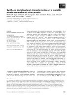
Fig. 2. Electrophoretic migration of AEC fractions. F0.5v, and AEC subfractions
(F0.3, F0.5, F0.7, F1, F1.5, F2 and F3 M) (50 μg each) were applied at agarose
gel electrophoresis from the origin (Or) to the positive pole (+).
as a single concentric band, indicating that F2 and F3 were obtained
with high purity (Fig. 2). Furthermore, these two fractions yielded 43%
and 39% (w/w) of the total SPs present in F0.5v.
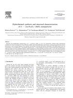
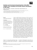
Both F2 and F3 were further analyzed by gel permeation chromatography (GPC) in a Sephadex G-100 column to determine their molecular weights (Fig. 3). The chromatogram showed a single and symmetrical peak, indicating that the SPs were successfully purified from
F0.5v by AEC. Furthermore, the chromatogram obtained from GPC was
used to calculate the apparent molecular weight using a regression
equation determined using different molecular weight standards
(Fig. 3). Thus, the molecular weights of F2 and F3 were estimated to be
approximately 62 and 61 kDa, respectively.
Results of the chemical analyses are plotted in Table 1. F2 and F3
are both composed only of galactose, sulfate, and pyruvate. However,
the SPs showed differences in the proportions of sulfate and pyruvate;
F3 showed higher sulfate/sugar and pyruvate/sugar ratios than F2. F2
showed a sulfate/sugar ratio of 1:4 and pyruvate/sugar ratio of 1:12.
On the other hand, F3 had a sulfate/sugar ratio of approximately 1:2.6
and pyruvate/sugar ratio of 1:8. No protein traces were detected for
both SPs (Table 1).
4
Carbohydrate Polymers 222 (2019) 115010
D.A. Sabry, et al.
Fig. 3. Homogeneity and molecular weight determination of SP, F2, and F3, from C. isthmocladum. The GPC chromatogram of F2 and F3 on a Sephadex G-100 column
and the standard curve of molecular weight (kDa).
glycosidic linkage bonding different units. A weak cross-peak H3 (B)/
C1 (A) allowed the determination of the bonding between unit A and B,
as a 3,4-Pyr-β-D-Galp-(1→3)-β-D-Galp4S-(1→ fragment, with some of
these pyruvylated units being sulfated at C6 and absence of sulfation at
C4. Meanwhile, a strong cross-peak H2 (C)/C1 (B) allowed the determination of unit B and C bond as the fragment →3)-β-D-Galp4S-(1→
3,6)-β-D-Galp-(1→. Fig. 5 shows the proposed structure of F2 and F3
based on GCeMS and NMR data analysis.
Similar to the current findings, Farias et al. (2008) successfully
purified two similar pyruvylated SGs, named SG1 and SG2, from C.
isthmocladum using the same procedure. Therefore, combined ice-cold
acetone precipitation, anion exchange chromatography, and gel electrophoresis in PDA buffer is an effective method for the purification of
SPs from seaweed. SG1 (14 kDa) and SG2 (20 kDa) belong to the same
SG family, which are known to be predominantly composed of 4-sulfated, 3-linked β-D-Galp units. In addition, SG1/SG2 contain pyruvate
groups that form five-membered cyclic ketals as 3,4-O-(1-carboxyethylidene)-β-D-galactose residues. These structural features were also
identified in F2 and F3, indicating that belong to the same SG family.
However, we observed differences between F1/F2 and SG1/SG2 polysaccharides. F1/F2 and SG1/SG2 were obtained from two different
fractions, namely, F0.5 and F0.9, respectively. Moreover, the molecular
weight of SG1/SG2 is lower than that determined for F1/F2, indicating
differences in structure although they belong to the same SG family. In
fact, SG1/SG2 showed some 4,6-sulfated β-D-Galp units, whereas F1/F2
did not. On the other hand, F1/F2 was found to contain pyruvylated 6sulfated β-D-Galp units, which were not detected in SG1/SG2. Other
representatives of this SG family were also described from several
species of the genus Codium (Ciancia et al., 2007; Estevez et al., 2009;
Fernández et al., 2015; Li et al., 2015).
Farias et al. (2008) showed that acetone fractions from C. isthmocladum have different monosaccharide compositions. However, some
Table 1
Chemical composition of F2 and F3 from Codium isthmocladum.
SP
F2
F3
Sulfate/sugar ratio
1:4
1:2.6
Pyruvate/sugar ratio
1:12
1:8
Protein (μg)
nd
nd
Molar ratio
Gal
SO4
Pyr
1
1
0.6
0.8
0.15
0.17
nd = not detected; Gal = galactose; Pyr = pyruvate; SO4 = sulfate.
the presence of a pyruvate group linked to this unit. Moreover, some
H6/C6 were shifted to high-frequency (δ 4.33/67.4 ppm), which is typically observed in the presence of sulfate group as confirmed by HSQC
spectrum of DSF2 (data not shown), in which the signal was not detected, indicating that some units A also contain sulfation at C6, named
A6S. The above findings were confirmed by HMBC spectra analysis,
which shows a cross-peak between H6 (δ 4.33 ppm) from sulfated residues and C4 (δ 75.2 ppm) from pyruvylated residues (Fig. 4).
Unit B showed an anomeric correlation at δ 4.54/103.4 ppm. The
cross-peak of H4/C4 (δ 4.86/77.8 ppm) was observed at the high frequency region, indicating the presence of sulfation at this position.
Furthermore, H3/C3 (δ 4.09/77.4 ppm) and H5/C5 (δ 3.85/74.8 ppm)
signals were also shifted to higher ppm values because of the presence
of a nearby sulfate group. Determination of the remaining chemical
shifts resulted in defining unit B as →3)-β-D-Galp4S-(1→ residue based
on the results obtained by Fernández et al. (2015).
Unit C showed an anomeric cross-peak at δ 4.71/103.8 ppm.
Chemical shifts of H4/C4 (δ 4.30/70.9) and H6/C6 (δ 4.23/67.4) indicated the absence of sulfation in this residue because these signals are
located in regions of lower frequency compared to sulfated residues
(Bilan, Vinogradova, Shashkov, & Usov, 2007).
In addition, the HMBC spectrum (1H/13C) shows the sequence of
5
Carbohydrate Polymers 222 (2019) 115010
D.A. Sabry, et al.
Fig. 4. NMR spectra of SGs from C. isthmocladum. First panel shows the comparison of the 1H-NMR spectra of F2 and F3. All other panels show the results of 2D-NMR
of F2. Letters refer to the spin system, and numbers refer to the position at spin system. A: 3,4-Pyr-β-D-Galp; A6S: 3,4-Pyr-β-D-Galp6S; B: →3)-β-D-Galp4S-(1→; C: →
3,6)-β-D-Galp4S-(1→. Galp: galactopyranose.
Table 2
Methylation analysis of sulfated galactan (F2) obtained from Codium isthmocladum and their desulfated derivative (mol %).
O-methylated alditol acetate
F2
DSF2
Linkage pattern
0-Me-Gal
2-Me -Gal
2,4-Me2-Gal
2,6-Me2-Gal
2,3,4-Me3-Gal
2,3,6-Me3-Gal
2,4,6-Me3-Gal
2,3,4,6-Me4-Gal
40
28
4
6
3
6
7
6
0
0
27
16
27
6
9
15
→2,3,4,6)-Galp-(1→
→3,4,6)-Galp-(1→
→3,6)-Galp-(1→
→3,4)-Galp-(1→
→6)-Galp-(1→
→4)-Galp-(1→
→3)-Galp-(1→
Non-reducing end
Table 3
Chemical shift assignments of the NMR spectra of SP (F2 and F3) from C.
isthmocladum based on 2D-NMR experiments.
Unit
A
fractions contained arabinose and mannose residues, in addition to
galactose residues. Furthermore, results from agarose gel electrophoresis demonstrated the presence of bands with different mobilities, indicating that Codium can synthesize other SGs or other types of SPs.
Considering that sulfated mannans (Fernández et al., 2012), and sulfated arabinans (Hayakawa et al., 2000) were described in Codium, the
current findings are consistent with those reported for other seaweeds,
wherein the same species were found to produce different SPs
(Skriptsova, 2015). We aim to evaluate the other fractions from C.
isthmocladum and verify the above findings in future studies.
Chemical shifts, δ (ppm)a
Structural unit
3,4-Pyr-β-D-Galpc
c
A6S
3,4-Pyr-β-D-Galp6S
B
→3)-β-D-Galp4S-(1→
C
→3,6)-β-D-Galp-(1→
Ref.b
H1
C1
H2
C2
H3
C3
H4
C4
H5
C5
H6
C6
4.84
102.7
4.84
102.7
4.54
103.4
4.71
103.8
3.55
73.6
3.55
73.6
3.76
70.8
3.76
70.8
4.26
78.8
4.26
78.8
4.09
77.4
3.86
82.4
4.18
75.2
4.18
75.2
4.86
77.8
4.30
70.9
4.05
73.3
4.05
73.3
3.85
74.8
3.85
74.8
3.89
61.1
4.33
67.4
3.80
61.4
4.23
67.4
1
1
2
2
a
Chemical shifts are relative to the internal standard, trimethylsilyl propionic acid (δ =0 ppm).
b
References used to elucidate structural units based on similarity in chemical shifts: (1) Fernández et al. (2015) and (2) Bilan et al. (2007).
c
Signals at δ 107.8 ppm and 1.63/23.4 correspond to C2 and C3/H3 of
pyruvic acid ketal linked to O-3 and O-4 of terminal galactose units.
ductal adenocarcinoma cell line (PANC-1) were evaluated by MTT
assay after 24 and 48 h of treatment. The viabilities of PANC-1 cells
were reduced in the presence of F2 in a dose-dependent manner
(Fig. 6). However, we observed no time-dependent effects on PANC-1
viability in the presence of F2. We observed approximately 40% inhibition of PANC-1 viability using 1 mg/mL F2 at both treatment
3.4. Cell viability in the presence of F2 and F3
The antiproliferative activities of F2 and F3 against the pancreas
6
Carbohydrate Polymers 222 (2019) 115010
D.A. Sabry, et al.
Fig. 5. Proposed structure of galactans F2 and F3 from Codium isthmocladum. Unit A: 3,4-Pyr-β-D-Galp; Unit B: →3)-β-D-Galp4S-(1→; Unit C: →3,6)-β-D-Galp-(1→.
Radicals position (R) can represent a hydrogen nucleus (H) or SO3 group. In cases where unit A contains an OSO3 group, this unit is called A6S.
fine structure such as the type of glycosidic linkage, molecular weight,
and the sulfate pattern, are important characteristics for the bioactivity
of sulfated polysaccharides.
3.5. Antioxidant activities of F2 and F3
We evaluated the antioxidant activities of F2 and F3 by the TAC
assay. F2 and F3 did not effectively prevent molybdenum reduction,
exhibiting less than 5 ascorbic acid equivalents (mg/g) of F2 or F3 (data
not shown). Costa et al. (2010), observed antioxidant activity below 10
ascorbic acid equivalents (mg/g) of crude SPs from C. isthmocladum
using the same assay, which is not a strong result. However, comparing
the results of F2 and F3 with crude SPs, it is likely that components
present in the crude SPs (other than both F2 and F3) exhibit slightly
stronger capacity to inhibit molybdenum reduction.
The amount and distribution of sulfate groups in the polysaccharide
molecule are very important for it to exhibit strong antioxidant activity
(Patel, 2012). However, the structural requirements, such as the presence of disulfated monosaccharides or specific sulfation in a specific
position, have not been defined yet. In this context, the contribution of
this study is that the presence of C4 and C6 sulfation, besides the presence of 3,4-O-linked pyruvate are not important requirements for the
antioxidant activity of sulfated galactans.
3.6. Anticoagulant activities of F2 and F3
The anticoagulant activities of F2 and F3 were evaluated by aPTT
and PT assays. The clotting times of both SPs did not change based on
the PT test, indicating that these SPs did not act on the extrinsic
pathway of coagulation. Meanwhile, F2 and F3 were effectively in
prolonging the clotting time on intrinsic pathway of coagulation in a
dose-dependent manner based on the aPTT test. At the concentration of
10 μg/mL, both SPs showed 1.7-fold longer clotting times compared to
Clexane®, a commercial heparin (Fig. 7A). Furthermore, converting F2,
F3, and Clexane® to molar concentrations at the dose of 10 μg/mL, and
analyzing the [Clexane®]/[SP] molar ratio, we observed that Clexane®
reached about 50% of F2 or F3 activity, using almost 14-fold higher
molar concentration than F2 or F3 (Fig. 7B).
Matsubara et al. (2001) evaluating the anticoagulant properties of
SGs extracted from C. cylindricum, using 15 μg/mL prolonged clotting
time over 300 s. F2 and F3 from C. isthmocladum reached the same
prolongation using 10 μg/mL. However, Matsubara samples showed
high protein content and the authors did not rule out whether proteins
influenced this activity. On the other hand, F2 and F3 were not
Fig. 6. Cell viability of PANC-1 cells in the presence of F2 and F3 after 24 h
(Panel A) and 48 h of treatment (Panel B). * symbol indicates significant difference between F2 and F3 treatment from negative control (p ≤ 0.05).
periods. On the other hand, F3 reduced the cell viability of PANC-1 in a
dose- and time-dependent manner. However, F3 exerted weaker effects
on cell viability compared to F2 at the same concentrations. Sulfated
galactans from green seaweed Udotea flabellum (from 0.1 to 1 mg/mL)
did not affect B16-F10 melanoma cells viability (Marques et al., 2019).
This indicates that only the type of monosaccharide and the presence of
sulfate are not determinant requirements for the antiproliferative activity of SPs, which strengthen that other factors into polysaccharide
7
Carbohydrate Polymers 222 (2019) 115010
D.A. Sabry, et al.
Higher Level Personal Development Coordination, in loose translation),
Programa Ciências do Mar (AUXPE-CIMAR-1956/2014), Programa
Nacional de Cooperaỗóo Acadờmica (CAPES/PROCAD n 2965/2014),
and Ministộrio de Ciờncia, Tecnologia, Inovaỗừes e Comunicaỗừes
(MCTIC) (Science and Technology Ministry in Brazil, in loose translation) for financial support. H. A. O Rocha, H. B. Nader, and G. L. Sassaki
are CNPq fellowship honored researchers. D. Sabry had a Ms.C. and
pH.D. scholarship from CNPq and currently has a post-doctoral fellowship from PNPD-CAPES. C. Silva has a Ms.C. scholarship from
CAPES.
References
Arata, P. X., Quintana, I., Canelón, D. J., Vera, B. E., Compagnone, R. S., & Ciancia, M.
(2015). Chemical structure and anticoagulant activity of highly pyruvylated sulfated
galactans from tropical green seaweeds of the order Bryopsidales. Carbohydrate
Polymers, 122, 376–386. />Bilan, M. I., Vinogradova, E. V., Shashkov, A. S., & Usov, A. I. (2007). Structure of a highly
pyruvylated galactan sulfate from the Pacific green alga Codium yezoense
(Bryopsidales, Chlorophyta). Carbohydrate Research, 342(3–4), 586–596. https://doi.
org/10.1016/j.carres.2006.11.008.
Bradford, M. M. (1976). A rapid and sensitive method for the quantitation of microgram
quantities of protein utilizing the principle of protein-dye binding. Analytical
Biochemistry, 72(1–2), 248–254. />Ciancia, M., Alberghina, J., Arata, P. X., Benavides, H., Leliaert, F., Verbruggen, H., ...
Estevez, J. M. (2012). Characterization of cell wall polysaccharides of the coencocytic
green seaweed Bryopsis plumosa (Bryopsidaceae, Chlorophyta) from the argentine
coast. Journal of Phycology, 48, 326–335. />01131.x.
Ciancia, M., Quintana, I., Vizcargüénaga, M. I., Kasulin, L., de Dios, A., Estevez, J. M., ...
Cerezo, A. S. (2007). Polysaccharides from the green seaweeds Codium fragile and C.
vermilara with controversial effects on hemostasis. International Journal of Biological
Macromolecules, 41(5), 641–649. />Ciucanu, I., & Kerek, F. (1984). Rapid and simultaneous methylation of fatty and hydroxy
fatty acids for gas-liquid chromatographic analysis. Journal of Chromatography A, 284,
179–185. />Costa, L. S., Fidelis, G. P., Cordeiro, S. L., Oliveira, R. M., Sabry, D. A., Câmara, R. B. G., ...
Rocha, H. A. O. (2010). Biological activities of sulfated polysaccharides from tropical
seaweeds. Biomedecine & Pharmacotherapy, 64(1), 21–28. />biopha.2009.03.005.
De Oliveira-Carvalho, M. D. F., Oliveira, M. C., Pereira, S. M. B., & Verbruggen, H. (2012).
Phylogenetic analysis of Codium species from Brazil, with the description of the new
species C. pernambucensis (Bryopsidales, Chlorophyta). European Journal of Phycology,
47(4), 355–365. />De Oliveira-Carvalho, M., de, F., Pereira, S. M. B., & Pedroche, F. F. (2010). Taxonomy
and distribution of the green algal genus Codium (Bryopsidales, Chlorophyta) in
Brazil. Nova Hedwigia, 91(1), 87–109. />0091-0087.
Dietrich, C. P., & Dietrich, S. M. C. (1976). Electrophoretic behaviour of acidic mucopolysaccharides in diamine buffers. Analytical Biochemistry. />0003-2697(76)90496-6.
Dodgson, K. S., & Price, R. G. (1962). A note on the determination of the ester sulphate
content of sulphated polysaccharides. The Biochemical Journal, 84, 106–110 Retrieved
from />Dubois, M., Gilles, K. A., Hamilton, J. K., Rebers, P. A., & Smith, F. (1956). Colorimetric
method for determination of sugars and related substances. Analytical Chemistry,
28(3), 350–356. />Estevez, J. M., Fernández, P. V., Kasulin, L., Dupree, P., & Estevez, J. M. (2009). Chemical
and in situ characterization of macromolecular components of the cell walls from the
green seaweed Codium fragile. Glycobiology, 19(3), 212–228. />1093/glycob/cwn101.
Farias, E. H. C., Pomin, V. H., Valente, A. P., Nader, H. B., Rocha, H. A. O., & Mourão, P.
A. S. (2008). A preponderantly 4-sulfated, 3-linked galactan from the green alga
Codium isthmocladum. Glycobiology, 18(3), 250–259. />glycob/cwm139.
Fernández, P. V., Estevez, J. M., Cerezo, A. S., & Ciancia, M. (2012). Sulfated β-d-mannan
from green seaweed Codium vermilara. Carbohydrate Polymers, 87(1), 916–919.
/>Fernández, P. V., Raffo, M. P., Alberghina, J., & Ciancia, M. (2015). Polysaccharides from
the green seaweed Codium decorticatum. Structure and cell wall distribution.
Carbohydrate Polymers, 117, 836–844. />039.
Garegg, P. J., Lindberg, B., & Kvarnström, I. (1979). Preparation and N.M.R. studies of
pyruvic acid and related acetals of pyranosides: Configuration at the acetal carbon
atoms. Carbohydrate Research, 77(1), 71–78. />Hayakawa, Y., Hayashi, T., Lee, J. B., Srisomporn, P., Maeda, M., Ozawa, T., ...
Sakuragawa, N. (2000). Inhibition of thrombin by sulfated polysaccharides isolated
from green algae. Biochimica et Biophysica Acta – Protein Structure and Molecular
Enzymology, 1543(1), 86–94. />Kolsi, R. B. A., Jardak, N., Hajkacem, F., Chaaben, R., Jribi, I., El Feki, A., ... Belghith, K.
(2017). Anti-obesity effect and protection of liver-kidney functions by Codium fragile
Fig. 7. Anticoagulant activities of F2 and F3. Panel A: aPTT test of F2 and F3
compared to Clexane®. Aliquots (1, 2.5, 5, 10 and 20 μg/mL) of each SP were
analyzed by anticoagulant evaluation. Panel B: [Clexane®]/[SP] molar ratio
based on F2, F3, and Clexane® at the dose of 10 μg/mL. [Clexane®]/[Clexane®]
ratio correspond to 1 (data not shown).
contaminated by proteins.
Arata et al. (2015) evaluating the anticoagulant potential of F1, a
sulfated and pyruvylated galactan from green seaweed Penicillus capitatus, observed a weak anticoagulation activity. F1 from P. capitatus is
composed of galactose residues 3-, 6-, and 3,6-linked, showing 4,6-Olinked pyruvate groups in part sulfated at C2. Furthermore, F1 prolonged weakly the aPTT anticoagulant test time, did not reach 100 s
using 100 μg/mL. Meanwhile, F2 and F3 from Codium isthmocladum
prolonged clotting time over 100 s, using only 2.5 μg/mL. This stronger
anticoagulant response might be related to the presence of sulfation at
C4 and 3,4-O-linked pyruvate groups in part sulfated at C6.
4. Conclusions
Two sulfated galactans, namely, F2 and F3, were purified from the
green seaweed Codium isthmocladum using combination of ice-cold
acetone precipitation and anion exchange chromatography. F2 and F3
were found to be pyruvylated and formed five-membered cyclic ketals
(S configuration) as 3,4-O-(1′carboxy)-ethylidene-β-D-galactose residues. The only significant difference between F2 and F3 is the sulfate/
sugar ratio, in which F3 showed 1/2.6 while F2 showed 1/4.
Furthermore, NMR analysis showed that both SGs are linked-(1→3)-βD-galactans, with branching at C6 and sulfation at O4. None of the SGs
presented significant activity on MTT or TAC essay. On the other hand,
both F2 and F3 showed stronger anticoagulant activity than Clexane®, a
commercial heparin. Taken together, the current findings highlight the
potential application these of SGs as anticoagulants agents.
Acknowledgements
The authors wish to thank Conselho Nacional de Desenvolvimento
Científico e Tecnolúgico-CNPq (Edital Universal, n 408369/2016-7),
Coordenaỗóo de Aperfeiỗoamento Pessoal de Nớvel Superior (CAPES 8
Carbohydrate Polymers 222 (2019) 115010
D.A. Sabry, et al.
sulphated polysaccharide on high fat diet induced obese rats. International Journal of
Biological Macromolecules, 102, 119–129. />04.017.
Lee, J. B., Ohta, Y., Hayashi, K., & Hayashi, T. (2010). Immunostimulating effects of a
sulfated galactan from Codium fragile. Carbohydrate Research, 345(10), 1452–1454.
/>Leite, E. L., Medeiros, M. G. L., Rocha, H. A. O., Farias, G. G. M., Da Silva, L. F., Chavante,
S. F., ... Nader, H. B. (1998). Structure and pharmacological activities of a sulfated
xylofucoglucuronan from the alga Spatoglossum schroederi. Plant Science, 132(2),
215–228. />Li, N., Mao, W., Yan, M., Liu, X., Xia, Z., Wang, S., ... Cao, S. (2015). Structural characterization and anticoagulant activity of a sulfated polysaccharide from the green
alga Codium divaricatum. Carbohydrate Polymers, 121, 175–182. />1016/j.carbpol.2014.12.036.
Matsubara, K., Matsuura, Y., Bacic, A., Liao, M. L., Hori, K., & Miyazawa, K. (2001).
Anticoagulant properties of a sulfated galactan preparation from a marine green alga,
Codium cylindricum. International Journal of Biological Macromolecules, 28(5),
395–399. />Marques, M., Presa, F., Viana, R., Costa, M., Amorim, M., Bellan, D., ... Rocha, H. (2019).
Anti-thrombin, anti-adhesive, anti-migratory, and anti-proliferative activities of sulfated galactans from the tropical green seaweed, Udotea flabellum. Marine Drugs,
17(1), 5. />Mosmann, T. (1983). Rapid colorimetric assay for cellular growth and survival:
Application to proliferation and cytotoxicity assays. Journal of Immunological Methods,
65(1–2), 55–63. />Nagasawa, K., Inoue, Y., & Tokuyasu, T. (1979). An improved method for the preparation
of chondroitin by solvolytic desulfation of chondroitin sulfates. Journal of
Biochemistry, 86(5), 1323–1329. />a132648.
Ohta, Y., Lee, J. B., Hayashi, K., & Hayashi, T. (2009). Isolation of sulfated galactan from
Codium fragile and its antiviral effect. Biological & Pharmaceutical Bulletin, 32(5),
892–898. />Patel, S. (2012). Therapeutic importance of sulfated polysaccharides from seaweeds:
Updating the recent findings. 3 Biotech, 2(3), 171–185. />s13205-012-0061-9.
Presa, F., Marques, M., Viana, R., Nobre, L., Costa, L., & Rocha, H. (2018). The protective
role of sulfated polysaccharides from green seaweed Udotea flabellum in cells exposed
to oxidative damage. Marine Drugs, 16(4), 135. />md16040135.
Sassaki, G. L., Gorin, P. A. J., Souza, L. M., Czelusniak, P. A., & Iacomini, M. (2005). Rapid
synthesis of partially O-methylated alditol acetate standards for GC-MS: Some relative activities of hydroxyl groups of methyl glycopyranosides on Purdie methylation. Carbohydrate Research, 340(4), 731–739. />2005.01.020.
Sassaki, G. L., Souza, L. M., Serrato, R. V., Cipriani, T. R., Gorin, P. A. J., & Iacomini, M.
(2008). Application of acetate derivatives for gas chromatography-mass spectrometry: Novel approaches on carbohydrates, lipids and amino acids analysis. Journal
of Chromatography A, 1208(1–2), 215–222. />08.083.
Skriptsova, A. V. (2015). Fucoidans of brown algae: Biosynthesis, localization, and physiological role in thallus. Russian Journal of Marine Biology, 41(3), 145–156. https://
doi.org/10.1134/S1063074015030098.
Verbruggen, H., Leliaert, F., Maggs, C. A., Shimada, S., Schils, T., Provan, J., ... Coppejans,
E. (2007). Species boundaries and phylogenetic relationships within the green algal
genus Codium (Bryopsidales) based on plastid DNA sequences. Molecular Phylogenetics
and Evolution, 44(1), 240–254. />Wynne, M. J. (1986). A checklist of benthic marine algae of the tropical and subtropical
western Atlantic. Canadian Journal of Botany, 64(10), 2239–2281. />1139/b86-298.
9