Báo cáo khoa học: Discriminating between the activities of human cathepsin G and chymase using fluorogenic substrates potx
Bạn đang xem bản rút gọn của tài liệu. Xem và tải ngay bản đầy đủ của tài liệu tại đây (818.71 KB, 12 trang )
Discriminating between the activities of human cathepsin
G and chymase using fluorogenic substrates
Brice Korkmaz
1,2
, Gwenhael Je
´
got
1,2
, Laurie C. Lau
3
, Michael Thorpe
4
, Elodie Pitois
1,2
, Luiz
Juliano
5
, Andrew F. Walls
3
, Lars Hellman
4
and Francis Gauthier
1,2
1 Unite
´
INSERM U-618 ‘Prote
´
ases et Vectorisation pulmonaires’, Tours, France
2 Universite
´
Franc¸ois Rabelais de Tours, France
3 Immunopharmacology Group, Sir Henry Wellcome Laboratories, Southampton General Hospital, UK
4 Department of Cell and Molecular Biology, The Biomedical Center, Uppsala University, Sweden
5 Departamento de Biofı
´
sica, Escola Paulista de Medicina, Universidade Federal, Sa˜o Paulo, Brazil
Introduction
Cathepsin G (CG) (EC 3.4.21.20) and chymase (EC
3.4.21.39) are monomeric chymotrypsin-like serine pro-
teases that display a high degree of sequence similarity
and highly similar substrate specificity [1–3]. They are
located predominantly in the primary granules of neu-
trophils and mast cells, respectively, although CG may
also be found in mast cells [4]. The understanding of
their distinctive roles in inflammatory events involving
both neutrophils and mast cells can represent a chal-
lenge as a result of their closely-related substrate
specificities. No substrate has been identified to date
that allows differentiation of their activities when both
Keywords
cathepsin G; chymase; FRET substrate;
kinetics; mast cell; serine protease
Correspondence
B. Korkmaz, Unite
´
INSERM U-618
‘Prote
´
ases et Vectorisation pulmonaires’,
Universite
´
Franc¸ois Rabelais de Tours,
37032 Tours, France
Fax: +33 2 47 36 60 46
Tel: +33 2 47 36 62 53
E-mail:
(Received 4 April 2010, revised 11 May
2011, accepted 16 May 2011)
doi:10.1111/j.1742-4658.2011.08189.x
Cathepsin G (CG) (EC 3.4.21.20) and chymase (EC 3.4.21.39) are two clo-
sely-related chymotrypsin-like proteases that are released from cytoplasmic
granules of activated mast cells and ⁄ or neutrophils. We investigated the
potential for their substrate-binding subsites to discriminate between their
substrate specificities, aiming to better understand their respective role dur-
ing the progression of inflammatory diseases. In addition to their prefer-
ence for large aromatic residues at P1, both preferentially accommodate
small hydrophilic residues at the S1¢ subsite. Despite significant structural
differences in the S2¢ subsite, both prefer an acidic residue at that position.
The Ala226 ⁄ Glu substitution at the bottom of the CG S1 pocket, which
allows CG but not chymase to accommodate a Lys residue at P1, is the
main structural difference, allowing discrimination between the activities of
these two proteases. However, a Lys at P1 is accommodated much less effi-
ciently than a Phe, and the corresponding substrate is cleaved by b2-tryp-
tase (EC 3.4.21.59). We optimized a P1 Lys-containing substrate to
enhance sensitivity towards CG and prevent cleavage by chymase and b2-
tryptase. The resulting substrate (ABZ-GIEPKSDPMPEQ-EDDnp) [where
ABZ is O-aminobenzoic acid and EDDnp is N-(2,4-dinitrophenyl)-ethy-
lenediamine] was cleaved by CG but not by chymase and tryptase, with a
specificity constant of 190 m
M
)1
Æs
)1
. This allows the quantification of active
CG in cells or tissue extracts where it may be present together with chym-
ase and tryptase, as we have shown using a HMC-1 cell homogenate and a
sputum sample from a patient with severe asthma.
Abbreviations
ABZ, O-aminobenzoic acid; ACT, antichymotrypsin; CG, cathepsin G; CMK, chloromethyl ketone; EDDnp, N-(2,4-dinitrophenyl)-
ethylenediamine; FRET, fluorescence resonance energy transfer; HNE, human neutrophil elastase; PR3, proteinase 3; Z, benzyloxycarbonyl.
FEBS Journal 278 (2011) 2635–2646 Journal compilation ª 2011 FEBS. No claim to original French government works 2635
proteases are present. Moreover, CG is weaker than
chymase at hydrolyzing most substrates currently
employed to quantify their activity and, accordingly,
this has hampered studies of their enzymatic properties
[4,5].
CG and chymase genes are located on chromosome
14 together with the genes of granzymes B and H [6].
The two proteases are synthesized as a prepro-protein,
containing a peptide signal, a prodipeptide and a
C-terminal propeptide [7]. Mast cell chymase and CG
convert angiotensin I to the vasoactive peptide angio-
tensin II in human tissues [8], and this reaction may be
important in the progression to heart failure [9] and
aortic stenosis [10]. Both proteases can also convert
the CXC chemokine connective tissue-activating pep-
tide III into active chemokine neutrophil-activating
peptide 2 through limited proteolysis [11], and both
are secretagogues for cultured serous cells [12]. Mast
cell chymase and CG can also inactivate hepatocyte
growth factor [13] and both can degrade connective tis-
sue components such as fibronectin and vascular endo-
thelial cadherin [14]. A close relationship between CG
and chymase is highlighted by the recent development
of a dual inhibitor, the administration of which has
been reported to be efficacious in the treatment of lung
inflammation in animal models [15].
The selective presence of CG in neutrophils confers
a destructive role on this protease with respect to the
degradation of pathogens within the phagolysosomes
[16]. CG may also be secreted on neutrophil activa-
tion, and may remain associated with the neutrophil
membrane as an active protease [17]. Soluble and
membrane-bound extracellular CG may participate in
the regulation of inflammatory processes through the
processing of chemokines ⁄ cytokines and activation of
specific cell surface receptors [16,18]. This protease
is also likely to contribute to the proteolysis of con-
nective tissue components in chronic inflammatory
disease [19].
Measuring protease-specific activities in situ is criti-
cal for the understanding of their distinctive functions,
as well as for the design of drugs that may be able to
regulate their activity. Fluorescence resonance energy
transfer (FRET) substrates have proven to be valuable
alternatives to classical chromogenic and fluorogenic
substrates, both in terms of specificity and sensitivity.
This is because FRET substrates allow an investigation
of protease specificity on both sides of the cleavage
site, unlike peptides with 4-nitroanilides, peptide thiob-
enzyl esters, 4-methyl-7-coumarylamide or naphthyla-
mides, which release chromophores or fluorophores
from the C-terminus [20,21]. Moreover, FRET
substrates are particularly appropriate for a kinetic
investigation of neutrophil serine proteases because
these proteases have an extended binding site on both
the S and S¢ sides, as shown by X-ray analysis of the
complex with inhibitors [22]. Furthermore, synthesis of
FRET substrates does not require sophisticated chemi-
cal procedures and may be applied readily in the
routine measurement of proteolytic activity in biologi-
cal fluids or in fractionated cell suspensions [20]. We
and others have previously developed FRET substrates
that are sufficiently sensitive to measure subnanomolar
concentrations of human neutrophil elastase (HNE)
(
EC 3.4.21.37) and proteinase 3 (PR3) (EC 3.4.21.76)
and CG [20,21,23]. However, to date, no in depth
investigation of the S¢ specificity of CG has been
carried out that could aid the understanding of its
pathophysiological function, and distinguish its activity
from that of mast cell chymase. Ultimately, a better
knowledge of CG specificity should help in the devel-
opment of a selective inhibitor of therapeutic interest.
Results and Discussion
The crystal structure of CG in complex with the pept-
idyl phosphonate inhibitor Suc-Val-Pro-Phe
P
(OPh)
2
exhibited the characteristic fold of chymotrypsin-like
serine proteases and was very similar to that of human
chymase [1]. Preferential accommodation of a large
hydrophobic residue in the S1 subsite of the two
proteases is a result of the absence of a disulfide bond
between Cys191 and Cys220, which is conserved in the
neutrophil serine proteases HNE and PR3. The
presence of a Glu at position 226 at the bottom of the
CG S1 subsite explains the accommodation of a posi-
tively-charged P1 residue [1,24]. Similar to other
chymotrypsin-like serine proteases, CG and chymase
preferentially accommodate a Pro at P2, and most of
the commonly used chromogenic and fluorogenic
substrates contain the Pro-Phe pair at P2–P1 [25,26].
A prolyl residue at the P2 position allows a change in
the substrate chain as it threads through the active site,
leading to an optimal positioning of the scissile bond
in the active site [25]. Lys192 in CG and chymase has
been suggested to favour interaction with a negatively-
charged P3 residue [1]. These observations explain the
very similar substrate specificity of CG and chymase
with both synthetic and natural substrates, although
CG generally cleaves synthetic substrates more slowly
than do chymase and chymotrypsin-like proteases [5].
The S¢ specificity of both CG and chymase is less well
documented than S specificity; thus, a better knowl-
edge of the combination between S and S¢ specificities
could help to distinguish between the specificities of
the two proteases.
Cathepsin G versus chymase specificity B. Korkmaz et al.
2636 FEBS Journal 278 (2011) 2635–2646 Journal compilation ª 2011 FEBS. No claim to original French government works
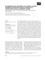
S1¢ specificity of CG and chymase
The crystallographic data reported by Hof et al. [1]
indicate that the side-chain of Arg41 located on the
30S insertion loop in CG projects from the molecular
surface to the east of the active site in accordance with
the standard orientation (Fig. 1). Thus, the S1¢ pocket
in CG appears as a narrow crevice stabilized by the
Cys42-Cys58 disulfide bridge that defines the 30S loop
in both CG and chymase. The S1¢ pocket is bordered
by His57 of the catalytic triad, Ser40 and Arg41,
whose flexibility allows it to be close to both the S1¢
and S2¢ subsites (Fig. 1). Interestingly, an Arg residue
at position 41 is specific to CG and is shared only by
human and chimpanzee CG, suggesting a recent
appearance over the course of evolution (not shown).
In chymase, as in many other serine proteases, residue
41 is a Phe but, unlike other serine proteases, it
projects from the surface of the molecule and is proxi-
mal to the substrate P2¢ side-chain [27]. Thus, the
chymase S1¢ pocket on the top of the Cys42-Cys58
loop is bordered by His57 to the west and the aliphatic
part of the Lys40 side-chain to the east [2] (Fig. 1). As
a result, the P1¢ specificity of chymase could be
different from that of CG on account of the Lys40 in
chymase helping to accomodate a negatively-charged
P1¢ residue. This could explain why chymase is more
efficient than CG at inactivating bradykinin
(RPPGFSPFRCOO
)
) upon cleavage of the C-terminal
F–R bond [28] and it is likely that the Lys40 in chym-
ase will form an electrostatic interaction with the
negatively-charged carboxyl group of bradykinin. To
confirm this hypothesis, we raised two FRET sub-
strates that had either an Asp or an Arg at P1¢. The
peptidyl backbone of these substrates was that of a
previously described FRET substrate: ABZ-GIA-
TFCMLMPEQ-EDDnp (substrate 1) [where ABZ is
O-aminobenzoic acid and EDDnp is N-(2,4-dinitrophe-
nyl)-ethylenediamine], which was derived from the
inhibitory loop sequence of serpinB1 (previously called
Arg143
Arg143
Lys40
Lys40
Phe41
P1
P2
P3
P4
S1’
S2’
Arg143
Arg143
Lys217
Lys192
Lys192
Arg217
Arg41
Arg41
Ser40
P1
P2
P3
P4
S1’
S2’
Cathepsin G
Chymase
Ser40
Arg143
Arg41
Lys192
Lys192
Phe41
His57
Ile99
Ile99
Cys42
Ser40
His57
Arg143
Lys40
Cathepsin G Chymase
S1’
S2’
S2’
A
B
S1’
Fig. 1. Structural differences between CG
and chymase. (A) The solvent accessible
surface based on the atom coordinates of
CG (1CGH) [1] and chymase (1PJP) [2] is
coloured to show positive (blue) and nega-
tive (red) electrostatic potentials. The irre-
versible phosphonate inhibitors Suc-Val-Pro-
Phe
P
-(OPh)
2
and Suc-Ala-Ala-Pro-Phe-chlo-
romethylketone complexed to CG and to
chymase, respectively, are shown as cyan
stick models. The serine of the catalytic
triad is yellow. (B) Ribbon plot of CG and
chymase in irreversible complexes with syn-
thetic inhibitors showing ball-and-stick mod-
els for the seven residues located in the
vicinity of the active site. The molecular sur-
faces were generated using
YASARA software
().
B. Korkmaz et al. Cathepsin G versus chymase specificity
FEBS Journal 278 (2011) 2635–2646 Journal compilation ª 2011 FEBS. No claim to original French government works 2637
monocyte neutrophil elastase inhibitor) and can be
cleaved at the F–C bond by CG and by chymase
[29,30]. We found that the specificity constants,
k
cat
⁄ K
m
, for cleavage by CG and by chymase of ABZ-
GIATFDMLMPEQ-EDDnp (substrate 2) and ABZ-
GIATFRMLMPEQ-EDDnp (substrate 3) were similar
in the 2 · 10
2
mm
)1
Æs
)1
range (Table 1), indicating that
the Lys40 in chymase does not act as a discriminating
structural determinant of P1¢ specificity.
The two best substrates developed previously for
CG, and which are also cleaved by chymase, differ
mainly in the size of the P1¢ residue. One is derived
from the antichymotrypsin (ACT) sequence (ABZ-
TPFSGQ-EDDnp) and bears a Ser at P1¢ [31] and the
other, from a CG-cleaved sequence in protease-acti-
vated receptor-1, PAR-1 (ABZ-EPFWEDQ-EDDnp),
bears a Trp at this position [31,32]. Because of the small
size of the S1¢ pocket in CG, we hypothesized that
small-sized residues are preferred by CG and that they
could possibly help to discriminate between CG and
chymase. We introduced either a Ser or a Trp residue
at P1¢ in substrate 1 to obtain substrate 4 (ABZ-GI-
ATFSMLMPEQ-EDDnp) and substrate 5 (ABZ-GI-
ATFWMLMPEQ-EDDnp) and tested these substrates
with CG and chymase. As expected, cleavage sites iden-
tified by HPLC fractionation of the proteolysis prod-
ucts remained unchanged after the P1 Phe residue, and
a Trp residue at P1¢ significantly decreased the k
cat
⁄ K
m
;
however, this result was obtained for both proteases
(Table 1), which strongly suggests that S1¢ subsites in
CG and chymase are too closely related structurally to
allow discrimination between these two proteases.
S2¢ specificity of CG and chymase
Crystallographic data show that the S2¢ subsite of CG
is highly polar as a result of the presence of three posi-
tively-charged residues: Arg41, Arg143 and Lys192 [1]
(Fig. 1). In chymase, the Arg ⁄ Phe substitution at posi-
tion 41 projects the Phe side-chain into the active site
cleft, resulting in partial obstruction at the bottom of
the S2¢ subsite. However, the crystal structure of chym-
ase also indicates that the orientation of Arg143 in
chymase differs from that in CG and is more proximal
to the S2¢ subsite. This probably explains why, despite
the Arg ⁄ Phe substitution, chymase accomodates a neg-
atively-charged P2¢ residue, as shown using a phage
display random nonapeptide library (Fig. 1) [33–35].
Thus, CG and chymase could accommodate a negative
P2¢ residue, although via a different mechanism that
involves Arg41 and Lys192 in CG and Arg143 and
Lys192 in chymase. We have tested the influence of
negative and positive residues at P2¢ in the serpinB1-
derived FRET substrate to possibly take advantage of
this different mechanism for discriminating between
the two proteases. We observed a significant increase
in specificity constant value using ABZ-GIATFCD-
LMPEQ-EDDnp (substrate 6) compared to substrate 1
and a significant decrease in this rate constant using
ABZ-GIATFCRLMPEQ-EDDnp (substrate 7) but,
again, similar results were obtained with both chymase
and CG. Nevertheless, this demonstrates the impor-
tance of the S2¢ subsite for both proteases, and also
that Arg143 in chymase has a function similar to that
of Arg41 in CG (Table 1). This finding is in agreement
with our observation that mouse CG, in which Arg41
is replaced by an Ala residue, cleaves substrates 6 and
7 at the same rate [24]. Thus, despite the significantly
different structure of their S2¢ subsite, CG and chym-
ase have a similar preference for negatively-charged
P2¢ residues. We have previously shown that PR3 and
HNE poorly accommodate a Pro at P2¢, which empha-
sizes the importance of the S2¢ subsite in neutral serine
proteases [36]. Unlike PR3 and HNE, CG accommo-
dates a P2¢ prolyl residue, as shown using substrate 8
(ABZ-GIATFCPLMPEQ-EDDnp), that is cleaved
approximately twice as fast as control substrate 1
(Table 1). Again, however, the same result was
obtained with chymase, further confirming the similar
specificity of these two proteases.
Table 1. Influence of residues at P1, P1¢ and P2¢ on the specificity
of CG and chymase as deduced from the specificity constant
k
cat
⁄ K
m
with FRET substrates derived from the serpinB1 and ACT-
reactive site loops. Values (m
M
)1
Æs
)1
) are the mean of ‡ 3 experi-
ments. The error for k
cat
⁄ K
m
is < 15%. The arrow indicates cleav-
age sites by CG and chymase. NSH, no significant hydrolysis.
Number Substrates
k
cat
⁄ K
m
CG Chymase
Derived from SERPINB1
S1¢ specificity
1 ABZ-GIATFCMLMPEQ-EDDnp 263
a
238
2 ABZ-GIATFDMLMPEQ-EDDnp 175 221
3 ABZ-GIATFRMLMPEQ-EDDnp 162 203
4 ABZ-GIATFSMLMPEQ-EDDnp 217 209
5 ABZ-GIATFWMLMPEQ-EDDnp 69 96
S2¢ specificity
6 ABZ-GIATFCDLMPEQ-EDDnp 817 1035
7 ABZ-GIATFCRLMPEQ-EDDnp 68 144
8 ABZ-GIATFCPLMPEQ-EDDnp 560 287
Derived from ACT
S1 specificity
9 ABZ-TPFSALQ-EDDnp 153.8
b
136
10 ABZ-TPKSALQ-EDDnp 8.1
b
NSH
11 ABZ-TPWSALQ-YNO
2
69 97
a
Value from Korkmaz et al. [29].
b
Value from Re
´
hault et al. [25].
Cathepsin G versus chymase specificity B. Korkmaz et al.
2638 FEBS Journal 278 (2011) 2635–2646 Journal compilation ª 2011 FEBS. No claim to original French government works
S1 specificity of CG and chymase
The dual specificity of CG for cleaving after large
hydrophobic or positively-charged residues has been
explained by the presence of a Glu residue at
position 226 at the bottom of the S1 pocket [1,24].
This idea has received support using mouse CG that
has an Ala at position 226 and does not cleave P1-
Lys containing substrates [24] and, more recently, as
a result of a phylogenetic analysis of mammalian
CGs [37]. Human chymase also has an Ala residue
at position 226 and this could be exploited to raise
a specific CG substrate (Fig. 2A). However, the
specificity constant for the reaction between CG and
a P1 Lys-containing substrate is far lower than
that of the corresponding substrate with a Phe at P1
[25].
The presence of an Ala residue at position 226 in
chymase also makes the S1 subsite wider, and this
could favour the accommodation of a P1 Trp residue
by chymase, as recently shown using a phage-displayed
selection of peptides susceptible to chymase cleavage
[34]. We compared the hydrolysis by CG and chymase
of ABZ-TPFSALQ-EDDnp (substrate 9), ABZ-TP
KSALQ-EDDnp (substrate 10) and ABZ-TPWSALQ-
YNO
2
(substrate 11) (Table 1). As expected, CG and
chymase prefered a Phe at P1 (substrate 9), although
both also accommodated a Trp in their S1 subsite and
only CG cleaved the P1Lys-containing substrate
(Table 1). However, this occurred at a very low rate,
in accordance with previous findings [25]. Because no
other subsites from S2 to S3¢ in CG and chymase
demonstrated a specificity that would allow discrimina-
tion between the two proteases, we next attempted to
improve the specificity constants of P1 Lys-containing
substrates, aiming to measure subnanomolar amounts
of CG specifically.
Design of specific and sensitive substrates for CG
and chymase
A first step was to improve the k
cat
⁄ K
m
value of P1
Phe-containing substrates before substituting the
P1-Phe by Lys. Accordingly, we started from our most
sensitive but not specific CG ⁄ chymase FRET substrate
ABZ-GIATFCDLMPEQ-EDDnp (substrate 6) and
replaced the Thr residue by Pro [ABZ-GIAP FCDLM-
PEQ-EDDnp (substrate 12)], aiming to prevent cleav-
age at the C–D bond by HNE and PR3 with a Pro at
P3 [38,39] and to improve cleavage by CG and chym-
ase, although the latter prefers aliphatic residues at P2
[34]. The Pro-Phe pair at P2–P1 is present in most of
Cathepsin G Chymase
S1
S1
Arg41
Lys192
Gln192
Arg143
Asp143
Asp147
Glu217
Arg217
S2’
S2’
S3
Cathepsin G
P3
β2-Tryptase (monomer)
A
B
Fig. 2. Structural differences between CG,
chymase and b2-tryptase. (A) Ribbon plot of
CG and chymase in a complex with syn-
thetic inhibitors. The irreversible phospho-
nate inhibitors Suc-Val-Pro-Lys
P
-(OPh)
2
and
Suc-Ala-Ala-Pro-Phe-chloromethyl ketone in
a complex with cathepsin G and to chym-
ase, respectively, are shown as cyan stick
models. Glu
226
and Ala
226
residues at the
bottom of the S1 subsite are shown in
green. (B) Electrostatic surface potential of
human CG and b2-tryptase [50]. Solvent-
accessible surfaces with a positive electro-
static potential are shown in dark blue, and
these with a negative electrostatic potential
are shown in red. The serine of the catalytic
triad is shown in yellow. The molecular sur-
faces were generated using using
YASARA
software ().
B. Korkmaz et al. Cathepsin G versus chymase specificity
FEBS Journal 278 (2011) 2635–2646 Journal compilation ª 2011 FEBS. No claim to original French government works 2639
the commonly used chromogenic and fluorogenic
substrates of CG that are also cleaved by chymase
[31]. As expected, k
cat
⁄ K
m
of substrate 12 was
increased significantly using CG and chymase, and was
resistant to HNE cleavage (Table 2). However, this
substrate was still cleaved by PR3 at the C–D bond
(Table 2). Total resistance to PR3 hydrolysis was
obtained by substituting Ser for Cys in substrate 12 as
a result of the higher electronegative charge of the O
atom of the Ser side-chain compared to that of the
sulfur atom in the Cys side-chain; P3¢ Leu for Pro
because a Pro is not well accommodated by the PR3
S2¢ subsite [36]; and Ala for Glu at P3 because this
improves interaction with Lys192 at the S3 subsite of
CG. The resulting substrate (ABZ-GIEPFSDPMPEQ-
EDDnp (substrate 13) fulfils most of the requirements
for CG, as well as for chymase cleavage (i.e. a nega-
tively-charged residue at P3 and P2¢, a Pro-Phe pair at
P2–P1, and a Ser and a Pro at P1¢ and P3¢, respec-
tively), and this represents one of the most sensitive
substrates to have been reported for these two prote-
ases (Table 2). Finally, substituting Phe for Lys in this
optimized substrate [ABZ-GIEPKSDPMPEQ-EDDnp
(substrate 14)] totally abolished cleavage by chymase,
at the same time as maintaining specificity constant in
the 10
5
m
)1
Æs
)1
range (i.e. sufficiently high to allow
specific measurements of nanomolar concentrations of
CG) (Table 2). As expected, HPLC analysis showed
that CG cleaved this substrate at the K–S bond
(Fig. 3A). Furthermore, this substrate was not hydro-
lyzed by b2-tryptase (EC 3.4.21.59), despite the Lys at
P1 that is a preferential cleavage site for trypsin-like
proteases (Fig. 3B). This was a result of the presence
of negatively-charged residues at P2 and P2¢ that are
not accommodated within the b2-tryptase active site
because of the presence of Asp147 and Asp143
within the S2 and the S2¢ subsites, respectively [40,41]
(Fig. 2B). We cannot exclude the possibility, how-
ever, that trypsin-like protease(s) other than tryptase
are present in cells, tissues or biological fuids, such as
lung secretions and skin exudates, where CG and
chymase have been identified as critical pathophysio-
logical actors. Trypsin-like activities, however, could
be easily detected using broad spectrum inhibitors
such as leupeptin or N-tosyl-l-lysine chloromethyl ke-
tone that do not affect chymotrypsin-like proteases.
Nevertheless, we used a lysate of cells from a mast cell
line and also sputum from a patient with severe
asthma to measure hydrolysis of the newly-described
substrate.
Measurement of CG activity in a mast cell line
extract and in sputum
Mast cells contain substantial amounts of a variety of
proteases, including chymase, tryptase, carboxypepti-
dase A3 and dipeptidyl peptidase I (cathepsin C), that
participate in host defence and homeostasis [3]. The
qualitative and quantitative importance of CG or a
CG-like protease in mast cells and mast cell lines
remains unclear because the substrate specificity of
CG is close to that of chymase [42] and the corre-
sponding mRNA has not been detected in the cell
extracts [43]. We used a mast cell line (HMC-1) extract
to measure CG activity using ABZ-GIEPKSDPM-
PEQ-EDDnp (substrate 14) and evaluate its concentra-
tion in comparison with that of chymase. Accordingly,
we compared the rate of hydrolysis of the specific CG
substrate and a CG ⁄ chymase substrate by the cell
extract, as well as by purified CG and chymase. Opti-
mized kinetic conditions were first determined to
ensure that both substrates were cleaved at approxi-
mately V
max
. We measured CG activity in the HMC-1
cell line, which confirms previous results obtained
using a specific trypsin-like fluorophosphonate probe
[44]. We ensured that the activity measured with
ABZ-GIEPKSDPMPEQ-EDDnp was only a result of
CG by adding the irreversible chloromethylketone
inhibitor Z-GLF-CMK (where Z is benzyloxycarbonyl
and CMK is chloromethyl ketone), which specifically
targets chymotrypsin-like proteases. Full inhibition
was obtained under these conditions, confirming
Table 2. Specificity constant k
cat
⁄ K
m
for the hydrolysis of the FRET substrates derived from serpinB1 by CG, chymase, HNE and PR3.
Values (m
M
)1
Æs
)1
) are the means of ‡ 3 experiments. The error for k
cat
⁄ K
m
is < 15%. NSH, no significant hydrolysis.
Number Substrates derived from serpinB1
k
cat
⁄ K
m
CG Chymase HNE PR3
6 ABZ-GIATFCDLMPEQ-EDDnp 817 1035 1100 2230
12 ABZ-GIAPFCDLMPEQ-EDDnp 2054 1963 < 1 488
13 ABZ-GIEPFSDPMPEQ-EDDnp 1700 1648 NSH NSH
14 ABZ-GIEPKSDPMPEQ-EDDnp 190 NSH NSH NSH
Cathepsin G versus chymase specificity B. Korkmaz et al.
2640 FEBS Journal 278 (2011) 2635–2646 Journal compilation ª 2011 FEBS. No claim to original French government works
the specific role of CG in cleavage (Fig. 4A). We
checked that this inhibitor did not alter cleavage by
the cell lysate of the trypsin-like substrate ABZ-
TPRSALQ-EDDnp at the R–S bond (not shown). We
also found that chymase activity was only twice as
high as that of CG in HMC-1 cells, in accordance
with preliminary observations made using MC
TC
mast
cells [4].
We also measured the hydrolysis of ABZ-GIEP
FSDPMPEQ-EDDnp (substrate 13) and ABZ-GIEP
KSDPMPEQ-EDDnp (substrate 14) by a sample of
whole sputum from a patient with severe asthma. Both
substrates were rapidly cleaved at a single site identi-
fied at the F-S bond and the K–S bond, respectively,
by HPLC analysis (Fig. 4B). Cleavage was completely
abolished after incubation with the chymotrypsin-
like-specific Z-GLF-CMK inhibitor, which clearly
demonstrates that no trypsin-like protease cleaved
substrate 14 in the sputum (not shown). However, the
resulting EDDnp-containing fragments from CG
ABZ-GIEPKSDPMPEQ-EDDnp
Fluorescence
10 12 14 16 18 20 22 24
0
50
100
150
200
250
10 12 14 16 18 20 22 24
0
100
200
300
400
500
600
2000
1000
0
0 200 400 600 800 1000 1200 1400
Elution time (min)
Absorbance
220 nm
320 nm
360 nm
SDPMPEQ-EDDnp
SDPMPEQ-EDDnp
ABZ-GIEPF
ABZ-GIEPK
Time (s)
ABZ-GIEPKSDPMPEQ-Y
Cathepsin G
+
ABZ-GIEPKSDPMPEQ-Y + Tryptase
ABZ-TPKSALQ-EDDnp + Tryptase
NO2
NO2
ABZ-GIEPFSDPMPEQ-EDDnp
A
B
Fig. 3. Hydrolysis of ABZ-GIEPFSDPMPEQ-
EDDnp and ABZ-GIEPKSDPMPEQ-EDDnp
by CG. (A) Demonstration of identical cleav-
age sites within the two substrates as visu-
alized by reverse-phase HPLC and recording
at 360 nm of the EDDnp-containing frag-
ments. (B) Control experiment showing no
cleavage of the Lys-containing CG substrate
ABZ-GIEPKSDPMPEQ-Y
NO2
(20 lM)by
b2-tryptase (10
)7
M final concentration) but
a rapid cleavage of ABZ-TPKSALQ-EDDnp
(20 l
M)by10
)9
M b2-tryptase. Hydrolysis of
ABZ-GIEPKSDPMPEQ-Y
NO2
(20 lM)by
10
)9
M CG is shown for comparison. Assays
were carried out at 37 °Cin50m
M Hepes
buffer (pH 7.4), 100 m
M NaCl, 0.01% Igepal
CA-630 (v ⁄ v).
B. Korkmaz et al. Cathepsin G versus chymase specificity
FEBS Journal 278 (2011) 2635–2646 Journal compilation ª 2011 FEBS. No claim to original French government works 2641
hydrolysis (SDPMPEQ-EDDnp) were sequentially
degraded in a time-dependent manner. This could be a
result of the presence of amino peptidase activity(ies)
in asthma sputum, although further work is required
using larger numbers of sputum samples to confirm
this hypothesis.
Time (s)
Elution time
(
min
)
FluorescenceAbsorbance
ABZ-GIEPKSDPMPEQ-EDDnp
(substrate 14)
ABZ-GIEPKSDPMPEQ-EDDnp
(substrate 14)
ABZ-GIEPFSDPMPEQ-EDDnp
(substrate 13)
0 200 400 600 800 1000 1200 1400
0
100
000
200
000
300
000
400
000
500
000
+ HMC-1 cells lysate
+ Asthma sputum
+ Asthma sputum
+ [HMC-1 cells lysate + Z-GLF-CMK]
Time (s)
Fluorescence (10 x)
0 400 800 1200 1600 2000
600
500
400
300
200
100
0
+ substrate 14
+ ABZ-GIEPFSY
Purified chymase
-3
10 12 14 16 18 20 22 24
0
50
100
150
200
250
300
350
10 12 14 16 18 20 22 24
0
50
100
150
200
250
300
350
220 nm
320 nm
360 nm
SDPMPEQ-EDDnp
ABZ-GIEPF
ABZ-GIEPK
B
A
SDPMPEQ-EDDnp
NO2
Fig. 4. Hydrolysis of the CG substrate by a cell line extract and by a biological sample. (A) Monitoring of ABZ-GIEPKSDPMPEQ-EDDnp
hydrolysis by a HMC-1 mast cell lysate before and after incubation with the chymotrypsin-like protease inhibitor Z-GLF-CMK (3 m
M final con-
centration). The total inhibition observed in the presence of inhibitor indicates that the cleavage of the P1 Lys-containing substrate was a
result of CG. The insert shows the peptidase activity of purified chymase on a polyvalent substrate and its inability to cleave substrate 14
under the same experimental conditions. (B) Hydrolysis of ABZ-GIEPFSDPMPEQ-EDDnp and ABZ-GIEPKSDPMPEQ-EDDnp by sputum from
a patient with severe asthma as visualized by reverse-phase HPLC and recording at 360 nm for the EDDnp-containing fragments. Identical
cleavage sites are observed within the two substrates but their cleavage was abolished after previous incubation with Z-GLF-CMK (not
shown), indicating that only CG was involved in these cleavages. Further degradation of the EDDnp-containing fragment, most probably by
aminopeptidase activity present in the sputum, is observed for both peptides. Assays were carried out at 37 °Cin50m
M Hepes buffer
(pH 7.4), 100 m
M NaCl, 0.01% Igepal CA-630 (v ⁄ v).
Cathepsin G versus chymase specificity B. Korkmaz et al.
2642 FEBS Journal 278 (2011) 2635–2646 Journal compilation ª 2011 FEBS. No claim to original French government works
The reason why two closely-related proteases such
as chymase and CG are co-stored within the same cell
type remains unclear. Mast cells are involved in a
variety of biological functions [45,46] and are mediated
by a range of potent mediators and proteases of differ-
ent specificities whose roles require clarification. Using
a specific CG substrate such as that described in the
present study should help to define the roles of these
two proteases in diseases associated with mast cell
activation and facilitate the development of specific
inhibitors that could control their activity.
Materials and methods
Materials
Purified CG (EC 3.4.21.20), HNE (EC 3.4.21.37) and ACT
were obtained from Biocentrum (Krakow, Poland). Purified
PR3 (
EC 3.4.21.76) and b2-tryptase (EC 3.4.21.59) were
provided by Athens Research & Technology Inc. (Athens,
GA, USA) and Merck (Nottingham, UK), respectively. Ige-
pal CA-630 was obtained from Sigma (St Louis, MO,
USA). Z-GLF-CMK was obtained from Enzyme System
Products (Livermore, CA, USA). N,N-dimethylformamide
and acetonitrile were obtained from Merck (Darmstad,
Germany). Electrophoresis chemicals were obtained from
Bio-Rad (Marnes-la-Coquette, France). All other chemical
reagents were of analytical grade.
Design and synthesis of quenched fluorescent
substrates
Quenched fluorogenic substrates were either obtained from
Genecust-Europe (Dudelange, Luxembourg) or prepared by
solid phase synthesis with Fmoc methodology [47]. Sub-
strate purity was checked by MS (TofSpec-E; Micromass,
Manchester, UK) and by reversed-phase chromatography
on a C18 column. The purified ABZ-peptidyl-EDDnp con-
centration was determined by measuring A
365
with
e
365
= 17 300 m
)1
Æcm
)1
for EDDnp [where ABZ is O-am-
inobenzoic acid and EDDnp is N-(2,4-dinitrophenyl)-ethy-
lenediamine]. Stock substrate solutions (2–5 mm) were
prepared in 30% (v ⁄ v) N,N-dimethylformamide and diluted
to 0.5 with 50 mm Hepes buffer (pH 7.4).
Enzyme assays
HNE, PR3 and CG were titrated with a
1
-proteinase inhibi-
tor, as described previously [48]. Recombinant chymase,
produced and activated as described previously [34], was
titrated with ACT, the titre of which had been determined
by titration with CG. Assays were carried out at 37 °Cin
50 mm Hepes buffer (pH 7.4), 100 m m NaCl and 0.01%
Igepal CA-630 (v ⁄ v) for CG; in 0.1 m Tris ⁄ HCl (pH 8.0)
and 50 mm Hepes (pH 7.4) for chymase; and in 750 mm
NaCl and 0.05% Igepal CA-630 (v ⁄ v) for HNE and PR3.
The hydrolysis of ABZ-peptidyl-EDDnp substrates was
monitored by measuring fluorescence at k
ex
= 320 nm and
k
ex
= 420 nm in a Hitachi F-2000 spectrofluorometer (Hit-
achi, Tokyo, Japan). Specificity constants (k
cat
⁄ K
m
) were
determined under first-order conditions, using a substrate
concentration far below the estimated K
m
as described pre-
viously [31].
HMC-1 cells, kindly provided by Dr J. H. Butterfield
(Mayo Clinic, Rochester, MN, USA) were cultured as
described previously [42]. Suspensions of 30–60 million cells
were lysed in 2 mL of NaCl ⁄ P
i
supplemented with 1% Ige-
pal CA-630 (v ⁄ v). Proteolytic activity was measured at
37 °C using 50 lL of the cell lysates with ABZ-GIE-
PFSDPMPEQ-EDDnp (25 lm) or ABZ-GIEPKSDPM-
PEQ-EDDnp (25 lm) and 5 lL of cell lysate with
ABZ-TPRSALQ-EDDnp (25 lm) in a total volume of
70 lL using a microplate fluorescence reader (Spectra Max
Gemini; Molecular Devices, Sunnyvale, CA, USA) under
continuous stirring. A sample of induced sputum from a
patient with severe asthma was kindly provided by Dr Peter
H. Howarth (University of Southampton, Southampton,
UK). Written informed consent was obtained from the
patient from whom the sputum sample was obtained.
Chromatographic procedures and analysis of
peptide products
Once the enzyme–substrate reaction was complete, the reac-
tion medium was incubated with four volumes of absolute
ethanol for 15 min on ice and centrifuged at 13 000 g for
10 min. The supernatant containing the hydrolysis products
was recovered, air-dried under vacuum and dissolved in
200 lL of 0.0075% trifluoroacetic acid (v ⁄ v). Hydrolysis
fragments were fractionated by reversed-phase HPLC and
eluted peaks were monitored at three wavelengths (220, 320
and 360 nm) simultaneously, which allowed direct identifi-
cation of EDDnp-containing peptides before sequencing or
MS analysis to identify cleavage sites.
Nomenclature
The nomenclature used for the individual amino acid resi-
dues (e.g. P2, P1, P1¢,P2¢, etc.) of a substrate and corre-
sponding residues of the enzyme subsites (e.g. S2, S1, S1¢,
S2¢, etc.) follows that of Schechter and Berger [49].
Acknowledgements
This work was supported by ‘Region Centre’ and the
‘Fonds Europe
´
en de De
´
veloppement Re
´
gional’ (Projet
INFINHI) and Agence Nationale pour la Recherche
(project ANR-07-PHYSIO-029-01). The authors thank
B. Korkmaz et al. Cathepsin G versus chymase specificity
FEBS Journal 278 (2011) 2635–2646 Journal compilation ª 2011 FEBS. No claim to original French government works 2643
Miche
`
le Brillard-Bourdet for sequence analyses; Chris-
tophe Epinette and Lise Vanderlynden for technical
support; Dr Peter H. Howarth, University of South-
ampton, for providing a sputum sample; and the
‘Plate-forme d’Analyse Inte
´
grative des Biomarqueurs’
for MALDI-TOF MS analyses.
References
1 Hof P, Mayr I, Huber R, Korzus E, Potempa J, Travis
J, Powers JC & Bode W (1996) The 1.8 A crystal
structure of human cathepsin G in complex with Suc-
Val-Pro-PheP-(OPh)2: a Janus-faced proteinase with
two opposite specificities. EMBO J 15, 5481–5491.
2 Pereira PJ, Wang ZM, Rubin H, Huber R, Bode W,
Schechter NM & Strobl S (1999) The 2.2 A crystal
structure of human chymase in complex with succinyl-
Ala-Ala-Pro-Phe-chloromethylketone: structural expla-
nation for its dipeptidyl carboxypeptidase specificity.
J Mol Biol 286, 163–173.
3 Trivedi NN & Caughey GH (2010) Mast cell peptidases:
chameleons of innate immunity and host defense. Am
J Respir Cell Mol Biol 42, 257–267.
4 Schechter NM, Irani AM, Sprows JL, Abernethy J,
Wintroub B & Schwartz LB (1990) Identification of a
cathepsin G-like proteinase in the MCTC type of
human mast cell. J Immunol 145, 2652–2661.
5 Schechter NM, Choi JK, Slavin DA, Deresienski DT,
Sayama S, Dong G, Lavker RM, Proud D & Lazarus
GS (1986) Identification of a chymotrypsin-like protein-
ase in human mast cells. J Immunol 137, 962–970.
6 Fellows E, Gil-Parrado S, Jenne DE & Kurschus FC
(2007) Natural killer cell-derived human granzyme H
induces an alternative, caspase-independent cell-death
program. Blood 110, 544–552.
7 Salvesen G, Farley D, Shuman J, Przybyla A, Reilly
C & Travis J (1987) Molecular cloning of human
cathepsin G: structural similarity to mast cell and
cytotoxic T lymphocyte proteinases. Biochemistry 26,
2289–2293.
8 Reilly CF, Tewksbury DA, Schechter NM & Travis J
(1982) Rapid conversion of angiotensin I to angiotensin
II by neutrophil and mast cell proteinases. J Biol Chem
257, 8619–8622.
9 Jahanyar J, Youker KA, Loebe M, Assad-Kottner C,
Koerner MM, Torre-Amione G & Noon GP (2007)
Mast cell-derived cathepsin g: a possible role in the
adverse remodeling of the failing human heart. J Surg
Res 140, 199–203.
10 Helske S, Syvaranta S, Kupari M, Lappalainen J, Laine
M, Lommi J, Turto H, Mayranpaa M, Werkkala K,
Kovanen PT et al. (2006) Possible role for mast cell-
derived cathepsin G in the adverse remodelling of ste-
notic aortic valves. Eur Heart J 27, 1495–1504.
11 Schiemann F, Grimm TA, Hoch J, Gross R, Lindner B,
Petersen F, Bulfone-Paus S & Brandt E (2006) Mast
cells and neutrophils proteolytically activate chemokine
precursor CTAP-III and are subject to counterregula-
tion by PF-4 through inhibition of chymase and cathep-
sin G. Blood 107, 2234–2242.
12 Sommerhoff CP, Nadel JA, Basbaum CB & Caughey
GH (1990) Neutrophil elastase and cathepsin G stimulate
secretion from cultured bovine airway gland serous cells.
J Clin Invest 85, 682–689.
13 Raymond WW, Cruz AC & Caughey GH (2006) Mast
cell and neutrophil peptidases attack an inactivation
segment in hepatocyte growth factor to generate NK4-
like antagonists. J Biol Chem 281, 1489–1494.
14 Mayranpaa MI, Heikkila HM, Lindstedt KA, Walls
AF & Kovanen PT (2006) Desquamation of human
coronary artery endothelium by human mast cell prote-
ases: implications for plaque erosion. Coron Artery Dis
17, 611–621.
15 Maryanoff BE, de Garavilla L, Greco MN, Haertlein
BJ, Wells GI, Andrade-Gordon P & Abraham WM
(2009) Dual inhibition of cathepsin G and chymase is
effective in animal models of pulmonary inflammation.
Am J Respir Crit Care Med 181, 247–253.
16 Pham CT (2006) Neutrophil serine proteases: specific
regulators of inflammation. Nat Rev Immunol 6, 541–
550.
17 Owen CA & Campbell EJ (1999) The cell biology
of leukocyte-mediated proteolysis. J Leukoc Biol 65 ,
137–150.
18 Meyer-Hoffert U (2009) Neutrophil-derived serine pro-
teases modulate innate immune responses. Front Biosci
14
, 3409–3418.
19 Korkmaz B, Horwitz M, Jenne DE & Gauthier A
(2010) Neutrophil elastase, proteinase 3 and cathepsin
G as therapeutic targets in human diseases. Pharmacol
Rev 62, 726–759.
20 Carmona AK, Juliano MA & Juliano L (2009) The use
of fluorescence resonance energy transfer (FRET) pep-
tides for measurement of clinically important proteolytic
enzymes. An Acad Bras Cienc 81, 381–392.
21 Korkmaz B, Attucci S, Juliano MA, Kalupov T, Jourdan
ML, Juliano L & Gauthier F (2008) Measuring elastase,
proteinase 3 and cathepsin G activities at the surface of
human neutrophils with fluorescence resonance energy
transfer substrates. Nat Protoc 3, 991–1000.
22 Korkmaz B, Moreau T & Gauthier F (2008) Neutrophil
elastase, proteinase 3 and cathepsin G: physicochemical
properties, activity and physiopathological functions.
Biochimie 90, 227–242.
23 Lesner A, Wysocka M, Guzow K, Wiczk W, Legowska
A & Rolka K (2008) Development of sensitive cathepsin
G fluorogenic substrate using combinatorial chemistry
methods. Anal Biochem 375, 306–312.
Cathepsin G versus chymase specificity B. Korkmaz et al.
2644 FEBS Journal 278 (2011) 2635–2646 Journal compilation ª 2011 FEBS. No claim to original French government works
24 Kalupov T, Brillard-Bourdet M, Dade S, Serrano H,
Wartelle J, Guyot N, Juliano L, Moreau T, Belaaouaj
A & Gauthier F (2009) Structural characterization of
mouse neutrophil serine proteases and identification of
their substrate specificities: relevance to mouse models
of human inflammatory diseases. J Biol Chem 284,
34084–34091.
25 Re
´
hault S, Brillard-Bourdet M, Juliano MA, Juliano L,
Gauthier F & Moreau T (1999) New, sensitive
fluorogenic substrates for human cathepsin G based on
the sequence of serpin-reactive site loops. J Biol Chem
274, 13810–13817.
26 Tanaka T, Minematsu Y, Reilly CF, Travis J & Pow-
ers JC (1985) Human leukocyte cathepsin G. Subsite
mapping with 4-nitroanilides, chemical modification,
and effect of possible cofactors. Biochemistry 24, 2040–
2047.
27 McGrath ME, Mirzadegan T & Schmidt BF (1997)
Crystal structure of phenylmethanesulfonyl fluoride-
treated human chymase at 1.9 A. Biochemistry 36 ,
14318–14324.
28 Reilly CF, Schechter NB & Travis J (1985) Inactiva-
tion of bradykinin and kallidin by cathepsin G and
mast cell chymase. Biochem Biophys Res Commun 127,
443–449.
29 Korkmaz B, Attucci S, Hazouard E, Ferrandiere M,
Jourdan ML, Brillard-Bourdet M, Juliano L & Gauthi-
er F (2002) Discriminating between the activities of
human neutrophil elastase and proteinase 3 using
serpin-derived fluorogenic substrates. J Biol Chem 277,
39074–39081.
30 Korkmaz B, Hajjar E, Kalupov T, Reuter N, Brillard-
Bourdet M, Moreau T, Juliano L & Gauthier F (2007)
Influence of charge distribution at the active site surface
on the substrate specificity of human neutrophil prote-
ase 3 and elastase. A kinetic and molecular modeling
analysis. J Biol Chem 282, 1989–1997.
31 Attucci S, Korkmaz B, Juliano L, Hazouard E, Girardin
C, Brillard-Bourdet M, Rehault S, Anthonioz P & Gau-
thier F (2002) Measurement of free and membrane-
bound cathepsin G in human neutrophils using new sen-
sitive fluorogenic substrates. Biochem J 366, 965–970.
32 Renesto P, Si-Tahar M, Moniatte M, Balloy V, Van
Dorsselaer A, Pidard D & Chignard M (1997) Specific
inhibition of thrombin-induced cell activation by the
neutrophil proteinases elastase, cathepsin G, and pro-
teinase 3: evidence for distinct cleavage sites within the
aminoterminal domain of the thrombin receptor. Blood
89, 1944–1953.
33 Bastos M, Maeji NJ & Abeles RH (1995) Inhibitors of
human heart chymase based on a peptide library. Proc
Natl Acad Sci USA 92, 6738–6742.
34 Andersson MK, Enoksson M, Gallwitz M & Hellman
L (2009) The extended substrate specificity of the
human mast cell chymase reveals a serine protease with
well-defined substrate recognition profile. Int Immunol
21, 95–104.
35 Andersson MK, Thorpe M & Hellman L (2010) Arg143
and Lys192 of the human mast cell chymase mediate
the preference for acidic amino acids in position P2¢ of
substrates. FEBS J 277, 2255–2267.
36 Korkmaz B, Attucci S, Moreau T, Godat E, Juliano L
& Gauthier F (2004) Design and use of highly specific
substrates of neutrophil elastase and proteinase 3. Am J
Respir Cell Mol Biol 30, 801–807.
37 Raymond WW, Trivedi NN, Makarova A, Ray M,
Craik CS & Caughey GH (2010) How immune peptid-
ases change specificity: cathepsin g gained tryptic func-
tion but lost efficiency during primate evolution.
J Immunol 185, 5360–5368.
38 Segal DM, Powers JC, Cohen GH, Davies DR & Wil-
cox PE (1971) Substrate binding site in bovine chymo-
trypsin A-gamma. A crystallographic study using
peptide chloromethyl ketones as site-specific inhibitors.
Biochemistry 10, 3728–3738.
39 Thompson RC & Blout ER (1973) Elastase-catalyzed
amide hydrolysis of tri- and tetrapeptide amides. Bio-
chemistry 12, 66–71.
40 Harris JL, Niles A, Burdick K, Maffitt M, Backes BJ,
Ellman JA, Kuntz I, Haak-Frendscho M & Craik CS
(2001) Definition of the extended substrate specificity
determinants for beta-tryptases I and II. J Biol Chem
276, 34941–34947.
41 Spichalska B, Lesner A, Wysocka M, Sledz M, Lego-
wska A, Jaskiewicz A, Miecznikowska H & Rolka K
(2008) The influence of substrate peptide length on
human beta-tryptase specificity. J Pept Sci 14, 917–
923.
42 Sheth PD, Pedersen J, Walls AF & McEuen AR (2003)
Inhibition of dipeptidyl peptidase I in the human mast
cell line HMC-1: blocked activation of tryptase, but not
of the predominant chymotryptic activity. Biochem
Pharmacol 66, 2251–2262.
43 Nilsson G, Blom T, Kusche-Gullberg M, Kjellen L,
Butterfield JH, Sundstrom C, Nilsson K & Hellman L
(1994) Phenotypic characterization of the human mast-
cell line HMC-1. Scand J Immunol 39, 489–498.
44 Pan Z, Jeffery DA, Chehade K, Beltman J, Clark
JM, Grothaus P, Bogyo M & Baruch A (2006)
Development of activity-based probes for trypsin-fam-
ily serine proteases. Bioorg Med Chem Lett 16,
2882–2885.
45 Bischoff SC (2007) Role of mast cells in allergic and
non-allergic immune responses: comparison of human
and murine data. Nat Rev Immunol 7, 93–104.
46 Bradding P, Walls AF & Holgate ST (2006) The role of
the mast cell in the pathophysiology of asthma.
J Allergy Clin Immunol 117, 1277–1284.
47 Hirata IY, Cezari MHS, Nakaie CR, Boschcov P, Ito
AS & Juliano MA (1994) Internally quenched
B. Korkmaz et al. Cathepsin G versus chymase specificity
FEBS Journal 278 (2011) 2635–2646 Journal compilation ª 2011 FEBS. No claim to original French government works 2645
fluorogenic protease substrates: solid phase synthe-
sis and fluorescence spectroscopy of peptides contain-
ing ortho-aminobenzoyl ⁄ dinitrophenyl groups as
donor-acceptor pairs. Lett Pept Sci 1, 299–308.
48 Serveau C, Moreau T, Zhou GX, ElMoujahed A, Chao
J & Gauthier F (1992) Inhibition of rat tissue kallikrein
gene family members by rat kallikrein-binding protein
and alpha 1-proteinase inhibitor. FEBS Lett 309,
405–408.
49 Schechter I & Berger A (1967) On the size of the active
site in proteases. I. Papain. Biochem Biophys Res Com-
mun 27, 157–162.
50 Pereira PJ, Bergner A, Macedo-Ribeiro S, Huber R,
Matschiner G, Fritz H, Sommerhoff CP & Bode W
(1998) Human beta-tryptase is a ring-like tetramer with
active sites facing a central pore. Nature 392, 306–311.
Cathepsin G versus chymase specificity B. Korkmaz et al.
2646 FEBS Journal 278 (2011) 2635–2646 Journal compilation ª 2011 FEBS. No claim to original French government works