Báo cáo khoa học: A kinetic model for the burst phase of processive cellulases pptx
Bạn đang xem bản rút gọn của tài liệu. Xem và tải ngay bản đầy đủ của tài liệu tại đây (846.91 KB, 14 trang )
A kinetic model for the burst phase of processive
cellulases
Eigil Praestgaard
1
, Jens Elmerdahl
1
, Leigh Murphy
1
, Søren Nymand
1
, K. C. McFarland
2
, Kim Borch
3
and Peter Westh
1
1 Roskilde University, NSM, Research Unit for Biomaterials, Roskilde, Denmark
2 Novozymes Inc., Davis, CA, USA
3 Novozymes A ⁄ S, Bagsværd, Denmark
Introduction
The enzymatic hydrolysis of cellulose to soluble sugars
has attracted increasing interest, because it is a critical
step in the conversion of biomass to biofuels. One
major challenge for both the fundamental understand-
ing and application of cellulases is that their activity
tapers off early in the process, even when the substrate
is plentiful. Typically, the rate of hydrolysis decreases
by an order of magnitude or more at low cellulose
conversion, and experimental analysis has led to quite
divergent interpretations of this behavior. One line of
evidence has suggested that the slowdown is a result of
the heterogeneous nature of the insoluble substrate.
Keywords
burst phase; calorimetry; cellulase; kinetic
equations; slowdown of cellulolysis
Correspondence
P. Westh, Roskilde University, Building 18.1,
PO Box 260, 1 Universitetsvej, DK-4000
Roskilde, Denmark
Fax: +45 4674 3011
Tel: +45 4674 2879
E-mail:
(Received 30 October 2010, revised 21
February 2011, accepted 25 February
2011)
doi:10.1111/j.1742-4658.2011.08078.x
Cellobiohydrolases (exocellulases) hydrolyze cellulose processively, i.e. by
sequential cleaving of soluble sugars from one end of a cellulose strand.
Their activity generally shows an initial burst, followed by a pronounced
slowdown, even when substrate is abundant and product accumulation is
negligible. Here, we propose an explicit kinetic model for this behavior,
which uses classical burst phase theory as the starting point. The model is
tested against calorimetric measurements of the activity of the cellobiohy-
drolase Cel7A from Trichoderma reesei on amorphous cellulose. A simple
version of the model, which can be solved analytically, shows that the burst
and slowdown can be explained by the relative rates of the sequential reac-
tions in the hydrolysis process and the occurrence of obstacles for the pro-
cessive movement along the cellulose strand. More specifically, the
maximum enzyme activity reflects a balance between a rapid processive
movement, on the one hand, and a slow release of enzyme which is stalled
by obstacles, on the other. This model only partially accounts for the
experimental data, and we therefore also test a modified version that takes
into account random enzyme inactivation. This approach generally
accounts well for the initial time course (approximately 1 h) of the hydroly-
sis. We suggest that the models will be useful in attempts to rationalize the
initial kinetics of processive cellulases, and demonstrate their application to
some open questions, including the effect of repeated enzyme dosages and
the ‘double exponential decay’ in the rate of cellulolysis.
Database
The mathematical model described here has been submitted to the Online Cellular Systems
Modelling Database and can be accessed at
/>index.html free of charge.
Abbreviations
CBH, cellobiohydrolase; Cel7A, cellobiohydrolase I; ITC, isothermal titration calorimetry; RAC, reconstituted amorphous cellulose.
FEBS Journal 278 (2011) 1547–1560 ª 2011 The Authors Journal compilation ª 2011 FEBS 1547
Thus, if various structures in the substrate have differ-
ent susceptibility to enzymatic attack, the slowdown
may reflect a phased depletion of the preferred types
of substrate [1,2]. Other investigations have empha-
sized enzyme inactivation as a major cause of the
decreasing rates [3]. This inactivation could reflect
the formation of nonproductive enzyme–substrate
complexes [4–6] or the adsorption of cellulases on
noncellulosic components, such as lignin [7,8],
although the role of lignin remains controversial [9].
Recently, Bansal et al. [10] have provided a compre-
hensive review of theories for cellulase kinetics, and it
was concluded that no generalization could be made
regarding the origin of the slowdown. In particular,
so-called ‘restart’ or ‘resuspension’ experiments, in
which a substrate is first partially hydrolyzed, then
cleared of cellulases and finally exposed to a second
enzyme dose, have alternatively suggested that enzyme
inactivation and substrate heterogeneity are the main
causes of decreasing hydrolysis rates (see refs. [10,11]).
Further analysis of different contributions to the
slowdown appears to require a better theoretical
framework for the interpretation of the experimental
material. In this study, we introduce one approach and
test it against experimental data for the cellobiohydro-
lase Cel7A (formerly CBHI) from Trichoderma reesei.
Our starting point is classical burst phase theory for
soluble substrates [12], and we extend this framework
to account for the characteristics of cellobiohydrolases,
such as adsorption onto insoluble substrates, irrevers-
ible inactivation and processive action. The latter
implies a propensity to complete many catalytic cycles
without the dissociation of enzyme and substrate. For
cellobiohydrolases, the processive action may involve
the successive release of dozens or even hundreds of
cellobiose molecules from one strand [13], and some
previous reports have suggested a possible link
between this and the slowdown in hydrolysis [8,13,14].
Results and Discussion
Theory
Burst phase for soluble substrates and nonprocessive
enzymes
The concept of a burst phase was introduced more
than 50 years ago, when it was demonstrated that an
enzyme reaction with two products may show a rapid
production of one of the products in the pre-steady-
state regime [15,16]. Later work has shown that this is
quite common for hydrolytic enzymes with an ordered
‘ping–pong bi–bi’ reaction sequence [12]. At a constant
water concentration, this type of hydrolysis may be
described by Eqn (1), which does not explicitly include
water as a substrate (the process is considered as an
ordered uni–bi reaction):
E+S¡
k
1
k
À1
ES À!
k
2
EP
2
þ P
1
À!
k
3
E+P
2
ð1Þ
In an ordered mechanism, the product P
1
is always
released from the complex before the product P
2
, and
it follows that, if k
3
is small (compared with k
1
S
0
and
k
2
), there will be a rapid production of P
1
(a burst
phase) when E and S are first mixed. Subsequently, at
steady state, a large fraction of the enzyme population
will be trapped in the EP
2
complex, which is only
slowly converted to P
2
and free E, and the (steady
state) rate of P
1
production will be lower. The result is
a maximum in the rate of production of P
1
but not P
2
(see Fig. 1). To analyze this maximum, we need an
expression for the rate of P
1
production: P
1
¢(t). Here,
and in the following analyses of the reaction schemes,
we first try to derive analytical solutions, as this
approach provides rigorous expressions that may help
to identify the molecular origin of the burst and slow-
down. In cases in which analytical expressions cannot
P
1
′(t) and P
2
′(t) (nM·s
–1
)
P
1
(t) and P
2
(t) (nM)
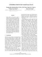
Fig. 1. Initial time course of the concentrations P
1
(t) and P
2
(t) (A)
and the rates P
1
¢(t) and P
2
¢(t) (B) calculated from Eqns <10>–<13>
in Data S1. Full and broken lines indicate P
1
and P
2
, respectively,
and the dotted line shows the steady-state condition with constant
concentrations of the intermediates ES and EP2, and hence con-
stant rates. The intersection p is a measure of the extent of the
burst (see text for details). The parameters were S
0
=20lM,
E
0
= 0.050 lM, k
2
=0.3s
)1
, k
1
= 0.002 s
)1
ÆlM
)1
, k
)1
= k
3
= 0.002 s
)1
;
these values are similar to those found below for Cel7A.
Burst phase of processive cellulases E. Praestgaard et al.
1548 FEBS Journal 278 (2011) 1547–1560 ª 2011 The Authors Journal compilation ª 2011 FEBS
be found, we use numerical treatment of the rate equa-
tions. The results based on analytical solutions were
also tested by the numerical treatment, and no differ-
ence between the two approaches was found. The
equation for P
1
¢(t) has previously been solved on
the basis of different simplifications, such as merging
the first two steps in Eqn (1) [17,18] or using a steady-
state approximation for the intermediates [15,19]. The
equations may also be solved numerically without
resorting to any assumptions, or solved analytically if
it is assumed that the change in S is negligible. If the
initial substrate concentration S
0
is much larger than
E
0
, the assumption of a constant S during the burst is
very good, and we have used this approach to derive
expressions for both the rates P
1
¢(t) and P
2
¢(t), and the
concentrations P
1
(t) and P
2
(t) (see Data S1). Figure 1
shows an example of how these functions change in
the pre-steady-state regime, when parameters similar to
those found below for Cel7A are inserted.
The initial slopes in Fig. 1A are zero and, after
about 100 s, both functions asymptotically reach the
steady-state value, where the concentrations of both
intermediates ES and EP
2
, and hence the rates P
1
¢(t)
and P
2
¢(t), become independent of time (Fig. 1B). For
P
2
(t), the slope in Fig. 1A never exceeds the steady-
state level, but P
1
(t) shows a much higher intermediate
slope that subsequently falls off towards the steady-
state level. This behavior is more clearly illustrated by
the rate functions in Fig. 1B, and it follows that a
method that directly measures the reaction rate (rather
than the concentrations) may be particularly useful in
the investigation of burst phase kinetics. This is the
rationale for using calorimetry in the current work.
Experimental analysis of the burst phase often utilizes
the intersection p of the ordinate and the extrapolation
of the steady-state condition for P
1
(t) (dotted line in
Fig. 1A). This value is used as a measure of the amount
of P
1
produced during the burst, i.e. the excess of P
1
with respect to the steady-state production rate, and it is
therefore a measure of the magnitude of the burst. An
expression for p is readily obtained by inserting t =0in
the (asymptotic) linear expression for P
1
(t), which
results from considering t ޴(see Data S1). Under
the simplification that k
)1
= k
3
, p may be written:
p ¼ E
0
k
2
k
1
S
0
ðÀk
2
3
þ k
2
k
1
S
0
Þ
ðk
2
þ k
3
Þ
2
ðk
3
þ k
1
S
0
Þ
2
ð2Þ
If Eqn (2) is considered for the special case in which
the first two steps in Eqn (1) are much faster than the
third step (i.e. k
1
S
0
>> k
3
+ k
)1
and k
2
>> k
3
), it
reduces to the important relationship p =E
0
, which is
the basis for so-called substrate titration protocols [20],
in which the concentration of active enzyme is derived
from experimental assessments of p. The intuitive con-
tent of this is that each enzyme molecule quickly
releases one P
1
molecule, as described by the first two
steps in Eqn (1), before it gets caught in a slowly dis-
sociating EP
2
complex.
Burst phase for processive enzymes
Kipper et al. [13] studied the hydrolysis of end-labelled
cellulose by Cel7A, and found that the release of the
first (fluorescence-labelled) cellobiose molecule from
each cellulose strand showed a burst behavior, which
was qualitatively similar to that shown in Fig. 1. This
suggests that this first hydrolytic cycle may be
described along the lines of Eqn (1). Unlike the exam-
ple in Eqn (1), however, Cel7A is a processive enzyme
that completes many catalytic cycles before it dissoci-
ates from the cellulose strand [13]. This dissociation
could occur by random diffusion, but some reports
have suggested that processivity may be linked to the
occurrence of obstacles and imperfections on the cellu-
lose surface [4,6,14]. These observations may be cap-
tured in an extended version of Eqn (1) that takes
processivity and obstacles into account. Thus, we con-
sider a cellulose strand C
n
, which has no obstacles for
the processive movement of Cel7A between the reduc-
ing end (the attack point of the enzyme) and the nth
cellobiose unit [i.e. there is a ‘check-block’ that pre-
vents processive movement from the nth to the
(n + 1)th cellobiose unit]. The processive hydrolysis of
this strand may be written as:
2221
21
3
kkkk
xnnnn
k
3
k
3
k
3
k
EC EC EC C EC C
EC
−−
+ + +
↓↓ ↓
21
xnnn
EC EC EC EC
−−
↓
++ + +
(3)
We note that this reaction reduces to Eqn (1) when
n = 2 and k
)1
= k
3
. In Eqn (3), the free cellulase (E)
first combines with a cellulose strand (C
n
) to form an
EC
n
complex. This process, which will also include a
possible diffusion on the cellulose surface and the
‘threading’ of the strand into the active site, is gov-
erned by the rate constant k
1
at a given value of S
0
.
The EC
n
complex is now allowed to decay in one of
two ways. Either the enzyme makes a catalytic cycle
in which a cellobiose molecule (C) is released whilst
the enzyme remains bound in a slightly shorter EC
n)1
complex. Alternatively, the EC
n
complex dissociates
back to its constituents E and C
n
. The rate constants
for hydrolysis and dissociation are k
2
and k
3
, respec-
tively. This pattern continues so that any enzyme–sub-
strate complex EC
n)i
(where i enumerates the number
of processive steps) can either dissociate [vertical step
E. Praestgaard et al. Burst phase of processive cellulases
FEBS Journal 278 (2011) 1547–1560 ª 2011 The Authors Journal compilation ª 2011 FEBS 1549
in Eqn (3)] or enter the next catalytic cycle [horizontal
step to the right in Eqn (3)], which releases one more
cellobiose. A typical cellulose strand is hundreds or
thousands of glycosyl units long, and it follows that
the local environment experienced by the cellulase
may be similar for many sequential catalytic steps.
Therefore, we use the same rate constants k
2
and k
3
for consecutive hydrolytic or dissociation steps. This
version of the model neglects the fact that the C
n)1
,
C
n)2,
strands are also substrates (free E is not
allowed to associate with these partially hydrolyzed
strands). This simplification is acceptable in the early
part of the process where C
n
>> E
0
. After n proces-
sive steps, the enzyme reaches the ‘check block’, and
this necessitates a (slow) desorption from the remain-
ing cellulose strand (designated C
x
) before the enzyme
can continue cellobiose production from a new C
n
strand. In other words, the strand consists of n + x
cellobiose units in total, but because of the ‘check
block’, only the first n units are available for enzy-
matic hydrolysis. This interpretation of obstacles and
processivity is similar to that recently put forward by
Jalak & Valjamae [14].
A kinetic treatment of Eqn (3) requires the specifica-
tion of the substrate concentration. This is not trivial
for an insoluble substrate, but, as the enzyme used
here attacks the reducing end of the strand, we use the
molar concentration of ends for S
0
throughout this
work. This problem may be further addressed by intro-
ducing noninteger (fractal) kinetic orders that account
for the special limitations of the heterogeneous reac-
tion (see refs. [31,32]). For this model, this is readily
performed by introducing apparent orders in Eqn (5).
However, the current treatment is limited to the simple
case in which the kinetic order is equal to the molecu-
larity of the reactions in Eqn (3). This implies that the
adsorption of enzyme onto the substrate is described
by a kinetic (rather than equilibrium) approach (c.f.
Ref. [21]). Based on this and the simplifications men-
tioned above, the kinetic equations for each step in
Eqn (3) were written and solved with respect to the
EC
n)i
intermediates as shown in Data S1. As cellobi-
ose production in Eqn (3) comes from these EC
n)i
complexes, which all decay with the same rate constant
k
2
, the rate of cellobiose production C¢(t) follows the
equation:
C
0
ðtÞ¼k
2
X
nÀ1
i¼0
EC
nÀi
ðtÞð4Þ
Using the expressions in Data S1, the sum in Eqn (4)
may be written as:
where Gamma½n; xt¼
R
1
x
t
nÀ1
e
Àt
dt is the so-called
upper incomplete gamma function [22]. Equations (4)
and (5) provide a description of the burst phase for
processive enzymes. In the simple case, this approach
will eventually reach steady state with constant concen-
trations of all EC
n)i
complexes and hence constant
C¢(t). We emphasize, however, that there are no
steady-state assumptions in the derivation of Eqn (5)
and, indeed, we use it to elucidate the burst in the pre-
steady-state regime. As discussed below, Eqn (3) is
found to be too idealized to account for experimental
data, and some modifications are introduced. Never-
theless, Eqn (5) is the main result of the current work
and is the backbone in the subsequent analyses.
Examination of a processive burst phase as specified
by Eqns (4) and (5) reveals some similarity to the sim-
ple burst behaviour in Fig. 1. Hence, if we insert the
same rate constants as in Fig. 1, and use an obstacle-
free path length of n = 100 cellobiose units, the rate
of cellobiose production C¢(t) (full curve in Fig.2)
exhibits a maximum akin to that observed for P
1
¢(t)in
Fig. 1B. However, the occurrence of fast sequential
steps in the processive model produces a more pro-
nounced maximum in both duration and amplitude.
Figure 2 also illustrates the meaning of the three terms
that are summed in Eqn (5). The chain line shows the
contribution from the first (simple exponential) term
on the right-hand side of Eqn (5), which describes the
kinetics devoid of any effect from obstacles (corre-
sponding to n ޴). The broken line is the sum of
the last two terms (the terms with gamma functions)
and quantifies the (negative) effect on the hydrolysis
rate arising from the ‘check blocks’. For the parame-
ters used in Fig. 2, this contribution only becomes
important above t % 300 s, and this simply reflects the
minimal time required for a significant population of
enzyme to bind and perform the 100 processive steps
to reach the ‘check block’. After about 600 s, essen-
tially all enzymes have reached their first encounter
X
nÀ1
i¼0
EC
nÀi
ðtÞ¼
1 Àe
À½ðk
3
þk
1
S
0
Þt
ÂÃ
E
0
k
1
S
0
ðk
3
þ k
1
S
0
Þ
þ
E
0
ð
k
2
k
2
þk
3
Þ
n
k
1
S
0
À1 þ
Gamma½ðnÞ;ðk
2
þk
3
Þt
Gamma½n
ðk
3
þ k
1
S
0
Þ
þ
1
ðk
3
þ k
1
S
0
Þ
e
À½ðk
3
þk
1
S
0
Þt
E
0
k
1
S
0
k
2
k
2
À k
1
S
0
n
1 À
Gamma½ðnÞ; ðk
2
À k
1
S
0
Þt
Gamma½n
ð5Þ
Burst phase of processive cellulases E. Praestgaard et al.
1550 FEBS Journal 278 (2011) 1547–1560 ª 2011 The Authors Journal compilation ª 2011 FEBS
with a ‘check block’ and we observe an abeyance with
reduced C¢(t) because a significant (and constant) frac-
tion of the enzyme is unproductively bound in front of
a ‘check block’.
The extent of the processive burst may be assessed
from the intersect p
processive
defined in the same way as
p for the simple reaction (see Fig. 1A). As shown in
Data S1, p
processive
may be written as:
p
processive
¼ E
0
S
0
k
1
k
2
À1 þ
k
2
k
2
þk
3
n
1 þ
nðk
3
þk
1
S
0
Þ
k
2
þk
3
hi
k
3
þ k
1
S
0
ðÞ
2
ð6Þ
We note that p
processive
is proportional to E
0
and, if
we again consider the case in which adsorption and
hydrolysis are fast compared with desorption (i.e.
k
1
S
0
>> k
3
and k
2
>> k
3
), Eqn (6) reduces to p
pro-
cessive
= nE
0
. This implies that, under these special
conditions, every enzyme rapidly makes one run
towards the ‘check block’, and thus produces the num-
ber of cellobiose molecules n which are available to
hydrolysis in the obstacle-free path.
Modifications of the model
In analogy with the simple case in Eqn (1), the rate
C¢(t) specified by Eqn (3) runs through a maximum
and falls towards a steady-state level (Fig. 2) in which
the concentrations of all intermediates EC
n)i
and the
rate C¢(t) are independent of time. This behavior, how-
ever, is at odds with countless experimental reports, as
well as the current measurements, which suggest that
the activity of Cel7A does not reach a constant rate.
Instead, the reaction rate continues to decrease. This
suggests that, in addition to the burst behavior
described in Eqn (3), other mechanisms must be
involved in the slowdown. The nature of such inhibi-
tory mechanisms has been discussed extensively and
much evidence has pointed towards product inhibition,
reduced substrate reactivity or enzyme inactivation
(see, for example, refs. [10,11,23] for reviews). In the
current work, we observed this continuous slowdown
even in experiments with very low substrate conversion
(< 1%), where the hydrolysis rates are unlikely to be
affected by inhibition or substrate modification (an
inference that is experimentally supported in Fig.9
below). In the coupled calorimetric assay used here,
the product (cellobiose) is converted to gluconic acid.
The concentration is in the micromolar range, and pre-
vious tests have shown that this is not inhibitory to
cellulolysis or the coupled reactions (see Ref. [48]).
Therefore, the continuous decrease in the rate of
hydrolysis was modeled as protein inactivation. To this
end, we essentially implemented the conclusions of a
recent experimental study by Ma et al. [24] in the
model. As with earlier reports [3,14,25–27], Ma et al.
discussed unproductively bound cellulases, and found
that substrate-associated Cel7A could be separated
into two populations of reversibly and irreversibly
adsorbed enzyme. The latter population, which grew
gradually over time, was found to lose most catalytic
activity. This behavior was introduced into the model
through a new rate constant k
4
, which pertains to the
conversion of an active enzyme–cellulose complex
(EC
n)i
) into a complex of cellulose and inactive protein
(IC
n)i
). In other words, any EC
n)i
complex in Eqn (7)
is allowed three alternative decay routes, namely
hydrolysis (k
2
), dissociation (k
3
) or irreversible inacti-
vation (k
4
). We also introduced a separate rate con-
stant k
)1
for the dissociation of substrate and enzyme
EC
n
before the first hydrolytic step. With these modifi-
cations, we may write the reaction:
444
1
1
21 xnn
kkk
k
n
k
IC IC IC
EC
−
−−
↑↑ ↑
+
222
21
3
1
kkk
xnnn
k
3
k
3
k
n
EC EC C EC C
EC
EC
−−
−
+ +
↓↓↓
+
2 xn
EC EC
−
++
(7)
We were not able to find an analytical solution for
C¢(t) on the basis of Eqn (7), and we instead used a
numerical treatment with the appropriate initial condi-
tions [i.e. all initial concentrations except E(t) and
C
n
(t) are zero].
—
C′(t) (nM·s
–1
)
Fig. 2. The rate of cellobiose production C¢(t) (solid curve) calcu-
lated according to Eqns (4) and (5) and plotted against time. The
rate constants are the same as in Fig. 1 and the initial concentra-
tions were E
0
= 0.050 lM and S
0
=5lM reducing ends. The obsta-
cle-free path n was set to 100 cellobiose units. The chain curve
shows the first term in Eqn (5), which signifies the rate of cellobi-
ose production on an ‘obstacle-free’ substrate (i.e. for n fi¥).
The broken curve, which is the sum of the last two terms in
Eqn (5), signifies the inhibitory effect of the obstacles. The two
curves sum to the full curve.
E. Praestgaard et al. Burst phase of processive cellulases
FEBS Journal 278 (2011) 1547–1560 ª 2011 The Authors Journal compilation ª 2011 FEBS 1551
One final modification of the model was introduced
to examine the effect of ‘polydispersity’ in n. Thus, n
as defined in Eqns (3) and (7) is a constant, and this
implies that all enzymes must perform exactly n
catalytic cycles before running into the ‘check block’.
This is evidently a rather coarse simplification and, to
consider the effects of this, we also tested an approach
which used a distribution of different n values. For
example, the substrate was divided into five equal sub-
sets (i.e. each 20% of S
0
) with n values ranging from
40% to 160% of the average value. We also analyzed
different distributions and subsets of different sizes
(with a larger fraction close to the average n and less
of the longest ⁄ shortest strands). In all of these analy-
ses, the rate of cellobiose production from each subset
was calculated independently and summed to obtain
the total C¢(t).
Experimental
Two parameters from the model, namely the substrate
and enzyme concentrations (E
0
and S
0
), can be readily
varied in experiments, and we therefore firstly com-
pared measurements and modeling in trials in which S
0
and E
0
were systematically changed. Figure 3A shows a
family of calorimetric measurements in which Cel7A
was titrated to different initial substrate concentrations
(S
0
in lm of reducing ends – this unit can be readily
converted into a weight concentration using the molar
mass of a glycosyl unit and the average chain length
for the current substrate, DP = 220 glycosyl units).
The concentration of Cel7A was 50 nm in these experi-
ments and the experimental temperature was 25 °C.
Figure 3B shows model results for the same values of
E
0
and S
0
. Here, we used the model in Eqn (3)
[Eqns (4) and (5)] and manually adjusted the kinetic
constants and n by trial and error. The parameters in
Fig. 3B are k
1
= 0.0004 s
)1
Ælm
)1
, k
2
= 0.55 s
)1
,
k
3
= 0.0034 s
)1
and n = 150. Comparison of the two
panels shows that the idealized description of proces-
sive hydrolysis in Eqn (3) cannot account for the over-
all course of the process, but some characteristics, both
qualitative and quantitative, are captured by the model.
For example, the model accounts well for the dimin-
ished burst (i.e. the disappearance of the maximum) at
low S
0
(below 5–10 lm). In these dilute samples, the
rate of cellobiose production C¢(t) increases slowly to a
level which is essentially constant over the time consid-
ered in Fig. 3. At higher S
0
, a clear maximum in C ¢ (t)
signifies a burst phase in both model and experiment.
On a quantitative level, comparisons of the maximal
rate at the peak of the burst (t = 150 s in Fig. 3C) and
after the burst (t = 1400 s in Fig. 3C) showed a rea-
sonable accordance between experiments and model. In
addition, the substrate concentration that gives half the
maximal rate (5–10 mm) is similar to within experimen-
tal scatter (Fig. 3C). Conversely, two features of the
experiments do not appear to be captured by Eqn (3).
Firstly, the model predicts a sharp termination of the
A
µ
µ
µ
M
M
M
µ
M
µ
M
µ
M
µ
M
µ
M
µ
M
µ
M
µ
M
B
C
S
0
(μM)
Time (s)
C′(t) (nM·s
–1
)
C′(t) (n
M·s
–1
)
Fig. 3. Comparison of the results from experiment and model
[Eqn (3)] for different substrate concentrations (S
0
in lM reducing
ends). The enzyme concentration E
0
was 50 nM. Experimental (A)
and model (B) C¢(t) results from Eqns (4) and (5) using the para-
meters k
1
= 0.0004 s
)1
ÆlM
)1
, k
2
= 0.55 s
)1
, k
3
= 0.003 s
)1
and n =
150 cellobiose units. (C) Experimental (circles) and modeled (lines)
rates at two time points plotted as a function of S
0
.
Burst phase of processive cellulases E. Praestgaard et al.
1552 FEBS Journal 278 (2011) 1547–1560 ª 2011 The Authors Journal compilation ª 2011 FEBS
burst phase, which tends to produce a rectangular
shape of the C ¢(t) function at high S
0
(Fig. 3B). This is
in contrast with the experiments which all show a grad-
ual decrease in C¢(t) after the maximum. Secondly, the
model suggests a constant C¢(t) well within the time
frame covered in Fig. 3, but no constancy was observed
in the experiments. We return to this after discussing
the effect of changing E
0
.
Figure 4 shows a comparison of the calorimetric
measurements and model results for a series in which
the enzyme load was varied and S
0
was kept constant
at 40.8 lm reducing ends. The model calculations were
based on the same parameters as in Fig. 3 without any
additional fitting, and it appears that C¢( t) increases
proportionally to E
0
. This behavior, which was seen in
both model and experiment, implies that the turnover
number C¢(t) ⁄ E
0
is constant over the studied range of
time and concentration, and this, in turn, suggests that
the extent of the burst scales with E
0
. To analyze this
further, p
processive
was estimated from the data in
Fig. 4. For the model results (Fig. 4B), this is simply
done by inserting the kinetic parameters in Eqn (6).
For the experimental data, we first numerically inte-
grated the rates in Fig. 4A to obtain the concentration
of cellobiose C(t), and then extrapolated linear fits to
the data between 1400 and 1600 s to the ordinate as
illustrated in the inset of Fig. 5. In analogy with the
procedure used for nonprocessive enzymes (Fig. 1A),
this intercept between the extrapolation and the C(t)
axis was taken as a measure of the experimental
p
processive
.
The proportionality of the theoretical p
processive
and
E
0
seen in Fig. 5 follows directly from Eqn (6). The
slope of the theoretical curve is about 42, suggesting
that each enzyme molecule completes 42 catalytic
cycles (produces 42 cellobiose molecules) during the
burst phase. This is about three times less than the
obstacle-free path (n), which is 150 in these calcula-
tions, and this discrepancy simply reflects that k
1
S
0
is
too small for the simple relationship p
processive
= nE
0
to be valid (see Theory section). Thus, low k
1
and the
concomitant slow ‘on rate’ tend to smear out the burst
and, consequently, p
processive
⁄ E
0
< n. This is a general
weakness of the extrapolation procedure [17,18], also
visible in Fig. 1, where the dotted line intersects the
ordinate at a value slightly less than E
0
. It occurs when
the rate constants and S
0
attain values that make the
fractions on the right-hand side of Eqns (2) and (6)
smaller than unity (this implies that the criteria for
a simple p expression, k
1
S
0
>> k
3
+ k
)1
and k
2
>>
k
3
, discussed in the Theory section, are not met
[17,18]). More importantly, the experimental data also
show proportionality between p
processive
and E
0
with a
comparable slope (about 65), and this supports the
general validity of Eqn (3).
nM
nM
A
B
C′(t) (nM·s
–1
)
Fig. 4. Comparison of experimental and model results for different
enzyme concentrations (E
0
). The substrate concentration was
40.8 l
M reducing ends. Experimental (A) and model (B) C¢(t) results
using the same parameters as in Fig. 3.
C(t) (µM)
Fig. 5. Theoretical (open symbols) and experimental (filled symbols)
estimates for the extent of the burst (p
processive
) based on the
results in Fig. 4. Theoretical values were obtained by insertion of
the kinetic constants from Fig. 3 into Eqn (4), and the experimental
values represent extrapolation of the C¢(t) function to t = 0 as illus-
trated in the inset. The extrapolations were based on linear fits to
C¢(t) from 1400 to 1600 s.
E. Praestgaard et al. Burst phase of processive cellulases
FEBS Journal 278 (2011) 1547–1560 ª 2011 The Authors Journal compilation ª 2011 FEBS 1553
We now return to the two general shortcomings of
Eqn (3) which were identified above: (a) the abrupt
termination of the modeled burst phase (Fig. 3B),
which is evident for high S
0
and not seen in the experi-
ments; and (b) the regime with constant C¢(t) (see, for
example, t > 500 s in Fig. 4B and inset in Fig. 6),
which is also absent in the measurements. We suggest
that, at least to some extent, (a) is a consequence of the
‘polydispersity’ in n in a real substrate and (b) depends
on the random inactivation of the enzyme. As discussed
in the Theory section, simplified descriptions of these
properties may be included in the model, and these
modifications considerably improve the concordance
between theory and experiment. To illustrate this, we
considered a substrate distribution with five subsets
(each 20% of S
0
) with n = 40, 70, 100, 130 and 160,
respectively. We analyzed the initial 1700 s of all trials
in Fig. 3 using Eqn (5) and the nonlinear regression
routine in Mathematica 7.0. It was found that, above
S
0
$ 15 lm, the parameters derived from each calori-
metric experiment were essentially equal, and we con-
clude that one set of parameters can describe the
results in this concentration range. The parameters
were k
2
= 1.0 ± 0.2 s
)1
, k
3
= 0.0015 ± 0.0003 s
)1
and k
1
S
0
= 0.0052 ± 0.001 s
)1
, and some examples of
the results are shown in Fig. 6. Parameter interdepen-
dence was evaluated partly by the confidence levels
given by Mathematica and partly by ‘grid searches’,
which provide an unambiguous measure of parameter
dependence [28,29] and hence reveal possible overpa-
rameterization. In the latter procedure, the standard
deviation of the fit was determined in sequential
regressions, where two of the rate constants were
allowed to change, whilst the third was inserted as a
constant with values slightly above or below the maxi-
mum likelihood parameter [28,29]. These analyses
showed moderate parameter dependence with 95%
confidence intervals of about ±10% (slightly asym-
metric with larger margins upwards). This limited
parameter interdependence is also illustrated in the
correlation matrix in Data S1, which shows that all
correlation coefficients are below 0.7, and we conclude
that it is realistic to extract three rate constants from
the experimental data. The parameters from this
regression analysis may be compared with recent work
[30], which used an extensive analysis of reducing ends
in both soluble and insoluble fractions to estimate
apparent first-order rate constants for processive
hydrolysis and enzyme–substrate disassociation, respec-
tively. Values for the system investigated in Fig. 6 (i.e.
T. reesei Cel7A and amorphous cellulose) were
1.8 ± 0.5 s
)1
(hydrolysis) and 0.0032 ± 0.0006 s
)1
(dissociation) at 30 °C [30]. The concordance of these
values, which were derived by a completely different
approach, and k
2
and k
3
from Fig. 6 provides strong
support of the molecular picture in Eqn (3). With
respect to the ‘on rate’, it is interesting to note that a
constant value of k
1
provided very poor concordance
between theory and experiment (not shown), whereas
constant k
1
S
0
gave satisfactory agreement (Fig. 6).
This suggests that the initiation of hydrolysis (adsorp-
tion to the insoluble substrate and ‘threading’ of the
cellulase) exhibits apparent first-order kinetics. This
may reflect the reduced dimensionality or fractal kinet-
ics, which has previously been proposed for cellulase
activity on insoluble substrates [31,32], and it appears
C′(t) (nM·s
–1
)
Fig. 6. Experimental data (symbols) and model results (lines) based
on Eqn (3). In this case, the substrate was treated as a mixture
with different obstacle-free path lengths. Specifically, S
0
was
divided into five subsets with n = 40, 70, 100, 130 and 160. The
nonlinear regression was based on the data for the first 1700 s.
The inset shows an enlarged picture of the course after 1700 s and
illustrates that, for the simple model [Eqn (3)], the experimental val-
ues fall below the model beyond the time frame considered in the
regression.
Burst phase of processive cellulases E. Praestgaard et al.
1554 FEBS Journal 278 (2011) 1547–1560 ª 2011 The Authors Journal compilation ª 2011 FEBS
that the current approach holds some potential for sys-
tematic investigations of this phenomenon.
The model could not account for the measurements
at the lowest S
0
, and this may reflect the fact that the
assumption S
0
>> E
0
, used in the derivation of the
expression for C¢(t), becomes unacceptable. Thus, the
concentration of reducing ends S
0
:E
0
ranges from 30
to 2200 in this work (for S
0
=15lm, it is 300). If,
however, we use instead the accessible area of amor-
phous cellulose, which is about 42 m
2
Æg
)1
[33], and a
footprint of 24 nm
2
for Cel7A [34], we find an S
0
:E
0
area ratio (total available substrate area divided by
monolayer coverage area of the whole enzyme popula-
tion) which is an order of magnitude smaller (3–240).
These latter numbers are rough approximations as the
average area of randomly adsorbed enzymes will be
larger than the footprint, and only a certain fraction
of the enzyme will be adsorbed in the initial stages.
Nevertheless, the analysis suggests that not all reducing
ends are available in amorphous cellulose, and hence
the deficiencies of the model at substrate concentra-
tions below 15 lm could reflect the fact that the pre-
mise S
0
>> E
0
becomes increasingly unrealistic.
The results in Fig. 6 are for the fixed average and
distribution of n mentioned above. We also tried wider
or narrower distributions with five subsets, distribu-
tions with 10 subsets and distributions with a predomi-
nance of n values close to the average (e.g. 5%, 20%,
50%, 20%, 5%, instead of equal amounts of the five
subsets). The regression analysis with these different
interpretations of n polydispersity gave comparable fits
and parameters. In addition, average n values of
100 ± 50 were found to account reasonably for the
measurements, and we conclude that detailed informa-
tion on the obstacle-free path n will require a broader
experimental material, particularly investigations of
different types of substrate.
We consistently found that the experimental C¢(t) fell
below the model towards the end of the 1-h experiments
(see inset in Fig. 6). For a series of 4-h experiments (not
shown), this tendency was even more pronounced. This
was interpreted as protein inactivation, as discussed in
the Theory section. Numeric analysis with respect to
Eqn (7) showed that the inclusion of inactivation and
the same polydispersity as in Fig. 6 enabled the model
to fit the data reasonably over the studied time frame
for S
0
above approximately 15 lm. Some examples of
this for different S
0
are shown in Fig.7.
The parameters from the analysis in Fig. 7 were
k
1
S
0
= (5.2 ± 1.6) · 10
)3
s
)1
, k
2
= 1 ± 0.3 s
)1
, k
3
=
k
)1
= (1.2 ± 0.6) · 10
)3
s
)1
and k
4
= (2 ± 0.7) ·
10
)4
s
)1
. The parameter dependence of these fits is illus-
trated in the correlation matrix in Data S1. It appears
that k
3
and k
4
show some interdependence, with an aver-
age correlation coefficient of 0.88, whereas other correla-
tion coefficients are low or very low. This result
supports the validity of extracting four parameters from
the analysis in Fig. 7. The parameters for k
1
S
0
, k
2
and k
3
are essentially equal to those from the simpler analysis
in Fig. 6, and the inactivation constant k
4
is about an
order of magnitude lower than k
3
. The rates in Fig. 7
were integrated to give the concentration C(t), and two
examples are shown in Fig. 8. In this presentation, the
accordance between model and experiment appears to
be better, and this underscores the fact that the rate
function C¢(t) provides a more discriminatory parameter
for modeling than does the concentration C(t). Figure 8
also shows that the percentage of cellulose converted
during the experiment (right-hand ordinate) ranges from
a fraction of a percent for the higher to a few percent for
the lower S
0
values.
The qualitative interpretation of Fig. 7 is that Cel7A
produces a burst in hydrolysis when enzymes make
their initial ‘rush’ down a cellulose strand towards the
first encounter with a ‘check block’, and then enters a
μM
μM
μM
C′(t) (nM·s
–1
)
Fig. 7. Experimental data (full lines) and results from the model in
Eqn (7) (broken lines) at different substrate concentrations. The
concentration of Cel7A was 50 n
M. The parameters were
k
1
S
0
= 5.2 · 10
)3
s
)1
, k
2
=1s
)1
, k
3
= k
)1
=1.2· 10
)3
s
)1
and k
4
=
2 · 10
)4
s
)1
. The obstacle-free path lengths were 40, 70, 100, 130
and 160, respectively, for the five substrate subsets so that the
average n was 100. It appears that inclusion of the inactivation rate
constant k
4
enables the model to account for 1-h trials.
E. Praestgaard et al. Burst phase of processive cellulases
FEBS Journal 278 (2011) 1547–1560 ª 2011 The Authors Journal compilation ª 2011 FEBS 1555
second phase with a slow, single-exponential decrease
in C¢(t) as the enzymes gradually become inactivated.
In this latter stage, all enzymes have encountered a
‘check block’ and, in this sense, it corresponds to the
constant rate regime in Fig. 2. Unlike in Fig. 2, how-
ever, C¢(t) is not constant, but decreasing, as dictated
by the rate constant of the inactivation process k
4
.In
this interpretation, the extent of inactivation scales
with enzyme activity (number of catalytic steps) and
not with time. Hence, for any enzyme–substrate com-
plex EC
n)i
, the probability of experiencing inactivation
when it moves one step to the right in Eqn (7) is
k
4
=
ðk
2
þ k
3
þ k
4
Þ.For the parameters in Fig. 7, this
translates to about one inactivation for every 5000
hydrolytic steps, which is consistent with the frequency
of inactivation (1 : 6000) suggested for a cellobiohy-
drolase working on soluble cello-oligosaccharides [35].
As the final C(t) is about 40 lm in Fig. 8, and we used
E
0
=50nm, each enzyme has performed about 800
hydrolytic steps in these experiments. With a probabil-
ity of 2 · 10
)4
, some inactivation can be observed
within the experimental time frame used here, and this
is further illustrated in Fig. 11. It is also interesting to
note that the probability of hydrolysis of an EC
n)i
complex (k
2
) is about 800 times larger than the proba-
bility of disassociation (k
3
), and hence a processivity of
that magnitude would be e xpected for an i deal, ‘obstacle-
free’ cellulose strand.
The notion of two partially overlapping phases of
the slowdown is interesting in the light of the experi-
mental observations of a ‘double exponential decay’
reported for the rate of cellulolysis [6,36–38]. In these
studies, hydrolysis rates for quite different systems
were successfully fitted to empirical expressions of the
type C¢(t)=Ae
)at
+Be
)bt
. This behavior has been
associated with two-phase substrates (high and low
reactivity) [37], but, in the current interpretation, it
relies on the properties of the enzyme. The first (rapid)
time constant a reflects the gradual termination of the
burst as the enzymes encounter their first ‘check
block’, and the second (slower) constant b represents
inactivation and is related to k
4
in Eqn (7). As the
extent of the first phase will scale with the amount of
protein, this interpretation is congruent with the pro-
portional growth of p
processive
with E
0
shown in Fig. 5.
This enzyme-based interpretation of the double expo-
nential decay predicts that a second injection of
enzyme to a reacting sample would generate a second
burst (whereas a second burst in C ¢ (t) would not be
expected if the slowdown relied on the depletion of
good substrate). Figure 9 shows that a second dosage
of Cel7A after 1 h indeed gives a second burst, which
is similar to the first, and this further supports the cur-
rent explanation of the double exponential slowdown.
In the last section, we show two examples of how
the analysis of the kinetic parameters may elucidate
certain aspects of the activity of Cel7A. First, we con-
sider changes in the ratio k
1
S
0
⁄ k
3
. This reflects the
ratio of the ‘on rate’ and ‘off rate’. At a fixed k
2
,a
change in this ratio may be interpreted as a change in
the affinity of the enzyme for the substrate. Hence, we
can assess relationships of this ‘affinity parameter’ and
the hydrolysis rate C¢(t). The results of such an analy-
sis using S
0
=25lm and the simple model [Eqn (3)]
are illustrated in Fig. 10. The black curve, which is the
same in all three panels, represents the cellobiose pro-
duction rate C¢(t), calculated using the parameters
from Fig. 3. Figure 10A illustrates the effects of
increased ‘affinity’, inasmuch as k
1
⁄ k
3
is enlarged by
factors of two, three and five for the red, green and
blue curves, respectively. This was performed by both
multiplying the original k
1
and dividing the original k
3
by
ffiffiffi
2
p
,
ffiffiffi
3
p
and
ffiffiffi
5
p
, respectively. It appears that these
changes strongly promote the initial burst, but also
decrease the rate later in the process (the curves cross
over around t = 300 s). This decrease in C¢(t)is
mainly a consequence of smaller k
3
values (‘off rates’),
which make the release of enzymes stuck in front of a
‘check block’ the rate-limiting step [the population of
inactive EC
x
in Eqn (3) increases]. Figure 10B shows
the results when the k
1
⁄ k
3
ratio is decreased in an
analogous fashion. This reduces C¢(t) over the whole
time course, and this is mainly because the population
of unbound (aqueous) enzyme becomes large when k
1
(the ‘on rate’) is diminished. The blue curves in
Fig. 10B, C also illustrate how a moderate increase in
Fig. 8. Concentration of cellobiose produced by 50 nM Cel7A at
25 °C plotted as a function of time. These results for
S
0
= 110.9 lM (filled symbols) and 7.5 lM (open symbols) and for
the model in Eqn (7) (lines) were obtained by integration of the data
in Fig. 7. The broken and chain lines show the conversion in per-
cent of the initial amount of cellulose.
Burst phase of processive cellulases E. Praestgaard et al.
1556 FEBS Journal 278 (2011) 1547–1560 ª 2011 The Authors Journal compilation ª 2011 FEBS
k
3
tends to abolish the burst (maximum) in C¢(t) alto-
gether. This is because the inhibitory effect of the
‘check block’, as defined by the broken line in Fig. 2,
becomes unimportant when the release rate is
increased. Multiplying both k
1
and k
3
by
ffiffiffi
2
p
,
ffiffiffi
3
p
and
ffiffiffi
5
p
, respectively, will obviously not change the ratio
(or ‘affinity’), but will speed up both adsorption and
desorption, and hence increase the rate of hydrolysis
(Fig. 10C).
For the model in Eqn (7), the enzyme is distributed
between four states: aqueous (E), catalytically active
(EC
n)i
), stuck at ‘check block’ (EC
x
) or inactivated
(IC
n)i
). These enzyme concentrations can be numeri-
cally derived from the parameters found in Fig. 7. Fig-
ure 11 shows an example of such an analysis for
E
0
=50nm and S
0
= 37.4 lm (i.e. corresponding to
the middle panel in Fig. 6). It appears that the concen-
tration of free enzyme (E) decreases for about 10 min
and then reaches a near-constant (slowly decreasing)
level which is about 20% of E
0
. This calculated course
of E(t) is in line with earlier experimental results on
different types of substrate [39–43]. In addition, an
80% reduction in free enzyme after about 10 min
matches our own adsorption measurements for a
mixture of T. reseei cellulases on amorphous cellulose
(L. Murphy, unpublished data). The population of cat-
alytically active enzyme is highest (and about 25% of
E
0
) after a few minutes, but decreases at later stages,
as a growing fraction of the enzyme becomes stuck in
front of a ‘check block’. After about 12 min, this pop-
ulation is well over half of E
0
and this transition from
active EC
n)i
to stuck EC
x
is the origin of the burst in
cellobiose production. As the inactivation of enzyme in
Eqn (7) is modeled as an irreversible transition, the
concentration of this species grows monotonically.
This behavior also appears from Fig. 11, but further
analysis of IC
n)i
is postponed until calorimetric trials
over extended time frames (and hence more precise
values of k
4
) become available.
In summary, we have proposed an explicit model
that describes the initial burst and subsequent slow-
down in the rate of cellobiose production for proces-
sive enzymes such as Cel7A. The focus is on the initial
T
A
B
C
C′(t) (µM·s
–1
)
Fig. 10. Parameter dependence of the rate C¢(t) calculated from the simple model [Eqn (3)] using S
0
=25lM. The black curves are identical
in the three panels and were calculated from the parameters listed in Fig. 3. The other curves represent C¢(t) when the ratio k
1
⁄ k
3
is
increased (A) or decreased (B) by a factor of two (red), three (green) or five (blue), respectively. (C) Ratio k
1
⁄ k
3
is constant, but the values of
both k
1
and k
3
are multiplied by
ffiffiffi
2
p
,
ffiffiffi
3
p
and
ffiffiffi
5
p
, respectively.
C′(t) (µM·s
–1
)
Fig. 9. Rate of cellobiose production C¢(t) as a function of time for
S
0
=70lM. One aliquot of 50 nM Cel7A was added at t = 0 and a
second dose (bringing the total enzyme concentration to 100 n
M)
was added at t = 3600 s.
Fig. 11. Time-dependent distribution of enzyme between the four
states defined in Eqn (7). The values were calculated at different
time points using the kinetic parameters listed in Fig. 7. The total
enzyme concentration (E
0
) was 50 nM and S
0
was 37.4 lM (hence
corresponding to the middle panel of Fig. 7).
E. Praestgaard et al. Burst phase of processive cellulases
FEBS Journal 278 (2011) 1547–1560 ª 2011 The Authors Journal compilation ª 2011 FEBS 1557
phase of the process, where inhibition from accumu-
lated product and ⁄ or the depletion of good attack
points on the substrate are of minor importance. We
found that a burst and slowdown may indeed occur as
a consequence of obstacles to processive movement, on
the one hand, and the relative size of rate constants
for adsorption, processive hydrolysis and desorption,
on the other. This interpretation is analogous to that
conventionally used for the description of burst phases
in systems with soluble substrates and nonprocessive
enzymes. The theory was tested against calorimetric
measurements of the hydrolysis of amorphous cellulose
by T. reesei Cel7A. No other enzymes or substrates
were investigated, and the conclusions thus only per-
tain directly to this system. We note, however, that, if
the origin of the slowdown is linked to low dissocia-
tion rates (low k
3
), as suggested here, an analogous
burst behavior should be expected on other substrates,
and it appears relevant to conduct such measurements.
We found that some experimental hallmarks were
reproduced in a simple burst model, where the only
cause of the slowdown was a protracted release of
enzyme that had reached the obstacle on the cellulose
chain. However, to account more precisely for the
experimental data, it was necessary to consider enzyme
inactivation as well as some heterogeneity in the obsta-
cle-free path length. We implemented the former as an
irreversible inactivation step that competed with the
production of cellobiose in each hydrolytic cycle. The
result was a more complex model which could explain
the ‘double exponential decay’ in the rate of cellobiose
production which has been reported in several earlier
studies. Thus, in this interpretation, the fast compo-
nent in the double exponential decay reflects the first
sweep of each cellulase down a cellulose strand,
whereas the slow component is ascribed to random
inactivation which is unrelated to the stage of the pro-
cess. It has recently been stated that ‘processivity is
more about disassociation than about the rate of
hydrolysis’ [44], and a pronounced improvement in
activity has indeed been observed in an enzyme variant
with diminished processivity [45]. We suggest that the
models presented here may be useful in attempts to
elucidate and rationalize such interrelationships of
activity and processivity.
Materials and methods
All mathematical analysis and numerical fitting were per-
formed using the software package Mathematica 7.0 (Wol-
fram Research, Inc. Champaign, IL, USA).
The substrate in the calorimetric measurements was
reconstituted amorphous cellulose (RAC) prepared essen-
tially as described by Zhang et al. [46] Briefly, 0.4 g cellu-
lose (Sigmacell 20) was suspended in 0.6 mL MilliQ-water
and placed on ice before adding 8 mL cold 85% phospho-
ric acid with vigorous stirring. After a few minutes, an
additional 2-mL aliquot of phosphoric acid was added.
This mixture was incubated for 40 min on ice with continu-
ous stirring. Then, 40 mL of MilliQ-water was slowly
added with vigorous stirring. The suspension was trans-
ferred to a 50-mL centrifuge tube and centrifuged at
2500 g for 15 min. The cellulose was washed in water and
spun down three times, and then resuspended in 50 mL of
0.05 m Na
2
CO
3
to neutralize traces of acid. The carbonate
was removed by four washes in water and four in buffer
(50 mm sodium acetate, pH 5.00 + 2 mm CaCl
2
), and the
final product was then suspended in 50 mL of acetate buf-
fer. RAC was blended for 5 min in an coaxial mixer.
The number of reducing ends (i.e. attack points for
Cel7A) in the produced RAC was determined by the BCA
method [47]. The BCA stock reagents A (1.942 gÆL
)1
disodi-
um-2,2¢-bicinchoniate + 54.28 gÆL
)1
Na
2
CO
3
+ 24.2 gÆL
)1
NaHCO
3
) and B (1.248 gÆL
)1
CuSO
4
.5H
2
O + 1.262
gÆL
)1
l-serine) were mixed 1 : 1. RAC was diluted 20 times
before mixing 0.75 mL RAC and 0.75 mL BCA (working
solution) in a 2-mL Eppendorf tube. After 30 min at 75 °C
in a thermomixer, the cellulose was centrifuged down at
9000 g for 5 min, and the absorbance at 560 nm was mea-
sured (Shimadzu UV1700, Kyoto, Japan) and quantified
against a 0–50-lm cellobiose standard curve.
Trichoderma reesei Cel7A was purified by column chro-
matography. Desalted concentrated culture broth from a
T. reesei strain with deletion of the Cel7A gene was applied
in 20 mm Tris, pH 8.5, to a Q-Sepharose Fast Flow column
(GE Healthcare Lifesciences, Little Chalfont, UK) and
eluted in the same buffer with a gradient to 1 m NaCl.
Fractions containing purified Cel7A were identified by
SDS ⁄ PAGE and pooled. The fraction with Cel7A was
mixed with ammonium sulfate to 1 m, and applied to Phe-
nyl Sepharose (GE Healthcare Lifesciences), and eluted in a
gradient from 1 to 0 m ammonium sulfate in 20 mm Tris,
pH 7.5. Fractions containing purified Cel7A were identified
by SDS ⁄ PAGE, pooled, concentrated and buffer exchanged
to 20 mm Tris, $150 mm NaCl, pH 7.5.
The enzymatic activity was measured by the calorimetric
method recently described in detail by Murphy et al. [48].
RAC at different concentrations was loaded into the cell of
the isothermal titration calorimeter (VP-ITC, Microcal, Pis-
cataway, NJ, USA) at 25 °C and titrated with Cel7A from
the syringe. All samples were dissolved in 50 mm sodium
acetate with 2 mm calcium chloride, pH 5.00. In addition
to the substrate, the calorimetric cell also contained
0.3 mgÆmL
)1
b-glucosidase, 25 GODUÆmL
)1
glucose oxi-
dase and 25 CIUÆmL
)1
catalase [48]. As a result, the cello-
biose produced by the hydrolysis of RAC is first cleaved
into two glucose molecules, and then oxidized to two
d-glucono-d-lactone molecules. This strongly amplifies the
Burst phase of processive cellulases E. Praestgaard et al.
1558 FEBS Journal 278 (2011) 1547–1560 ª 2011 The Authors Journal compilation ª 2011 FEBS
heat signal and hence allows measurements at low enzyme
dosages such as those used here. The advantages and limi-
tations of the coupled calorimetric assay are discussed
elsewhere [48]. The raw result from the calorimetric measure-
ments is the heat flow in JÆs
)1
(W), and this is readily
converted to the rate of cellobiose production (in MÆs
)1
)by
division with the molar enthalpy change of the coupled reac-
tion ()360 kJÆmol
)1
) [48] and the volume of the calorimetric
cell (1.42 mL). The response time of the calorimeter is about
15 s and no correction for this was introduced in the analysis.
Acknowledgements
This work was supported by The Danish Council for
Strategic Research (grants 09-063210 and 2104-07-0028).
Expert experimental assistance from David Osborne
and Erik L. Rasmussen is gratefully acknowledged.
References
1 Nidetzky B & Steiner W (1993) A new approach for
modeling cellulase–cellulose adsorption and the kinetics
of the enzymatic-hydrolysis of microcrystalline cellulose.
Biotechnol Bioeng 42, 469–479.
2 Zhang S, Wolfgang DE & Wilson DB (1999) Substrate
heterogeneity causes the nonlinear kinetics of insoluble
cellulose hydrolysis. Biotechnol Bioeng 66, 35–41.
3 Yang B, Willies DM & Wyman CE (2006) Changes in
the enzymatic hydrolysis rate of avicel cellulose with
conversion. Biotechnol Bioeng 94, 1122–1128.
4 Eriksson T, Karlsson J & Tjerneld F (2002) A model
explaining declining rate in hydrolysis of lignocellulose
substrates with cellobiohydrolase I (Cel7A) and endo-
glucanase I (Cel7B) of Trichoderma reesei. Appl Bio-
chem Biotechnol 101, 41–60.
5 Igarashi K, Wada M, Hori R & Samejima M (2006) Sur-
face density of cellobiohydrolase on crystalline celluloses
– a critical parameter to evaluate enzymatic kinetics at a
solid–liquid interface. FEBS J 273, 2869–2878.
6 Valjamae P, Sild V, Pettersson G & Johansson G
(1998) The initial kinetics of hydrolysis by cellobiohy-
drolases I and II is consistent with a cellulose surface-
erosion model. Eur J Biochem 253, 469–475.
7 Berlin A, Maximenko V, Bura R, Kang KY, Gilkes N
& Saddler J (2006) A rapid microassay to evaluate
enzymatic hydrolysis of lignocellulosic substrates.
Biotechnol Bioeng 93, 880–886.
8 Eriksson T, Borjesson J & Tjerneld F (2002) Mecha-
nism of surfactant effect in enzymatic hydrolysis of lig-
nocellulose. Enzyme Microb Technol 31, 353–364.
9 Meunier-Goddik L & Penner MH (1999) Enzyme-
catalyzed saccharification of model celluloses in the
presence of lignacious residues. J Agric Food Chem 47,
346–351.
10 Bansal P, Hall M, Realff MJ, Lee JH & Bommarius AS
(2009) Modeling cellulase kinetics on lignocellulosic
substrates. Biotechnol Adv 27, 833–848.
11 Lynd LR, Weimer PJ, van Zyl WH & Pretorius IS
(2002) Microbial cellulose utilization: fundamentals and
biotechnology. Microbiol Mol Biol Rev 66, 506–577.
12 Segel IH (1975) Enzyme Kinetics: Behavior and Analysis
of Rapid Equilibrium and Steady-State Enzyme Systems.
John Wiley & Sons Inc., New York.
13 Kipper K, Valjamae P & Johansson G (2005) Processive
action of cellobiohydrolase Cel7A from Trichoderma
reesei is revealed as ‘burst’ kinetics on fluorescent poly-
meric model substrates. Biochem J 385, 527–535.
14 Jalak J & Valjamae P (2010) Mechanism of initial rapid
rate retardation in cellobiohydrolase catalyzed cellulose
hydrolysis. Biotechnol Bioeng 106, 871–883.
15 Gutfreund H & Sturtevant JM (1956) Mechanism of
the reaction of chymotrypsin with para-nitrophenyl ace-
tate. Biochem J 63, 656–661.
16 Hartley BS & Kilby BA (1954) The reaction of para-ni-
trophenyl esters with chymotrypsin and insulin. Bio-
chem J 56, 288–297.
17 Cornish-Bowden A (2004) Fundamentals of Enzyme
Kinetics, 3rd edn. Portland Press, London, UK.
18 Fersht A (1977) Enzyme Structure and Mechanism, 2nd
edn. W.H. Freeman and Company, New York.
19 Gutfreund H & Sturtevant JM (1956) The mechanism
of chymotrypsin-catalyzed reactions.
Proc Natl Acad
Sci USA 42, 719–728.
20 Knight CG (1995) Active-site titration of peptidases. In
Proteolytic Enzymes: Aspartic and Metallo Peptidases
(Barrett AJ eds), pp 85–101. Methods in Enzymology
248, Academic Press, San Diego, USA.
21 Gan Q, Allen SJ & Taylor G (2003) Kinetic dynamics
in heterogeneous enzymatic hydrolysis of cellulose: an
overview, an experimental study and mathematical
modelling. Process Biochem 38, 1003–1018.
22 Abramowitz M & Stegun IA (1972) Handbook of
Mathematical Functions with Formulas, Graphs, and
Mathematical Tables, 9th edn. Dover Publication Inc.,
New York.
23 Zhang YHP, Himmel ME & Mielenz JR (2006) Out-
look for cellulase improvement: screening and selection
strategies. Biotechnol Adv 24, 452–481.
24 Ma AZ, Hu Q, Qu YB, Bai ZH, Liu WF & Zhuang
GQ (2008) The enzymatic hydrolysis rate of cellulose
decreases with irreversible adsorption of cellobiohydro-
lase I. Enzyme Microb Technol 42, 543–547.
25 Beldman G, Voragen AGJ, Rombouts FM, Searlevan-
leeuwen MF & Pilnik W (1987) Adsorption and kinetic-
behavior of purified endoglucanases and exoglucanases
from Trichoderma viride. Biotechnol Bioeng 30, 251–
257.
26 Converse AO, Matsuno R, Tanaka M & Taniguchi M
(1988) A model of enzyme adsorption and hydrolysis of
E. Praestgaard et al. Burst phase of processive cellulases
FEBS Journal 278 (2011) 1547–1560 ª 2011 The Authors Journal compilation ª 2011 FEBS 1559
microcrystalline cellulose with slow deactivation of the
adsorbed enzyme. Biotechnol Bioeng 32, 38–45.
27 Xu F, Ding HS, Osborn D, Tejirian A, Brown K,
Albano W, Sheehy N & Langston J (2008) Partition of
enzymes between the solvent and insoluble substrate
during the hydrolysis of lignocellulose by cellulases.
J Mol Catal B-Enzym 51, 42–48.
28 Johnson ML (1992) Why, when, and how biochemists
should use least-squares. Anal Biochem 206, 215–225.
29 Johnson ML & Frasier SG (1985) Nonlinear least-
squares analysis. Methods Enzymol 117, 301–342.
30 Kurasin M & Valja
¨
ma
¨
e P (2011) Processivity of cello-
biohydrolases is limited by the substrate. J Biol Chem
286, 169–177.
31 Valjamae P, Kipper K, Pettersson G & Johansson G
(2003) Synergistic cellulose hydrolysis can be described
in terms of fractal-like kinetics. Biotechnol Bioeng 84,
254–257.
32 Xu F & Ding HS (2007) A new kinetic model for heter-
ogeneous (or spatially confined) enzymatic catalysis:
contributions from the fractal and jamming (overcrowd-
ing) effects. Appl Catal A-Gen 317, 70–81.
33 Hong J, Ye XH & Zhang YHP (2007) Quantitative
determination of cellulose accessibility to cellulase based
on adsorption of a nonhydrolytic fusion protein con-
taining CBM and GFP with its applications. Langmuir
23, 12535–12540.
34 Stahlberg J, Johansson G & Pettersson G (1991) A new
model for enzymatic-hydrolysis of cellulose based on
the 2-domain structure of cellobiohydrolase-I. Bio-Tech-
nology 9, 286–290.
35 Harjunpaa V, Teleman A, Koivula A, Ruohonen L,
Teeri TT, Teleman O & Drakenberg T (1996) Cello-oli-
gosaccharide hydrolysis by cellobiohydrolase II from
Trichoderma reesei – association and rate constants
derived from an analysis of progress curves. Eur J Bio-
chem 240, 584–591.
36 Murphy L, Borch K, McFarland KC, Bohlin C & Westh
P (2010) A calorimetric assay for enzymatic saccharifica-
tion of biomass. Enzyme Microb Technol 46, 141–146.
37 Sattler W, Esterbauer H, Glatter O & Steiner W (1989)
The effect of enzyme concentration on the rate of
the hydrolysis of cellulose. Biotechnol Bioeng 33,
1221–1234.
38 Olsen SN, Lumby E, McFarland KC, Borch K &
Westh P (2010) Kinetics of enzymatic high-solid
hydrolysis of ligno-cellulosic biomass studied by
calorimetry. Appl. Biochem. Biotech. 163, 626–635.
39 Medve J, Karlsson J, Lee D & Tjerneld F (1998)
Hydrolysis of microcrystalline cellulose by cellobiohy-
drolase I and endoglucanase II from Trichoderma reesei:
adsorption, sugar production pattern, and synergism of
the enzymes. Biotechnol Bioeng 59, 621–634.
40 Medve J, Stahlberg J & Tjerneld F (1994) Adsorption
and synergism of cellobiohydrolase-I and cellobiohy-
drolase-II of Trichoderma reesei during hydrolysis of
microcrystalline cellulose. Biotechnol Bioeng 44,
1064–1073.
41 Nidetzky B, Steiner W & Claeyssens M (1994) Cellulose
hydrolysis by the cellulases from Trichoderma reesei
–
adsorptions of 2 cellobiohydrolases, 2 endocellulases
and their core proteins on filter-paper and their relation
to hydrolysis. Biochem J 303, 817–823.
42 Palonen H, Tenkanen M & Linder M (1999) Dynamic
interaction of Trichoderma reesei cellobiohydrolases
Ce16A and Ce17A and cellulose at equilibrium and dur-
ing hydrolysis. Appl Environ Microbiol 65, 5229–5233.
43 Tomme P, Heriban V & Claeyssens M (1990)
Adsorption of 2 cellobiohydrolases from Trichoderma
reesei to Avicel – evidence for exo–exo synergism and
possible loose complex-formation. Biotechnol Lett 12,
525–530.
44 Vuong TV & Wilson DB (2009) Processivity, synergism,
and substrate specificity of Thermobifida fusca Cel6B.
Appl Environ Microbiol 75, 6655–6661.
45 Horn SJ, Sikorski P, Cederkvist JB, Vaaje-Kolstad G,
Sorlie M, Synstad B, Vriend G, Varum KM & Eijsink
VGH (2006) Costs and benefits of processivity in enzy-
matic degradation of recalcitrant polysaccharides. Proc
Natl Acad Sci USA 103, 18089–18094.
46 Zhang YHP, Cui JB, Lynd LR & Kuang LR (2006) A
transition from cellulose swelling to cellulose dissolution
by o-phosphoric acid: evidence from enzymatic hydroly-
sis and supramolecular structure. Biomacromolecules 7,
644–648.
47 Zhang YHP & Lynd LR (2005) Determination of the
number-average degree of polymerization of cellodext-
rins and cellulose with application to enzymatic hydro-
lysis. Biomacromolecules 6, 1510–1515.
48 Murphy L, Baumann MJ, Borch K, Sweeney M &
Westh P (2010) An enzymatic signal amplification sys-
tem for calorimetric studies of cellobiohydrolases. Anal
Biochem 404, 140–148.
Supporting information
The following supplementary material is available:
Data S1. Derivations of the expressions for P
1
(t), P
2
(t),
P
1
¢(t) and P
2
¢(t) used in Fig. 1 and derivations of
Eqns (2) and (4)–(6).
This supplementary material can be found in the
online version of this article.
Please note: As a service to our authors and readers,
this journal provides supporting information supplied
by the authors. Such materials are peer-reviewed and
may be re-organized for online delivery, but are not
copy-edited or typeset. Technical support issues arising
from supporting information (other than missing files)
should be addressed to the authors.
Burst phase of processive cellulases E. Praestgaard et al.
1560 FEBS Journal 278 (2011) 1547–1560 ª 2011 The Authors Journal compilation ª 2011 FEBS