Báo cáo khoa học: A cryptochrome-based photosensory system in the siliceous sponge Suberites domuncula (Demospongiae) docx
Bạn đang xem bản rút gọn của tài liệu. Xem và tải ngay bản đầy đủ của tài liệu tại đây (2.26 MB, 20 trang )
A cryptochrome-based photosensory system in the
siliceous sponge Suberites domuncula (Demospongiae)
Werner E. G. Mu
¨
ller
1
, Xiaohong Wang
2
, Heinz C. Schro
¨
der
1
, Michael Korzhev
1
, Vladislav A.
Grebenjuk
1
, Julia S. Markl
1
, Klaus P. Jochum
3
, Dario Pisignano
4
and Matthias Wiens
1
1 Institute for Physiological Chemistry and Pathobiochemistry, Johannes Gutenberg University, Mainz, Germany
2 National Research Center for Geoanalysis, Beijing, China
3 Max-Planck-Institute for Chemistry, Mainz, Germany
4 Scuola Superiore ISUFI, Universita
`
del Salento and National Nanotechnology Laboratory, Istituto Nazionale di Fisica della Materia-Consiglio
Nazionale Delle Ricerche, Lecce, Italy
Keywords
optical waveguide; photosensor; Porifera;
sponges; Suberites domuncula
Correspondence
W. E. G. Mu
¨
ller, Institute for Physiological
Chemistry and Pathobiochemistry, Johannes
Gutenberg University, Medical School,
Duesbergweg 6, D-55099 Mainz, Germany
Fax: +49 6131 39 25243
Tel: +49 6131 39 25910
E-mail:
Website: />Database
Sequences CRYPTO_SUBDO (Suberites
domuncula) and CRYPTO_CRAME (Cratero-
morpha meyeri) have been submitted to the
EMBL ⁄ GenBank database under the acces-
sion numbers FN421335 (CRYPTO_SUBDO)
and FN421336 (CRYPTO_CRAME).
Sequence HPRT_SUBDO (hypoxanthine
phosphoribosyl-transferase 1) has been
submitted to the EMBL ⁄ GenBank database
under the accession number FN564031
Note
This contribution is dedicated to
Professor M. Pavans de Ceccatty (Universite
´
Claude Bernard, Lyon/Montpellier) in
memory of his groundbreaking studies on
the ‘coordination in sponges’
(Received 19 September 2009, revised 8
November 2009, accepted 17 December
2009)
doi:10.1111/j.1742-4658.2009.07552.x
Based on the light-reactive behavior of siliceous sponges, their intriguing
quartz glass-based spicular system and the existence of a light-generating
luciferase [Mu
¨
ller WEG et al. (2009) Cell Mol Life Sci 66, 537–552], a pro-
tein potentially involved in light reception has been identified, cloned and
recombinantly expressed from the demosponge Suberites domuncula. Its
sequence displays two domains characteristic of cryptochrome, the N-ter-
minal photolyase-related region and the C-terminal FAD-binding domain.
The expression level of S. domuncula cryptochrome depends on animal’s
exposure to light and is highest in tissue regions rich in siliceous spicules;
in the dark, no cryptochrome transcripts ⁄ translational products are seen.
From the experimental data, it is proposed that sponges might employ a
luciferase-like protein, the spicular system and a cryptochrome as the light
source, optical waveguide and photosensor, respectively.
Abbreviations
CPD, cyclobutane pyrimidine dimer; GAPDH, glyceraldehyde-3-phosphate dehydrogenase; GBS, giant basal spicule; HPRT, hypoxanthine
phosphoribosyl-transferase 1.
1182 FEBS Journal 277 (2010) 1182–1201 ª 2010 The Authors Journal compilation ª 2010 FEBS
Introduction
During the evolutionary transition from unicellular to
multicellular organisms, the common metazoan ances-
tor acquired most of the structural ⁄ functional regula-
tory systems and molecular pathways present in
‘modern’ Metazoa [1]. Sponges (phylum Porifera), con-
sidered to belong to the most basal Metazoa, have a
surprisingly complex genetic repertoire with an intri-
cate network of highly differentiated interacting cells
[2]. Even though some characteristics of diploblasts
and triploblasts, for example, the neuronal basis for
contraction or light perception [3–6], are missing in
sponges, coordinated reactions to light and mechanical
stimuli can be observed [7]. Increasing experimental
evidence indicates that at least some molecular basal
components of a neuron-like system exist in Porifera,
such as metabotropic glutamate ⁄ 4-aminobutyrate-like
receptors [8], protosynaptic protein homologs or post-
synaptic scaffold proteins [9]. These findings suggest
that the phylum Porifera possesses a sophisticated
intercellular communication and signaling system
which nevertheless differs from the integrated neuronal
network of other Metazoa [8,9].
In particular, reactions to light observed in sponges
have led to studies on potential sensor systems for light
[5] and mechanical stimuli [10]; it is proposed that such
signal transmissions are based on electric signal propa-
gation [11]. To elucidate the phototactic behavior of
sponge larvae [11], it has been shown that larvae of the
demosponge Reniera sp. respond to blue light (440 nm),
and to a lesser extent to orange–red light (600 nm), with
coordinated reactions. Interestingly, the study suggests
the involvement of photoreceptive pigments and several
candidate photoreactive pigments, including carote-
noids, have been identified in demosponges [12].
In 1921, endogenous light formation after tactile
stimulation was observed in the demosponge Grantia
sp. [13]. It was proposed that light in sponges might be
generated either by symbiotic bacteria [14] or by a
sponge-specific endogenous photoprotein [15]. In this
line, a sponge luciferase was very recently cloned and
expressed. In the presence of the substrate luciferin,
the poriferan enzyme generates light with emission
peaks at 548 and 590 nm [16]. The existence of a corre-
sponding poriferan light-guiding system, which is
based upon siliceous skeletal elements (spicules), is well
established. This spicular framework of the classes
Demospongiae and Hexactinellida [17,18] is composed
of biosilica [19]. Its inorganic polymerous component,
poly(silicate), is formed enzymatically via the enzyme
silicatein in demosponges and in hexactinellids [20–22].
Poriferan biosilica reaches a purity similar to that of
quartz glass [23] and allows for the transmission of
light as an optical fiber [24]. More specifically, spicules
can act as single-mode, few-mode or multimode fibers
[14]. They efficiently transmit light between wave-
lengths of 615 and 1310 nm [15].
To date, no poriferan genes or gene products related
to those that usually control the morphogenesis of
visual systems in triploblasts (e.g. Pax 6) [25] have
been discovered. Recently, the molecular basis of an
alternative photoreceptor system was identified in trip-
loblastic Metazoa in general [26], and corals in particu-
lar, as a representative taxon of early-branching,
diploblastic Metazoa [27]. This photoreceptor system is
based upon cryptochrome(s) and has been described as
a flavoprotein-signaling receptor [28]. Cryptochromes
control the circadian rhythm in plants and animals
[28]. They belong to the protein family of photolyases,
which is divided into three groups, according to their
functions in repairing light-induced DNA damage
[27,29,30]. First, cyclobutane pyrimidine dimers (CPD)
are repaired by the CPD photolyases; second, 6,4-
pyrimidine-pyrimidones (6,4 photoproducts, induced
by UV irradiation) are mended by (6-4) photolyases,
only known to be present in eukaryotes [31]; and third,
CPDs in single-stranded DNA are excised by photoly-
ases present in bacteria, plants and animals.
Structurally, photolyase proteins are composed of
a ⁄ b domains and the helical domain [32] that bind
cofactor(s), the chromophore(s) [32]. Usually, the cata-
lytic chromophore is FADH
2
, which is tethered to the
helical domain. A second chromophore, working as
a light-harvesting antenna in plants, for example
8-hydroxy-5-deazaflavin, 5,10-methenyltetrahydro-folic
acid or again flavin mononucleotide ⁄ FAD [33], is
bound to the cryptochromes.
Cryptochromes can be divided into three classes
according to sequence similarities: (a) metazoan cryp-
tochromes, (b) plant cryptochromes and (c) crypto-
chrome-DASH proteins of bacteria, fungi, plants and
animals. Cryptochrome-DASH proteins display DNA-
specific photolyase activity [34]. By contrast, members
of the first cryptochrome subfamily are not part of any
DNA repair mechanism even though they are closely
related to (6-4) photolyases [30]. Major progress in our
understanding of the role(s) of metazoan crypto-
chromes derived from the studies of Levy et al.
[27] and Hoang et al. [26]. By analyzing (potential)
blue-light-sensing photoreceptors in the coral Acro-
pora millepora, the authors showed that expression
levels of two cryptochrome genes, cry1 and cry2, were
significantly upregulated during exposure to light [27].
W. E. G. Mu
¨
ller et al. Cryptochrome-based photosensory system of sponges
FEBS Journal 277 (2010) 1182–1201 ª 2010 The Authors Journal compilation ª 2010 FEBS 1183
Based on this finding, a dominant role for cry1 and
cry2 in controlling the circadian rhythm in Cnidaria
has been assumed. This assumption is supported by
the observation that insect cells, transfected with
human or Drosophila cryptochrome genes, respond to
blue light [26]. In addition, it has been shown that
light causes a change in the redox state of flavin bound
to cryptochrome receptors [26]. In view of these data,
it is proposed that the vertebrate cryptochrome system
might represent a hitherto unknown light-activated
nonvisual perception system [35].
In this study, we report cryptochromes of siliceous
sponges (consisting of the two classes Demospongiae
and Hexactinellida). Because the demosponge Sube-
rites domuncula can be cultivated under controlled lab-
oratory conditions [36], and a cell culture system
(primmorphs) has been established [37], functional
studies were performed with this species. Primmorphs
are 3D cell aggregates, comprising both rapidly prolif-
erating and differentiating cells. Furthermore, light
transmission of spicules can be studied exemplarily
with the macroscopic spicules of hexactinellids [14,15].
In particular, the giant basal spicules (GBS) of Mono-
rhaphis chuni can reach 3 m in length, with a diameter
of 12 mm [38]. Comparative analyses of sequence data
of the poriferan cryptochrome genes isolated from
S. domuncula (demosponge) and Crateromorpha meyeri
(hexactinellid) revealed a considerable phylogenetic
relationship to the coral cry1 and cry2 genes. In addi-
tion, the gene products display characteristic structural
features, the N-terminal photolyase-related region, pro-
posed to bear two chromophore-binding domains and
the C-terminal FAD-binding domain. Having prepared
recombinant S. domuncula cryptochrome and antibod-
ies against this protein, it was possible to prove that
S. domuncula cryptochrome expression is increased in
tissue regions that had been exposed to light, in partic-
ular in spicule-rich layers. Therefore, we propose that
poriferan siliceous spicules represent a network of light
waveguides with the luciferase molecule as the light
producing element and cryptochrome as the photo-
receptor.
Results
Spicules as optical glass fibers
S. domuncula (Demospongiae) specimens are usually
associated with a hermit crab, living in a mollusk shell
(Fig. 1A), that provides free motility. However,
10% of the animals used in this study had lost
the crab, which forced them into sessile behavior
(Fig. 1A). The specimens were 5–6 cm in size.
A B
C
E
F
G
JK
H
I
D
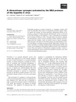
Fig. 1. Spicules as optical glass fibers. (A) Specimens of the demo-
sponge Suberites domuncula. Although most specimens are associ-
ated with hermit crabs, allowing the sponge to live on a ‘mobile’
substrate, some have lost the crab, consequently forcing them into
a sessile way of life. (B) Giant basal spicule (GBS) of Monorha-
phis chuni. (C,D) Localization of tylostyles at the surface of S. do-
muncula. Colloidal gold particles were used to highlight spicules
that protrude with their knobs from the surface of the animals (<;
><). (C) Transversal section of S. domuncula tissue; the packed
zones of spicules are marked (><; sz). (D) Sponge surface. (E)
S. domuncula tylostyle illuminated by a white light source (wl) that
was coupled to its knob. (F) GBS illuminated using a green laser
light source (gl). Epibiontic corals (co), surrounding the spicule,
remain opaque. (G–I) The majority of the tylostyles from S. domun-
cula spicules have perfect terminal knobs (k) (G), whereas some
tylostyles (H) display a more complex morphology with a collar (c)
between the knob (k) and the monaxonal spicule rod (sp). (I) After
etching with HF the different building blocks, knob (k), collar (c) and
spicule (sp), become more prominent. (J,K) Net of fused choano-
somal spicules (Euplectella aspergillum), highlighting that light
guided within spicules is split at fusion sites (fs).
Cryptochrome-based photosensory system of sponges W. E. G. Mu
¨
ller et al.
1184 FEBS Journal 277 (2010) 1182–1201 ª 2010 The Authors Journal compilation ª 2010 FEBS
S. domuncula comprises relatively small spicules
(< 400 lm). By contrast, some hexactinellid spicules
are gigantic, reaching a length of up to 3 m and a
diameter of 12 mm, for example the GBS of M. chuni,
around which the sponge tissue grows (Fig. 1B).
In S. domuncula, the tylostyles (spicules with a globu-
lar swelling at one end and a sharp tip at the other;
150–320 lm in length) are regularly arranged in pali-
sade-like arrays at the periphery of the poriferan body
(Fig. 1C,D). There, zones of packed spicules reach a
thickness of up to 5 mm. By contrast, tylostyles in the
central part of the body, the medulla, are oriented in a
slanted direction along the aquiferous canal system [39].
All tylostyles display a globular knob, which is located
almost exclusively at the end of the monaxonial spicules
(Fig. 1G). In rare cases, it is fixed to a narrow collar
(Fig. 1H). By using a nanopositioning and nanomeasur-
ing machine, analyses of such globular knobs were pos-
sible at the nanometer scale. The majority of terminal
knobs, with a spherical ⁄ elliptical geometry, have a sur-
prisingly regular shape, reminiscent of a collecting lens.
Their diameters vary slightly between 6.53 and 7.28 lm
(in the longitudinal direction of the spicule) and 8.54
and 9.21 lm (in the perpendicular direction) (n = 12).
These globular knobs are fused to monaxonial rods
with a diameter of 6.14–6.57 lm. The outer circumfer-
ences of the subterminal collars range between 6.9 and
7.2 lm. Limited dissolution of the silica mantel indi-
cates that terminal knobs and subterminal collars are
formed as independent units (Fig. 1I).
Siliceous spicules of hexactinellids have the potential
to guide light [15]. For example, GBS of M. chuni (the
syntypus deposited by Schulze [40]) showed that coher-
ent light is guided through the spicule associated with
the siliceous rod, but not through epibiontic corals
(Fig. 1F). In some hexactinellids, for example Euplec-
tella aspergillum, secondary fusion of spicules is obser-
ved. By illuminating this choanosomal spicular network,
it can be seen that the light beam is split at the fusion
sites of the choanosomal skeletal spicules (Fig. 1J,K).
Similarly, illumination of the tylostyles of the demo-
sponge S. domuncula with a white light source demon-
strates that the light beam is transmitted and directed
along their longitudinal axis (Fig. 1E).
Spicules in sponge tissue
In general, demosponge tissues contain small microscl-
eres (siliceous skeletal elements of sizes < 10 lm) and
larger macroscleres (between > 10 and < 300 lm).
All spicules are initially formed intracellularly and,
after having reached sizes of > 8 lm, are completed
extracellularly [41,42]. S. domuncula primmorphs repre-
sent a highly suitable model to study the organization
of spicules within sponge tissue, because this species
generates exclusively tylostyles. In this study, prim-
morphs were used 5 days after re-aggregation of disso-
ciated, single cells to investigate the establishment of
contact between spicules and cells. The cells involved
in spiculogenesis, termed sclerocytes, release both the
silica precursors ⁄ enzyme substrate and the enzyme sili-
catein [43]. Silicatein and silica are required for the
appositional layering of biosilica during spicule
growth, in order to reach the final spicular morphol-
ogy. TEM showed that the cells are scattered along
the spicule surface (Fig. 2), but are mainly present at
t
t
k
1 µm
5 µm
1 µm
5 µm
-ac
sp
sp
col
1 µm
sp
ac
sp
sp
sp
m
m
m
A
B
C
D
E
F
-ac
1 µm
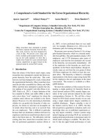
Fig. 2. Localization of spicules within Suberites domuncula primmorphs. Primmorphs were formed over a 5-day period and then used for
sectioning and SEM analysis (A–C) Sections through the knobs (k) and the spiny tips (t) of tylostyles (sp). The cells, sclerocytes, are scat-
tered along the surface of tylostyles. (m) Mesohyl (intercellular matrix). (D) Immature spicule, comprising a large oval axial canal (ac) contain-
ing the axial filament. The spicule is embedded in the bulky mesohyl, which is traversed by collagen fibers (co). (E) At a later stage the axial
filament is contracted and adopts a triangular profile. (F) At the final stage of spiculogenesis, the 3.5 lm spicule contains a small (0.5 lm
diameter) axial canal.
W. E. G. Mu
¨
ller et al. Cryptochrome-based photosensory system of sponges
FEBS Journal 277 (2010) 1182–1201 ª 2010 The Authors Journal compilation ª 2010 FEBS 1185
both ends of spicules, the knob (Fig. 2A) and the
pointed tip (Fig. 2B,C). Cross-sections through imma-
ture spicules revealed a large oval axial canal (1 lm
diameter), homogeneously filled with the proteinaceous
axial filament (Fig. 2D). During maturation, this axial
canal develops a triangular form, whereas the axial fil-
ament concurrently contracts to < 0.2 lm in diameter
(Fig. 2E). In adult spicules, the diameter of the axial
canal reduces and, in most cases, it becomes round
again (Fig. 2F).
Notably, sclerocytes are not intimately associated
with spicules. Instead, there is a gap of 50–100 nm
between them (Fig. 2F). Cells and spicules are embed-
ded in a bulky extracellular matrix, the mesohyl, which
is composed of structural proteins, for example colla-
gen and soluble proteins such as galectin [1].
Cloning and analysis of sponge cDNA encoding
cryptochromes
Complete cDNAs coding for putative cryptochrome
homologues were isolated from the demosponge
S. domuncula and the hexactinellid C. meyeri. The
S. domuncula cDNA (SDCRYPTO; 1565 nucleotides)
comprises an ORF (CRYPTO_SUBDO) from nucleo-
tides 1-3(Met) to 1552-1554 (Fig. 3A). Northern blot-
ting confirmed that the cDNA was completely isolated,
with a size of 1.9 kb (see below). The deduced polypep-
tide (518 amino acids) had a predicted molecular mass
of 59 070 Da (isoelectric point 6.47). Domain search
analysis ( />revealed two main features, the N-terminal photolyase-
related region (photolyase) (amino acids 20–200) and
the FAD-binding domain (amino acids 237–507). Both
domains showed a high similarity score (Expect value
[E]) [44] of E = 3.1e
)25
and 1e
)42
, respectively.
CRYPTO_SUBDO had highest sequence similarity to
the cryptochrome 3 sequence of Danio rerio
(BAA96850.1; E =1e
)95
) and cryptochrome CRY1 of
Acropora millepora (ABP97098.1; E =4e
)87
).
The C. meyeri cDNA (CMCRYPTO; 1675 nucleo-
tides) comprised an ORF from nucleotides 22-24(Met)
to 1584-1586, encoding the putative polypeptide
CRAME_CRYPTO (521 amino acids). The calculated
size of CRAME_CRYPTO is 59 070 Da (isoelectric
point 6.47). Again, transcript size (1.9 kb) was con-
firmed on northern blots (not shown). The two afore-
mentioned domains were found between amino acids 3
and 134 (photolyase-related region; E =2e
)06
) and
from amino acids 205 to 475 (FAD-binding domain;
E = 3.2e
)35
). In general, the hexactinellid sequence
had a lower similarity to other cryptochromes than
CRYPTO_SUBDO, for example D. rerio Cry4
(AAI64413.1; E =5e
)53
)orA. millepora CRY2
(ABP97099.1; E =3e
)49
).
For phylogenetic analysis, we used an extended data
set that had originally been applied to the study of
coral cryptochromes [27]. The resulting phylogenetic
tree was rooted with the blue light photoreceptor cryp-
tochrome 1 of Arabidopsis thaliana. The tree revealed a
distinct branch near the root that contained all mem-
bers of the class II photolyases, including distantly
related bacterial enzymes. By contrast, the molecules
of C. meyeri, A. vastus and S. domuncula were grouped
at the base of those branches that include metazoan
cryptochromes (Fig. 3B). The close relationship
between CRYPTO_SUBDO and the coral crypto-
chrome CRY2 was remarkable.
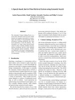
Fig. 3. Poriferan cryptochromes. (A) The deduced poriferan cryptochrome protein sequences CRYPTO_SUBDO (Suberites domuncula) and
CRYPTO_CRAME (Crateromorpha meyeri), and the photolyase-related protein from Aphrocallistes vastus (PHL64_APHVA; NCBI accession
no. 28625001), were aligned with the two coral (Acropora millepora) cryptochromes, CRY1 (CRY1_ACRO; 145881069) and CRY2 (CRY2_
ACRO; 145881071). Residues conserved (identical or similar with respect to their physicochemical properties) in all sequences are shown in
white on black; those which share similarity in four sequences are shown in black on gray. The characteristic domains, the N-terminal photol-
yase-related region (photolyase) and the FAD-binding domain, are marked. (B) For phylogenetic analyses, the aforementioned sequences
were used in combination with other representative members of the metazoan cryptochrome family, Danio rerio cryptochrome 4 (CRY4_
DARE; 8698594), cryptochrome 3 (CRY3_DARE; 8698592), cryptochrome 2a (CRY2a_DARE; 8698588), cryptochrome 1a (CRY1a_DARE;
8698584); Gallus gallus cryptochrome 1 (CRY1_ CHICKEN; 19550963), cryptochrome 2 (CRY2_CHICKEN; 19550965); Homo sapiens crypto-
chrome 2 (CRY2_HUMAN; 27469701); Mus musculus cryptochrome 2 (CRY2_MOUSE; 5670009); Anopheles gambiae cryptochrome 1
(CRY1_ANOGA; 78191295); Drosophila melanogaster blue light photoreceptor (CRY_DROME; 3986298) and Bactrocera tryoni cryptochrome
(CRY_BACTR; 51944883). In addition, the following photolyase sequences were integrated, the 6 : 4-type photolyases of D. rerio
(PHL64_DARE; 8698596) and Xenopus laevis (PHL64_XENLA; 8809676) and the D. melanogaster photolyase (PHL_DROME;1304062).
Finally, members of the class II photolyases were included, the DNA photolyase from Rhodopirellula baltica (PHL_RHOBA; 32447829),
Methanobacterium thermoautotrophicum (2507184; PHR_METTH), Arabidopsis thaliana (PHR-CPD_ARATH; 1617219), the D. rerio crypto-
chrome-DASH (CRYda_DARE; 41688004), and the cryptochrome 1 blue-light photoreceptor of A. thaliana (CRY1_ARATH; 2499553). The
latter sequence was used as outgroup to root the resulting phylogenetic tree. The degree of support of internal branches was assessed by
bootstrapping (1000 replicates) and the evolutionary distance calculated (0.1 amino acid substitutions per position in the sequence).
Cryptochrome-based photosensory system of sponges W. E. G. Mu
¨
ller et al.
1186 FEBS Journal 277 (2010) 1182–1201 ª 2010 The Authors Journal compilation ª 2010 FEBS
A
B
W. E. G. Mu
¨
ller et al. Cryptochrome-based photosensory system of sponges
FEBS Journal 277 (2010) 1182–1201 ª 2010 The Authors Journal compilation ª 2010 FEBS 1187
Recombinant S. domuncula cryptochrome and
cryptochrome antibodies
To facilitate functional analyses, recombinant
S. domuncula cryptochrome and cryptochrome anti-
bodies had to be generated. For this purpose, a partial
SDCRYPTO cDNA was expressed in Escherichia coli,
using l-arabinose as the transcription-inducing agent.
The bacterial crude extract was prepared and analyzed
by SDS ⁄ PAGE (Fig. 4A). In l-arabinose-induced sam-
ples (Fig. 4A, lane a), as well as in noninduced samples
(because of leaky expression; Fig. 4A, lane b), a prom-
inent band was detected at 19 kDa. This band was
also detected after purification of protein extracts
through affinity chromatography (Fig. 4A, lane c). The
size of this protein corresponded to the calculated size
of the recombinant protein, including His-tag and vec-
tor-specific sequences (18 970 Da). Subsequently, poly-
clonal antibodies (termed PoAb-aCRYPTO_SUBDO)
were raised against the purified recombinant protein
and used to detect wild-type cryptochrome in poriferan
protein extracts. Thus, on western blots PoAb-aCRYP-
TO_SUBDO recognized a 60-kDa protein (Fig. 4B,
lanes a,b), which matched the calculated size of
CRYPTO_SUBDO (59 070 Da). Controls demon-
strated that the preimmune serum did not cross-react
with the 60-kDa protein (Fig. 4B, lane c).
Light-induced expression of cryptochrome
To investigate the light-induced expression of crypto-
chrome (transcription and translation) in S. domuncula,
specimens that had lost their hermit crabs and hence
turned to a sessile living form (Fig. 1A), were adapted
to complete darkness over a 5-day period. In addition,
the poriferan 3D cell-culture system (primmorphs) was
used. For this purpose, dissociated single cells (Fig. 5A)
were first transferred to a Ca
2+
-containing medium, in
which primmorphs subsequently formed (Fig. 5B,C,
after 3 and 5 days, respectively). In order to stimulate
spiculogenesis, primmorphs were transferred to a silicate
cushion for an additional 6 days (Fig. 5D).
Ultimately, all samples were exposed to light for
1–8 h, using a short-pass filter (spectral range, 330–900
nm) or long-pass filter (spectral range, 700–1100 nm).
Afterwards, RNA was extracted. Subsequent north-
ern blot analyses revealed that after dark adaptation
(5 days) of primmorphs and tissues, no expression of
cryptochrome was detectable (Fig. 6). However, after
2 h of light exposure (330–900 nm), an increased
SDCRYPTO expression level could be seen which
increased further after 4 or 8 h of light exposure,
both in tissue (Fig. 6A) and in primmorphs
(Fig. 6B). Interestingly, a change in light quality
(700–1100 nm) did not affect the expression pattern
AB
Fig. 4. Protein detection of the Suberites domuncula crypto-
chrome. (A) Preparation of recombinant cryptochrome. Escherichia
coli was transformed with SDCRYPTO cDNA, as described in
Materials and methods. Protein expression was analyzed in the
presence (+) or absence (–) of
L-arabinose, using 15% polyacryl-
amide gel containing SDS (lanes a and b); equal amounts of protein
were loaded onto the gel. The His-tagged recombinant protein
(19 kDa) was purified by affinity chromatography on Ni-IDA col-
umns and then applied to SDS ⁄ PAGE (lane c). M, size marker.
(B) Immunodetection of cryptochrome in crude protein extracts
from S. domuncula via western blots. Proteins of crude extracts
were size-separated by SDS ⁄ PAGE and stained with Coomassie
Brilliant Blue (lane a). In parallel, proteins were transferred to
membranes. There, PoAb-aCRYPTO_SUBDO detected the 60-kDa
cryptochrome protein (lane b). As a control, preimmune serum was
applied to the blots (lane c). M, size marker.
AB
CD
Fig. 5. Suberites domuncula primmorphs. (A) Suspension of single
cells obtained after dissociation of sponge tissue in Ca
2+
⁄ Mg
2+
-free
artificial seawater. Formation of primmorphs after 3 days (B) or
5 days (C), respectively, in Ca
2+
⁄ Mg
2+
-supplemented medium. The
3D cell aggregates were transferred to a silicate cushion (D) for fur-
ther experiments. Size bars are given.
Cryptochrome-based photosensory system of sponges W. E. G. Mu
¨
ller et al.
1188 FEBS Journal 277 (2010) 1182–1201 ª 2010 The Authors Journal compilation ª 2010 FEBS
of SDCRYPTO (Fig. 6C). The expression of tubulin,
which was used as an internal control, remained
unchanged, irrespective of the duration of light expo-
sure (Fig. 6E). Alternatively, an animal was exposed
to light for 4 h (330–900 nm) and then immediately
transferred to darkness. After 2 h of dark adapta-
tion, significantly reduced transcription was seen,
whereas after 8 h of darkness no transcripts could be
identified using this method (Fig. 6D).
In a final set of RNA experiments, qPCR was
applied to determine cryptochrome transcription over
24 h, including light–dark transition (Fig. 7). Subse-
quently, cryptochrome expression was correlated to the
expression of the housekeeping gene tubulin. Thus,
during 12 h light exposure, cryptochrome expression
increased to 0.53 (± 0.02; n = 5) and then decreased,
until after 12 h of darkness a ratio of 0.15 (± 0.025)
was calculated. Accordingly, cryptochrome expression
during light exposure was up to 3.5-fold higher than
in darkness. This ratio remained invariant when the
housekeeping genes glyceraldehyde-3-phosphate dehy-
drogenase (GAPDH) or hypoxanthine phosphoribosyl-
transferase 1 (HPRT) were used as a reference.
In a parallel approach, cryptochrome protein expres-
sion was analyzed by immunodetection on western
blots. In these studies, expression of CRYPTO_SUB-
DO could not be detected in dark-adapted prim-
morphs (Fig. 8B, lane a). However, extracts from
primmorphs that had been exposed to light for 2, 4 or
8 h showed the characteristic 60 kDa band of crypto-
chrome (Fig. 8B, lanes b to days, respectively). To
demonstrate the specificity of PoAb-aCRYPTO_SUB-
DO, preimmune serum was used in parallel with pro-
tein extracts of primmorphs that had been exposed to
light for 8 h. Whereas preimmune serum did not
immunodetect any proteins (Fig. 8A, lane a) PoAb-
aCRYPTO_SUBDO elicited a positive signal at
60 kDa.
In situ localization of cryptochrome
Immobile S. domuncula specimens were exposed to
light (330–900 nm) for 24 h. For immunohistological
analyses, tissue sections were reacted with anti-crypto-
chrome IgG. The resulting immunostaining displayed a
distinct zonation. The brightest reactions were seen
50 lm below the surface of the animals in a thick
(500 lm) zone that was characterized by the ordered
accumulation of a spicule (tylostyle) phalanx
A
B
C
D
E
Fig. 6. Gene expression analyses of Suberites domuncula tissue
and primmorphs. Dark-adapted specimens were exposed to light
(330–900 or 700–1100 nm). RNA was extracted, size-separated,
blotted and probed for SDCRYPTO, using a digoxigenin-labeled
probe. RNA was analyzed from tissue (A) or primmorphs (B) that
had remained in the dark (0 h) or been exposed to light (330–
900 nm) for 2, 4 or 8 h (0 h, +2 h, +4 h, +8 h). (C) RNA was used
from the tissues of animals challenged with light of longer wave-
lengths (700–1100 nm) for the same times. (D) Animals were
exposed to light (330–900 nm) for 4 h [+4 h), followed by a period
of darkness for 2 or 8 h ()2h,)8 h). (E) Internal control. To ensure
that the same amount of RNA was loaded onto the gels, size-sepa-
rated RNA was probed for transcripts of the housekeeping gene
b-tubulin (SDTUB). Transcript sizes are indicated.
Fig. 7. Cryptochrome expression analyses of Suberites domuncula
tissue. Dark-adapted samples were exposed to light (330–900 nm)
for 8 h (08:00 to 16:00) and then kept in darkness for 12 h (16:00
to 04:00). Following RNA isolation, expression levels of crypto-
chrome and tubulin (housekeeping gene) were determined through
quantitative real-time PCR and subsequently correlated to deter-
mine relative expression levels.
W. E. G. Mu
¨
ller et al. Cryptochrome-based photosensory system of sponges
FEBS Journal 277 (2010) 1182–1201 ª 2010 The Authors Journal compilation ª 2010 FEBS 1189
(Fig. 9A,B). Close inspection showed that in addition
to the cells surrounding the spicules, the extracellular
matrix was also stained. This observation suggests that
cryptochrome not only exists intracellularly, but is also
present in the extracellular matrix, in which cells and
adjacent spicules are embedded. The staining of cellu-
lar and spicular structures was specific, because appli-
cation of preimmune serum did not elicit any
immunostaining (Fig. 9C,D).
In a further series of experiments, sections of near-
surface tissue that had been exposed to light were
reacted with anti-cryptochrome IgG. Micrographs show
the strongest accumulation of immunocomplexes adja-
cent to the spicules (Fig. 10A,E,G). In parallel, the cell
nuclei were visualized by 4¢,6-diamidino-2-phenylindole
AB
Fig. 8. Cryptochrome protein expression in primmorphs. (A) Speci-
ficity of PoAb-aCRYPTO_SUBDO. Protein extracts of primmorphs
that had been exposed to light (330–900 nm) for 8 h were size
separated and blotted onto membranes. Lane a, application of
preimmune serum (pi) to the membranes; lane b, PoAb-aCRYPTO_
SUBDO (i) binding the Suberites domuncula cryptochrome 60 kDa
protein (resulting immunocomplexes were detected with labeled
secondary antibodies). (B) Protein extracts of dark-adapted prim-
morphs (lane a) or primmorphs exposed to light (330–900 nm) for 2
(lane b), 4 (lane c) or 8 h (lane d) were analyzed on western blots,
using PoAb-aCRYPTO_SUBDO. Size markers are indicated.
AB
CD
Fig. 9. Immunohistological detection of cryptochrome in Suberites
domuncula tissue. After adaptation to light (330–900 nm), animals
were irradiated for 24 h; the direction of the light emission is indi-
cated by an arrow. (A) Immunostaining of a tissue section (sp) with
anti-cryptochrome IgG PoAb-aCRYPTO_SUBDO. (B) Corresponding
Nomarsky interference image. (C) Application of preimmune serum
to an adjacent section (control). (D) Corresponding Nomarsky inter-
ference image. The surface of the sponge is marked (s). Size bars
are given.
AB
CD
EF
GH
Fig. 10. Localization of cryptochrome in Suberites domuncula
tissue. Slices of S. domuncula tissue (following adaptation to light
at 330–900 nm for 24 h) were prepared and (A,E,G) reacted with
antibodies (PoAb-aCRYPTO_SUBDO); (B,F,H) corresponding views
are shown in which the cell nuclei had been visualized by 4¢,6-
diamidino-2-phenylindole. Control sections were incubated with
preimmune serum and Cy3-conjugated IgG and inspected (C) or
illuminated with fluorescence light to identify 4¢,6-diamidino-
2-phenylindole -stained nuclei (D). Size bars are given.
Cryptochrome-based photosensory system of sponges W. E. G. Mu
¨
ller et al.
1190 FEBS Journal 277 (2010) 1182–1201 ª 2010 The Authors Journal compilation ª 2010 FEBS
staining (Fig. 10B,F,H). Higher magnification reveals
staining of both the cells and the extracellular matrix
(Fig. 10E,G). In controls, preimmune serum and Cy3-
conjugated IgG were used, resulting in a very weak
background staining because of unspecific binding
(Fig. 10C). Concurrent staining with 4¢,6-diamidino-2-
phenylindole highlights the localization of nuclei and,
consequently, of cells in the vicinity of spicules
(Fig. 10D).
For in situ hybridization, labeled single-stranded
sense or antisense probes were applied to mounted
S. domuncula tissue samples. Animals that had been
dark adapted for 3 days revealed very weak staining
after application of the antisense probe (Fig. 11A).
Following exposure of animals to light (330–900 nm)
for 2 (Fig. 11B) and 8 h, binding of the antisense
probe elicited an increasingly strong staining pattern,
first observed in the region directly exposed to the light
source (Fig. 11D). By contrast, no staining was
observed through the sense probe (control; Fig. 11C).
Discussion
In bacteria, the sequence of the intermediate reaction
state of bacteriorhodopsin generated during the photo-
cycle has been elucidated to a large extent [45–47].
There is evidence that a single protein conformational
change in the cytoplasmic region occurs within a few
milliseconds after illumination, and is paralleled by
deprotonation of the Schiff base. In the protonated
state, this base covalently links a single molecule of
retinal to the protein. An analogous photocycle system
was studied in plants with the light-responsive protein.
This comprises the mononucleotide light-binding fla-
vin, oxygen and voltage domain proteins, which have
been implicated in phototropic movement [48,49], chlo-
roplast relocation [50] and stomatal opening of guard
cells [51]. Biochemical evidence that luciferase is
involved in circadian rhythms was found in the marine
dinoflagellate, Gonyaulax polyedra [52,53]. Subse-
quently, the molecular basis of these processes was elu-
cidated by Krieger et al. [54], and then completed by
molecular sequencing [55]. In mammals, the protein
cryptochrome is one regulator in the complex molecu-
lar system of the circadian clock [56].
Focusing on the phylum Porifera, the closest relative
of the common metazoan ancestor, seminal studies on
luminescence were performed by Harvey [57,58]. He
described a case of ‘doubtful’ luminous sponge with
the hexactinellid Crateromorpha meyeri [59] and the
demosponge Grantia sp. [13,57]. Whereas light produc-
tion in C. meyeri was attributed to an annelid and,
hence considered as a secondary luminescence, Grantia
sp. was classified as self-luminous. However, in view of
the recently gathered biochemical and molecular bio-
logical data, it seems likely that the sponges, with
S. domuncula as a potential reference species, are
inherently bioluminescent.
Siliceous sponges represent the only animal taxon
that comprises a complex array of fiber-optic like
structures. The biosiliceous material of these skeletal
elements not only confers unique physical and mechan-
ical properties, but also reaches quartz glass quality
[23], which is one reason for the exceptional potential
of spicules to operate as optical fibers ex vivo
[14,15,60]. Further, recent studies indicate fluorescence
properties of spicules in the long-wavelength region
[61]. These observations led to the assumption that the
poriferan spicular network might be the light-transmit-
ting part of an alternative photosensory system [15].
This was supported by the recent finding that sponges
themselves, and not their symbiotic bacteria [14], pro-
duce light which can be coupled into spicules [16].
This study aims to identify and characterize putative
poriferan photoreceptors. Recent studies of corals [27],
but also of human and insect cell models [26], suggest
the involvement of cryptochromes in a light-sensing
response via photoreduction of chromophores. The
existence of a poriferan protein homologue was
reported in 2003 for the hexactinellid Aphrocallistes
vastus [62], in which it was shown that the (6 : 4) pho-
tolyases-based system is expressed most highly at the
A
B
C
D
Fig. 11. In situ hybridization analyses of Suberites domuncula tis-
sue. The animals were exposed to light (330–900 nm) for 0 (A), 2
(B,C) or 8 h (D). They were then subjected to whole-mount hybrid-
ization, as described in Materials and methods. For hybridization of
samples A, B and D the digoxigenin-labeled antisense probe was
used, whereas specimen C was treated with the sense probe (con-
trol). The direction of light emission is indicated by an arrow. Size
bars, 5 mm.
W. E. G. Mu
¨
ller et al. Cryptochrome-based photosensory system of sponges
FEBS Journal 277 (2010) 1182–1201 ª 2010 The Authors Journal compilation ª 2010 FEBS 1191
tip of the animals, the place of most efficient light
exposure.
Sponge spicules guide light in a linear way [14,15,60]
and in branched skeletal networks, for example in the
hexactinellids Rosella racovitzae [24] and E. aspergillum,
in which the entire skeleton consists of fused spicules.
Thus, light penetrates several millimeters into the tissue
via the fused spicular network. In the demosponge
S. domuncula, investigated in this study, the putative
light-transmission matrix consists of solitary, monaxo-
nial spicules (tylostyles) that possess a knob-like struc-
ture at one end and a pointed tip at the other. The
almost perfectly round knobs with their uniform dimen-
sions might indicate a lens-like light-collecting function
to maximize the yield of photons that are, subsequently,
transported within the rod-like part of spicules.
S. domuncula spicules are localized in a highly
ordered pattern, immediately below the surface cell
layers, in palisade-like arrays. Interestingly, the orien-
tation of tylostyles is such that the pointed rods are
directed towards the center of the animal, the medulla,
whereas the knobs are directed towards the surface.
This directionality might imply light guidance from
outside into the sponge body (Fig. 12A). This is
supported by the light conditions of the habitat: the
animals are found exclusively in shallow waters (20–
30 m). In these coastal waters, light within a shorter
range of wavelengths (around 500 nm) is preferentially
transmitted, compared with offshore ocean water [63].
Interestingly, this range of wavelengths corresponds
to the bioluminescence emission spectrum of the
S. domuncula luciferase [16] (see below). Hence, it can
be deduced that spicules, exposed at the surface of the
sponge, absorb ⁄ harvest and transmit light to a crypto-
chrome-containing photoreceptive system (Fig. 12A).
It should be added here that the surfaces of (almost)
A
B
(a)
(b)
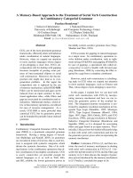
Fig. 12. Proposed photoreception ⁄ photo-
transduction system of sponges. (A) The
combination of cryptochrome and spicules
arranged at the surface of the sponge acts
as a sensory system for environmental light.
(Ba) Concurrently, biogeneous light gener-
ated within the interior of the animals by
luciferase might also be trans-
duced ⁄ detected by the same system. (Bb)
Schematic detail of this process with biolu-
minescence as the light source. Luciferase-
mediated oxidation of luciferin results in the
generation of bioluminescence. Photons are
transmitted through spicules and then
detected by a chromophore ⁄ redox system
associated with the cryptochrome. Concom-
itantly, oxyluciferin is the substrate for the
luciferin-regenerating enzyme.
Cryptochrome-based photosensory system of sponges W. E. G. Mu
¨
ller et al.
1192 FEBS Journal 277 (2010) 1182–1201 ª 2010 The Authors Journal compilation ª 2010 FEBS
all sponge species are clean because of the production
of biosurfactants by the animal itself or by associated
microorganisms [64].
Light emitted by the poriferan luciferase ranges
between 450 and 650 nm, with peaks at 548 and
590 ⁄ 620 nm [16]. This spectral range overlaps with
the lower range of light that can be transmitted
through sponge spicules (580–600 nm) [15,65].
Accordingly, light emitted through the enzymatic
activity of luciferase represents only a fraction of the
total wavelength range that can be transferred
through spicules. At present, we assume that this
observation has a specific biological relevance. First,
it is most efficient that in seawater the coupling of a
light-generating and a light-receiving system occurs
within a range far from maximally absorbed wave-
lengths (i.e. the red region of the electromagnetic
spectrum) [66]. Second, coupling of light emission and
absorption can be optimally regulated and at highest
sensitivity at wavelengths shorter and longer than
those corresponding to maximal emission ⁄ absorption.
Hence, it is plausible that fine-tuning in the system is
achieved by small changes in the luciferase emission
spectrum or by shifting light wavelengths during their
passage through the organic coupling system to the
spicular fibers.
At present, S. domuncula is the most suitable sili-
ceous sponge model for the study of structural and
functional properties of any given poriferan molecule,
because the combination of transcriptome, genomic
complexity and basic metabolic pathways has been
elucidated most comprehensively in this species [1].
Furthermore, controlled cell culture experiments using
the primmorph system can be performed [37].
Accordingly, following the rationale successfully
investigated in coral [27], human and insect systems
[26], poriferan EST-libraries were screened for the
presence of cryptochrome. Subsequently, candidate
molecules were isolated from representatives of
Demospongiae and Hexactinellida. Both deduced
polypeptides comprise the characteristic domains that
are also found in coral light-responsive crypto-
chromes, DNA photolyase and the FAD-binding
domain [27], and display a significant sequence
homology to the Acropora millepora molecules and
other light-sensing cryptochromes [67].
SEM analyses revealed that the spicule-forming cells
(sclerocytes) are scattered along the surface of spicules,
without firm contact to the biosilica-based skeletal ele-
ments. Concurrently, cryptochrome was immunohisto-
logically stained within the extracellular matrix. These
findings suggest that the potential cryptochrome-
containing photoreceptor might operate extracellularly.
The presence of functionally active cryptochromes in
the extracellular space of other model organisms is well
documented [68].
In order to evaluate the potentially light-inducible
expression of poriferan cryptochrome, both adult
sponge specimens and primmorphs were exposed to
light under different regimes. Spectral ranges of 330–
900 and 700–1100 nm were applied to cover the
emitted wavelength spectrum of poriferan luciferase
and the transmitted spectrum of spicules. Compara-
tive expression analyses (transcription, translation)
demonstrate that following dark adaptation crypto-
chrome expression is downregulated. However, 2 h of
light irradiation are sufficient to upregulate gene
expression, which is increased further with exposition
time. Under the experimental conditions chosen, both
filter types (330–900 and 700–1100 nm) elicited a
comparable response. This might suggest that some
additional poriferan proteins ⁄ factors cause a wave-
length shift in the light generated by the lucif-
erin ⁄ luciferase system, that is, to the longer range
spectrum. Furthermore, quantitative real-time PCR
analyses strongly indicate a diurnal expression pat-
tern. Moreover, immunohistological analyses dis-
played highest cryptochrome expression in the
vicinity of the spicules, particularly in the subsurface
regions of the animals. In this context, signals were
detected intra- and extracellularly. Finally, using
whole-mount in situ hybridization, it was shown that
those regions of the sponge body that are exposed
to light contain the highest levels of cryptochrome
transcripts.
Taken together, our data demonstrate that exposure
of S. domuncula (tissue and primmorphs) to light stim-
ulates cryptochrome expression. In contrast to the stud-
ies performed with corals [27], the light-induced
response in sponges occurs regardless of the spectral
range. Because cryptochrome is particularly localized
around spicules, skeletal elements might operate as
waveguides for environmental light that penetrates
with significant intensity water depths of 20 m in
the northern Adriatic Sea, the natural habitat of
S. domuncula. Thus, spicules at the surface of the ani-
mal might function as collectors that transmit light to
cryptochrome, the putative photosensory receptor of
sponges (Fig. 12A). Concurrently, light produced intra-
cellularly (luciferase-mediated) might be collected and
transmitted via the same optical system (Fig. 12B-1,
B-2). In both cases, cryptochrome is the light photo-
receptor. It remains to be studied whether flavin
(Fig. 12B-2) or another chromophore is associated with
cryptochrome, for example, avarol ⁄ avarone frequently
found in demosponges [69,70].
W. E. G. Mu
¨
ller et al. Cryptochrome-based photosensory system of sponges
FEBS Journal 277 (2010) 1182–1201 ª 2010 The Authors Journal compilation ª 2010 FEBS 1193
Experimental procedures
Chemicals, materials and enzymes
The sources of most chemicals and reagents used are given
elsewhere [16]; others were obtained from Sigma-Aldrich
(Taufkirchen, Germany).
Sponges and primmorphs
Specimens of the marine sponge S. domuncula (Porifera,
Demospongiae, Hadromerida) were collected in the northern
Adriatic, near Rovinj (Croatia), from depths of 20–30 m,
and kept in aquaria in Mainz (Germany) at 17 °C for
> 5 months. Although most sponge specimens harbor a
hermit crab, Pagurites oculatus (Decapoda: Paguridea),
which resides predominantly in shells of the mollusk Truncu-
lariopsis trunculus (Gastropoda: Muricidae), some had been
freed of crabs ⁄ mollusks for experimental purposes; such
specimens remained sessile on the substratum [71].
Extracts from sponge tissue were prepared in equal
amounts (v ⁄ w) of 20 mm Tris ⁄ HCl buffer (pH 7.5; contain-
ing 100 mm KCl, 5 mm MgCl
2
and 5% v ⁄ v glycerol). After
two cycles of freezing to )10 °C, thawing and subsequent
homogenization (Downs homogenizer) the suspension was
centrifuged (10 000 g; 10 min; 4 °C) and the supernatant
collected (1.5 mg proteinÆmL
)1
).
Primmorphs were obtained from single cells, as described
previously [37]. Cells were transferred to natural seawater,
supplemented with 0.2% v ⁄ v RPMI-1640 medium, contain-
ing 60 lm silicate [21], and incubated at 16 °C. After 5 days
the 3D cell aggregates (primmorphs) reached 5–7 mm. They
were transferred to a silicate cushion that had been pre-
pared from orthosilicate in sterile six-well plates (Nunc,
Langenselbold, Germany), as described previously [16], and
incubation was continued.
During cultivation, both sponges and primmorphs were
exposed to 50 lux using a Lumi-Lux-840 (4000 K) lamp
(Osram, Mu
¨
nchen, Germany) in a 12 h light and dark cycle.
For the experiments described, the specimens remained
either in complete darkness or under irradiation of a
General Electric 29208 projector white light bulb (50 W).
One of the following two optical filters (Schott - Advanced
Optics, Mainz, Germany) was placed in front of the light
source: either a short-pass filter with a spectral range of
330–900 nm (KG3; Tau
max
330–900 nm) or a long-pass
filter with a spectral range 700–1100 nm (RG9; 780 nm).
The exposure intervals are given with the respective
experiment.
Spicules and light transmission studies
Spicules of S. domuncula (tylostyles; 150–320 lm), M. chuni
(Amphidiscosida; GBS 720 mm long with a diameter of
6 mm) and E. aspergillum (Lyssacinosida; fused silica net-
works formed from choanosomal spicules in a synapticular
manner; size 15 · 0.1 mm) were cleaned by soaking in nitric
acid ⁄ sulfuric acid (1 : 4 v ⁄ v) for 2 days, followed by wash-
ing in distilled water until the pH value was 6. The hexacti-
nellid samples were obtained from J. Li (Institute of
Oceanology, Academia Sinica, Qingdao, China) and from
the Museum fu
¨
r Naturkunde (Berlin, Germany). In one
experiment, tylostyles were exposed to hydrofluoric acid
vapor to display the inner architecture [23]. The dimensions
of the spicules were determined using a nanopositioning
and nanomeasuring machine, as described previously [72].
For light-transmission studies, spicules were coupled to a
light source with their larger diameter, that is, the knob of
S. domuncula tylostyles or the base of M. chuni GBS. Tylo-
styles were illuminated with a white light source (AQ-4303B
from Ando Electronics, Kawasaki, Japan) and the GBS
with a green laser (CrystaLaser, Reno, NV, USA; 15 W,
532 nm). Images of the S. domuncula and E. aspergillum
spicules were taken with a VHX-100 digital microscope
(Keyence, Neu-Isenburg, Germany), equipped with a
VH-Z100 zoom lens (·100–1000) and the M. chuni spicule
with a D2 camera (Nikon, Tokyo, Japan).
Transmission electron microscopy
For TEM, primmorph samples were fixed in 0.1 m phos-
phate buffer (supplemented with glutaraldehyde), as
described previously [41]. Samples were subsequently trea-
ted with OsO
4
. After drying, primmorphs were incubated
with propylene oxide, fixed in propylene oxide ⁄ Araldite,
covered with pure Araldite and hardened at 60 °C, prior to
the preparation of ultrathin slices (60 nm). Samples were
placed onto coated copper grids and analyzed with a
Tecnai 12 microscope (FEI Electron Optics, Eindhoven,
The Netherlands).
Exposure of sponges and primmorphs to light
Prior to exposure, sponge specimens were adapted to com-
plete darkness for 3 days. During that time, samples were
kept under optimal aeration and water quality conditions
[36]. Sponge specimens that had been living sessile (without
hermit crabs) were taken and exposed to filtered light
(short-pass filter: 330–900 nm; long-pass filter: 700–
1100 nm). Two different regimes were employed: specimens
were either exposed to light continuously for up to 8 h
(termed +8 h) or exposed to light for only 4 h (+4 h)
before they were shifted to darkness for up to 8 h ()8 h).
Control samples remained in complete darkness.
Molecular cloning of S. domuncula cryptochrome
A fragment of a cryptochrome-like transcript (accession
no. sd002_020b_e01.q1cb.nt_raw) was identified in the
Cryptochrome-based photosensory system of sponges W. E. G. Mu
¨
ller et al.
1194 FEBS Journal 277 (2010) 1182–1201 ª 2010 The Authors Journal compilation ª 2010 FEBS
S. domuncula EST database (-
mainz.de/login.cgi) via homology searches. The sequence,
which comprises 810 nucleotides, contained an ORF
between nucleotides 232 and 517 of the final sequence.
PCR was used to identify the complete cDNA, termed
SDCRYPTO. Thus, two insert-specific reverse primers were
designed (SD_RV_Crypto_1: 5¢-GGATGACCCGACTAG
AAAAGCA-3¢, and SD_RV_Crypto_2: 5¢-CATTGTTGC
TCTCCCAAGGTAG-3¢, 346 and 412 bp to the 5¢-end of
the SDCRYPTO fragment, respectively) and ‘nested’ PCR
were performed in combination with two standard vector-
specific forward primers (pTriplEx2 and 5¢-CapFish) in
order to complete the sequence. PCR conditions were as
follows: 95 °C for 3 min, followed by 35 amplification
cycles at 95 °C for 30 s, 63 °C for 30 s, 72 °C for 90 s and
a final extension at 72 °C for 7 min. PCR fragments were
isolated, cloned into the pCR2.1-TOPO vector (Invitrogen,
Karlsruhe; Germany) and used to transform E. coli TOP10
cells (Invitrogen). Sequencing was performed with prim-
ers directed to the SP6 and T7 promoters. The com-
plete clone (SDCRYPTO), encoding the deduced protein
CRYPTO_SUBDO, was 1565 nucleotides, excluding the
poly(A) tail.
Molecular cloning of C. meyeri cryptochrome
A fragment of a cryptochrome sequence was found in
a C. meyeri cDNA library (izin.
uni-mainz.de/login.cgi; accession number cm 015c d07.p1ca.
exp.nt_raw), which had been prepared earlier [22]. Again,
PCR was used to identify the complete cryptochrome cDNA
(CMCRYPTO). Two insert-specific forward primers were
designed (CM_FRW_Crypto_1: 5¢-CTTCCAGAAGAATT
AACAGAGTTCCTA-3¢ and CM_FRW_Crypto_2: 5¢-TAG
AGAAGTTAATTGCAAATCGCTG-3¢, 499 and 662 bp to
the 3¢-end of the fragment). Nested PCRs were performed
with two vector-specific reverse primers (T7 and M13). The
reaction conditions were as follows: 95 °C for 3 min, fol-
lowed by 35 amplification cycles at 95 °C for 30 s, 61 °C
for 30 s, 72 °C for 90 s and a final extension at 72 °C for
7 min. Fragments were isolated, cloned into the pCR2.1-
TOPO vector and then used to transform E. coli TOP10
cells. Sequencing was performed as described above. The
complete clone (CMCRYPTO) has 1675 nucleotides,
excluding the poly(A) tail, and encodes the putative protein
CRAME_CRYPTO.
Sequence analyses
Sequences were analyzed with blast (.
nih.gov/blast/blast.cgi) and fasta ( />fasta33/). Multiple alignments were performed with
clustal w v. 1.6 [73]. Phylogenetic trees were constructed
on the basis of amino acid sequence alignments by neighbor
joining, as implemented in the neighbor program from the
phylip package [74]. Distance matrices were calculated
using the Dayhoff PAM matrix model as described previ-
ously [75]. The degree of support for internal branches was
further assessed by bootstrapping [73]. The graphic presen-
tations were prepared with genedoc [76].
Preparation of recombinant S. domuncula
cryptochrome
The partial ORF (amino acids 227–238) of SDCRYPTO
was recombinantly expressed in E. coli, using Gateway-
Technology in combination with the pDEST17 vector, as
described previously [44,77]. For this purpose, SDCRYPTO
cDNA was first inserted into the donor vector pDONR221
(Invitrogen) by BP-recombination after amplification via
two primers (SDCRYPTO-specific sequences underlined):
attB1_SP ⁄ Crypto_dom 5¢-GGGGACAAGTTTGTACAAA
AAAGCAGGCTTA
GAGTTTGCACTCTATACG-3¢ and
attB2_ASP ⁄ Crypto_dom 5¢-GGGGACCACTTTGTACAA
GAAAGCTGGGTACTA
TTGCCTGATTTGACGTAT-3¢
at an initial denaturation at 95 °C for 3 min, followed by
35 amplification cycles at 95 °C for 30 s, 56 °C for 35 s,
72 °C for 45 s, with a decreasing temperature of 0.1 °Cin
every cycle, and a final extension at 72 °C for 7 min. After
recombination (in frame with Met
start
and 6· His-tag of the
expression vector pDEST17) the clone SDCRYPTO was
expressed in the host strain BL21-AI (Invitrogen), growing
in Luria–Bertani medium with 50 lgÆmL
)1
carbenicillin,
in the presence or absence of 0.2% (w ⁄ v) l-arabinose
(overnight at 37 °C). The bacterial pellet was then lysed
in ‘BugBuster (primary amine-free) Protein Extraction
Reagent’ (Novagen) with 1 lLÆmL
)1
benzonase (Novagen ⁄
Merck KGaA, Darmstadt, Germany), supplemented with
a protease inhibitor cocktail (Roche Applied Science,
Mannheim, Germany) for 1 h at 20 °C, according to the
manufacturer’s instructions (Novagen ⁄ Merck). After soni-
cation on ice (3 · 15 s, with 15-s cooling periods in between
each), cell extracts were centrifuged for 30 min with
15 000 g at 4 °C. The insoluble fraction obtained was solu-
bilized with lysis buffer (50 mm KH
2
PO
4
pH 8.0, 6 m urea,
300 mm KCl, 5 mm imidazole). After further sonication on
ice (3 · 15 s, with 15-s cooling periods), the suspension was
centrifuged for 30 min at 15 000 g at 4 °C and the 6·
His-tagged protein was purified by affinity chromatography
on Ni-IDA columns, according to the manufacturer’s
instructions (Protino Ni-IDA Packed Columns; Macherey-
Nagel, Du
¨
ren, Germany). The size of the protein was
calculated to be 18.8 kDa (including His-tag and vector-
specific sequences). The recombinant protein was termed
rCRYPTO_SUBDO.
Preparation of antibodies
The recombinant protein rCRYPTO_SUBDO was used for
the production of rabbit polyclonal antibodies, as described
W. E. G. Mu
¨
ller et al. Cryptochrome-based photosensory system of sponges
FEBS Journal 277 (2010) 1182–1201 ª 2010 The Authors Journal compilation ª 2010 FEBS 1195
previously [41,78]. Per injection, 10 lg of recombinant pro-
tein (in 500 lL of NaCl ⁄ P
i
) was used and supplemented
with 500 lL of Freud’s adjuvant (Sigma, Taufkirchen, Ger-
many). For the first immunization, complete adjuvant was
used, whereas for the boosts it was replaced by incomplete
adjuvant. The serum was collected after three boosts. The
polyclonal antibody against cryptochrome was termed
PoAb-aCRYPTO_SUBDO and had a titer of 1 : 10 000.
For controls, preimmune serum was taken from the same
animal. This preimmune serum did not result in cross-
reactions with the immunogen.
SDS/PAGE and western blot analyses
SDS ⁄ PAGE was routinely performed as follows: 5 lgof
protein was dissolved in loading buffer (Roti-Load; Roth,
Karlsruhe, Germany), boiled for 5 min, then subjected to
SDS ⁄ PAGE (10% v ⁄ v polyacrylamide and 0.1% w ⁄ v
SDS), and finally electrophoresed as described previously
[79]. The gels were washed in 10% (v ⁄ v) methanol (sup-
plemented with 7% v ⁄ v acetic acid) for 30 min. Finally,
size-separated proteins were stained with Coomassie Bril-
liant Blue, as described previously [80]. Alternatively, size-
separated proteins were transferred onto poly(vinylidene
difluoride) membranes (Millipore, Schwalbach, Germany),
using the Trans-Blot SD system (Bio-Rad Laboratories,
Mu
¨
nchen, Germany). The membranes were rinsed in
TBS-T (20 mm Tris ⁄ HCl pH 7.6, 137 mm NaCl, 0.1% v ⁄ v
Tween-20) and incubated for 1 h with rabbit polyclonal
anti-cryptochrome serum (PoAb-aCRYPTO_SUBDO;
1 : 1000), as described earlier [41]. Membranes were
washed three times in TBS-T and incubated for 1 h with
horseradish peroxidase-conjugated goat secondary anti-
(rabbit IgG) serum (Jackson ImmunoResearch, Newmar-
ket, UK). For visualization, the peroxidase substrate kit
TMB (Linaris Biologische Produkte, Wertheim, Germany)
was used.
Immunohistological analyses
Sponge tissue was fixed in paraformaldehyde, embedded in
Technovit 8100 (Heraeus Kulzer, Wehrheim, Germany),
and sliced, essentially as described previously [81]. Spicules
were not removed from the tissue prior to sectioning. The
5-lm slices were incubated with PoAb-aCRYPTO_SUBDO
(1 : 1000 dilution) overnight and treated with Cy3-conju-
gated donkey anti-rabbit IgG serum for 2 h. Preimmune
rabbit serum was used as a control. Slices were inspected
with an Olympus AHBT3 microscope, using an excitation
wavelength of 546 nm to identify Cy3-stained structures, or
with Nomarsky interference contrast optics. The procedure
for labeling with colloidal gold particles has been described
previously [41]. Where indicated, the immunostained slices,
reacted with PoAb-aCRYPTO_SUBDO (1 : 1000 dilution) ⁄
Cy3-labeled secondary antibodies, were also stained with
4¢,6-diamidino-2-phenylindole (360 nm excitation wave-
length), as described [16].
Northern blotting analyses
S. domuncula total RNA was isolated through lysis of
homogenized tissue (Precellys 24 homogenizer; PeqLab Bio-
technologie, Erlangen, Germany) in the presence of TRIzol
reagent (Invitrogen). Total RNA was extracted according
to the manufacturer’s instructions and checked for integrity
via the Experion automated electrophoresis system (Bio-
Rad). Subsequently, 5 lg of total RNA was size-separated
through formaldehyde ⁄ agarose gel electrophoresis and blot-
ted on Hybond N
+
membranes. Hybridization was per-
formed either with a S. domuncula SDCRYPTO probe
(nucleotides 124–298) or a b-tubulin probe (SDTUB, nucle-
otides 83–483; accession number AJ550806), to detect the
expression of a housekeeping gene as internal reference
[82]. Prior to hybridization, the probes were labeled with
digoxigenin-11–dUTP through the ‘PCR-DIG-Probe Syn-
thesis Kit’ (according to the manufacturer’s instructions).
On northern blots, hybridized probes were detected with
anti-digoxigenin Fab fragments (conjugated to alkaline
phosphatase) and visualized by the chemiluminescence tech-
nique using CDP-Star as substrate. Signals were quantified
using molecular imaging software (Bio-Rad).
Quantitative RT-PCR
To remove possible DNA contamination, RNA samples
(see above) were treated with 1 UÆmL
)1
DNAse in 50 mm
Tris ⁄ HCl buffer (pH 8.3, 75 mm KCl, 3 mm MgCl
2
and
10 mm dithiothreitol) at 37 °C for 30 min. Subsequently,
DNAse was inactivated by DNAse inactivation reagent
(‘DNA-free’ kit; Ambion Inc, Austin, TX, USA). First-
strand cDNA was synthesized by M-MLV reverse trans-
criptase (Promega, Madison, WI, USA). Each reaction
(40 lL) contained 10 lg of total RNA, 0.5 mm dNTPs,
100 pmol of oligo(dT)
18
and 400 U reverse transcriptase in
50 mm Tris ⁄ HCl buffer. Reactions were incubated at 42 °C
for 1 h, followed by inactivation of the reverse transcriptase
(65 ° C, 15 min).
All qPCR experiments were performed in an iCycler
(Bio-Rad), using 1 ⁄ 10 serial dilutions and triplicates as
described previously [83,84]. The reverse transcriptase reac-
tion mixtures were diluted as required and 2 lL of the
appropriate dilution were used as a template for 30 lL
qPCR assays. Each qPCR contained ‘Absolute Blue Probe’
master mixture (ABgene, Hamburg, Germany), 5 pmol of
each primer and 2.5 pmol of TaqMan probe. qPCR was
run with an initial denaturation at 95 °C for 3 min, fol-
lowed by 40 cycles, each of 95 °C for 20 s and 60 °C for
30 s. Fluorescence data were collected at the 60 °C step.
Primers and probes were designed and purchased from
Eurofins MWG (Ebersberg, Germany). The following
Cryptochrome-based photosensory system of sponges W. E. G. Mu
¨
ller et al.
1196 FEBS Journal 277 (2010) 1182–1201 ª 2010 The Authors Journal compilation ª 2010 FEBS
primers for cryptochrome amplification were used: forward,
Crypto-F: 5¢-CCCTGTTTCGTCTGTTTGCTGG-3¢ (nucle-
otides 1227–1248); and reverse, Crypto-R: 5¢-CGGCTGAT
GAACATACTCGGAAGG-3¢ (nucleotides 1323–1301), as
well as the Crypto-Probe: FAM-5¢-ATGCAGTGGGTTCT
TTTGTACCCAGATTGAAAGC-3¢-TAMRA (nucleotides
1260–1293). The size of the resulting PCR product was
172 bp. As references, the following housekeeping genes
were selected: b-tubulin, GAPDH (accession number
AM902265) and HPRT (accession number FN564031),
according to Eisenberg & Levanon [85] and Pernas-Alonso
et al. [86]. For tubulin amplification the forward primer
TubB-F: 5¢-AACCGCTGTTTGCGACATCC-3¢ (nucleo-
tides 1126–1145), the reverse primer TubB-R: 5¢-CAATGC
AAGAAAGCCTTTCGCC-3¢ (nucleotides 1266–1245) and
the TubB-Probe FAM-5¢-TGTTGGCAACAGCACTGCC
ATCCAAGAG-3¢-TAMRA (nucleotides 1177–1204) were
used. The product size was 141 bp. For GAPDH, FAM-
5¢-CAAGAAGGCTTCA GAAGACCAGAC ATTGAAG A
AC-3¢-TAMRA (nucleotides 854–887), SdGAPDH-F 5¢-TC
CAAACCAGCCAAGTACGATG-3¢ (forward primer;
nucleotides 816–837) and SdGAPDH-R 5¢-AGTGAGTGT
CTCCCCTGAAGTC-3¢ (reverse; nucleotides 945–924) were
employed, resulting in a product of 130 bp. Finally, for
HPRT, FAM-5¢-CCAGCCAATGTCAAAGTTGCCAGTT
TGT-3¢-TAMRA (nucleotides 610–637), SdHPRT-F 5¢-TA
CTGGAGCCACGATGACCAAG-3¢ (forward; nucleotides
477–498) and SdHPRT-R 5¢-TGGTCTGTATCCCACACT
GAGG-3¢ (reverse; nucleotides 591–570) were used to
amplify a product of 115 bp.
The threshold position was set to 50.0 RFU above PCR-
subtracted baseline for all runs. Mean Ct values and effi-
ciencies were calculated using icycler software (Bio-Rad).
The estimated PCR efficiencies were in a range of 92–103%
(cryptochrome), 97–107% (tubulin), 91–100% (GAPDH)
and 91–103% (HPRT). Expression levels of cryptochrome
and housekeeping genes were correlated to determine
relative expression levels: E
Hg
Ct Hg
⁄ E
Cry
Ct Cry
, where ‘E’
describes PCR efficiency, ‘Ct’ represents the threshold cycle
[87] and ‘Hg’ is one of the three housekeeping genes.
Whole-mount in situ hybridization studies
Whole-mount in situ hybridization [88] was performed with
digoxigenin-labeled single-stranded DNA probes. The
probes (286 nucleotides each) were generated according to
Perovic
´
et al. [89] and Wiens et al. [90] via the PCR-DIG-
Probe Synthesis Kit (Roche), using in two separate reac-
tions either forward primer 5¢-GCTCGCGTCGTGCAGC
CATTCCC-3¢ (sense ⁄ control probe) or reverse primer
5¢-AACACATTGTTGCTCTCCCAAG-3¢ (antisense probe)
in combination with a linear SDCrypto cDNA template.
Sponge tissue samples (2 · 10 mm) were fixed in 4% w ⁄ v
paraformaldehyde, washed in ethanol and stored at
)70 °C. Shortly before hybridization, samples were rehy-
drated in NaCl ⁄ P
i
(supplemented with 0.1% v ⁄ v Tween-
20). Following proteinase K treatment (10 lgÆmL
)1
NaCl ⁄ P
i
⁄ 0.1% v ⁄ v Tween-20 for 10 min, 37 °C), samples
were prehybridized in 50% (v ⁄ v) formamide, 5· NaCl ⁄ Cit,
containing 5 mm EDTA, 1· Denhardt’s solution,
50 lgÆmL
)1
heparin, 100 lgÆmL
)1
tRNA and 0.1% (v ⁄ v)
Tween-20. After 3 h (48 °C) purified and denatured digoxi-
genin-labeled probes were added (0.15 lgÆmL
)1
; sense or
antisense) for 24 h (50 °C). Subsequently, samples were
washed twice in 50% formamide, 4· NaCl ⁄ Cit (including
0.1% v ⁄ v Tween-20) (55 °C), then twice in 50% v ⁄ v form-
amide, 2· NaCl ⁄ Cit (0.1% v ⁄ v Tween-20) and twice in
50% v ⁄ v formamide, 1· NaCl ⁄ Cit (0.1% v ⁄ v Tween-20),
for 15 min each. Ultimately, samples were transferred into
0.1 m maleic acid (pH 7.5; containing 0.15 m NaCl and
0.1% v ⁄ v Tween-20) for antibody detection (anti-digoxige-
nin-AP, Roche) of bound probes with nitrotetrazolium
blue chloride (NBT)/5-bromo-4-chloro-3-indolyl phosphate
(BCIP) as described [91].
Further analytical method
For protein quantifications the Bradford method [92] (Roti-
Quant solution, Roth) was used.
Acknowledgements
We thank the Marine Biological Museum of the Insti-
tute of Oceanography, Chinese Academy of Sciences
(Qingdao, China), that provided us with the Monorha-
phis spicules for our research. This work was sup-
ported by grants from the Bundesministerium fu
¨
r
Bildung und Forschung Germany (project ‘Center of
Excellence BIOTECmarin’), the International Human
Frontier Science Program, the European Commission,
the International S & T Cooperat ion Program of
China (Grant No. 2008DFA00980), the National Nat-
ural Science Foundation of China (Grant No.
50402023), the European Commission (Grant MRTN-
CT-2004-512301; BIOCAPITAL) and the Deutsche
Forschungsgemeinschaft (WI 2116 ⁄ 2-2).
References
1Mu
¨
ller WEG, Wiens M, Adell T, Gamulin V, Schro
¨
der
HC & Mu
¨
ller IM (2004) The Bauplan of the Urmeta-
zoa: the basis of the genetic complexity of Metazoa
using the siliceous sponges [Porifera] as living fossils.
Int Rev Cytol 235, 53–92.
2Mu
¨
ller WEG (2006) The stem cell concept in sponges
(Porifera): metazoan traits. Semin Cell Dev Biol 17,
481–491.
3 Mackie GO (1979) Is there a conduction system in
sponges? Colloq Int CNRS 291, 145–151.
W. E. G. Mu
¨
ller et al. Cryptochrome-based photosensory system of sponges
FEBS Journal 277 (2010) 1182–1201 ª 2010 The Authors Journal compilation ª 2010 FEBS 1197
4 Leys SP & Degnan BM (2001) Cytological basis of
photoresponsive behavior in a sponge larva. Biol Bull
201, 323–338.
5 Leys SP, Cronin TW, Degnan BM & Marshall JN
(2002) Spectral sensitivity in a sponge larva. J Comp
Physiol A 188, 199–202.
6 Simpson TL (1984) The Cell Biology of Sponges.
Springer, New York, NY.
7 Meech RW (2008) Non-neural reflexes: sponges and the
origins of behaviour. Curr Biol 18, R70–R72.
8 Perovic
´
S, Prokic I, Krasko A, Mu
¨
ller IM & Mu
¨
ller
WEG (1999) Origin of neuronal-like receptors in Meta-
zoa: cloning of a metabotropic glutamate ⁄ GABA-like
receptor from the marine sponge Geodia cydonium. Cell
Tissue Res 296, 395–404.
9 Sakarya O, Armstrong KA, Adamska M, Adamski M,
Wang IF, Tidor B, Degnan BM, Oakley TH & Kosik
KS (2007) A post-synaptic scaffold at the origin of the
animal kingdom. PLoS ONE 6, e506.
10 Elliott GRD & Leys SP (2007) Coordinated contrac-
tions effectively expel water from the aquiferous system
of a freshwater sponge. J Exp Biol 210, 3736–3748.
11 Leys SP & Meech RW (2006) Physiology of coordina-
tion in sponges. Can J Zool 84, 288–306.
12 Biesalski HK, Doepner G, Tzimas G, Gamulin V,
Schro
¨
der HC, Batel R, Nau H & Mu
¨
ller WEG (1992)
Modulation of myb gene expression in sponges by
retinoic acid. Oncogene 7, 1765–1774.
13 Harvey EN (1921) Studies on bioluminescence. XIII.
Luminescence in the coelenterates. Biol Bull 41,
280–284.
14 Aizenberg J, Sundar V, Yablon AD, Weaver JC &
Chen G (2004) Biological glass fibers: correlation
between optical and structural properties. Proc Natl
Acad Sci USA 101, 3358–3363.
15 Mu
¨
ller WEG, Wendt K, Geppert C, Wiens M, Reiber
A & Schro
¨
der HC (2006) Novel photoreception system
in sponges? Unique transmission properties of the stalk
spicules from the hexactinellid Hyalonema sieboldi.
Biosens Bioelectron 21, 1149–1155.
16 Mu
¨
ller WEG, Kasueske M, Wang XH, Schro
¨
der HC,
Wang Y, Pisignano D & Wiens M (2009) Luciferase a
light source for the silica-based optical waveguides
(spicules) in the demosponge Suberites domuncula. Cell
Mol Life Sci 66
, 537–552.
17 Uriz MJ, Turon X, Becerro MA & Agell G (2003) Sili-
ceous spicules and skeletons frameworks in sponges:
origin, diversity, ultrastructural patterns and biological
functions. Microsc Res Tech 62 , 279–299.
18 Uriz MJ (2006) Mineral spiculogenesis in sponges. Can
J Zool 84, 322–356.
19 Sandford F (2003) Physical and chemical analysis of the
siliceous skeleton in six sponges of two groups (Demo-
spongiae and Hexactinellida). Microsc Res Tech 62,
336–355.
20 Cha JN, Shimizu K, Zhou Y, Christianssen SC, Chm-
elka BF, Stucky GD & Morse DE (1999) Silicatein fila-
ments and subunits from a marine sponge direct the
polymerization of silica and silicones in vitro. Proc Natl
Acad Sci USA 96, 361–365.
21 Krasko A, Batel R, Schro
¨
der HC, Mu
¨
ller IM & Mu
¨
ller
WEG (2000) Expression of silicatein and collagen genes
in the marine sponge Suberites domuncula is controlled
by silicate and myotrophin. Eur J Biochem 267,
4878–4887.
22 Mu
¨
ller WEG, Wang XH, Kropf K, Boreiko A,
Schloßmacher U, Brandt D, Schro
¨
der HC & Wiens M
(2008) Silicatein expression in the hexactinellid Cratero-
morpha meyeri: the lead marker gene restricted to
siliceous sponges. Cell Tissue Res 333, 339–351.
23 Mu
¨
ller WEG, Jochum K, Stoll B & Wang XH (2008)
Formation of giant spicule from quartz glass by
the deep sea sponge Monorhaphis. Chem Mater 20,
4703–4711.
24 Cattaneo-Vietti R, Bavestrello G, Cerrano C, Sara
`
A,
Benatti U, Giovine M & Gaino E (1996) Optical fibres
in an Antarctic sponge. Nature 383, 397–398.
25 Gehring WJ & Ikeo K (2009) Pax 6: mastering eye
morphogenesis and eye evolution. Trends Genet 15,
371–377.
26 Hoang N, Schleicher E, Kacprzak S, Bouly JP, Picot
M, Wu W, Berndt A, Wolf E, Bittl R & Ahmad M
(2008) Human and Drosophila cryptochromes are light
activated by flavin photoreduction in living cells. PLoS
Biol 6, e160, doi:10.1371/journal.pbio.0060160.
27 Levy O, Appelbaum L, Leggat W, Gothlif Y, Hayward
DC, Miller DJ & Hoegh-Guldberg O (2007) Light-
responsive cryptochromes from a simple multicellular
animal, the coral Acropora millepora. Science 318,
467–470.
28 Lin C & Todo T (2005) The cryptochromes. Genome
Biol 6, 220–228.
29 Kanai S, Kikuno R, Toh H, Ryo H & Todo T (1997)
Molecular evolution of the photolyase–blue-light
photoreceptor family. J Mol Evol 45, 535–548.
30 Lucas-Lledo JI & Lynch M (2009) Evolution of muta-
tion rates: phylogenomic analysis of the photolyase ⁄
cryptochrome family. Mol Biol Evol 26, 1143–1153.
31 Todo T, Takemori H, Ryo H, Ihara M, Matsunaga T,
Nikaido O, Sato K & Nomura T (1993) A new photo-
reactivating enzyme that specifically repairs ultraviolet
light-induced (6-4) photoproducts. Nature 272,
109–112.
32 Tamada T, Kitadokoro K, Higuchi Y, Inaka K, Yasui
A, de Ruiter PE, Eker APM & Miki K (1997) Crystal
structure of DNA photolyase from Anacystis nidulans.
Nat Struct Biol 4, 887–891.
33 Fujihashi M, Numoto N, Kobayashi Y, Mizushima A,
Tsujimura M, Nakamura A, Kawarabayasi Y & Miki
K (2007) Crystal structure of archaeal photolyase from
Cryptochrome-based photosensory system of sponges W. E. G. Mu
¨
ller et al.
1198 FEBS Journal 277 (2010) 1182–1201 ª 2010 The Authors Journal compilation ª 2010 FEBS
Sulfolobus tokodaii with two FAD molecules: implica-
tion of a novel light-harvesting cofactor. J Mol Biol
365, 903–910.
34 Selby CP & Sancar A (2006) A cryptochrome ⁄ photoly-
ase class of enzymes with single-stranded DNA-specific
photolyase activity. Proc Natl Acad Sci USA 103 ,
17696–17700.
35 O’Day KE (2008) Shedding light on animal crypto-
chromes. PLoS Biol 6, e168.
36 Le Pennec G, Perovic
´
S, Ammar MSA, Grebenjuk VA,
Steffen R & Mu
¨
ller WEG (2003) Cultivation of prim-
morphs from the marine sponge Suberites domuncula:
morphogenetic potential of silicon and iron. J Biotech-
nol 100, 93–108.
37 Mu
¨
ller WEG, Wiens M, Batel R, Steffen R, Schro
¨
der
HC, Borojevic R & Custodio MR (1999) Establishment
of a primary cell culture from a sponge: primmorphs
from Suberites domuncula. Mar Ecol Progr Ser 178,
205–219.
38 Wang XH, Schro
¨
der HC & Mu
¨
ller WEG (2009) Giant
siliceous spicules from the deep-sea glass sponge Mono-
rhaphis chuni: morphology, biochemistry and molecular
biology. Int Rev Cell Mol Biol 273, 69–115.
39 Van Soest RWM (2002) Family Suberitidae Schmidt,
1870. In Systema Porifera: A Guide to the Classification
of Sponges, Vol. 1 (Hooper JNA & van Soest RWM
eds), pp 227–244. Plenum, New York, NY.
40 Schulze FE (1904) Hexactinellida. Wissenschaftliche
Ergebnisse der Deutschen Tiefsee-Expedition auf dem
Dampfer ‘Valdivia’ 1898–1899. Fischer, Stuttgart.
41 Mu
¨
ller WEG, Rothenberger M, Boreiko A, Tremel W,
Reiber A & Schro
¨
der HC (2005) Formation of siliceous
spicules in the marine demosponge Suberites domuncula.
Cell Tissue Res 321, 285–297.
42 Simpson TL, Langenbruch PF & Scalera-Liaci L (1985)
Silica spicules and axial filaments of the marine sponge
Stelletta grubii (Porifera, Demospongiae). Zoomorpholo-
gy 105, 375–382.
43 Schro
¨
der HC, Natalio F, Shukoor I, Tremel W,
Schloßmacher U, Wang XH & Mu
¨
ller WEG (2007)
Apposition of silica lamellae during growth of spicules
in the demosponge Suberites domuncula: biological ⁄
biochemical studies and chemical ⁄ biomimetical
confirmation. J Struct Biol 159, 325–334.
44 Coligan JE, Dunn BM, Ploegh HL, Speicher DW &
Wingfield PT (2000) Current Protocols in Protein
Science, pp. 2.0.1–2.8.17. Wiley, Chichester.
45 Polland HJ, Franz MA, Zinth W, Kaiser W, Koelling E
& Oesterhelt D (1986) Early picosecond events in
the photo cycle of bacteriorhodopsin. Biophys J 49,
651–662.
46 Kottke T, Heberle J, Hehn D, Dick B & Hegemann P
(2003) Phot-LOV1: photocycle of a blue-light receptor
domain from the green alga Chlamydomonas reinhardtii.
Biophys J 84, 1192–1201.
47 Hirai T & Subramaniam S (2009) Protein conforma-
tional changes in the bacteriorhodopsin photocycle:
comparison of findings from electron and X-ray crystal-
lographic analyses. PLoS ONE 4, e5769. doi:10.1371/
journal.pone.0005769.
48 Christie JM, Reymond P, Powell GK, Bernasconi P,
Raibekas AA, Liscum E & Briggs WR (1998) Arabidop-
sis NPH1: a flavoprotein with the properties of a photo-
receptor for phototropism. Science 282, 1698–1701.
49 Huala E, Oeller PW, Liscum E, Han E, Larsen IS &
Briggs WR (1997) Arabidopsis NPH1: a protein kinase
with a putative redox-sensing domain. Science 278,
2120–2123.
50 Jarillo JA, Gabrys H, Capel J, Alonso JM, Ecker JR &
Cashmore AR (2001) Phototropin-related NPL1
controls chloroplast relocation induced by blue light.
Nature (Lond) 410, 952–954.
51 Kinoshita T, Doi M, Suetsugu N, Kagawa T, Wada M
& Shimazaki KI (2001) Phot1 and phot2 mediate blue
light regulation of stomatal opening. Nature (Lond)
414, 656–660.
52 Hastings JW & Sweeney BM (1958) A persistent diurnal
rhythm of luminescence in Gonyaulax polyedra. Biol
Bull 115, 440–458.
53 Hastings JW & Bode VC (1962) Biochemistry of rhyth-
mic systems. Ann NY Acad Sci 98, 876–889.
54 Krieger N, Njus D & Hastings JW (1974) An active
proteolytic fragment of Gonyaulax luciferase?
Biochemistry 13, 2871–2877.
55 Gooch VD, Mehra A, Larrondo LF, Fox J, Tourou-
toutoudis M, Loros JJ & Dunlap JC (2008) Fully
codon-optimized luciferase uncovers novel temperature
characteristics of the Neurospora clock. Eukaryot Cell 7,
28–37.
56 Lamia KA, Sachdeva UM, DiTacchio L, Williams EC,
Alvarez JG, Egan DF, Vasquez DS, Juguilon H, Panda
S, Shaw RJ et al. (2009) AMPK regulates the circadian
clock by cryptochrome phosphorylation and degrada-
tion. Science 326, 437–440.
57 Harvey EN (1940) Living Light. Princeton, Princeton
University Press.
58 Harvey EN (1952) Bioluminescence
. Academic Press,
New York, NY.
59 Okada YK (1925) Luminescence in sponges. Science 62,
567–568.
60 Sundar VC, Yablon AD, Grazul JL, Ilan M & Aizen-
berg J (2003) Fibre-optical features of a glass sponge.
Nature 424, 899–900.
61 Kul’chin YuN, Bukin OA, Voznesenskiy SS, Galkina
AN, Gnedenkov SV, Drozdov AL, Kuryavyi VG,
Mal’tseva TL, Nagornyi IG, Sinebryukhov SL et al.
(2008) Optical fibres based on natural biological miner-
als – sea sponge spicules. Quantum Electron 38, 51–55.
62 Schro
¨
der HC, Krasko A, Gundacker D, Leys SP,
Mu
¨
ller IM & Mu
¨
ller WEG (2003) Molecular and
W. E. G. Mu
¨
ller et al. Cryptochrome-based photosensory system of sponges
FEBS Journal 277 (2010) 1182–1201 ª 2010 The Authors Journal compilation ª 2010 FEBS 1199
functional analysis of the (6-4) photolyase from the
hexactinellid Aphrocallistes vastus. Biochim Biophys Acta
1651, 41–49.
63 Losey GS, Cronin TW, Goldsmith TH, Hydes D,
Marshall NJ & McFarland WN (1999) The UV visual
world of fishes: a review. J Fish Biol 54, 921–943.
64 Gandhimathi R, Kiran SG, Hema TA, Selvin J, Raviji
TR & Shanmughapriya S (2009) Production and
characterization of lipopeptide biosurfactant by a
sponge-associated marine actinomycetes Nocardiop-
sis alba. Bioprocess Biosyst Eng 32, 825–835.
65 Wang XH, Li J, Qiao L, Schro
¨
der HC, Eckert C, Kropf
K, Wang YM, Feng QL & Mu
¨
ller WEG (2007) Struc-
ture and characteristics of giant spicules of the deep sea
hexactinellid sponges of the genus Monorhaphis (Hexac-
tinellida: Amphidiscosida: Monorhaphididae). Acta
Zool Sinica 53, 557–569.
66 D’Sa E, Steward R, Vodacek A, Blough N & Phinney
D (1999) Determining optical absorption of colored dis-
solved organic matter in seawater with a liquid capillary
waveguide. Limnol Oceanogr 44, 1142–1148.
67 Li Z, Wakao S, Fischer BB & Niyogi KK (2009) Sens-
ing and responding to excess light. Annu Rev Plant Biol
60, 239–260.
68 Fan Y, Hida A, Anderson DA, Izumo M & Johnson
CH (2007) Cycling of cryptochrome proteins is not nec-
essary for circadian-clock function in mammalian fibro-
blasts. Curr Biol 17, 1091–1100.
69 Sarma AS, Daum T & Mu
¨
ller WEG (1993) Secondary
Metabolites from Marine Sponges: Part I: Origin and
Chemistry of New Metabolites and Synthetic Studies;
Part II: Biological Properties of New Metabolites and
Physiological Activities of Avarol and Related
Compounds Isolated from Dysidea sp. Akademie
gemeinnu
¨
tziger Wissenschaften zu Erfurt. Ullstein-
Mosby Verlag, Berlin.
70 Batke E, Ogura R, Vaupel P, Hummel K, Kallinowski
F, Gasic MJ, Schro
¨
der HC & Mu
¨
ller WEG (1988)
Action of the antileukemic and anti-HTLV-III (anti-
HIV) agent Avarol on the levels of superoxide dismuta-
ses and glutathione peroxidase activities in L5178y
mouse lymphoma cells. Cell Biochem Funct 6, 123–129.
71 Schro
¨
der HC, Sudek S, De Caro S, De Rosa S,
Perovic
´
S, Steffen R, Mu
¨
ller IM & Mu
¨
ller WEG (2002)
Synthesis of the neurotoxin quinolinic acid in apoptotic
tissue from Suberites domuncula: cell biological, mole-
cular biological and chemical analyses. Mar Biotechnol
4, 546–558.
72 Schmidt I, Hausotte T, Gerhardt U, Manske E & Ja
¨
ger
G (2007) Investigations and calculations into decreasing
the uncertainty of a nanopositioning and nanomeasur-
ing machine (NPM-Machine). Meas Sci Technol 18,
482–486.
73 Thompson JD, Higgins DG & Gibson TJ (1994)
CLUSTAL W: improving the sensitivity of progressive
multiple sequence alignment through sequence weight-
ing, positions-specific gap penalties and weight matrix
choice. Nucleic Acids Res 22, 4673–4680.
74 Felsenstein J (1993) PHYLIP, ver. 3.5. University of
Washington, Seattle.
75 Dayhoff MO, Schwartz RM & Orcutt BC (1978) A
model of evolutionary change in protein. In Atlas of
Protein Sequence and Structure (Dayhoff MO ed), pp.
345–352. National Biomedical Research Foundation,
Washington, DC.
76 Nicholas KB & Nicholas HB Jr & Deerfield DW (1997)
GeneDoc: analysis and visualization of genetic
variation. EMBNET News 4, 1–4.
77 Ausubel FM, Brent R, Kingston RE, Moore DD,
Smith JA, Seidmann JG & Struhl K (2004)
Current Protocols in Molecular Biology. Wiley,
New York, NY.
78 Schu
¨
tze J, Skorokhod A, Mu
¨
ller IM & Mu
¨
ller WEG
(2001) Molecular evolution of metazoan extracellular
matrix: cloning and expression of structural proteins
from the demosponges Suberites domuncula and
Geodia cydonium. J Mol Evol 53, 402–415.
79 Laemmli UK (1970) Cleavage of structural proteins
during the assembly of the head of bacteriophage T4.
Nature 227, 680–685.
80 Wiens M, Koziol C, Hassanein HMA, Batel R &
Mu
¨
ller WEG (1998) Induction of gene expression of the
chaperones 14-3-3 and HSP70 induced by PCB 118
(2,3¢,4,4¢,5-pentachlorobiphenyl) in the marine sponge
Geodia cydonium: novel biomarkers for polychlorinated
biphenyls. Mar Ecol Prog Ser 165, 247–257.
81 Wagner C, Steffen R, Koziol C, Batel R, Lacorn M,
Steinhart H, Simat T & Mu
¨
ller WEG (1998) Apoptosis
in marine sponges: a biomarker for environmental stress
(cadmium and bacteria). Mar Biol 131, 411–421.
82 Wiens M, Bausen M, Natalio F, Link T, Schlossmacher
U&Mu
¨
ller WEG (2009) The role of the silicatein-a
interactor silintaphin-1 in biomimetic biomineralization.
Biomaterials 30 , 1648–1656.
83 Livak KJ & Schmittgen TD (2001) Analysis of relative
gene expression data using real-time quantitative PCR
and the 2
-DDCT
method. Methods 25, 402–408.
84 Velarde E, Haque R, Iuvone PM, Azpeleta C, Alonso-
Go
´
mez AL & Delgado MJ (2009) Circadian clock
genes of goldfish, Carassius auratus: cDNA cloning
and rhythmic expression of Period and Cryptochrome
transcripts in retina, liver and gut. J Biol Rhythms 24,
104–113.
85 Eisenberg E & Levanon EY (2003) Human housekeep-
ing genes are compact. Trends Genet 19, 362–365.
86 Pernas-Alonso R, Morelli F, di Porzio U & Perrone-
Capano C (1999) Multiplex semiquantitative reverse
transcriptase-polymerase chain reaction of low
abundance neuronal mRNAs. Brain Res Protoc 4,
395–406.
Cryptochrome-based photosensory system of sponges W. E. G. Mu
¨
ller et al.
1200 FEBS Journal 277 (2010) 1182–1201 ª 2010 The Authors Journal compilation ª 2010 FEBS
87 Pfaffl MW (2001) A new mathematical model for rela-
tive quantification in real-time RT-PCR. Nucleic Acids
Res 29, 2002–2007.
88 Hauptmann G (1999) Two-color detection of mRNA
transcript localizations in fish and fly embryos using
alkaline phosphatase and b-galactosidase conjugated
antibodies. Dev Genes Evol 209, 317–321.
89 Perovic
´
S, Schro
¨
der HC, Sudek S, Grebenjuk VA, Batel
R, Stifanic
´
M, Mu
¨
ller IM & Mu
¨
ller WEG (2003)
Expression of one sponge Iroquois homeobox gene in
primmorphs from Suberites domuncula during canal for-
mation. Evol Dev 5, 240–250.
90 Wiens M, Korzhev M, Perovic
´
-Ottstadt S, Luthringer
B, Brandt D, Klein S & Mu
¨
ller WEG (2007) Toll-like
receptors are part of the innate immune defense system
of sponges (Demospongiae: Porifera). Mol Biol Evol 24,
792–804.
91 Shain DH & Zuber MX (1996) Sodium dodecyl sulfate
(SDS)-based whole-mount in situ hybridization of Xeno-
pus laevis embryos. J Biochem Biophys Methods 31,
185–188.
92 Compton S & Jones CG (1985) Mechanism of dye
response and interference in the Bradford protein assay.
Anal Biochem 151, 369–374.
W. E. G. Mu
¨
ller et al. Cryptochrome-based photosensory system of sponges
FEBS Journal 277 (2010) 1182–1201 ª 2010 The Authors Journal compilation ª 2010 FEBS 1201