Báo cáo khoa học: Copines-1, -2, -3, -6 and -7 show different calcium-dependent intracellular membrane translocation and targeting pdf
Bạn đang xem bản rút gọn của tài liệu. Xem và tải ngay bản đầy đủ của tài liệu tại đây (2.76 MB, 16 trang )
Copines-1, -2, -3, -6 and -7 show different
calcium-dependent intracellular membrane
translocation and targeting
Pavel V. Perestenko, Amy M. Pooler*, Maryam Noorbakhshnia, Adrian Grayà, Charlotte Bauccio§
and Robert Andrew Jeffrey McIlhinney
Medical Research Council Anatomical Neuropharmacology Unit, Oxford, UK
Introduction
The copines are a family of proteins that share a com-
mon structure, with two N-terminal C2-domains and a
C-terminal von Willebrand factor A (vWA)-domain.
The former has similarity with the C2-domains found
in protein kinase C, phospholipase C, synaptotagmin
and rabphilin, which are known to be responsible for
calcium-dependent phospholipid binding [1,2]. The
vWA-domain has a distant similarity to the vWA-
domain of certain integrins, which can bind other pro-
teins, usually in a Ca
2+
-, Mg
2+
-orMn
2+
-dependent
Keywords
C2-domains; copines; HEK-293; intracellular
calcium; vWA-domain
Correspondence
P. V. Perestenko, Medical Research Council
Anatomical Neuropharmacology Unit,
Mansfield Road, Oxford, OX1 3TH, UK
Fax: 44(1865)271647
Tel: 44(1865)271866
E-mail:
*Present addresses
Medical Research Council Centre for
Neurodegeneration Research Institute of
Psychiatry, Department of Neuroscience,
King’s College London, UK
Department of Biology, Faculty of Science,
Isfahan University, Iran
àSir William Dunn School of Pathology,
Oxford, UK
§Trinity College, Oxford, UK
(Received 21 June 2010, revised 15 October
2010, accepted 22 October 2010)
doi:10.1111/j.1742-4658.2010.07935.x
The copines are a family of C2- and von Willebrand factor A-domain-con-
taining proteins that have been proposed to respond to increases in intra-
cellular calcium by translocating to the plasma membrane. The copines
have been reported to interact with a range of cell signalling and cytoskele-
tal proteins, which may therefore be targeted to the membrane following
increases in cellular calcium. However, neither the function of the copines,
nor their actual movement to the plasma membrane, has been fully estab-
lished in mammalian cells. Here, we show that copines-1, -2, -3, -6 and -7
respond differently to a methacholine-evoked intracellular increase in cal-
cium in human embryonic kidney cell line-293 cells, and that their mem-
brane association requires different levels of intracellular calcium. We
demonstrate that two of these copines associate with different intracellular
vesicles following calcium entry into cells, and identify a novel conserved
amino acid sequence that is required for their membrane translocation in
living cells. Our data show that the von Willebrand factor A-domain of the
copines modulates their calcium sensitivity and intracellular targeting.
Together, these findings suggest a different set of roles for the members of
this protein family in mediating calcium-dependent processes in mamma-
lian cells.
Structured digital abstract
l
MINT-8049236: Copine-6 (uniprotkb:Q9Z140) and transferrin (uniprotkb:P02787) colocalize
(
MI:0403)byfluorescence microscopy (MI:0416)
l
MINT-8049176: CD2 (uniprotkb:P06729) and Copine-2 (uniprotkb:P59108) colocalize
(
MI:0403)byfluorescence microscopy (MI:0416)
Abbreviations
2-APB, 2-aminoethyldiphenyl borate; C2A6, chimaera of the C2C2-domains of copine-2 and the vWA-domain of copine-6; C2A6*, chimaera of
the C2C2-domains of copine-2 and the vWA-domain of copine-6 with the copine-6 linker; C6A2, chimaera of the C2C2-domains of copine-6 and
the vWA-domain of copine-2; COS-7, CV-1 cells stably transformed with the large SV40 T antigen; EGFP, enhanced green fluorescent protein;
EYFP, enhanced yellow fluorescent protein; HEK-293, human embryonic kidney cell line-293; vWA, von Willebrand factor type A domain.
5174 FEBS Journal 277 (2010) 5174–5189 ª 2010 The Authors Journal compilation ª 2010 FEBS
manner. The copine vWA-domain has the residues
required for metal binding and, in the case of copine-1,
has been demonstrated to bind Mn
2+
[3–5]. The
copines were first described in Paramecium tetraurelia
[4] and, subsequently, in Caenorhabditis elegans,
Arabidopsis and Dictyostelium [6–10]. Mutations in
genes coding for the copines cause dwarfing, cell
death phenotypes and alterations in the expression of
the disease resistance gene SNCI in Arabidopsis,as
well as defects in differentiation and vacuolation in
Dictyostelium [9–14]. Copine expression has been
found in many mammalian tissues, including brain,
heart, lung, liver and kidney [5]. Screening of human
tissues for human copines-1–6 has shown that copines-
1, -2 and -3 are ubiquitous, whereas copine-4 has a
more restricted distribution in brain, heart and pros-
tate gland, and copine-6 is brain specific [15]. Interest-
ingly, the levels of copine-6 have been shown to
increase after the induction of kindling or long-term
potentiation in the rat hippocampus [16,17].
The precise role of copines in cells remains unclear,
although there is evidence that the copines may be
involved in the regulation of plasma membrane protein,
or lipid, content. Thus, in C. elegans, a copine has been
implicated in the insertion, or removal, of a transient
receptor potential channel [7], and the synaptic target-
ing of the levamisole receptor was reduced following
RNAi-mediated knockdown of a copine [18]. Another
example of such potential regulation is the involvement
of OS-9, a copine-6-interacting protein and the product
of a gene frequently amplified in osteosarcoma [6,19],
in the trafficking of the membrane protease meprin and
as a transient receptor potential channel [20,21].
The domain structure of the copines has led to the
suggestion that they can target proteins to the plasma
membrane in response to an intracellular increase in
calcium, with the C2-domains acting as the calcium
sensor and directing the copine to the plasma
membrane. The vWA-domain is thought to bind the
copine’s target protein(s) [8]. Potential target proteins
for human copines-1, -2 and -4 include transcription
factors, cytoskeletal-associated proteins, phosphoryla-
tion regulators, proteins associated with protein ubiq-
uitinylation [22] and members of the calcium-binding
protein family, the neuronal calcium-binding proteins
[23]. It should be noted, however, that, although there
is evidence for calcium-dependent interaction of
human copine-6 with OS-9, this interaction appears to
be with the C2-domain and not the vWA-domain [19].
If the copines do act to target specific proteins to the
cell membranes in response to increases in intracellular
calcium, they should show calcium-dependent membrane
binding. In vitro studies using phospholipid vesicles have
shown that some copines, or their C2-domains, can exhi-
bit calcium-dependent phospholipid binding [4,5,11,16].
However, in vivo evidence for such behaviour is limited,
with a single report in Dictyostelium showing transient
membrane binding of enhanced green fluorescent protein
(EGFP)-tagged copine A in response to starvation and
subsequent expression of cAMP receptors [11].
We have therefore characterized the calcium
responses of copines-1, -2, -3, -6 and -7 with respect to
their calcium-dependent intracellular movement, when
expressed in human embryonic kidney cell line-293
(HEK-293) cells. Our results show that, in these cells,
after ionomycin treatment, all of the copines exhibit
calcium concentration-dependent translocation to the
plasma membrane, and copines-1, -2, -3 and -7 also
translocate to the nucleus. However, only copine-2 and
copine-7 respond to a methacholine-induced intracellu-
lar increase in calcium. We also show that the
C2-domains alone are not sufficient to cause the trans-
location of the proteins to the plasma membrane, and
that their membrane association requires a conserved
22-amino-acid sequence that immediately follows the
last C2-domain. In addition, we demonstrate that the
vWA-domains of these proteins modulate both their
calcium responses and intracellular targeting. The
C2- and vWA-domains therefore have distinct and cru-
cial roles in the translocation and targeting of the
copines. Together, these findings suggest that the
copines may have other roles in addition to targeting
proteins to cell membranes.
Results
Expression of copines in mammalian cells
In order to examine the behaviour of copines in cul-
tured HEK-293 and COS-7 (CV-1 cells stably trans-
formed with the large SV40 T antigen) cells, a number
of N-terminal antigen-tagged (myc- or HA-), as well as
N- and C-terminal EGFP- or enhanced yellow fluores-
cent protein (EYFP)-tagged, variants of full-length
copines, their domains and cross-domain fusions were
made (illustrated in Fig. 1). Western blot analysis of
lysates from cells expressing the myc- and EGFP- or
EYFP-tagged copines showed robust expression of the
recombinant proteins in HEK-293 cells (Fig. 2A) and
COS-7 cells (not shown). Immunocytochemical analy-
sis of the expressed copines displayed a diffuse cyto-
plasmic distribution (Fig. 2B). However, in HEK-293
cells, copines-1, -2, -3, and -7, but not copine-6, also
exhibited nuclear staining (Fig. 2B). Similar patterns of
intracellular localization were seen with the myc- and
EYFP-tagged constructs, and none of the copines had
P. V. Perestenko et al. Calcium-dependent translocation of copines
FEBS Journal 277 (2010) 5174–5189 ª 2010 The Authors Journal compilation ª 2010 FEBS 5175
significant effects on cell morphology after 24–48 h of
expression (see also Fig. S1).
Copines show different plasma membrane
translocation responses to increases in
intracellular calcium and require extracellular
calcium to show maximal responses
To examine the responses of the different copines to
changes in intracellular calcium, HEK-293 cells were
transiently transfected with individual copines and
treated with ionomycin, an ionophore from Streptomy-
ces conglobatus, which increases intracellular calcium by
making both endoplasmic reticulum and plasma mem-
branes of the cell permeable to Ca
2+
. In preliminary
experiments, myc-tagged copine-2 was found to translo-
cate to the periphery of the cell within 90 s of ionomy-
cin treatment (5 lm; Fig. 3A), where it colocalized with
the plasma membrane protein CD2. In addition, an
increase in the nuclear immunoreactivity of myc–
copine-2 was observed. Thus, ionomycin treatment of
the cells caused the translocation of myc–copine-2 from
the cytoplasm to both the plasma membrane and
nucleus.
To quantify the translocation of the copines, we
made use of the different copine–EYFP constructs and
monitored the change in the amount of copine in a
region of interest following ionomycin treatment (as
shown in Fig. 3E, G). Copines-2, -3 and -6 all translo-
cated to the membrane in response to increases in
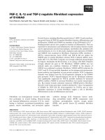
Fig. 1. Cloned fluorescent protein-tagged
copines and their domain chimaeras.
(A) Schematic diagrams of the domain struc-
ture of copines, with the position of the tag
in myc- or HA-tagged copines indicated (1),
and the fluorescent-protein tagged full-
length copines-2, -3 and -6 prepared for this
study (2,3). In addition to truncated versions
of copines-2 and -6 containing only specific
domains (4–8), domain swaps of copines-2
and -6 (9–11) were also constructed as
copine-2 C2-domain chimaeras with the
copine-6 vWA-domain connected through
the copine-2 (9) or copine-6 (11) linker.
(B) Alignment of the linker (grey
background) between the end of the
C2C2-domains (black background) and the
beginning of the vWA-domains for
copines-2, -6 and their derivatives, with the
conserved sequences boxed. (C) Alignment
of the linker area of copines-2 and -6 against
the corresponding sequences of C2A6 and
C2A6* constructs.
Calcium-dependent translocation of copines P. V. Perestenko et al.
5176 FEBS Journal 277 (2010) 5174–5189 ª 2010 The Authors Journal compilation ª 2010 FEBS
intracellular calcium; however, they did so at different
rates (Fig. 3B), with the movement of copine-2 being
the most rapid, followed by copine-6 and then copine-
3. To determine whether extracellular calcium is
necessary for the translocation of the copines, the
experiments were repeated in calcium-free medium.
Under these conditions, ionomycin caused a small
increase in intracellular calcium (Fig. 3C), but did not
lead to the translocation of copine-2 or copine-6
(Fig. 3D). The addition of 2 mm calcium to the iono-
mycin-treated cells in calcium-free medium, however,
caused a large increase in intracellular calcium
(Fig. 3C) and the rapid translocation of copine-2 and
copine-6 to the membrane (Fig. 3D). Copine-1 and
copine-7 showed similar ionomycin responses, as did
N-terminally tagged EYFP–copine-2 (Fig. S2A). Thus,
the ionomycin-induced translocation of the copines was
dependent on the presence of extracellular calcium.
We next characterized copine-2 and copine-6 in
greater detail with respect to their responses to an
increase in intracellular calcium. Treatment of HEK-
293 cells with thapsigargin caused a marked increase in
intracellular calcium because of its release from intra-
cellular stores, as well as the influx of extracellular cal-
cium through calcium channels. Calcium added to cells
treated for 2–3 min with thapsigargin in calcium-free
medium produced a dramatic increase in calcium
caused by its entry through store-operated calcium
channels. This calcium influx can be blocked by the
addition of 2-aminoethyldiphenyl borate (2-APB) or
2 lm Gd
3+
(Fig. 4A). In calcium-free medium, treat-
ment of cells, transfected with either copine-2 or
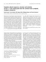
Fig. 2. Expression of recombinant copines-1,
-3, -6 and -7 in cultured mammalian cells.
(A) Western blots of myc- ⁄ HA- and EYFP-
tagged full-length copines in cultured HEK-
293 cells. The top bands in the anti-HA
panel represent nonspecific bands that were
present in nontransfected cells. (B) Expres-
sion patterns of myc- ⁄ HA- and EYFP-tagged
full-length copines in cultured COS-7 and
HEK-293 cells. Apart from the weak nuclear
staining of anti-HA IgG, the antibodies
showed no nonspecific binding in cells (see
also Fig. S1). Scale bars, 10 lm.
P. V. Perestenko et al. Calcium-dependent translocation of copines
FEBS Journal 277 (2010) 5174–5189 ª 2010 The Authors Journal compilation ª 2010 FEBS 5177
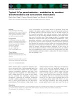
Fig. 3. Ionomycin treatment of HEK-293 cells causes translocation of the copines to the plasma membrane. HEK-293 cells were transfected
with the different copines and treated with ionomycin in medium containing 1.8 m
M CaCl
2
. Cells were either fixed with paraformaldehyde,
permeabilized and immunostained for the copines (A, H), or the localization of EYFP-tagged copines was visualized by confocal microscopy
of live cells (E, G). (A) HEK-293 cells expressing the lymphocyte membrane protein CD2 and myc-tagged copine-2 were treated with ionomy-
cin and immunostained for both proteins. Copine-2 (red) showed rapid movement to the plasma membrane where it colocalized with CD2
(green). (B) Fluorescence levels of cytosolic EYFP-tagged copines-2, -3 and -6 were monitored in HEK-293 cells (30–40 cells) expressing the
copines, using circular regions of interest as illustrated in (E) and (G). (C) The effect of ionomycin on EYFP fluorescence in these areas over
time, in Ca
2+
-containing medium, was calculated, and the results were plotted. (D) The effect of ionomycin on cytoplasmic calcium levels in
HEK-293 cells (30–40 cells) in calcium-free medium was visualized using the fluorescent calcium indicator Fluo-4FF. In the absence of extra-
cellular calcium, ionomycin had no effect on the cytoplasmic fluorescence of EYFP-tagged copines-2 and -6. (E) Confocal images of the
ionomycin responses of copine-2–EYFP and its C2C2-domain constructs in HEK-293 cells. (G) Typical responses of copine-6–EYFP and its
C2C2–EYFP construct to ionomycin treatment. The average ionomycin responses of EYFP-tagged copines-2 and -6 and their C2C2–EYFP
constructs are summarized in (F) (30–50 cells), where the grey bars are the responses in calcium-free medium and the open bars are those
in medium containing calcium. For all the constructs, the response in medium containing Ca
2+
was significantly greater than that in calcium-
free medium (P > 0.001, U-test). All the quantitative data are expressed as F ⁄ F
0
, and the data represent the means from at least 10 cells
per experiment. HEK-293 cells were cotransfected with myc-tagged copine-2 and HA-tagged copine-6 and treated with ionomycin for 3 min.
The cells were fixed, permeabilized and stained for the two different epitopes. The results show that copine-2 is not associated with copine-
6 when the latter is internalized (H). Scale bars represent 10 lm.
Calcium-dependent translocation of copines P. V. Perestenko et al.
5178 FEBS Journal 277 (2010) 5174–5189 ª 2010 The Authors Journal compilation ª 2010 FEBS
copine-6, with thapsigargin, did not stimulate their
movement to the membrane, despite the increase in
intracellular calcium as a result of release from intra-
cellular stores. However, the addition of calcium to the
medium of treated cells caused a rapid shift in both
copines to the membrane, although copine-6 required
significantly greater extracellular calcium concentra-
tions to initiate membrane translocation (Fig. 4B). In
calcium-containing medium, the copine responses were
also dependent on the opening of the store-operated
calcium channels, as the inhibitors 2-APB and 2 lm
Gd
3+
reduced both the number of cells responding
and the extent of their response, as shown for copine-2
(Fig. 4C).
In order to examine the response of the copines to a
more physiological stimulus, we took advantage of the
expression of the muscarinic acetylcholine receptor in
HEK-293 cells [24]. Selective muscarinic agonists, such
as acetyl-b-methylcholine (methacholine), can activate
these receptors and induce extracellular calcium influx,
as well as its intracellular release, in HEK-293 cells.
We observed that only copine-2 and copine-7 showed
robust responses to treatment of the cells with 10 lm
methacholine (63 ± 6.5% and 78.4 ± 8.7% of the
transfected cells, respectively) (see both Figs 5 and 6).
Copines-1, -3 and -6 showed little response to meth-
acholine treatment, with fewer cells responding and a
reduced extent of translocation. For example, only
2.4 ± 1.1% of copine-3-transfected cells weakly
responded to methacholine treatment (Fig. 6B, top
right). Unlike the ionomycin or thapsigargin responses,
the responses of copine-2 and copine-7 to methacho-
line were transient because of the transient increase in
intracellular calcium induced by methacholine, as
shown in Fig. 6A. The methacholine-induced translo-
cation of all of the copines, and the copine constructs,
was blocked by cotreatment with the muscarinic recep-
tor antagonist atropine (data not shown).
In order to confirm that the intracellular increase in
calcium caused by methacholine was sufficient to
translocate copine-2 or its C2-domain construct to the
membrane, cells expressing these proteins were treated
Fig. 4. Calcium-dependent intracellular translocation of the copines depends on the opening of store-operated calcium channels. (A) Fluo-
4FF fluorescence in the cell cytoplasm was used to visualize the changes in calcium levels in HEK-293 cells in response to thapsigargin
treatment. Measurements were made first in calcium-free medium, and then in medium to which calcium was restored. Changes in the
intracellular calcium levels were recorded over time. The effects of 2-ABP or Gd
3+
ions on the entry of calcium to the cells were also exam-
ined. The traces shown represent the average results from 250–300 cells. (B) Changes in the localization of copine-2–EYFP and copine-6–
EYFP were imaged using confocal microscopy of live cells. The localization of both copines was affected by thapsigargin treatment, but only
when the levels of extracellular calcium were increased. Here, each plot represents the average ( 50 cells) reduction in cytoplasmic
copine–EYFP at the indicated time points. (C) The inhibitory effects of 2-APB and Gd
3+
on the copine-2–EYFP responses to extracellular
calcium in cells with Ca
2+
stores depleted by thapsigargin are shown. Each plot shows the decrease in cytosolic copine as a fraction of the
original fluorescence for an individual cell, and the results are from approximately 20–25 cells per coverslip (three coverslips each). The chart
in (C) shows the decrease in cytosolic copine-2 fluorescence as an average of the data from multiple experiments, approximately 150–200
cells in total (P < 0.001*** for Ca
2+
and P 0.05** for 2-APB or Gd
3+
, U-test).
P. V. Perestenko et al. Calcium-dependent translocation of copines
FEBS Journal 277 (2010) 5174–5189 ª 2010 The Authors Journal compilation ª 2010 FEBS 5179
with methacholine in the presence or absence of extra-
cellular calcium, and in the presence of calcium
and 2-APB. The treatment of HEK-293 cells with
methacholine caused a robust transient increase in
intracellular calcium that could be reduced either by
removing extracellular calcium or by blocking the
Fig. 5. Methacholine-induced translocation of different copines and C2C2-constructs to the plasma membrane in response to transient ele-
vation of Ca
2+
in cultured HEK-293 cells. HEK-293 cells were transfected with copine-3–EYFP, -6–EYFP, -7–EYFP and copine-2–EYFP ⁄ EGFP
and its different C2C2-constructs as indicated above each panel. Methacholine was added to the medium containing extracellular calcium, at
time point 0 s, and the changes in cytoplasmic fluorescence were imaged at the indicated time points. Scale bar corresponds to 5 lm.
Calcium-dependent translocation of copines P. V. Perestenko et al.
5180 FEBS Journal 277 (2010) 5174–5189 ª 2010 The Authors Journal compilation ª 2010 FEBS
store-operated channels and IP3 receptors with 2-APB
(Fig. 6A). In the absence of extracellular calcium or in
the presence of both calcium and 2-APB, the weak
responses of copines-3 and -6 to methacholine were
completely inhibited (Fig. 6B–D). In contrast with
thapsigargin treatment, in calcium-free medium, the
responses of copine-2 and its C2C2-linker construct to
methacholine were not ablated. The response was
reduced significantly, however, with fewer cells
responding to treatment and, in the cells that did
respond, the extent of translocation being attenuated
(Fig. 6C), suggesting that a maximal response required
Fig. 6. Full methacholine-induced copine
responses depend on extracellular calcium.
(A) Intracellular levels of calcium in HEK-293
cells were increased by 10 l
M methacholine
treatment and the calcium levels were mon-
itored using Fluo-4FF. The stimulatory effect
of methacholine was prevented by either
cotreating the cells with 2-APB or Gd
3+
,or
by incubating the cells in calcium-free med-
ium. The peak value for the intracellular rise
in calcium was significantly lower when the
cells were exposed to 2-APB relative to
2 l
M Gd
3+
(P 0.05, n
1–2
> 300, U-test).
(B) Methacholine application affects the
level of cytoplasmic fluorescence of copine-
2–EYFP, -3–EYFP and -6–EYFP, as well as
the C2C2–EYFP domains of copine-2, to
varying degrees, in the presence of extracel-
lular calcium. The effect of methacholine is
attenuated for copine-2–EYFP and copine-6–
EYFP in the absence of extracellular Ca
2+
(C) and in the presence of 2-APB (D). The
experiments show the traces from 20–25
cells per coverslip (three coverslips in total).
Copine-3–EYFP did not respond to methach-
oline in the absence of Ca
2+
or the presence
of 2-APB (data not shown). (E) The cumula-
tive representation of three to five indepen-
dent experiments (160–250 cells in total)
of copine-2–EYFP and its C2C2-EYFP
domain response to methacholine. The
graphs show sorted minimal cytoplasmic
fluorescence (i.e. the maximum response to
methacholine) for each individual cell. Full-
length copine-2–EYFP and its C2C2-EYFP
domain responded similarly to methacholine
in the presence (P = 0.59; n
1
= 254,
n
2
= 187; U-test) or absence (P = 0.31;
n
1
= 240, n
2
= 173; U-test) of extracellular
Ca
2+
, suggesting that the vWA-domain is
not important for the translocation of
copine-2–EYFP to the plasma membrane.
P. V. Perestenko et al. Calcium-dependent translocation of copines
FEBS Journal 277 (2010) 5174–5189 ª 2010 The Authors Journal compilation ª 2010 FEBS 5181
the influx of extracellular calcium. However, further
investigation revealed that store-operated channels
were also involved, as methacholine-induced transloca-
tion of copine-2 was reduced by 2-APB treatment.
Moreover, this reduction was even greater than the
reduction produced by the elimination of extracellular
calcium (P < 0.001; n
1
= 188, n
2
= 161; U-test;
Fig. 6D, E), reflecting the inhibition of release of cal-
cium from intracellular stores by 2-APB [25]. In con-
trast, the C2C2-linker domains (copine-2) gave similar
responses to methacholine whether in calcium-free
medium or in the presence of calcium plus 2-APB
(P = 0.074; n
1
= 160, n
2
= 177; U-test; Fig. 6D, E).
Thus, in the full-length protein, the presence of the
vWA-domain may modulate the intracellular translo-
cation of the copines by reducing the sensitivity of the
C2-domains to calcium. Together, these results show
that the copines have different sensitivities to increases
in intracellular calcium, and that they require extracel-
lular calcium to exhibit their maximal translocation
responses.
The copine C2-domains and linker region are
crucial for ionomycin-induced membrane
translocation
The predicted domain structure of the copines suggests
that the C2-domains might be responsible for the cal-
cium-mediated membrane association of copines
[4,8,16]. In order to test this hypothesis, the
C2-domains of copine-2 alone were fused with EYFP
at both the N- and C-termini and in the presence and
absence of the linker region between the last
C2-domain and the start of the vWA-domain (see
schematic diagrams 4–7 in Fig. 1A). The response of
these EYFP-tagged domains to ionomycin treatment
was compared with that of full-length copine-2–EYFP.
The results showed clearly that all of the constructs
containing the linker region behaved similarly to co-
pine-2–EYFP. However, if the linker region was
removed, the protein did not associate with the plasma
membrane (Figs 3E and S2B), indicating the impor-
tance of the linker region in mediating this interaction.
Similar results were obtained with copine-6 (Figs 3G
and S2B). Quantitative analysis of several experiments
showed that, for copines-2 and -6, the C2-domain con-
structs behaved similarly to the full-length copine–
EYFP following ionomycin treatment (Fig. 3F). In
addition, the C2-domain constructs of copine-2
containing the copine-2 linker region, tagged at the
N-terminus with EGFP or at the C-terminus with
EYFP, responded robustly to methacholine, whereas if
the linker region was removed no response was
observed (see both Figs 5 and 6). In contrast, the
EYFP–vWA-domains of the copines showed no
response to ionomycin, despite the presence of the
linker region (data not shown).
Taken together, the investigation of the behaviour
of the different truncations and domain swap con-
structs showed that the C2-domains are essential for
calcium-mediated membrane binding, but that the
binding requires the presence of the linker region,
proximal to the vWA-domain (see Fig. S3).
Copine-6 associates with clathrin-coated
vesicles in a calcium-dependent manner which is
regulated by both the C2- and vWA-domains
During the course of these experiments, we noted that
ionomycin treatment of copine-6 (but not copine-2)-
expressing cells appeared to show copine-6-containing
vesicles in cytoplasm after 3 min of exposure to iono-
mycin (Fig. 3G, H). Indeed, when myc-tagged copine-2
and HA-tagged copine-6 were co-expressed in the same
cells, and the cells were exposed to ionomycin, only
HA-tagged copine-6 was found in intracellular vesicles
(Fig. 3H). A fusion construct of C2-domains of
copine-2 (including the linker of copine-2) and the
vWA-domain of copine-6 behaved similarly (Fig. 3G),
whereas the C2-domains of copine-2 alone exhibited a
pattern identical to full-length copine-2 (Fig. 3E).
Thus, the association of copine-6 with vesicles appears
to require the vWA-domain of copine-6.
In order to investigate this further, HEK-293 cells
expressing either HA- or EYFP-tagged copines-2, -3 or
-6 were stimulated for 3–5 min with ionomycin, and
immunostained using markers for clathrin-mediated
endocytosis (transferrin), caveolar endocytosis (caveo-
lin) or a late endosome marker (mannose-6-phosphate
receptor). Neither caveolin nor mannose-6-phosphate
receptor staining colocalized with any of the copines
(data not shown). However, the copine-6-containing
vesicles (Fig. 7A1), but not copines-2 or -3 (Fig. 7A2,
A3), colocalized with Alexa-Fluor-568-conjugated
transferrin. To visualize the effect of ionomycin on the
formation of copine-6-containing vesicles, we imaged
live cells, transfected with either copine-6–EYFP or
copine-2–EYFP and incubated with fluorescent trans-
ferrin. In untreated cells, transferrin was associated
with internalized clathrin-coated vesicles and was
partially diffused throughout the cell cytoplasm
(Fig. 7A1, A2, top row). Ionomycin treatment of the
cells caused fast translocation of both copines to the
plasma membrane, with copine-6, but not copine-2,
bound to the internalized clathrin-coated vesicles con-
taining transferrin. Ionomycin therefore did not cause
Calcium-dependent translocation of copines P. V. Perestenko et al.
5182 FEBS Journal 277 (2010) 5174–5189 ª 2010 The Authors Journal compilation ª 2010 FEBS
the internalization of copine-6, but rather stimulated
its association with clathrin-coated membranes of
internalized early endosomes and with the plasma
membrane (Fig. 7A1, A2). To determine which
domains of the copines contribute to the endosomal
association of copine-6, different chimaeric copine con-
structs were examined following ionomycin treatment
of cells labelled with transferrin. Myc-tagged copine-6
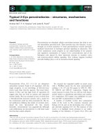
Fig. 7. Copine-6 associates with clathrin-coated vesicles following increases in intracellular calcium. HEK-293 cells expressing copines were
pre-incubated with Alexa546-conjugated transferrin, washed and treated with ionomycin for 5 min in the presence of 1.8 m
M extracellular
Ca
2+
. Green corresponds to EYFP or EGFP fluorescence, red to transferrin fluorescence. Live HEK-293 cells expressing copine-6–EYFP (A1)
or copine-2–EYFP (A2) were imaged before, 10 s and 3 min after ionomycin application. Alternatively, cells were fixed after ionomycin treat-
ment and immunofluorescence was used to visualize copine-3–EYFP (A3). Similar experiments were performed with the cells fixed after
5 min using N-terminally tagged copine-6 (EGFP–copine-6) (B1), the C2C2–EYFP domains of copine-2 (B2) and copine-6 (B4) or the domain
recombination constructs C2A6–EYFP (B3) and C6A2–EYFP (B5). Scale bars, 5 lm.
P. V. Perestenko et al. Calcium-dependent translocation of copines
FEBS Journal 277 (2010) 5174–5189 ª 2010 The Authors Journal compilation ª 2010 FEBS 5183
and copine-6–EYFP, but not the N-terminally tagged
EGFP–copine-6, colocalized with clathrin-mediated
internalized vesicles following ionomycin treatment
(Fig. 7B1, B2, B3). The domain swap C2A6–EYFP
construct and the C2C2–copine-6 derivative also
bound to the transferrin-containing vesicles (Fig. 7B3,
B4), unlike the C6A2–EYFP construct (Fig. 7B5). In
contrast, neither copine-2 nor its C2-domain–EYFP
construct bound to the transferrin-containing vesicles
(Fig. 7A2, B2). Thus, the N-terminal copine-6
C2-domains appear to contribute to endosomal vesicle
binding, but are not sufficient to confer this property
to the copine-2 vWA-domain. Thus, the copine-6
vWA-domain seems to carry an endosomal targeting
sequence which can confer endosomal binding to the
C2-domains of copine-2.
Discussion
In the present study, we have shown for the first time
that, in mammalian cell lines, copines-1, -2, -3, -6 and
-7 can move to the plasma membrane following
increases in intracellular Ca
2+
triggered by ionomycin
treatment of cells in medium containing 1.8 mm cal-
cium. Structural considerations [4,8] and in vitro bind-
ing studies [5] had suggested that the copines should
associate with cell membranes in a calcium-dependent
manner. To date, this hypothesis had only been dem-
onstrated in Dictyostelium, where a small percentage
(1–4%) of cells showed copine-A translocated to the
plasma membrane after starvation [11]. Our finding
that copine-3 can translocate to the plasma membrane
in response to ionomycin treatment has been con-
firmed in a recent study on the role of copine-3 in cell
migration [26]. In that study, and in the present study,
ionomycin also caused nuclear translocation of copine-
3, as well as copines-1, -2, and -7. In contrast, copine-
6 did not show nuclear translocation after exposure of
cells to ionomycin, but did bind to intracellular vesi-
cles, identified as early endosomes, as well as the
plasma membrane. The copines showed different rates
of calcium-induced membrane translocation, with
copine-2 and copine-7 moving most rapidly, followed
by copines-1, -6 and -3.
In calcium-free medium, neither ionomycin nor
thapsigargin caused movement of the copines to the
plasma membrane, despite both agents causing
increases in intracellular calcium. The addition of
2mm calcium to the medium of cells treated with
either agent caused rapid movement of copine-2 to the
cell membrane. However, after thapsigargin, but not
ionomycin, treatment, copine-6 required the addition
of higher concentrations of extracellular calcium
(5 mm) than did copine-2 to trigger its movement. This
may reflect the fact that ionomycin forms calcium-
permeable pores in the cell membrane, permitting a
rapid increase in intracellular calcium from the extra-
cellular medium [27,28]. In contrast, calcium enters the
thapsigargin-treated cells through store-operated cal-
cium channels, opened by the release of calcium from
intracellular stores by the drug [29–31], and this release
can be blocked by 2-APB and Gd
3+
(Fig. 4), as has
been reported previously for HEK-293 cells [32,33].
Therefore, the rate of calcium entry into the thapsigar-
gin-treated cells may be slower than that in ionomycin-
treated cells, and more dependent on the calcium
concentration difference across the membrane. Thus,
the higher extracellular concentrations of calcium
needed to mobilize copine-6 following thapsigargin
treatment suggest that it has a lower affinity for calcium
than does copine-2. A higher affinity for calcium of
copine-2 may also explain its more rapid rate of mem-
brane translocation in response to ionomycin treatment
and its stronger response to methacholine stimulation,
compared with the responses of copine-6. Such differ-
ences in calcium sensitivity have been observed in other
families of C2-domain-containing proteins, such as, for
example, DOC2A and DOC2B [34], and even between
different C2-domains from within a protein family
(e.g. the protein kinase C family [35]).
Following the activation of the endogenous musca-
rinic receptor in HEK-293 cells by methacholine, the
copines again exhibited different levels of membrane
translocation, with copine-2 and copine-7 showing a
rapid and robust movement to the cell membrane, with
weaker responses from copine-6 and copine-3. In
HEK-293 cells, the full response of the proteins to
methacholine was dependent on calcium entry through
store-operated calcium channels. Thus, regardless of the
stimulus used to increase intracellular calcium, the
copines showed different rates of membrane association
and different sensitivities to the levels of intracellular
calcium. Nevertheless, in order to exhibit a full response
to either ionomycin or methacholine, the copines
require calcium entry from the extracellular medium.
The calcium-dependent association of the copines
required the presence of the C2-domains, as these
contain the calcium-binding sites [36]. However, inter-
estingly, we found that both copine-2 and copine-6
required a specific region between the second
C2-domain and the vWA-domain for membrane asso-
ciation, which we termed the ‘linker region’ (Figs 1C
and S4). The linker region contains two sections that
are strongly conserved in all mouse copines. The first
is proximal to the C-terminus of the second
C2-domain, which contains several positively charged
Calcium-dependent translocation of copines P. V. Perestenko et al.
5184 FEBS Journal 277 (2010) 5174–5189 ª 2010 The Authors Journal compilation ª 2010 FEBS
amino acids, and the second is situated near the start
of the vWA-domain. The positively charged sequence
is preserved in the C2-domain constructs that did not
show calcium-dependent membrane association, and
therefore the membrane-binding site must lie in the
C-terminal segment of the linker region. Lipid binding
of C2-domains of other proteins has been attributed to
two regions: the calcium-binding region and a cationic
b-groove located in strands b3 and b4 of the protein
[35,36]. In the case of the copines, the linker region
identified here lies outside the canonical C2-domain. In
the only study to date on isolated copine-6 domains,
binding of both domains without the linker region to
phosphatidylserine vesicles was observed, with the first
C2-domain exhibiting calcium-independent vesicle
binding [16]. This indicates that the single C2-domains
fused to glutathione S-transferase and expressed in
Escherichia coli can bind to lipids in in vitro assays,
but our results show clearly that the linker is critical
for membrane association in vivo in cells, where
perhaps it acts to stabilize membrane binding follow-
ing a transient C2-domain-mediated initial interaction.
The linker appears to be insufficient to promote
membrane association, as the vWA-domains contain-
ing the linker do not bind to cell membranes. How-
ever, the vWA-domains can clearly modulate the
responses of the C2-domains to calcium, and possibly
play a role in intracellular targeting. The altered meth-
acholine response of the different constructs of copine-
2 indicates a role for the vWA-domain in modulating
its calcium responsiveness. Thus, replacement of the
copine-2 vWA-domain with that of copine-6 ablates
the methacholine response, but not the ionomycin
response, of the hybrid construct. Similarly, removing
the copine-2 vWA-domain renders the C2C2-linker
construct of copine-2 more responsive than full-length
copine-2 to methacholine challenge in both calcium-
free medium and in the presence of 2-APB (see Figs 5
and 6). A contribution of the vWA-domains to the
intracellular targeting of the copines is indicated by
our finding that the exchange of the vWA-domain of
copine-6 with that of copine-2 is sufficient to prevent
endosome association of the chimaeric construct
C6A2. Furthermore, unlike copine-2, the C2A6–EYFP
construct does not show nuclear localization, but does
show early endosome binding, following ionomycin
treatment of the cells (see Fig. 7). The fact that the
majority of the copines, but not copine-6, show
nuclear targeting suggests a role for these copines in
nuclear processes. The recent demonstration that
copine-3 appears to bind several nuclear proteins,
including interleukin enhancer-binding protein 2,
nucleolin and DNA topoisomerase 1, is consistent with
such a role for copine-3 [26], as is the finding that
copine-1 is involved in the regulation of NF-kappaB
transcriptional responses via endoproteolysis of the
p65 protein [37,38].
The physiological function of the majority of the
copines is currently unclear. However, a recent report
showing that copine-3 interacts with Erb-2, is upregu-
lated in breast and prostate tumours, and promotes
tumour migration by recruiting RACK1 to focal adhe-
sion plaques [26] indicates the importance of studying
this family of proteins. The data presented here show,
for the first time, that the copines will move to the
plasma membrane in response to increases in intracel-
lular calcium. However, it is clear that they show dif-
ferent sensitivities to calcium. The finding that copines
have distinct properties suggests that each copine may
be tailored to respond to specific physiological stimuli:
for example, in the present study, copine-3 did not
respond to stimulation of the muscarinic receptor,
whereas previously it was found to respond to activa-
tion of the ErbB2 receptor by heregulin [26]. In addi-
tion, our data show that, although the C2-domains of
the copines are essential for calcium-dependent mem-
brane binding, they are not sufficient, and we have
identified a conserved linker region in the copines,
between the C2- and vWA-domains, that is necessary
for membrane binding in living cells. We have also
shown that the vWA-domains contain targeting infor-
mation, and can modulate the calcium sensitivity of
the proteins. Our data also indicate that many of the
copines, but not copine-6, show nuclear localization,
either normally (e.g. copines-2 and -7) or following ele-
vation of intracellular calcium (copines 1 and -3). We
conclude that the copines are likely to mediate cal-
cium-dependent targeting of proteins to various intra-
cellular locations, including the plasma membrane and
the nucleus. The rapid translocation of the copines in
response to changes in intracellular calcium suggests
that this family of proteins may play an important role
in calcium-dependent intracellular signalling.
Materials and methods
DNA constructs
Coding sequences of mouse copines-2 and -6, and human
copines-1, -3 and –7, were derived from image clones:
IMAGE:3985959, IMAGE:6591063, IMAGE:3502122,
IMAGE:5300530 and IMAGE:5727324, respectively (Gene-
Service Ltd.; ). N-terminally
HA-tagged copine-3 was amplified by PCR from the
IMAGE clone DNA in two steps, where the product of the
first step was used in the second round of PCR. Step 1
P. V. Perestenko et al. Calcium-dependent translocation of copines
FEBS Journal 277 (2010) 5174–5189 ª 2010 The Authors Journal compilation ª 2010 FEBS 5185
primers: 5¢-CCGTATGACGTCCCAGATTACGCATCGA
TGGCTGCCCAGTGTGTCAC-3¢ and 5¢-GGGGATCCT
CACTGCTTCTGTTGTTTCGTGG-3¢. In step 2, primer
5¢-CCTCTAGACGCCGCCACCATGCCGGATTACGCG
TCTTACCCGTATGACGTCCCAGATT-3¢ was used with
the second primer of step 1. The PCR product was cloned
into the XbaI and BamHI sites of the pcDNA3.1(–) vector
(Invitrogen, Paisley, Renfrewshire, UK). The same scheme
was applied to clone HA–copine-6 with primers 5¢-CCGTA
TGACGTCCCAGATTACGCATCGATGTCGGACCCA
GAGATGGGATG-3¢ and 5¢-GGGGATCCTCATGGG
CTAGGGCTGGGAGTC-3¢ used in the first round of
PCR. N-terminally myc-tagged copine-2 was amplified from
its IMAGE clone with the primers 5¢-CGAATTCGGATG
GCCTACATTCCGGATGG-3¢ and 5¢-CGCTCGAGTCA
GGCAGGCTCTGAGTTGGTG-3¢, and cloned between
the EcoRI and XhoI sites of pCMV-myc (Clontech, Moun-
tain View, California, USA). N-terminally myc-tagged
copine-6 was made by substitution of the SpeI–ClaI fragment
of HA–copine-6 with the DNA fragment amplified from
pcDNA3.1(–) with primers 5¢-GTTTCTGATTAT TGAC
TAGTTATTAATAGTAATCAATTACGGG-3¢ and 5¢-GT
TTCTATCGATGACAAGTCCTCTTCAGAAATGAGCT
TTTGCTCCATGGTGGCGGCGTCTAGAG-3¢. N-termi-
nally myc-tagged copines-1 and -7 were made by substitu-
tion of the HindIII–ClaI fragment in the myc–copine-6
construct with the respective products of PCR amplification
(5¢-GTTTCTATCGATGGCCCACTGCGTGACCTTGG-3¢,
5¢-GTTTCTAAGCTTTTAAGCCTGGGGGGCCTGTG C
AG-3¢ and 5¢-GTTTCTATCGATGAGCGCGGGCTCGG
AGCG-3¢,5¢-GTTTCTAAGCTTTCACGGTGTGCAGCC
TGGGCTG-3¢).
To clone EGFP fusion proteins, the PCR amplification
products (5¢-GTTTCTGAATTCCATGGCCTACATTCCG
GATGGG-3¢ and 5¢-GTTTCTGGATCCTCAGGCAGG
CTCTGAGTTGGTG-3¢) of the copine-2 IMAGE clone
were inserted into the pEGFP-C1 vector (Clontech) using
its EcoRI and BamHI sites. The EGFP–HA–copine-6 con-
struct was cloned by inserting the XbaI–BamHI fragment
of HA–copine-6 into the pEGFP-C1 vector. To clone C-ter-
minal EYFP-tagged copines-1, -2, -3, -6 and –7, PCR
amplification products of corresponding IMAGE clones
were inserted into the pEYFP-N1 (Clontech) vector (co-
pine-1–EYFP: 5¢-GTTTCTGAATTCGCCACCATGGCC
CACTGCGTGACCTTGG-3¢ and 5¢-GTTTCTACCGGT
CCTGAAGCCTGGGGGGCCTGTGCAG-3¢, EcoRI–
AgeI; copine-2–EYFP: 5¢-CCAGATCTCCATGGCCTAC
ATTCCGGATGGG-3¢ and 5¢-CCCTCGAGGGCAGGCT
CTGAGTTGGTG-3¢, BglII–XhoI; copine-3–EYFP: 5¢-GT
TTCTCTCGAGGCCGCCACCATGGCTGCCCAGTGTG
TCAC-3¢ and 5¢-GTTTCTGGATCCGTACTCTGCTTCT
GTTGTTTCGTGG-3¢, XhoI–BamHI; copine-6–EYFP:
5¢-GTTTCTGAATTCTAGCCACCATGTCGGACCCAG
AGATGGGATG-3¢ and 5¢-GTTTCTGGATCCG ATGGG
CTAGGGCTGGGAGTCATAG-3¢, EcoRI–BamHI; copine-
7–EYFP: 5¢-GTTTCTGCTAGCGCCACCATGAGC
GCGGGCTCGGAGCG-3¢ and 5¢-GTTTCTAAGCTTTG
ACGGTGTGCAGCCTGGGCTG-3¢, NheI–HindIII). The
C2C2-domains of copines-2 and -6 were amplified by PCR
from the corresponding IMAGE clones and inserted
between the EcoRI–BamHI sites of the pEYFP-N1 vector.
The primers used to produce the C2C2-domains lacking
the linker region were 5¢-GTTTCTGAATTCTGGCCAC
CATGGCCTACATTCCGGATGGG-3¢ and 5¢
-GTTTCT
GGATCCGCGCTTTTCTTCTTCCTCTGCTTCTTGG-3¢
(C2C2–copine-2), and 5¢-GTTTCTGAATTCTGGCCACC
ATGTCGGACCCAGAGATGGGATG-3¢ and 5¢-GTTTC
TGGATCCAGCTTGTAATTCTTCTTCTTGTCTCGGTA
CTTGG-3¢ (C2C2–copine-6). The reverse primers for the
C2C2-linker domains were 5¢-GTTTCTGGATCCCCGCA
GCCTCCCAGAATGTAGTCCAG-3¢ (C2C2-linker, co-
pine-2) and 5¢-GTTTCTGGATCCCCGCAGCCACCCAT
GATATAATCCAGG-3¢ (C2C2-linker, copine-6). To pro-
duce N-terminally EGFP-tagged C2C2-domains of copine-2
with ⁄ without the linker area, products of EGFP–copine-2
PCR amplification with primers 5¢-GTTTCTGCAGAGC
TGGTTTAGTGA-3¢ and 5¢-GTTTCTGGATCCCTAGCA
GCCTCCCAGAATGTA-3¢⁄5¢-GTTTCTGGATCCCTAG
CTTTTCTTCTTCCTCTG-3¢ were cloned into the pEGFP-
C1 vector using the AgeI and BamHI sites. The vWA-
domains of copines-2 and -6 were amplified by PCR and
inserted into the EcoRI–BamHI sites of the pEGFP-C1
vector using the primers 5¢-GTTTCTGAATTCAAAGA
AGCAGAGGAAGAAGAAAAGCTACAAG-3¢ and 5¢-GT
TTCTGGATCCTCAGGCAGGCTCTGAGTTGGT G-3¢
(copine-2), and 5¢-GTTTCTGAATTCAAAGTACCGAG
ACAAGAAGAAGAATTACAAGAG-3¢ and 5¢-GTTTCT
GGATCCTCATGGGCTAGGG CTGGGAG-3¢ (copine-6).
To clone EYFP-tagged chimaera of the C2C2-domains of
copine-2 with the vWA-domain of copine-6 (C2A6) with
the copine-2 linker, the copine-6 vWA-domain, amplified
with primers 5¢-GTTTCTGGATCCTCAGATCAGCTTC
ACGGTGGCTATC-3¢ and 5¢-GTTTCTGGATCCGATG
GGCTAGGGCTGGGAGTCATAG-3¢, was inserted into
the BamHI site of the C2C2–copine-2* construct. For the
C2A6* chimaera, containing the copine-6 linker between
the C2C2- and vWA-domains, the PCR amplification
product (5¢-GTTTCTGGATCCAAAGTACCGAGACAA
GAAGAAGAATTACAAGAG-3¢ and 5¢-GTTTCTGGAT
CCGATGGGCTAGGGCTGGGAG-3¢) was inserted into
the BamHI site of the C2C2–copine-2 construct. The C6A2
(fusion of the C2C2-domains of copine-6 and the vWA-
domain of copine-2) chimaera was obtained by insertion of
the PCR amplification product of the copine-6 vWA-
domain into the BamHI site of the C2C2-linker (copine-6)
construct (primers: 5¢-GTTTCTGGATCCTCAGCTCATG
TTCACCGTTGGAATAG-3¢ and 5¢-GTTTCTGGATCCG
AGGCAGGCTCTGAGTTGGTGGG-3¢). The assignment
of the domain boundaries of the different regions of the
copines was based on their analysis in the Simple Modular
Calcium-dependent translocation of copines P. V. Perestenko et al.
5186 FEBS Journal 277 (2010) 5174–5189 ª 2010 The Authors Journal compilation ª 2010 FEBS
Architecture Research Tool (smart: l-hei-
delberg.de/). The range and structure of the constructs
used in this study are illustrated schematically in Fig. 1A.
Cell culture and transfection
HEK-293 cells (ECACC Cat. No. 851120602) and COS-7
cells were grown in Dulbecco’s modified Eagle’s medium
(Sigma-Aldrich, Poole, Dorset, UK), supplemented with
10% (v ⁄ v) fetal bovine serum (GIBCO, Paisley, Renfrew-
shire, UK), 2 mml-glutamine, 50 UÆmL
)1
penicillin and
50 lgÆmL
)1
streptomycin (all from Sigma-Aldrich), at 37 °C
in 5% CO
2
, 100% humidity. Only the third to 15th passages
of HEK-293 cells were used for live imaging. For micros-
copy, cells were plated onto borosilicate glass coverslips
coated with poly-d-lysine for HEK-293 cells, and grown for
48 h prior to transfection. Cells were transiently transfected
with polyethyleneimine according to the protocol adopted
from Durocher et al. [39], 24 h prior to fixation or live imag-
ing. The transfection efficiency varied between 30 and 70%.
Cell imaging and microscopy
All live imaging was performed at 25 °C in HBS buffer
(10 mm Hepes, 150 mm NaCl, 5 mm KCl, 1.8 mm MgCl
2
,
5.3 mmd-glucose, pH 7.4), with 1.8 mm CaCl
2
added for
Ca
2+
studies.
For calcium imaging, HEK-293 cells were loaded with
1 lm Fluo-4FF (Invitrogen) in HBS for 30 min at room
temperature, followed by three rinses in HBS. Coverslips
were mounted in a slide-holder chamber in 0.5 mL HBS for
imaging. The fluorescent images (512 · 512 or 1024 · 1024
pixels, one scan per frame) were taken with an LSM510
inverted confocal microscope system and a Plan-NEOFL-
UAR 40·⁄1.3 oil DIC immersion lens (Carl Zeiss Ltd.,
Welwyn Garden City, Hertfordshire, UK; excitation 488-
nm and emission 530–550-nm bypass filter, or excitation
543-nm and emission 560-nm long-pass filter; optical slice,
0.1–0.3 lm). For live internalization imaging, cells were
pre-incubated with transferrin conjugated with ALEXA 568
(50 lgÆmL
)1
, Invitrogen) for 5 min at room temperature,
rinsed three times with HBS and imaged after 5 min iono-
mycin stimulation. The fluorescence intensity from selected
areas in each frame was calculated using Zeiss LSM510
software, and the data were then exported to Microsoft
Excel, sigmaplot 10 (Systat Software Inc., Chicago, IL,
USA) or spss 16 (SPSS Inc., Chicago, IL, USA) for further
analysis. The immunofluorescence was expressed as the flu-
orescence intensity in a defined region of interest divided by
that in the same region at the start of the experiment
(F ⁄ F
0
). For live imaging, calcium and all drugs were added
to the cell by bath application in HBS buffer. For intracel-
lular protein localization, cells were fixed in 4% (w ⁄ v) para-
formaldehyde in HBS (pH 7.4) for 5 min at room
temperature, washed 2 · 5 min in Tris-saline and, where
appropriate, permeabilized with 0.2% (v ⁄ v) Triton X-100
for 5 min. Nonspecific binding was blocked by incubating
the cells with 1% (w ⁄ v) bovine serum albumin for 30 min.
The cells were then incubated in blocking solution with pri-
mary antibody for 2 h at room temperature, washed in
HBS for 3 · 5 min, incubated for 1 h with the appropriate
secondary antibody, washed in HBS for 3 · 5 min and
mounted for imaging.
Western blotting
Cells were scraped into solubilization mixture [1% (v ⁄ v) Tri-
ton X-100, HBS buffer, pH 7.4, 10 mm EDTA] containing
the recommended concentration of CompleteÔ protease
inhibitor cocktail (Roche, Welwyn Garden City, Hertford-
shire, UK), triturated with a pipette and incubated at 4 °C
for 1 h. The lysate was cleared by centrifugation, subjected to
SDS ⁄ PAGE and blotted onto Polyscreen
Ò
poly(vinylidene
difluoride) membrane (Perkin Elmer, Cambridge, UK). Blots
were probed with antibodies as indicated in the figure legends.
Detection was by horseradish peroxidase-conjugated second-
ary antibodies (Promega, Southampton, Hampshire, UK)
and Super Signal
Ò
West Pico chemiluminescent substrate
(Thermo Scientific, Loughborough, Leicestershire, UK).
Other reagents and antibodies
Reagents: ionomycin from Streptomyces conglobatus,
thapsigargin, acetyl-b-methylcholine chloride, poly-d-lysine
hydrobromide, gadolinium(III) chloride hexahydrate and
2-APB (all from Sigma-Aldrich); carbamoylcholine chloride
(Fluka, Gillingham, Dorset, UK). Antibodies and conju-
gates: chicken anti-mannose-6-phosphate receptor (cation-
independent) (Chemicon, Temecula, CA, USA, AB3463);
mouse anti-c-adaptin (BD Transduction Labs, Oxford, UK,
A36120); rabbit anti-caveolin-1 (BD Transduction Labs,
610406); rhodamine-conjugated dextran (Invitrogen,
D-1824); transferrin from human serum Alexa-568 conjugate
(Invitrogen, T-23365); rabbit anti-HA (Abcam, Cambridge,
UK, 9119-100); monoclonal anti-myc 9E10, goat anti-
rabbit Alexa Fluor 488 or 568 IgG (Invitrogen); goat
anti-mouse Alexa Fluor 488 or 568 IgG (Invitrogen). All
other high-purity grade chemicals were purchased from
Sigma-Aldrich, BDH (West Chester, PA, USA) or Fluka.
Acknowledgements
We are grateful to Dr Antony Morgan for advising us
on all aspects of calcium imaging. This work was
supported by the Medical Research Council, UK.
A. Pooler’s work was supported by the Blaschko
European Visiting Fellowship. M. Noorbakhshnia’s work
was supported by the Iran Ministry of Science, Research
and Technology and British Council Scholarships.
P. V. Perestenko et al. Calcium-dependent translocation of copines
FEBS Journal 277 (2010) 5174–5189 ª 2010 The Authors Journal compilation ª 2010 FEBS 5187
References
1 Rizo J & Sudhof TC (1998) C2-domains, structure
and function of a universal Ca
2+
-binding domain.
J Biol Chem 273, 15879–15882.
2 Nalefski EA & Falke JJ (1996) The C2 domain cal-
cium-binding motif: structural and functional diversity.
Protein Sci 5, 2375–2390.
3 Michishita M, Videm V & Arnaout MA (1993) A novel
divalent cation-binding site in the A domain of the
beta 2 integrin CR3 (CD11b ⁄ CD18) is essential for
ligand binding. Cell 72, 857–867.
4 Creutz CE, Tomsig JL, Snyder SL, Gautier MC, Skouri
F, Beisson J & Cohen J (1998) The copines, a novel
class of C2 domain-containing, calcium-dependent,
phospholipid-binding proteins conserved from
Paramecium to humans. J Biol Chem 273,
1393–1402.
5 Tomsig JL & Creutz CE (2000) Biochemical charac-
terization of copine: a ubiquitous Ca
2+
-dependent,
phospholipid-binding protein. Biochemistry 39, 16163–
16175.
6 Gottschalk A, Almedom RB, Schedletzky T, Anderson
SD, Yates JR & Schafer WR (2005) Identification and
characterization of novel nicotinic receptor-associated
proteins in Caenorhabditis elegans. EMBO J 24, 2566–
2578.
7 Church DL & Lambie EJ (2003) The promotion of
gonadal cell divisions by the Caenorhabditis elegans
TRPM cation channel GON-2 is antagonized by GEM4
copine. Genetics 165, 563–574.
8 Tomsig JL & Creutz CE (2002) Copines: a ubiquitous
family of Ca
2+
-dependent phospholipid-binding pro-
teins. Cell Mol Life Sci 59, 1467–1477.
9 Jambunathan N, Siani JM & McNellis TW (2001) A
humidity-sensitive Arabidopsis copine mutant exhibits
precocious cell death and increased disease resistance.
Plant Cell 13, 2225–2240.
10 Hua J, Grisafi P, Cheng SH & Fink GR (2001) Plant
growth homeostasis is controlled by the Arabidopsis
BON1 and BAP1 genes. Genes Dev 15, 2263–2272.
11 Damer CK, Bayeva M, Hahn ES, Rivera J & Socec CI
(2005) Copine A, a calcium-dependent membrane-
binding protein, transiently localizes to the plasma
membrane and intracellular vacuoles in Dictyostelium.
BMC Cell Biol 6, 46–64.
12 Damer CK, Bayeva M, Kim PS, Ho LK, Eberhardt
ES, Socec CI, Lee JS, Bruce EA, Goldman-Yassen AE
& Naliboff LC (2007) Copine A is required for cyto-
kinesis, contractile vacuole function, and development
in Dictyostelium. Eukaryot Cell 6, 430–442.
13 Jambunathan N & McNellis TW (2003) Regulation of
Arabidopsis COPINE 1 gene expression in response to
pathogens and abiotic stimuli. Plant Physiol 132,
1370–1381.
14 Yang HJ, Li YQ & Hua J (2006) The C2 domain
protein BAP1 negatively regulates defense responses in
Arabidopsis. Plant J 48, 238–248.
15 Cowland JB, Carter D, Bjerregaard MD, Johnsen AH,
Borregaard N & Lollike K (2003) Tissue expression of
copines and isolation of copines I and III from the
cytosol of human neutrophils. J Leukoc Biol 74, 379–
388.
16 Nakayama T, Yaoi T & Kuwajima G (1999) Locali-
zation and subcellular distribution of N-copine in
mouse brain. J Neurochem 72, 373–379.
17 Nakayama T, Yaoi T, Yasui M & Kuwajima G (1998)
N-copine: a novel two C2-domain-containing protein
with neuronal activity-regulated expression. FEBS Lett
428, 80–84.
18 Garcia SM, Casanueva MO, Silva MC, Amaral MD &
Morimoto RI (2007) Neuronal signaling modulates pro-
tein homeostasis in Caenorhabditis elegans post-synaptic
muscle cells. Genes Dev 21, 3006–3016.
19 Nakayama T, Yaoi T, Kuwajima G, Yoshie O &
Sakata T (1999) Ca
2+
-dependent interaction of
N-copine, a member of the two C2-domain protein
family, with OS-9, the product of a gene frequently
amplified in osteosarcoma. FEBS Lett 453, 77–80.
20 Litovchick L, Friedmann E & Shaltiel S (2002) A selec-
tive interaction between OS-9 and the carboxyl-terminal
tail of meprin beta. J Biol Chem 277, 34413–34423.
21 Wheeler D, Sneddon WB, Wang B, Friedman PA &
Romero G (2007) NHERF-1 and the cytoskeleton regu-
late the traffic and membrane dynamics of G protein-
coupled receptors. J Biol Chem 282, 25076–25087.
22 Tomsig JL, Snyder SL & Creutz CE (2003) Identifica-
tion of targets for calcium signaling through the copine
family of proteins. Characterization of a coiled-coil
copine-binding motif. J Biol Chem 278, 10048–10054.
23 Sugita S, Ho A & Sudhof TC (2002) NECABs: a family
of neuronal Ca
2+
-binding proteins with an unusual
domain structure and a restricted expression pattern.
Neuroscience 112 , 51–63.
24 Thomas P & Smart TG (2005) HEK293 cell line:
a vehicle for the expression of recombinant proteins.
J Pharmacol Toxicol Methods 51, 187–200.
25 Bootman MD, Collins TJ, Mackenzie L, Roderick HL,
Berridge MJ & Peppiatt CM (2002) 2-Aminoethoxydi-
phenyl borate (2-APB) is a reliable blocker of store-
operated Ca
2+
entry but an inconsistent inhibitor of
InsP3-induced Ca
2+
release. FASEB J 16, 1145–1150.
26 Heinrich C, Keller C, Boulay A, Vecchi M, Bianchi M,
Sack R, Lienhard S, Duss S, Hofsteenge J & Hynes NE
(2010) Copine-III interacts with ErbB2 and promotes
tumor cell migration. Oncogene 29, 1598–1610.
27 Morgan AJ & Jacob R (1994) Ionomycin enhances
Ca
2+
influx by stimulating store-regulated cation entry
and not by a direct action at the plasma membrane.
Biochem J 300(Pt 3), 665–672.
Calcium-dependent translocation of copines P. V. Perestenko et al.
5188 FEBS Journal 277 (2010) 5174–5189 ª 2010 The Authors Journal compilation ª 2010 FEBS
28 Kauffman RF, Taylor RW & Pfeiffer DR (1980) Cation
transport and specificity of ionomycin. Comparison
with ionophore A23187 in rat liver mitochondria. J Biol
Chem 255, 2735–2739.
29 Inesi G & Sagara Y (1994) Specific inhibitors of intracel-
lular Ca
2+
transport ATPases. J Membr Biol 141, 1–6.
30 Lytton J, Westlin M & Hanley MR (1991) Thapsigargin
inhibits the sarcoplasmic or endoplasmic reticulum Ca-
ATPase family of calcium pumps. J Biol Chem 266,
17067–17071.
31 Takemura H, Hughes AR, Thastrup O & Putney JW
Jr (1989) Activation of calcium entry by the tumor pro-
moter thapsigargin in parotid acinar cells. Evidence that
an intracellular calcium pool and not an inositol phos-
phate regulates calcium fluxes at the plasma membrane.
J Biol Chem 264, 12266–12271.
32 Jung S, Muhle A, Schaefer M, Strotmann R, Schultz G
& Plant TD (2003) Lanthanides potentiate TRPC5 cur-
rents by an action at extracellular sites close to the pore
mouth. J Biol Chem 278, 3562–3571.
33 Luo D, Broad LM, Bird GS & Putney JW Jr (2001)
Signaling pathways underlying muscarinic receptor-
induced [Ca
2+
]
i
oscillations in HEK293 cells. J Biol
Chem 276, 5613–5621.
34 Groffen AJ, Friedrich R, Brian EC, Ashery U &
Verhage M (2006) DOC2A and DOC2B are sensors for
neuronal activity with unique calcium-dependent and
kinetic properties. J Neurochem 97, 818–833.
35 Guerrero-Valero M, Marin-Vicente C, Gomez-Fernan-
dez JC & Corbalan-Garcia S (2007) The C2 domains of
classical PKCs are specific PtdIns(4,5)P2-sensing
domains with different affinities for membrane binding.
J Mol Biol 371 , 608–621.
36 Cho W & Stahelin RV (2006) Membrane binding and
subcellular targeting of C2 domains. Biochim Biophys
Acta 1761, 838–849.
37 Ramsey CS, Yeung F, Stoddard PB, Li D, Creutz CE
& Mayo MW (2008) Copine-I represses NF-kappaB
transcription by endoproteolysis of p65. Oncogene 27,
3516–3526.
38 Tomsig JL, Sohma H & Creutz CE (2004) Calcium-
dependent regulation of tumour necrosis factor-alpha
receptor signalling by copine. Biochem J 378, 1089–
1094.
39 Durocher Y, Perret S & Kamen A (2002) High-level
and high-throughput recombinant protein production
by transient transfection of suspension-growing human
293-EBNA1 cells. Nucleic Acids Res 30, E9.
Supporting information
The following supplementary material is available:
Fig. S1. Expression of myc- ⁄ HA-tagged copines in cul-
tured mammalian cells.
Fig. S2. Effect of sustained Ca
2+
influx into HEK-293
cells, triggered by application of 5 lm ionomycin, on
some EGFP- ⁄ EYFP-tagged copines and their C2C2-
domains.
Fig. S3. Schematic diagram of the methacholine and
ionomycin responses of copine-2 and copine-6.
Fig. S4. Alignment of linker region sequences of co-
pine family members.
This supplementary material can be found in the
online version of this article.
Please note: As a service to our authors and readers,
this journal provides supporting information supplied
by the authors. Such materials are peer-reviewed and
may be re-organized for online delivery, but are not
copy-edited or typeset. Technical support issues arising
from supporting information (other than missing files)
should be addressed to the authors.
P. V. Perestenko et al. Calcium-dependent translocation of copines
FEBS Journal 277 (2010) 5174–5189 ª 2010 The Authors Journal compilation ª 2010 FEBS 5189