Báo cáo khoa học: Ca2+ ⁄ H+ antiporter-like activity of human recombinant Bax inhibitor-1 reconstituted into liposomes pdf
Bạn đang xem bản rút gọn của tài liệu. Xem và tải ngay bản đầy đủ của tài liệu tại đây (341.58 KB, 7 trang )
Ca
2+
⁄
H
+
antiporter-like activity of human recombinant Bax
inhibitor-1 reconstituted into liposomes
Taeho Ahn
1
, Chul-Ho Yun
2
, Ho Zoon Chae
2
, Hyung-Ryong Kim
3
and Han-Jung Chae
4
1 Department of Biochemistry, College of Veterinary Medicine, Chonnam National University, Gwangju, Korea
2 School of Biological Sciences and Technology, Chonnam National University, Gwangju, Korea
3 Department of Dental Pharmacology, School of Dentistry, Wonkwang University, Iksan, Korea
4 Department of Pharmacology and Institute of Cardiovascular Research, Medical School, Chonbuk National University, Jeonju, Korea
Bax inhibitor-1 (BI-1; also known as ‘testis-enhanced
gene transcript’) is an antiapoptotic protein capable of
inhibiting Bax activation and translocation to mito-
chondria [1]. Cells isolated from BI-1
) ⁄ )
mice exhibited
hypersensitivity to apoptosis induced by endoplasmic
reticulum (ER) stress [2]. In BI-1
) ⁄ )
mice, the ische-
mia ⁄ reperfusion-induced unfolded protein response
increased significantly, leading to increased cell death
[3]. This ubiquitously expressed protein has 237 amino
acids and a molecular mass of 26 kDa. Computer
predictions and experimental observations suggest that
BI-1 is a membrane-spanning protein with six to seven
transmembrane domains and a cytoplasmic C-terminus
localizing to the ER and nuclear envelope [4].
Sequence homology among several species indicates
that the characteristic hydrophobicity and ER mem-
brane localization have been evolutionarily conserved
[5]. Functionally, BI-1 affects the leakage of calcium
ions from the ER, as measured with Ca
2+
-sensitive,
ER-targeted fluorescent proteins and Ca
2+
-sensitive
dyes [2]. BI-1 also regulates the production of reactive
oxygen species through functional inhibition of Bax
[6,7]. In the BI-1 overexpression system, the increase in
heme oxygenase-1 expression was also suggested as a
Keywords
antiporter; Bax inhibitor-1; Ca
2+
-release;
proteoliposome; reconstitution
Correspondence
T. Ahn, Department of Biochemistry,
College of Veterinary Medicine, Chonnam
National University, Gwangju 500-757, Korea
Fax: +82 62 530 2809
Tel: +82 62 530 2823
E-mail:
H J. Chae, Department of Pharmacology
and Institute of Cardiovascular Research,
Medical School, Chonbuk University, Jeonju,
Chonbuk 561-181, Korea
Fax: +82 63 275 2855
Tel: +82 63 270 3092
E-mail:
(Received 11 November 2008, revised 30
December 2008, accepted 10 February
2009)
doi:10.1111/j.1742-4658.2009.06956.x
We investigated the functional activity of recombinant Bax inhibitor-1
reconstituted into liposomes. When proteoliposomes were suspended in
acidic solutions, encapsulated Ca
2+
was released from the membranes, as
previously suggested [Kim HR, Lee GH, Ha KC, Ahn T, Moon JY, Lee
BJ, Cho SG, Kim S, Seo YR, Shin YJ et al. (2008) J Biol Chem 283,
15946–15955]. Concomitantly, proton ions were internalized when assayed
using the time-dependent change in the fluorescence of the pH-sensitive dye
oxonol V entrapped in the proteoliposomes. The influx of proton ions was
confirmed by observing tritium accumulation in the membranes. However,
the external acidity of the membranes per se did not induce proton ion
influx without internalized Ca
2+
. These results suggest that reconstituted
Bax inhibitor-1 has a Ca
2+
⁄ H
+
antiporter-like activity.
Abbreviations
BI-1, Bax inhibitor-1; ER, endoplasmic reticulum; InsP
3
, inositol 1,4,5-trisphosphate; PICR, proton ion-induced Ca
2+
release.
FEBS Journal 276 (2009) 2285–2291 ª 2009 The Authors Journal compilation ª 2009 FEBS 2285
regulatory mechanism for reactive oxygen species
through the activation of Nrf2 transcription factor [8].
Recently, we also suggested that BI-1 acts as a
pH-dependent Ca
2+
channel in the ER, which
increases Ca
2+
leakage via a mechanism dependent on
both the pH and the C-terminal cytosolic region of the
protein [9]. However, the precise role of BI-1 remains
unknown. Our results, following the reconstitution of
recombinant BI-1 into membranes, support a role for
BI-1 as a Ca
2+
⁄ H
+
antiporter.
Results and Discussion
Proton ion-induced Ca
2+
release measurement
using indo-1 fluorescence and
45
Ca
2+
We have previously suggested that BI-1 is a pH-depen-
dent regulator of Ca
2+
channel activity [9]. To ascer-
tain the validity of the proper refolding and
reconstitution of recombinant BI-1 into lipid bilayers,
a functional assay was performed using proteolipo-
somes to monitor proton ion-induced Ca
2+
release
(PICR) from the liposomes. Rapid dilution of BI-1-
reconstituted vesicles in acidic solution induced the
release of entrapped Ca
2+
(Fig. 1), confirming BI-1
activity as a Ca
2+
channel. In kinetic analyses
(Fig. 1A), the relaxation time was 37 s. Using other
pH values (e.g. pH 6.5 and 5.0), relaxation times were
calculated to be in the region of 35)42 s (results not
shown), in which any tendency for acidity was not
observed. The amounts of Ca
2+
released were similar
when assayed using a fluorescent dye and
45
Ca
2+
(Fig. 1B). These results also suggest that increasing
acidity is linked to increased Ca
2+
release by the pro-
teoliposomes, implying that concomitant refolding and
reconstitution of BI-1 can be used as a functional
assay of the recombinant protein. As a control experi-
ment, Ca
2+
efflux from proteoliposomes was measured
under alkaline conditions, such as pH 8.5 and 9.0, but
any remarkable Ca
2+
release was not observed. PICR
was also performed with liposomes in the absence of
reconstituted BI-1. Acidic or alkaline conditions did
not induce the release of encapsulated Ca
2+
in the
absence of BI-1, when assayed using indo-1 and
45
Ca
2+
. This suggests that the liposome was stable in
acidic or alkaline solutions and that the PICR
described in Fig. 1 was entirely mediated by reconsti-
tuted BI-1.
External Ca
2+
effect on PICR of reconstituted BI-1
External (cytosolic) Ca
2+
is a well-known modulator
of the channel activity of the inositol 1,4,5-trisphos-
phate (InsP
3
) receptor [10,11]. To compare the pattern
for stimulus-induced Ca
2+
release between BI-1 and
the InsP
3
receptor, we investigated the effect of external
Ca
2+
on the PICR. External Ca
2+
consistently inhib-
ited the PICR, regardless of the pH value outside the
liposomes, when the Ca
2+
concentration was increased
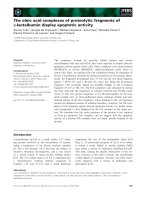
Fig. 1. Acidic or alkaline pH induced Ca
2+
release from BI-1 recon-
stituted into lipid vesicles. (A) The change in the fluorescence inten-
sity of indo-1 was recorded kinetically after the rapid dilution of
proteoliposomes into an acidic solution (pH 5.5). (B) The amounts
of Ca
2+
released by acidic or alkaline pH were expressed as a per-
centage of the maximum releasable Ca
2+
using the fluorescence
and the radioactivity of
45
Ca
2+
, and were compared with those
caused by the addition of 1% Triton X-100 (TX-100), which was set
to 100%. (C)pH 5.0 and (C)pH 9.0 represent the Ca
2+
release at pH
5.0 and 9.0 in the absence of reconstituted BI-1, respectively. Data
shown are the mean ± SE of five independent experiments.
Ca
2+
⁄ H
+
antiporter activity of Bax inhibitor-1 T. Ahn et al.
2286 FEBS Journal 276 (2009) 2285–2291 ª 2009 The Authors Journal compilation ª 2009 FEBS
to 5 lm (Fig. 2). It has been suggested that a biphasic
mode of external Ca
2+
on the channel activity of the
InsP
3
receptor is important in generating characteristic
Ca
2+
signaling within cells [12–15]. Moreover,
although the effect of external Ca
2+
differs according
to the type of InsP
3
receptor, Ca
2+
efflux exhibits a
biphasic pattern, with a maximal peak near
0.2)0.3 lm of cytoplasmic Ca
2+
[11,12]. Therefore, the
mode of regulation by external Ca
2+
suggests that
BI-1 shows Ca
2+
signaling different from that of the
conventional InsP
3
receptor, even though the other
regulation modes were not experimentally compared.
Proton influx into proteoliposomes by exchange
with Ca
2+
efflux
To obtain more insight into the functional influence of
proton ions on the PICR and to test the possibility
that BI-1 incorporated into membranes might exhibit a
Ca
2+
⁄ H
+
antiporter-like function, we measured the
changes in emission fluorescence for encapsulated oxo-
nol V (a pH-sensitive and non-permeable fluorescent
probe), which was entrapped in membranes in the
presence of internalized Ca
2+
, as described previously
[16]. Figure 3A shows the time course of the
decrease in fluorescence of the probe upon mixing
proteoliposomes with acidic solutions. Relaxation
times were calculated to be in the range of 32)45 s,
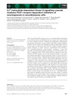
Fig. 2. Effect of external Ca
2+
on PICR of reconstituted BI-1. The
effects of external Ca
2+
on the PICR were plotted as external Ca
2+
concentration versus the amount of
45
Ca
2+
released from proteo-
liposomes. The external Ca
2+
concentration was buffered by the
EGTA–Ca
2+
chelating system and the experiment was performed
using the method described in Fig. 1. Inset: the radioactivity by
45
Ca
2+
efflux was normalized to 100% upon the absence of exter-
nal Ca
2+
and expressed as a relative percentage.
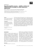
Fig. 3. Proton influx into proteoliposomes. (A) The time course for
the decrease in fluorescence of oxonol V encapsulated into proteo-
liposomes was measured. After rapid mixing of proteolipsomes
containing internalized Ca
2+
with acidic solutions, the emission fluo-
rescence at 630 nm (excitation at 610 nm) was measured as a
function of time. Lines a, b, c and d indicate an exterior pH of 6.5,
6.0, 5.5 and 5.0, respectively. Line e represents the emission fluo-
rescence in the absence of entrapped Ca
2+
. (B) The amount of pro-
ton uptake into liposomes as a result of the decrease in pH was
assayed by measuring the radioactivity of tritium, as described in
Materials and methods (In the figure, the y-axis represents the
cpm values of pellet). Black and hatched bars represent the radio-
activities of liposomes consisting of pure phospholipids without
reconstituted BI-1 protein and the cpm values of proteoliposomes
without internalized Ca
2+
, respectively, after the same procedure
described above was performed. White and gray bars represent
the cpm values of proteoliposomes after mixing the vesicles with
pH 7.4 and each indicated acidic solution, respectively.
T. Ahn et al. Ca
2+
⁄ H
+
antiporter activity of Bax inhibitor-1
FEBS Journal 276 (2009) 2285–2291 ª 2009 The Authors Journal compilation ª 2009 FEBS 2287
which were very similar to the rate of Ca
2+
release.
However, no correlation between the relaxation time
and the acidity was apparent. As a control experiment,
the change in fluorescence was also analyzed under the
conditions described above, without encapsulated
Ca
2+
. In this case, we did not observe any significant
decrease in the emission fluorescence of oxonol V
(Fig. 3A, line e). This indicates the importance of
entrapped Ca
2+
for the uptake of protons into proteo-
liposomes. However, the experiment was not per-
formed in the presence of various concentrations of
internalized Ca
2+
. Therefore, it is not, at present, pos-
sible to determine how many proton ions are moved
into liposomes in response to entrapped Ca
2+
ions.
Proton influx was also ascertained by measuring the
radioactivity of proteoliposomes containing encapsu-
lated Ca
2+
(Fig. 3B). When the proteoliposomes were
suspended in the indicated acidic buffer solutions
containing [
3
H], the radioactivity inside the proteolipo-
somes increased with decreasing external pH, indica-
tive of a pH-dependent proton uptake into the
proteoliposomes. However, at pH 5.0, the radioactivity
of the proteoliposomes decreased, compared with the
radioactivity measured at pH 5.5, which was not con-
sistent with the results obtained using oxonol V. As
expected, without reconstituted BI-1, the remarkable
proton influx could not be observed, regardless of the
external acidity. By contrast, significant tritium accu-
mulations were shown with proteoliposomes in the
absence of internal Ca
2+
, which is different from the
change in oxonol V-mediated fluorescence described in
Fig. 3A. Furthermore, the cpm values increased with
increasing acidity. This may be related to the finding
that the C-terminal region of BI-1, which contains a
cluster of charged residues, EKDKKKEKK, is impor-
tant in the protein function as a pH sensor [9], similar
to observations made for the TREK-1 potassium chan-
nel [17]. Therefore, this cytoplasmic motif may bind to
proton ions and result in the increased cpm values (as
shown in Fig. 3B), without proton uptake into proteo-
liposomes in the absence of internal Ca
2+
. However,
at present, there is no direct evidence for the associa-
tion of BI-1 with proton ions. Taken together, these
results suggest that encapsulated Ca
2+
facilitates
proton movement into proteoliposomes.
As a control experiment, liposomes without reconsti-
tuted BI-1 protein displayed background levels of
radioactivity, suggesting that proton uptake was not
induced by differences in the concentration of proton
ions between the interior and exterior of proteolipo-
somes per se (black bars in Fig. 3B). Collectively, these
results support the possible role of reconstituted BI-1
as a Ca
2+
⁄ H
+
antiporter.
Proteolytic cleavage of reconstituted BI-1
On the basis of the result described in Fig. 3, it was
considered that the proton influx associated with Ca
2+
efflux resulted from the membrane topology of BI-1,
because recombinant BI-1 proteins could be incorpo-
rated into artificial lipid bilayers with different confor-
mations during proteoliposome formulation. To test
this possibility and to obtain insight into the confor-
mation of reconstituted BI-1, proteolytic cleavage was
performed with the prepared proteoliposomes using
carboxypeptidase B, which was added externally. The
protease preferentially catalyzes the hydrolysis of the
basic amino acids lysine, arginine and ornithine from
the C-terminal end of polypeptides. After cleavage,
SDS ⁄ PAGE analysis revealed that nearly all the recon-
stituted BI-1 was partially digested by the protease and
intact BI-1 could not be observed (Fig. 4). This
suggests that most recombinant BI-1 proteins have a
protease-accessible structure in membranes during the
reconstitution procedure although the precise confor-
mation of BI-1 in a lipid bilayer is not currently
known. In relation to the membrane topology of BI-1,
previous observations also support that the C-terminus
of BI-1 is outside the ER [18,19]. After proteolytic
digestion, the PICR was examined with the resulting
proteoliposomes. However, we could not detect any
remarkable Ca
2+
release and proton influx (results not
shown). Considering the proteolytic pattern on
SDS ⁄ PAGE, these results imply that the C-terminal
region of BI-1 recognizing external acidity may be
exposed to the outer surface of proteoliposomes, where
they can be cleaved by the protease.
In terms of the BI-1-induced protective function,
the interplay of Ca
2+
and H
+
needs to be carefully
Fig. 4. Proteolytic cleavage of proteoliposomes. Reconstituted BI-1
was cleaved by carboxypeptide B and the products were analyzed
by SDS ⁄ PAGE. Lanes 1 and 2 represent the reconstituted BI-1 and
the digested sample, respectively.
Ca
2+
⁄ H
+
antiporter activity of Bax inhibitor-1 T. Ahn et al.
2288 FEBS Journal 276 (2009) 2285–2291 ª 2009 The Authors Journal compilation ª 2009 FEBS
interpreted. At normal pH, BI-1 has been suggested to
leak Ca
2+
, leading to a decrease in the Ca
2+
concen-
tration in the ER [9,20]. In this study, a unique func-
tion for the BI-1–Ca
2+
⁄ H
+
antiporter is suggested. At
normal pH, Ca
2+
homeostasis may be maintained
even in the presence of other stimuli such as ER stress,
because of Ca
2+
porter activity. In view of pH homeo-
stasis, proton uptake may be accelerated by a
Ca
2+
⁄ H
+
antiporter-like function of BI-1, leading to a
cell-protective function. Therefore, it may be antici-
pated that the antiporter activity of BI-1 contributes to
adaptation of the channel protein and ⁄ or ER Ca
2+
regulation. However, in exceptional surroundings, such
as at severely acidic pH, BI-1-overexpressed cells may
be highly sensitized to cell death, consistent with an
in vivo cell-based study [9]. More detailed studies are
needed to establish the functional relevance of BI-1
antiporter activity to cell death.
Conclusion
Proton ions induce Ca
2+
efflux from BI-1-reconsti-
tuted liposomes containing entrapped Ca
2+
, and con-
comitantly proton influx. Taking into account that the
movement of these ions was not observed without
encapsulated Ca
2+
and acidity, our results suggest that
the reconstituted recombinant BI-1 has a Ca
2+
⁄ H
+
antiporter-like function.
Materials and methods
All phospholipids were purchased from Avanti Polar Lipids
(Alabaster, AL, USA). The fluorescent Ca
2+
indicator,
indo-1, and the pH-sensitive dye, oxonol V, were purchased
from Invitrogen (Carlsbad, CA, USA). Radioactive materi-
als
45
Ca
2+
and
3
H were obtained from GE Healthcare Bio-
Sciences (Piscataway, NJ, USA).
Construction of Bax inhibitor-1 expression
plasmid
Human BI-1 cDNA was subcloned into the pET12a expres-
sion vector to generate pET12a ⁄ BI-1 using PCR. PCR
amplification was designed to include BamHI and HindIII
restriction enzyme sites for forward and reverse primer oli-
gonucleotides, respectively (forward: 5¢-GACGGATCCAT
GAACATATTTGATCGAAAG-3¢, reverse: 5¢-GACAAGC
TTTTATTTCTTCTCTTTCTTCTT-3¢). PCR was per-
formed with Pfu polymerase (Stratagene, La Jolla, CA,
USA). The mixture was preincubated for 5 min at 94 °C
before the addition of the polymerase, followed by amplifi-
cation for 30 cycles: 94 °C for 90 s, 60 °C for 90 s and
72 °C for 3 min. The resulting PCR product was purified,
digested with BamHI and HindIII, and then ligated into a
pET-12a vector treated with the same restriction enzymes.
The nucleotide sequence of the entire region including the
BI-1 gene was analyzed by dideoxy sequencing.
Expression of recombinant BI-1 protein
Cultures of Escherichia coli BL21(DE3) containing
pET12a ⁄ BI-1 were grown at 37 °C in 500 mL of Luria–Ber-
tani ⁄ ampicillin (50 lgÆmL
)1
) until an attenuance at 600 nm
of 0.5 was attained. Induction of the recombinant protein
was carried out by the addition of 0.5 mm isopropyl-b-d-
thiogalactopyranoside and further incubation for 4 h. To
obtain inclusion bodies of expressed BI-1, harvested bacte-
rial pellets were resuspended in lysis buffer consisting of
25 mm Tris ⁄ HCl (pH 8.0), 100 mm NaCl, 1 mm phen-
ylmethanesulfonyl fluoride and 5 lgÆmL
)1
of benzamidine,
leupeptin and pepstatin. The cells were lysed by passage
through an Amicon French pressure cell (Millipore, Billeri-
ca, MA, USA) and the lysates were centrifuged (12 000 g,
10 min, 4 °C) to recover the inclusion bodies. The pellets
were resuspended with 5 mL of the lysis buffer using probe
sonication and were centrifuged (12 000 g, 10 min, 4 °C).
The resuspension and centrifugation steps were repeated
five times. The final inclusion body-containing pellet was
dissolved in buffer A, which consisted of 20 mm Hepes (pH
7.4), 8 m urea, 0.1 mm dithiothreitol, 1 mm CaCl
2
and
1.5% Chaps by incubating the sample at 25 °C for 10 min
with gentle shaking.
Refolding and reconstitution of BI-1 inclusion
bodies into proteoliposomes
Chloroform solutions of lipids were stored in sealed amp-
ules under argon gas at )20 °C. Phosphatidylcholine, phos-
phatidylethanolamine and phosphatidylserine (all from
bovine brain) dissolved in chloroform were mixed to give a
respective molar ratio of 5 : 3 : 2. The phospholipid con-
centrations were determined using a phosphorus assay [21].
A phospholipid concentration of 5 mm was used to recon-
stitute the BI-1 protein. The solvent was evaporated under
a stream of argon gas and the residual chloroform was
removed by centrifugal lyophilization. The dry lipids were
hydrated with 1 mL of buffer A containing 20 lg BI-1.
The mixtures were dialyzed for 12 h against an excess vol-
ume of buffer B (buffer A without Chaps) containing 2 m
urea. The dialysis step was repeated for 12 h against an
excess volume of buffer C (buffer B without urea). The
resulting proteoliposomes were pelleted by centrifugation at
100 000 g for 30 min at 4 °C and washed with buffer D
(20 mm Hepes, pH 7.4, 100 mm NaCl, 0.1 mm dithiothrei-
tol, 0.5 mm EGTA, 1 m KCl) and then were dialyzed
against buffer E (buffer D without 1 m KCl and 0.5 mm
EGTA) for 12 h at 4 °C. The resulting proteoliposomes
T. Ahn et al. Ca
2+
⁄ H
+
antiporter activity of Bax inhibitor-1
FEBS Journal 276 (2009) 2285–2291 ª 2009 The Authors Journal compilation ª 2009 FEBS 2289
were passed through Chelex 100 (Bio-Rad, Hercules, CA,
USA) to remove free Ca
2+
. The formation of proteolipo-
somes was monitored spectrofluorometrically by measure-
ment of light scattering during dialysis at a wavelength of
450 nm. The amounts of reconstituted BI-1 protein were
determined using the NanoOrangeÒ protein quantitation
kit (Invitrogen).
Hydrogen ion-mediated Ca
2+
release from
proteoliposomes using indo-1 fluorescence and
45
Ca
2+
Ca
2+
release from the proteoliposomes was measured as
described previously [4]. Briefly, Ca
2+
efflux was observed
by measuring the fluorescence changes of external indo-1
(5 lm) after rapid dilution of the proteoliposomes with
acidic solutions (50 mm sodium phosphate pH 6.5 or
50 mm sodium citrate pH 6.0 and 5.5) at a ratio of 1 : 20
(v ⁄ v). The fluorescence intensity was measured at emission
and excitation wavelengths of 393 and 355 nm, respectively.
To quantify the proton ion-mediated release of Ca
2+
from
proteoliposomes, the acidity-induced fluorescence intensity
of indo-1 was compared with the fluorescence intensity
after addition of Triton X-100 to a final concentration of
1% (v ⁄ v). Proteoliposomes were also prepared in the pres-
ence of
45
Ca
2+
to include 20 000 cpm of
45
Ca
2+
under
the same conditions. After the dilution of proteoliposomes
with acidic solutions to induce Ca
2+
efflux, the sample was
centrifuged (100 000 g, 30 min, 30 °C). The amount of pro-
ton-mediated Ca
2+
release was determined by the radioac-
tivity of the pellet and supernatant using scintillation
counting.
Measurement of proton ion influx into
proteoliposome
To analyze proton influx into proteoliposomes coupling
Ca
2+
efflux, the pH-sensitive fluorescent probe oxonol V
was encapsulated inside proteoliposomes (pH 7.4) during
membrane formulation in the presence of Ca
2+
, as previ-
ously described [16]. After rapid mixing of the proteolipo-
somes with each of the indicated buffer solutions (pH 9.0,
8.5, 6.5, 6.0, 5.5, and 5.0 as described above), the decrease
in fluorescence was measured at an emission wavelength of
630 nm (excitation at 610 nm). Proton uptake was also
investigated by measuring the tritium radioactivities. Prote-
oliposomes with an interior pH of 7.4, in the presence or
absence of internalized Ca
2+
, were incubated with each
indicated buffer solution containing [
3
H] ( 20 000 cpm)
for 20 min at 30 ° C. The radioactivities of the pellet and
supernatant fractions were measured after centrifugation of
reaction samples using a Beckman TLA 100.2 rotor (Beck-
man Coulter, Palo Alto, CA, USA) at 70 000 rpm for
30 min.
Removal of Ca
2+
contamination
Removal of Ca
2+
contamination was conducted as
described previously [22]. Ca
2+
contamination during all
experiments was checked using the fluorescence of the
Ca
2+
indicator indo-1 before measurements.
Proteolytic cleavage of reconstituted BI-1 with
carboxypeptidase B
After reconstitution of BI-1, proteoliposomes were ultracen-
trifuged as described above. The pellet was suspended in
20 mm Hepes (pH 7.4) containing carboxypeptidase B at a
BI-1 ⁄ carboxypeptidase B ratio of 50 : 1 (w ⁄ w). The sample
was incubated at 25 °C for 20 min and the reaction was ter-
minated by the addition of 1 m m EDTA (final concentra-
tion). The products were analyzed by 12.5% SDS ⁄ PAGE
and followed by Coomassie Brilliant Blue staining of the
resolved proteins.
Acknowledgements
This work was supported by Korea Research Foun-
dation Grant funded by the Korean Government
(KRF-2008-314-C00231) and by Korea Science and
Engineering Foundation Grant (R01-2006-000-10422-0).
References
1 Xu Q & Reed JC (1998) Bax inhibitor-1, a mammalian
apoptosis suppressor identified by functional screening
in yeast. Mol Cell 1, 337–346.
2 Chae HJ, Kim HR, Xu C, Bailly-Maitre B, Krajewska
M, Krajewski S, Banares S, Cui J, Digicaylioglu M, Ke N
et al. (2004) BI-1 regulates an apoptosis pathway linked
to endoplasmic reticulum stress. Mol Cell 15, 355–366.
3 Bailly-Maitre MB, Fondevila C, Kaldas F, Droin N,
Luciano F, Ricci JE, Croxton R, Krajewska M, Zapata
JM, Kupiecweglinski JW et al. (2006) Cytoprotective
gene bi-1 is required for intrinsic protection from endo-
plasmic reticulum stress and ischemia–reperfusion
injury. Proc Natl Acad Sci USA 103, 2809–2814.
4Hu
¨
ckelhoven R (2004) BAX Inhibitor-1, an ancient cell
death suppressor in animals and plants with prokaryotic
relatives. Apoptosis 9 , 299–307.
5 Chae HJ, Ke N, Kim HR, Chen S, Godzik A, Dickman
M & Reed JC (2003) Evolutionarily conserved cytopro-
tection provided by Bax inhibitor-1 homologs from ani-
mals, plants, and yeast. Gene 323, 101–113.
6 Kawai-Yamada M, Ohori Y & Uchimiya H (2004) Dis-
section of Arabidopsis Bax inhibitor-1 suppressing Bax-,
hydrogen peroxide-, and salicylic acid-induced cell
death. Plant Cell 16, 21–32.
Ca
2+
⁄ H
+
antiporter activity of Bax inhibitor-1 T. Ahn et al.
2290 FEBS Journal 276 (2009) 2285–2291 ª 2009 The Authors Journal compilation ª 2009 FEBS
7 Baek D, Nam J, Koo YD, Kim DH, Lee J, Jeong JC,
Kwak SS, Chung WS, Lim CO, Bahk JD et al. (2004)
Bax-induced cell death of Arabidopsis is meditated
through reactive oxygen-dependent and -independent
processes. Plant Mol Biol 56, 15–27.
8 Lee GH, Kim HK, Chae SW, Kim DS, Ha KC,
Cuddy M, Kress C, Reed JC, Kim HR & Chae HJ
(2007) Bax inhibitor-1 regulates endoplasmic reticulum
stress-associated reactive oxygen species and heme
oxygenase-1 expression. J Biol Chem 282, 21618–
21628.
9 Kim HR, Lee GH, Ha KC, Ahn T, Moon JY, Lee BJ,
Cho SG, Kim S, Seo YR, Shin YJ et al. (2008) Bax
inhibitor-1 is a pH-dependent regulator of Ca
2+
chan-
nel activity in the endoplasmic reticulum. J Biol Chem
283, 15946–15955.
10 Taylor CW & Laude AJ (2002) IP
3
receptors and their
regulation by calmodulin and cytosolic Ca
2+
. Cell Cal-
cium 32, 321–334.
11 Miyakawa T, Maeda A, Yamazawa T, Hirose K, Kuro-
saki T & Iino M (1999) Encoding of Ca
2+
signals by
differential expression of IP
3
receptor subtypes. EMBO
J 18, 1303–1308.
12 Bezprozvanny I, Watras J & Ehrlich BE (1991)
Bell-shaped calcium-response curve of Ins(1,4,5)P
3
- and
calcium-gated channels from endoplasmic reticulum of
cerebellum. Nature 351, 751–754.
13 Finch EA, Turner TJ & Goldin SM (1991) Calcium as
a coagonist of inositol 1,4,5-trisphosphate-induced
calcium release. Science 252, 443–446.
14 Thomas AP, Bird GS, Hajnoczky G, Robb-Gaspers LD
& Putney JW Jr (1996) Spatial and temporal aspects of
cellular calcium signaling. FASEB J 10, 1505–1517.
15 Berridge MJ (1998) Neuronal calcium signaling. Neuron
21, 13–26.
16 Yoo SH & Jeon CJ (2000) Inositol 1,4,5-trisphosphate
receptor ⁄ Ca
2+
channel modulatory role of chromogra-
nin A, a Ca
2+
storage protein of secretory granules.
J Biol Chem 275, 15067–15073.
17 Kloda A, Ghazi A & Martinac B (2006) C-terminal
charged cluster of MscL, RKKEE, functions as a pH
sensor. Biophys J 90, 1992–1998.
18 Bolduc N, Ouellet M, Pitte F & Brisson LF (2003)
Molecular characterization of two plant BI-1 homo-
logues which suppress Bax-induced apoptosis in human
292 cells. Planta 216, 377–386.
19 Shikano S & Li M (2003) Membrane receptor traffick-
ing: Evidence of proximal and distal zones conferred by
two independent endoplasmic reticulum localization sig-
nals. Proc Natl Acad Sci USA 100, 5783–5788.
20 Xu C, Xu W, Palmer AE & Reed JC (2008) BI-1 regu-
lates endoplasmic reticulum Ca
2+
homeostasis down-
stream of Bcl-2 family proteins. J Biol Chem 283,
11477–11484.
21 Vaskovsky VE, Kostetsky EY & Vasendin IM (1975) A
universal reagent for phospholipid analysis. J Chroma-
togr 114, 129–141.
22 Meyer T, Wensel T & Stryer L (1990) Kinetics of cal-
cium channel opening by inositol 1,4,5-trisphosphate.
Biochemistry 29, 32–37.
T. Ahn et al. Ca
2+
⁄ H
+
antiporter activity of Bax inhibitor-1
FEBS Journal 276 (2009) 2285–2291 ª 2009 The Authors Journal compilation ª 2009 FEBS 2291