Báo cáo khoa học: Piecing together the structure of retroviral integrase, an important target in AIDS therapy pptx
Bạn đang xem bản rút gọn của tài liệu. Xem và tải ngay bản đầy đủ của tài liệu tại đây (1.26 MB, 21 trang )
REVIEW ARTICLE
Piecing together the structure of retroviral integrase,
an important target in AIDS therapy
Mariusz Jaskolski
1,2
, Jerry N. Alexandratos
3
, Grzegorz Bujacz
2,4
and Alexander Wlodawer
3
1 Department of Crystallography, Faculty of Chemistry, A. Mickiewicz University, Poznan, Poland
2 Center for Biocrystallographic Research, Institute of Bioorganic Chemistry, Polish Academy of Sciences, Poznan, Poland
3 Macromolecular Crystallography Laboratory, National Cancer Institute at Frederick, MD, USA
4 Institute of Technical Biochemistry, Technical University of Lodz, Poland
Although the existence of retroviruses and their ability
to cause diseases have been known for almost a cen-
tury [1], it was the emergence of AIDS in the early
1980s that provided a huge impetus to structural
studies of their protein and nucleic acid components.
Retroviruses, most notably HIV-1, are enveloped in a
glycoprotein coat and lack the high degree of internal
and external symmetry that makes it possible to crys-
tallize many relatively simple viruses, such as picornav-
iruses, exemplified by the viruses that cause common
cold and polio. It is thus unlikely that high-resolution
information about the structural organization of intact
retroviruses could be obtained with the currently avail-
able methods such as crystallography, although
Keywords
AIDS; antiretroviral drugs; DNA integration;
HIV; integrase
Correspondence
A. Wlodawer, Macromolecular
Crystallography Laboratory, National Cancer
Institute at Frederick, Frederick, MD 21702,
USA
Fax: +1 301 846 6322
Tel: +1 301 846 5036
E-mail:
Note
This review is dedicated to David Eisenberg
on the occasion of his 70th birthday.
(Received 13 January 2009, revised 17
February 2009, accepted 17 March 2009)
doi:10.1111/j.1742-4658.2009.07009.x
Integrase (IN) is one of only three enzymes encoded in the genomes of all
retroviruses, and is the one least characterized in structural terms. IN cata-
lyzes processing of the ends of a DNA copy of the retroviral genome and
its concerted insertion into the chromosome of the host cell. The protein
consists of three domains, the central catalytic core domain flanked by the
N-terminal and C-terminal domains, the latter being involved in DNA
binding. Although the Protein Data Bank contains a number of NMR
structures of the N-terminal and C-terminal domains of HIV-1 and HIV-2,
simian immunodeficiency virus and avian sarcoma virus IN, as well as
X-ray structures of the core domain of HIV-1, avian sarcoma virus and
foamy virus IN, plus several models of two-domain constructs, no structure
of the complete molecule of retroviral IN has been solved to date.
Although no experimental structures of IN complexed with the DNA sub-
strates are at hand, the catalytic mechanism of IN is well understood by
analogy with other nucleotidyl transferases, and a variety of models of the
oligomeric integration complexes have been proposed. In this review, we
present the current state of knowledge resulting from structural studies of
IN from several retroviruses. We also attempt to reconcile the differences
between the reported structures, and discuss the relationship between
the structure and function of this enzyme, which is an important, although
so far rather poorly exploited, target for designing drugs against HIV-1
infection.
Abbreviations
ASV, avian sarcoma virus; CCD, catalytic core domain; 5-CITEP, 1-(5-chloroindol-3-yl)-3-hydroxy-3-(2H-tetrazol-5-yl)-propenone; CTD,
C-terminal domain; FDA, US Food and Drug Administration; IBD, integrase-binding domain; IN, integrase; LEDGF, lens epithelium-derived
growth factor; NTD, N-terminal domain; PFV, prototype foamy virus; PIC, preintegration complex; PR, protease; RT, reverse transcriptase;
SIV, simian immunodeficiency virus; Y-3, 4-acetylamino-5-hydroxynaphthalene-2,7-disulfonic acid.
2926 FEBS Journal 276 (2009) 2926–2946 Journal compilation ª 2009 FEBS. No claim to original US government works
significant progress in lower-resolution studies by elec-
tron microscopy has given us excellent ideas about
global aspects of their structure [2].
A typical retrovirus such as HIV-1 has been
described as ‘Fifteen proteins and an RNA’ [3]. Three
of these proteins are enzymes that are retrovirus-spe-
cific and are encoded by all retroviral genomes [4],
although additional enzymes are found in some retro-
viruses. The structures of two of these enzymes, prote-
ase (PR) [5] and reverse transcriptase (RT) [6,7], have
been investigated in extensive detail during the last
20 years, using crystallography and NMR spectros-
copy. A very large number of such structures, solved
for both full-length apoenzymes and for complexes
with substrates, products, effectors, and inhibitors,
have been published [8–13]. The detailed structural
knowledge, based on low-resolution to medium-resolu-
tion structures of RT and medium-resolution to
atomic-resolution structures of PR, has been of consid-
erable use in the design of clinically relevant inhibitors
of these enzymes [13,14]. At this time, 18 nucleoside
and non-nucleoside inhibitors of RT, as well as 10
inhibitors of PR, have been approved by the US Food
and Drug Administration (FDA) for the treatment of
AIDS. By contrast, far less is known structurally about
the third retroviral enzyme, integrase (IN), and fewer
inhibitors of IN have been discovered so far. Only one
of them, raltegravir, has recently gained FDA approval
as an AIDS drug [15].
Although many anti-HIV drugs are already avail-
able, serious side effects and the emergence of drug-
resistant mutations necessitate the development of
novel compounds. The current drugs targeting RT and
PR are not without side effects. Significant side effects
include myopathy, hepatic steatitis, and lipodystrophy,
caused by anti-RT drugs alone, or a combination of
anti-RT and anti-PR drugs. Anti-RT drugs block sev-
eral mitochondrial proteins (DNA polymerase c,
uncoupling proteins), whereas anti-PR drugs such as
amprenavir or indinavir block the mechanistically
unrelated enzyme, mitochondrial processing PR [16].
Inhibitors of IN appear to be particularly promising
[17–19], because, unlike PR and RT, this enzyme does
not have direct human homologs. Although such
inhibitors might still affect the function of other
enzymes, such as RAG1 ⁄ 2 recombinase [20], they have
not as yet been shown to cause pathological effects.
Drugs against IN might be given in higher, more effec-
tive doses with better-tolerated side effects. The inhibi-
tors ⁄ drugs currently in animal experimental or human
clinical trials seem to be fulfilling this promise, having,
in the short term, fewer side effects than FDA-
approved anti-PR or anti-RT drugs. In consequence,
drugs targeting IN may be given in sufficiently high
doses to fully block the enzyme from integrating viral
DNA into the cell genome, thus allowing the host
immune system to fight off the infection completely.
Whereas HIV-1 IN is clearly the most medically
relevant IN, and has been extensively investigated for
over two decades, the enzyme encoded by avian
sarcoma virus (ASV) was studied much earlier [21]. In
addition, enzymes from other retroviruses, including
HIV-2, simian immunodeficiency virus (SIV), proto-
type foamy virus (PFV), Mason–Pfizer monkey virus,
and feline immunodeficiency virus, have been investi-
gated as well. Although a significant amount of work
has been performed with feline immunodeficiency virus
[22], it will not be further discussed here, as no crystals
have been obtained. Similarly, we will not discuss
Mason–Pfizer monkey virus IN further [23], as we are
not aware of any advanced structural studies involving
this protein.
As will be discussed later, no crystal structure of
full-length IN is available at this time. However, many
structures of fragments of this enzyme from several
different viral sources have been solved by crystallog-
raphy and NMR in the last 15 years (Table S1),
including several important structures that have
appeared since the last comprehensive review of this
subject was published [24]. These data will be discussed
below.
Functional properties of retroviral INs
In the present review, we focus predominantly on the
structural aspects of retroviral INs and not on the
enzymatic mechanism and other functional features of
these enzymes, which have been extensively reviewed
elsewhere [24–27]. However, a short introduction to
the basics of IN function is necessary to properly inter-
pret the importance of various structural features.
The retroviral genomic RNA is reverse transcribed
into a DNA copy by the previously mentioned retro-
viral enzyme, RT. The function of IN is to insert the
resulting viral DNA into the host genome, with the
reaction being accomplished in two distinct steps
(Fig. 1), both catalyzed by a triad of acidic residues in
a characteristic D,D(35)E motif (two aspartates and a
glutamate, the latter separated from the second aspar-
tate by 35 residues), found in all retroviral INs. In the
first processing step, IN removes the two terminal
nucleotides (GT in HIV-1, and TT in ASV) from each
3¢-end of the double-stranded viral DNA. The second
step, called ‘joining’ or ‘strand transfer’, involves a
nucleophilic attack by the free 3¢-hydroxyl of the viral
DNA on the target chromosomal DNA, resulting in
M. Jaskolski et al. Integrase – a target for AIDS therapy
FEBS Journal 276 (2009) 2926–2946 Journal compilation ª 2009 FEBS. No claim to original US government works 2927
covalent joining of the two molecules. If the reaction is
performed in a concerted manner, the second, coordi-
nated insertion is made into the complementary strand
of the target DNA, in a position five nucleotides away
from the site of the first insertion (in HIV and SIV; six
nucleotides in ASV). The subsequent removal of the
two unpaired nucleotides at each 5¢-overhanging end
of the viral DNA and filling of the gaps are most likely
performed by host enzymes.
Although the reactions described above require only
the viral and host DNA substrates and divalent metal
cofactors used by the IN during the catalytic mecha-
nism (physiologically Mg
2+
, but, in vitro, could also
be Mn
2+
), more components are included in the prein-
tegration complex (PIC), which is necessary for the
integration to take place in the nucleus [28,29]. PICs of
HIV-1 have been shown to also contain viral RT and
matrix proteins, as well as a number of host proteins.
One of the latter proteins, called barrier-to-autointe-
gration factor, appears to be crucial in preventing
autointegration (integration of viral DNA into viral
DNA) [30,31]. Whereas the structure of barrier-to-
autointegration factor complexed to DNA is known
[32], its mode of binding to IN (if any) is not. The only
cellular factor that has been shown experimentally to
bind directly to IN is lens epithelium-derived growth
factor (LEDGF), also known as PC4 and SFRS1
interacting protein 1 or transcriptional coactivator p75
[33–36]. Structural aspects of its interactions will be
discussed below. However, identification of all proteins
that participate in creating PICs and assignment of
their role is still not complete.
The amino acid sequence and domain
structure of retroviral INs
A single polypeptide chain of most retroviral INs com-
prises 290 residues and consists of three clearly iden-
tifiable domains [37], as well as interdomain linkers.
However, some important variations are present. For
example, PFV IN is significantly longer, comprising
392 residues, and ASV IN is encoded as a 323 amino
acid protein that is post-translationally processed to
the final polypeptide consisting of 286 residues, which
is fully enzymatically active [38]. It must be stressed,
however, that definition of the domain boundaries is,
to a certain extent, arbitrary, because of the differences
in the lengths of the linking sequences, as well as diffi-
culties in assignment of the residues at the borders
between the domains and the linkers. As shown in
Fig. 2, the N-terminal domain (NTD) of HIV-1 IN
contains residues 1–46, followed by a linker consisting
of residues 47–55. The catalytic core domain (CCD)
contains residues 56–202, and is followed by a linking
sequence comprising residues 203–219. Finally, the
C-terminal domain (CTD) contains residues 220–288.
The residue numbers at domain boundaries for
enzymes from HIV-2 and SIV are approximately the
same, whereas they differ for ASV IN (Fig. 2). For
PFV IN, a possibility exists that an additional domain
A
B
C
D
E
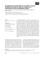
Fig. 1. A schematic representation of the reaction catalyzed by ret-
roviral IN during an infection cycle. This example shows the activity
of HIV-1 IN. The reaction catalyzed by enzymes from other retrovi-
ruses may differ in some details, but the general scheme is the
same. In the processing step (A fi B), the 3¢-ends of viral DNA
(colored molecule) are nicked (arrowheads) before the phosphate
group (diamond) of the conserved terminal GT dinucleotide (colored
beads; A, yellow; C, blue; G, green; T, red), leading to a DNA mole-
cule with a 5¢-overhang and a free 3¢-OH group on each strand. In
the joining step (B fi C), host DNA (black) is nicked with a five-
nucleotide stagger (vertical bars) on the two strands, and the free
3¢-ends of the viral substrate are joined to both host strands, pre-
serving DNA polarity. (D) and (E) are equivalent to (C), and are pre-
sented to illustrate the topology of the final DNA product (not
shown), which is created from molecule E by cellular DNA repair
enzymes, which remove the overhanging viral 5¢-dinucleotides and
seal the gaps on both sides of the integrated viral DNA. In the final
product, the viral insert is flanked by the repeated stagger
sequence, and begins with the conserved TG sequence at each
5¢-end.
Integrase – a target for AIDS therapy M. Jaskolski et al.
2928 FEBS Journal 276 (2009) 2926–2946 Journal compilation ª 2009 FEBS. No claim to original US government works
consisting of approximately 50 residues might be pres-
ent at the N-terminus, preceding the NTD. For practi-
cal reasons, slightly different start and end points have
been utilized for cloning of individual domains and ⁄ or
two-domain constructs that have been used in struc-
tural studies. The structures of representative isolated
domains of IN are shown in Fig. 3.
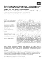
The sequence identity ⁄ similarity percentages for full-
length HIV-1 IN are 58% ⁄ 74% in comparison with
SIV IN, and 23% ⁄ 37% in comparison with ASV IN,
respectively (Fig. 2). These numbers are not completely
accurate, as they depend on the correctness of the
structure-based alignment of IN from different viral
sources. For individual domains, the identity ⁄ similarity
Fig. 2. Amino acid sequence alignment of retroviral INs. The secondary structure of HIV-1 IN is shown below the sequences (a-helices
marked as cylinders, b-strands indicated by arrows). Green: all residues identical; *, metal cation binding. Blue: at least three residues identi-
cal; :, structurally important. Yellow: similar residues; +, DNA binding. Red: active site residues; o, inhibitor binding.
M. Jaskolski et al. Integrase – a target for AIDS therapy
FEBS Journal 276 (2009) 2926–2946 Journal compilation ª 2009 FEBS. No claim to original US government works 2929
percentages are as follows: for the NTD, 55% ⁄ 76% in
comparison with HIV-1 and SIV IN, and 26% ⁄ 46% in
comparison with ASV IN; for the CCD, 61% ⁄ 77%
and 27% ⁄ 46%, respectively; and for the CTD,
53% ⁄ 68% and 14% ⁄ 25%, respectively. Clearly,
sequence conservation is the lowest for the CTD. It
should be stressed that the sequences included in
Fig. 2 are shown for enzymes encoded by specific ret-
roviral strains and that quite significant variations
between different strains have been observed [39]. In
addition, crystallographic studies of some CCDs of IN
or of two-domain constructs were only possible after
the introduction of mutations (see below).
Until now, no reports of crystallization of isolated
NTDs or CTDs have appeared. The first crystals of
the HIV-1 IN CCD [40] were only obtained after an
extensive mutagenesis study, which identified a protein
with an F185K mutation that had enhanced solubility
[41]. A protein with an F185H substitution, corre-
sponding to the structurally equivalent residue present
in ASV IN, was also crystallized [42]. A further muta-
tion, W131E, was introduced to the HIV-1 IN CCD to
enhance solubility even more [43]. The CCD of ASV
IN could be crystallized without mutations, although
special precautions in protein handling were necessary.
The NTD–CCD construct of HIV-1 IN was crystal-
lized using a soluble variant of the protein with the
above-mentioned mutation F185K, as well as with two
additional mutations, W131D and F139D [44]. The
combination of these mutations and use of a specific
buffer allowed the protein concentration to be
increased up to 10 mgÆmL
)1
, and resulted in the
growth of diffraction-quality crystals. The same three
mutations were also used in crystallization of the
CCD–CTD construct of HIV-1 IN, where they were
also introduced with the aim of increasing solubility
[45]. Two additional mutations, C56S and C286S, were
introduced to prevent nonspecific aggregation. How-
ever, the structure of the analogous two-domain con-
struct of SIV IN included only a single mutation,
F185H, implemented to improve protein solubility
[46].
The catalytic domain of IN
The central domain of IN (CCD) contains the com-
plete catalytic apparatus, and exhibits limited activity
even in the absence of the other domains. Although
the CCD by itself does not perform the joining reac-
tion, it does support processing, albeit with decreased
specificity [47]. The CCD also supports a reaction
called ‘disintegration’, in which donor and acceptor
DNA molecules are regenerated from a substrate with
a Y-letter topology [4]. Owing to its importance as the
core of the enzyme and because of the failure to crys-
tallize intact INs, the CCD was the first target for
structural investigation of these proteins.
The structures of the isolated CCDs (Fig. 3B) have
been determined in about three dozen crystallographic
studies of HIV-1 IN [40,42,43,45,48–51], ASV IN [52–
57], and PFV IN [58]. In addition, seven medium-reso-
lution to low-resolution structures of fusion constructs
with one of the terminal domains also included CCDs
of HIV-2 [59] and SIV [45]. As crystals of the ASV IN
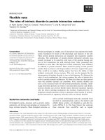
Fig. 3. The structures of the monomers of individual domains of HIV-1 IN. (A) The NTD (blue) with a Zn
2+
(large sphere) coordinated (thin
lines) by an HHCC motif (ball-and-stick) of an HTH fold is represented by the NMR structure 1WJC [75]. (B) The CCD (green), shown with
the D,D(35)E catalytic triad (ball-and-stick), an Mg
2+
(large sphere) coordinated in site I, and the flexible active site loop highlighted in gray, is
represented by the crystal structure 1BL3 [49]. The finger loop (red) extrudes from the body of the protein on the right, between helices a5
and a6 (C-terminus). (C) The CTD (red) is represented by the NMR structure 1IHV [80]. This and all subsequent figures were prepared with
PYMOL [107].
Integrase – a target for AIDS therapy M. Jaskolski et al.
2930 FEBS Journal 276 (2009) 2926–2946 Journal compilation ª 2009 FEBS. No claim to original US government works
CCD were easier to grow, they were studied more
extensively, yielding excellent structural data, such as
the atomic-resolution structure with the Protein Data
Bank code 1CXQ [57]. The CCD has been studied in
its apo-form and in various forms complexed with
metals, including the catalytically competent divalent
cations Mg
2+
and Mn
2+
. Again, ASV IN has pro-
vided a more exhaustive picture of metal coordination
by the CCD, including occupation of multiple metal
sites, or the presence of cations such as Zn
2+
that can
also act as inhibitors of IN activity. Whereas six struc-
tures of small-molecule inhibitor complexes of the
HIV-1 and ASV CCDs have been published [43,51,56],
it has not been possible to elucidate any structure of a
DNA complex, although some promising crystalliza-
tion results have been achieved. In contrast to the
situation concerning the structure of the peripheral
IN domains, no solution structure of the CCD is
available.
The CCD is built around a five-stranded mixed
b-sheet flanked by a-helices (Fig. 3B). The antiparallel
b1–b2–b3 hairpin-type arrangement is extended by two
parallel strands, b4 and b5, which form part of two
b–a–b crossovers, with the intervening helices a1 and
a3, plus a helical turn a2, all located on one side of
the b-sheet. The other side of the b-sheet is covered by
a long helix, a4, which runs across its face. A helix-
turn-helix motif leads to a long stretch of nearly 40
residues that has a helical conformation (a5 and a6),
except for a finger-like extrusion that is formed by
about 12 residues (Phe185–Ala196 in the HIV-1
sequence) in the middle. The finger has a peculiar con-
formation, extending away from the body of the
enzyme (Fig. 3B). Its general conformation is similar
in CCDs from different viruses, although it pivots on
its points of attachment as a semirigid body. Despite
its glycine-rich sequence, the finger is stabilized by con-
served interactions, for example by a salt bridge
(between Arg187 and Glu198 in HIV-1) anchored at
the beginning of helix a6. The finger sequence of the
ASV CCD is the least conserved and, for example, the
above salt bridge is not preserved. The amino acids of
the finger are hydrophilic, in accord with its solvent
exposure in the isolated CCD, except for the extreme
tip, which is occupied by a conserved isoleucine. (The
presence of Glu203 in an equivalent location in the
ASV IN sequence provides another exception in this
regard.) This unusual chemical character of the
exposed tip together with the lattice contacts formed
by the finger loop are most likely responsible for the
variations observed in different crystal structures. The
C-terminal helix a6 of the CCD is truncated in
the PFV IN CCD, and is completely absent in the
construct of an isolated ASV IN CCD used for crystal-
lographic studies [52,57]. However, the finger structure
is clearly seen in the two-domain construct of ASV IN
[60], where Lys199–Thr207 form an insert between
helices a5 and a6. These observations may indicate
that selection of Thr207 as the C-terminal boundary of
the ASV IN CCD on the basis of extensive studies of
many truncation constructs [47] might not represent
the situation in a complete CCD.
The catalytic residues of the D,D(35)E sequence sig-
nature found in all INs are presented by the middle of
chain b1 (Asp64), the loop connecting b4 and a2 (the
second aspartate), and the N-terminal segment of a4
(the glutamate). They are juxtaposed in a row within a
patch of negative charge on the surface of the rather
flat, slab-like molecule. The active site face of the slab
is opposite to the CCD dimerization face, and the two
active sites of the dimeric enzyme are therefore far
apart, nearly as far as the architecture of the dimer
allows. Dimerization of the CCD involves a tandem of
predominantly hydrophobic a1–a5¢ interactions, plus
hydrophobic contacts between helices a6 across the
dimer two-fold axis, and additional hydrophilic con-
tacts in the middle of the dimer. The latter interactions
are interesting because they are connected with the for-
mation of a hydrophilic cavity in the center of the
dimer, filled by a few water molecules.
Whereas the Ca traces of the ASV and HIV-1 CCDs
superpose quite well, the agreement between their
dimers is less optimal and reflects a slight but evident
difference in the dimer architecture. As a consequence
of this difference, the two active sites of the HIV-1 IN
CCD dimer are less distant (38.5 versus 42.5 A
˚
,as
measured by the separation of the catalytic magnesium
ions). The distance between the two active sites is
incommensurate with a 5–6 bp segment of double-heli-
cal B-DNA, and suggests that the host DNA must be
unwound for coordinated processing of the two
strands, or, more likely, that two distinct IN dimers
act each on only one insertion point. Until the struc-
ture of the complete IN enzyme is solved, it can only
be assumed that dimerization of the core domains of
the full-length proteins is not different from what has
been observed for the isolated CCD domains. This
assumption is supported by the consistent picture of
CCD dimerization revealed by all structures of two-
domain IN constructs and of complexes of IN with
LEDGF [35,59].
The CCD of HIV-1 IN used in the first structure
determination (1ITG [40]) contained the F185K muta-
tion introduced to enhance solubility. The cacodylate
residue from the crystallization buffer was found
attached to the cysteine side chains of the protein,
M. Jaskolski et al. Integrase – a target for AIDS therapy
FEBS Journal 276 (2009) 2926–2946 Journal compilation ª 2009 FEBS. No claim to original US government works 2931
including Cys65 located in the active site area [40]. The
constellation of the catalytic amino acids (Asp64,
Asp116, and Glu152) was found to be in an ‘inactive’,
non-native configuration (Fig. 4A). The distortion of
the catalytic apparatus became apparent only later, by
comparison with other, unperturbed, structures, nota-
bly the ASV IN CCD [52,53]. The non-native charac-
ter of the active site is manifested by the altered
conformations of the two aspartates, including a major
reorientation of the loop carrying the Asp116, and by
complete disorder of the helix fragment with the
Glu152 and the entire flexible active site loop in front
of it (13 residues in total, 141–153). It is unlikely that
the distortion of the active site was caused by the pres-
ence of the unnatural arsenic substituent, as in a
related structure of arsenic-free HIV-1 IN (2ITG [42]),
the catalytic aspartates are found in exactly the same
inactive conformation. Although the structure 1ITG
failed to map the functional state of the protein, it
provided the first chain tracing, and was important in
revealing the plasticity of the IN active site and its
ability to adopt different conformations.
Perhaps the most significant consequence of the
inactive conformation of the catalytic residues is the
inability of the two aspartate side chains to bind a cat-
alytic divalent metal cation in a coordinated fashion.
Such a cation, revealed by Mg
2+
and Mn
2+
complexes
of ASV IN [53,54] and later by Mg
2+
complexes of
HIV-1 IN [48,49] and PFV IN [58], has an octahedral
coordination sphere completed by four water mole-
cules (Fig. 4B). The catalytic triad can remain in the
active conformation even in the absence of metal
A
B
Fig. 4. The active site of retroviral INs. The figures show, in stereoview, the three essential amino acids of the D,D(35)E motif in selected,
least-squares-superposed crystallographic structures of the CCD in the (A) unliganded and (B) Mg
2+
-complexed form. The catalytic residues
are shown in the context of the protein secondary structure by which they are contributed, namely an extended b-ribbon (the first aspartate,
middle of figure), a loop (the second aspartate, left), and an a-helix (the glutamate, right). The residue numbering Asp64, Asp116 and Glu152
is for the HIV-1 IN sequence, and corresponds to Asp64, Asp121 and Glu157 in ASV IN. The three divalent metal cation-free active sites
shown in (A) correspond to the first HIV-1 IN structure (1ITG, orange) [40], solved in the presence of arsenic (part of cacodylate buffer),
which reacted with cysteine residues, including one within the active site area (orange sphere), to another medium-resolution structure of
HIV-1 IN (1BI4, molecule C, gray with red oxygen atoms) [49], and to the atomic-resolution structure of ASV IN (1CXQ, green) [57]. Note that
the aspartates in 1ITG have a completely different orientation than in the remaining structures, and the entire Asp116 loop has a different,
non-native conformation. Another symptom of active site disruption in the 1ITG structure is the absence in the model of Glu152, a conse-
quence of disorder in this helical segment. The active sites complexed with the catalytic cofactor Mg
2+
(large sphere) are shown (B) for HIV-1
IN, 1BL3 (molecule C, gray with red oxygen atoms) [49], ASV IN, 1VSD (green) [53], and PFV IN, molecule A of 3DLR (orange) [58]. The
structure of the ASV IN has the highest resolution, and its quality is reflected in the nearly ideal octahedral geometry (thin green lines) of the
Mg
2+
coordination sphere, which, in addition to interactions with the carboxylate groups of both active site aspartates, includes four pre-
cisely defined water molecules. The coordination geometry of the HIV-1 IN complex 1BL3 is significantly distorted. The view direction in
both figures is similar, with a small rotation around the horizontal axis.
Integrase – a target for AIDS therapy M. Jaskolski et al.
2932 FEBS Journal 276 (2009) 2926–2946 Journal compilation ª 2009 FEBS. No claim to original US government works
cations, but then the carboxylate groups are held in
place by water-mediated hydrogen bond bridges (AspÆ-
waterÆAsp64ÆwaterÆGlu). However, as revealed by the
atomic-resolution structures of ASV IN, and in agree-
ment with the requirement for basic conditions for IN
activity (peak endonuclease activity at pH 8.5 [55]),
conformational changes in the active site take place at
pH values below 6 and consist of protonation and a
concomitant swing of the Asp64 carboxylate group out
of its metal-coordinating position, and into a dual-
hydrogen-bond lock with a neighboring asparagine. In
addition, changes of pH influence the flexible active
site loop, which in HIV-1 IN is formed by residues
141–147, adjacent to the glutamate-bearing N-terminus
of helix a4, and which in all the crystal structures
shows a variable degree of disorder. The flexible active
site loop contains highly conserved residues and
appears to be involved directly in substrate contacts
[61].
There is little doubt that the metal-coordination site
formed between the two aspartate side chains (site I)
corresponds to a cation essential for catalysis. The per-
fect octahedral geometry of this site explains why
mutations of the catalytic aspartates cannot be toler-
ated. However, increasingly larger cations can still be
accommodated, from Mg
2+
(mean metal–O distance
2.11 A
˚
), to Mn
2+
(2.23 A
˚
), and even Cd
2+
(2.43 A
˚
)
and Ca
2+
(2.46 A
˚
for incomplete coordination sphere).
Estimation of the metal-binding geometry is more reli-
able from the ASV IN structures, which are in excel-
lent agreement with expected coordination
stereochemistry, for instance with valence parameters
[62] of the central ion, which for the structures listed
in Table S1 are calculated as 1.95 (1VSD), 1.92
(1A5V), or 1.79 (1VSJ), the ideal target being 2.00.
The corresponding values for the HIV-1 IN data indi-
cate a high level of error, e.g. 1.23 ⁄ 0.91 (1BL3) or even
1.08 ⁄ 0.80 ⁄ 0.79 (1QS4), presumably as a consequence
of poor data quality or structure refinement protocols.
There is an important difference between ASV and
HIV-1 IN in coordinating high-electron metals in site
I, connected with the presence of a cysteine at position
65 in the latter enzyme. The thiol group of this residue
is found in the coordination sphere of the cadmium
cations in 1EXQ [45]. As no such possibility exists in
ASV IN, where a phenylalanine immediately follows
the first catalytic aspartate, high-electron metals may
have different impacts on the catalytic properties of
INs from these two viruses. With light metals, such as
Mg
2+
, the thiol group of Cys65 in HIV-1 IN assumes
a totally different orientation, and, consequently, there
is no difference in the coordination chemistry between
ASV IN and HIV-1 IN.
Structural studies of inhibitor
complexes of IN
Structural data on inhibitor complexes of IN are
limited to a few structures of the CCD (Table S1).
The structure of an inhibitor, 1-(5-chloroindol-3-yl)-3-
hydroxy-3-(2H-tetrazol-5-yl)-propenone (5-CITEP)
(Fig. 5A), in complex with the Mg
2+
-containing
HIV-1 IN CCD [43] is the only one that includes a
compound capable of binding within the active site
area of the enzyme. The IC
50
value of 5-CITEP, mea-
sured in a reaction that monitors 3¢-end processing
together with DNA strand transfer, was reported to
be 2.1 lm. This inhibitor was observed in only one of
the three independent copies of the enzyme molecule
present in the crystal. The molecule of 5-CITEP is
located between the coordinated Mg
2+
and the cata-
lytic Glu152, with which it forms hydrogen bonds
(Fig. 5B). The active site of the molecule to which
the inhibitor is bound is located close to the crystallo-
graphic two-fold axis, raising the possibility that the
exact mode of binding might have been influenced by
crystal contacts. The inhibitor makes no direct con-
tacts with either Asp64 or Asp116, and has only an
indirect, water-mediated contact with the bound
Mg
2+
. Two symmetry-related molecules of 5-CITEP
interact directly with each other. In view of these
facts, it is doubtful whether this structure represents
the true mode of binding that would be present in an
IN–DNA complex.
Another IN inhibitor, 4-acetylamino-5-hydroxynaph-
thalene-2,7-disulfonic acid (Y-3) (Fig. 5A), was cocrys-
tallized with the ASV IN CCD in the absence and
presence of Mn
2+
[56]. This aromatic molecule, with
several hydrophilic substituents, does not bind in the
active site of the enzyme but rather on its surface,
where it participates in crystallographic contacts,
although there is no interference with CCD dimeriza-
tion. Its presence in the crystals is, however, not a
crystallographic artefact, as it is observed in the same
context at different pH conditions and regardless of
metal coordination. Although Y-3 undergoes no direct
interactions with the catalytic residues, it does seem to
influence the conformation of the flexible active site
loop by binding to Tyr143 and Lys159 (ASV number-
ing). Y-3 very likely directly interferes with DNA bind-
ing by hydrogen bonding to Lys119, a residue
corresponding to His114 in HIV-1 IN, which has been
shown to be capable of crosslinking to DNA. It is
quite possible that these interactions form the basis of
its inhibitory capacity.
The inhibitors discussed above, as well as
raltegravir (Fig. 5A), the only IN inhibitor approved
M. Jaskolski et al. Integrase – a target for AIDS therapy
FEBS Journal 276 (2009) 2926–2946 Journal compilation ª 2009 FEBS. No claim to original US government works 2933
for clinical use, are aryl diketo acid derivatives that
inhibit strand transfer much more efficiently than
3¢-end processing [63]. Such compounds are charac-
terized by the presence of a and c C=O groups in
the vicinity of a carboxylic acid moiety, although the
latter group can be replaced by a triazole or tetra-
zole ring [64]. No structure of raltegravir complexed
with IN has been published to date, but it is
expected that its mode of binding might involve
direct interactions with the divalent cation(s) present
in the active site.
A different class of inhibitors for which structural
data are available includes arsenic derivatives that were
cocrystallized with HIV-1 IN [51]. Crystal structures
have been solved for tetraphenylarsonium chloride and
3,4-dihydroxyphenyl-triphenylarsonium bromide. Both
compounds bind in a similar fashion at the interface of
the CCD dimer, and interact directly with Gln168 of
one of the molecules. Surprisingly, the quality of the
electron density maps is much better for the former
compound than for the latter, although only the latter
exhibits measurable inhibitory activity for the disinte-
gration reaction (IC
50
of 380 lm).
As IN must form at least a dimer to be catalyti-
cally active, prevention of dimerization offers an
interesting option for its inhibition [65]. Several
studies have reported inhibition of IN activity
through the use of peptides derived from amino acid
sequences responsible for the dimerization of the
CCD [66,67], although no structural data are avail-
able. In some cases, it was possible to confirm that
such peptides disrupted the association–dissociation
equilibrium [68] or the crosslinking of the IN dimer
[69]. On the other hand, Hayouka et al. [70] have
demonstrated that the opposite concept, namely forc-
ing IN to form higher-order oligomers, may be a
useful approach for rendering the IN inactive. Spe-
cifically, they used peptides (called ‘shiftides’),
derived from the cellular IN-binding protein
LEDGF, to inhibit the DNA-binding of IN by shift-
ing the enzyme’s oligomerization equilibrium from
the active dimer towards the tetramer, which,
according to their data, is incapable of catalyzing
the first step of integration, i.e. the 3¢-end
processing.
Development of these and other classes of IN inhibi-
tors is an ongoing process, and some very potent
inhibitors, with IC
50
values in the low nanomolar
range, are now available [71]. The process that led to
the FDA approval of raltegravir, as well as clinical
studies of other drug candidates, have been covered in
a number of recent reviews [72–74]. In view of the pau-
city of available structural data on IN inhibitors, the
wider subject of IN inhibitors in general cannot be
adequately treated within the scope of the current
review.
A
B
Fig. 5. Small-molecule inhibitors of the CCD of retroviral IN. (A)
Chemical diagrams of selected inhibitors discussed in this review.
(B) A dimer of the CCDs (colored silver and gold) of HIV-1 IN
shown in surface representation roughly down its two-fold axis.
The two active sites are marked by the magnesium ions (gray
spheres), with their octahedral coordination spheres formed by the
carboxylates of Asp64 and Asp116, and by four water molecules
(red spheres). Note that the active sites are located in shallow
depressions on the surface of the protein, with the magnesium
ions completely exposed to solvent. Next to the active site, a long
groove runs on the surface of the protein. In this structure, with
the Protein Data Bank code 1QS4 [43], one of the active site
groves is occupied by the 5-CITEP inhibitor, depicted here in ball-
and-stick representations, with C ⁄ N ⁄ O ⁄ Cl atoms shown in orange ⁄
blue ⁄ red ⁄ green. The two active sites are separated by 40.4 A
˚
,as
measured by the distance between the Mg
2+
centers.
Integrase – a target for AIDS therapy M. Jaskolski et al.
2934 FEBS Journal 276 (2009) 2926–2946 Journal compilation ª 2009 FEBS. No claim to original US government works
The NTD of IN
NMR structures of the isolated NTDs were solved for
INs from HIV-1 [75] and HIV-2 [76]. Multiple views of
the NTD are also available in medium-resolution crys-
tal structures of a two-domain construct of HIV-1 IN
that contains the NTD and CCD (1K6Y [44]) and of
the HIV-2 NTD–CCD–LEDGF complex (3F9K [59]).
The solution structure of the HIV-1 IN NTD showed
the existence of dimers consisting of two interconvert-
ing protein forms [75]. The two forms, denoted D
(1WJA) and E (1WJC), were observed together in the
NMR experiment, with the D form being seen mostly
above 300 K, and the E form below that tempera-
ture. A form intermediate between these two was
reported for an H12C mutant of the NTD (1WJE [77]).
The structure of a monomer of the NTD consists
principally of four helices (Fig. 3A). Helix 1 comprises
residues 2–14 in the E form and residues 2–8 in the D
form, helix 2 comprises residues 19–25, helix 3 com-
prises residues 30–39, and helix 4 comprises residues
41–45. The segment beyond residue 46 belongs to the
interdomain linker and is disordered. A Zn
2+
is tetra-
hedrally coordinated by His12, His16, Cys40, and
Cys43, although the details of the interactions with the
histidines differ between forms D and E.
The E form of the NTD is very similar to its coun-
terpart seen in the crystal structure of the two-domain
construct (1K6Y [44]), with an rmsd of 1.05 A
˚
between
molecules A of the models. By comparison, the rmsd
values between molecule A and the other three mole-
cules seen in the crystal range from 0.28 to 0.63 A
˚
.
Form D of the NTD deviates by almost 2 A
˚
from its
crystallographic counterpart. As expected, the interac-
tions of the Zn
2+
with its ligands in the crystal struc-
ture correspond to the structurally closer E form.
The structure of the NTD of HIV-2 IN [78,79] is very
similar to that of its HIV-1 counterpart. A comparison
between molecule A of the first model in the assembly
in 1E0E (no average structure available) and mole-
cule A of 1K6Y shows an rmsd of 0.86 A
˚
, although the
sequence identity between the two proteins is only 55%.
The details of the interactions with Zn
2+
are also
almost identical in the IN NTDs of HIV-1 (E form) and
HIV-2. The rmsd between NTD molecules A and B in
the structure of the HIV-2 IN NTD–CCD–LEDGF
complex (3F9K [59]) is 0.44 A
˚
, whereas the deviation
between NTD molecule A of 3F9K and 1E0E is 1.17 A
˚
.
The CTD of IN
The structure of the isolated CTD of HIV-1 IN (resi-
dues 220–270, the C-terminus truncated) was solved
independently by two groups using NMR (1IHV [80]
and 1QMC [78,81]). In addition, the structures of the
CCD–CTD constructs were determined by X-ray crys-
tallography for ASV IN (1C0M, 1C1A [60]), SIV IN
(1C6V [46]), and HIV-1 IN (1EX4 [45]). The structures
of the CTD show the presence of dimeric molecules
whose subunits were modeled as identical in 1IHV and
as very similar in 1QMC (rmsd 0.34 A
˚
calculated for
model 1, as no average structure is available). The
rmsd between these two structures is 1.2 A
˚
. The devia-
tions between the NMR structures of the isolated
CTD and the crystallographic models of the two-
domain constructs are larger, 1.65 A
˚
between 1IHV
and 1EX4 (both HIV-1 IN), 1.87 A
˚
for 1C6V (SIV
IN), and 2.05 A
˚
for 1C0M (ASV IN). The four CTDs
present in the crystal structure of ASV IN consist of
two very similar pairs (AB and CD, rmsd of
0.15 A
˚
), whereas the rmsd between molecules A and
C is 0.77 A
˚
.
A monomer of the CTD of HIV-1 IN consists of
five b-strands (residues 222–229, 232–245, 248–253,
256–262, and 266–270), arranged in an antiparallel
manner in a b-barrel (Fig. 3C). Eighteen residues that
were not included in the constructs used in the NMR
experiments are also not seen in the X-ray structures
of HIV-1 and SIV IN, and are presumed to be disor-
dered. The topology of the CTD is reminiscent of SH3
domains, which are found in many proteins that inter-
act with either other proteins or with nucleic acids,
although no sequence similarity to SH3 proteins could
be detected.
Two-domain constructs consisting of
the NTD and CCD
Two structures of the NTD–CCD constructs are
available. A 2.4 A
˚
resolution crystal structure of
NTD–CCD of HIV-1 IN offers multiple views, owing
to the presence of four molecules in the asymmetric
unit (1K6Y [44]), paired into AB and CD dimers, in
which the two-fold relationship between the catalytic
domains resembles that of the isolated CCDs. Mole-
cules A and D are very similar (rmsd of 0.43 A
˚
),
whereas molecules B and C are more distant (rmsd of
1.85 A
˚
), mostly owing to small changes in the inter-
domain angles. The interdomain linker region (residues
47–55) is disordered in all molecules, but the authors
have postulated a pattern of domain connectivity
taking into account the presence of NTD–CCD con-
tacts (involving the tip of the finger loop of the CCD
and one side of helix 20–24 in the NTD) and of
NTD–NTD¢ interactions in the dimer that would
M. Jaskolski et al. Integrase – a target for AIDS therapy
FEBS Journal 276 (2009) 2926–2946 Journal compilation ª 2009 FEBS. No claim to original US government works 2935
conserve the symmetry of the CCD–CCD¢ dimer, and
arguing that any other NTD–CCD connection would
be incompatible with the length of the linker (Fig. 4A).
In that interpretation, the distance between the end of
the NTD and the beginning of the CCD is about 9 A
˚
.
However, that view is contradicted by the 3.2 A
˚
resolu-
tion crystal structure of the NTD–CCD construct of
HIV-2 IN (3F9K), in which 24 IN molecules create 12
crystallographically independent dimers, each interact-
ing with a single molecule of LEDGF [59]. Whereas the
connection between the NTD and the CCD is broken
in the electron density map of one of the IN molecules
in each assembly, it is unambiguous in the other one,
forming an extended chain 18 A
˚
in length.
Surprisingly, careful analysis of the 1K6Y structure
allows reconnection of the separated NTDs and CCDs
in all four molecules in exactly the same manner as in
the 3F9K structure (Fig. 6C), by the use of symmetry-
related domains and of NTD–CCD linkers equivalent
to the intact linker from the 3F9K structure. In this
model, which differs significantly from the one origi-
nally proposed [44], the NTD forms a compact struc-
ture with the CCD, using the finger loop of the latter
as a docking site, with a number of hydrogen bond
and electrostatic points of attachment (Fig. 7). To rec-
oncile the two models of NTD–CCD arrangement,
Cherepanov et al. [59] have invoked the mechanism of
3D domain swapping. However, although this is cer-
tainly a possibility, it may be more prudent to con-
clude that the arrangement seen in the 3F9K structure
is the only model that is currently supported by experi-
ment. The relevance of the observed NTD–CCD inter-
actions to the functional properties of IN is not yet
clear.
Two-domain constructs consisting of
the CCD and CTD
The structures of two-domain constructs comprising
the CCD and CTD were solved independently for
HIV-1 IN at 2.8 A
˚
resolution (1EX4 [45]), for SIV IN
at 3.0 A
˚
resolution (1C6V [46]), and for two crystal
forms of ASV IN at 2.5 A
˚
(1C0M [60]) and 3.1 A
˚
(1C1A [60]) resolution. The crystals of HIV-1 IN con-
tain two molecules forming a dimer, although the two-
fold axis relating the CCDs differs from the operation
connecting the CTDs. In each molecule, the two
domains are connected by a long, well-defined helix
comprising residues 195–222. The helix separates the
CCD from the CTD by as much as 30 A
˚
(Fig. 6D).
The two crystal forms of ASV IN contain a single
dimer, or a pair of dimers. Similarly to what was
observed in HIV-1 IN, the symmetry operations
between the two domains of each dimer differ for the
CCD and the CTD. The linker between the CCD and
the CTD comprises residues 213–223 which assume a
completely extended conformation, and not the helical
form observed in HIV-1 IN. Thus, the number of
amino acids forming the linker in ASV IN is much
smaller than in HIV-1 IN, although the distance
between the start and end points of these linkers is not
very different, at least for one of the two crystallo-
graphically independent molecules of ASV IN.
Whereas the crystals of SIV IN also contain two
dimers in the asymmetric unit, only a single CTD
(denoted X) could be traced unambiguously. The chain
connecting it to the CCD could not be traced, and the
authors postulated a connection with chain A of the
catalytic domain [46]. If that were the case, the two
domains would form a fairly compact molecule, with
multiple interdomain contacts. However, an alternative
assignment of the visible CTD to the D chain of CCD
[44] would create an extended two-domain molecule
not unlike that of the other two enzymes, although the
interdomain angles would differ in each of the struc-
tures. In any case, a comparison of the three structures
makes it clear that the arrangement of the domains
shows considerable variability and may be influenced
by other parts of the molecular complex.
Interdomain contacts
One of the measures of the extent of interactions
between the domains of IN (dimerization of identical
domains, and oligomerization of different domains) is
the surface area buried in their interfaces. Calculations
of the buried surface area (in all buried surface calcu-
lations in this article, the reported surface refers to one
interacting protein partner, unless stated otherwise)
have been performed for a representative set of IN
structures (Table S2). The CCD–CCD interactions
extend over a fairly uniform area of about 1000–
1650 A
˚
2
. This area does not depend on the presence of
the linkers, at least with regard to the NTD–CCD lin-
ker (as shown by assigning the linker to either domain,
or removing it altogether for the structure 3F9K). The
most extensive association (largest buried surface area)
characterizes the CCD–CCD dimer of HIV-1 IN
(about 1500 A
˚
2
), and decreases in the order
HIV-1 > HIV-2 ( 1330 A
˚
2
)>SIV( 1250 A
˚
2
)>ASV
( 1080 A
˚
2
) > PFV ( 1000 A
˚
2
).
Homodimeric interactions between the CTDs range
between none to negligible (buried surface area at most
450 A
˚
2
). In most structures, the CTDs in the dimers
of CCD–CTD constructs are far away from each
other, possibly because of the influence of crystal
Integrase – a target for AIDS therapy M. Jaskolski et al.
2936 FEBS Journal 276 (2009) 2926–2946 Journal compilation ª 2009 FEBS. No claim to original US government works
contacts. However, even in the solution dimer of iso-
lated CTD, the area of interaction is very limited
( 330 A
˚
2
).
The interaction of isolated NTDs in solution is
slightly stronger ( 510 A
˚
2
) but still rather insubstan-
tial. In the dimer of the NTD–CCD construct with the
conformation substantiated by the 3F9K structure,
there are, of course, no direct NTD–NTD interactions,
because the NTDs fold back on their respective CCDs,
and thus are completely isolated from each other.
However, even in the model proposed speculatively for
the HIV-1 construct, the area of direct NTD–NTD
interaction is so small (260 A
˚
2
) that it can be safely
neglected.
As the NTD in a multidomain construct folds back
on the CCD, the calculation of the NTD–CCD inter-
action area will depend strongly on the treatment of
the linker peptide (residues 47–55 in the HIV-1 IN
sequence). When this sequence, which is in any case
disordered in most of the structures, is completely
AB
C
D
Fig. 6. Three-dimensional structures of dimeric two-domain constructs of HIV IN determined by X-ray crystallography. (A) In the NTD–CCD
structure (1K6Y [44]), the linker between the domains is disordered, and the speculative NTD (blue)–CCD (green) pairing (dashed line) has
been proposed from indirect reasoning, such as the existence of contacts between the NTDs. (B) Mutual orientation of the NTD (blue) and
CCD (green) as found experimentally in structure 3F9K of HIV-2 IN [59]. The linker, visible in molecule A, is shown in black. (C) This stereo-
view has been constructed by least squares superposition of the CCDs of molecules A (red) and B (orange) from the 2 : 1 complex of HIV-2
IN (3F9K) with the IBD of the LEDGF protein (molecule C, gray) onto the CCDs of molecules A (lemon) and B (green) of HIV-1 IN (1K6Y).
Note that the smaller, all-helical NTDs of the HIV-1 protein are lifted above (in this view, shooting to the right) the CCDs, whereas, in the
model of HIV-2 IN, they ‘fold back’ and adhere to the sides of the CCD dimer. The linkers connecting the NTD and CCD are not present in
any of the experimental models shown in this figure, except in molecule A (red) of 3F9K, for which clear electron density allowed unambigu-
ous connection of the domains. The asymmetric unit of the 1K6Y structure contains another NTD–CCD dimer, here represented in shades
of blue. Note that the blue NTD (of molecule D) superposes exactly on the NTD of molecule B (orange) of the HIV-2 NTD–CCD dimer. This
unexpected match is a strong indication that, with missing experimental evidence, the pairing of the NTD and CCD in the 1K6Y structure
does not correspond to the functional conformation of the protein. The ‘green’ NTD (chain B) and ‘blue’ CCD (chain D) of HIV-1 IN can be
assembled in a fashion similar to the ‘blue’ NTD (chain D)–’green’ CCD (chain B) pairing. By generating a symmetry-related copy of the
‘lemon‘ (chain A) NTD (upper yellow), one can complete the entire NTD–CCD dimer of HIV-1 IN with the blue catalytic cores. Likewise, a
crystallographic copy of the NTD attributed to chain C (bottom yellow), will complete the HIV-1 IN with the green catalytic cores. To guide
the eye in this complicated view, the missing connections between the NTD and CCD have been generated by copying the linker chain from
molecule A of the 3F9K model and grafting it into the remaining molecules (black Ca traces). In this way, four functional HIV NTD–CCD mol-
ecules, assembled into two dimers, have been generated. (D) In the CCD–CTD dimer (1EX4 [45]), the interdomain linker forms a long helix.
Because of different degrees of deformation of this helix, the relative orientation between the CCD (green) and the CTD (red) in the two
monomers is different. All of the NTD–CCD dimers considered in this figure have essentially a common two-fold axis for both domains. This
is not true for the CCD–CTD construct (D), where the two-fold axes relating the CCD and CTD are not identical.
M. Jaskolski et al. Integrase – a target for AIDS therapy
FEBS Journal 276 (2009) 2926–2946 Journal compilation ª 2009 FEBS. No claim to original US government works 2937
omitted, the value of the buried surface area is
530 A
˚
2
. Assigning the linker to the CCD yields a
slightly higher apparent buried surface area
( 670 A
˚
2
), but the linker certainly should not be trea-
ted as part of the NTD, as in that case the buried sur-
face area would be unreasonably large ( 1050 A
˚
2
).
The interaction between the CCD and CTD is very
limited, with buried surface area values falling below
400 A
˚
2
. In the published 1C6V model (SIV IN), the
buried surface area exceeds 600 A
˚
2
, but after a more
plausible reinterpretation of the assignment of the visi-
ble CTD to a CCD, the interaction area drops to
100 A
˚
2
, thus becoming insignificant.
As can be gleaned from these calculations, the sol-
vent-excluded buried areas of the homointeractions
and heterointeractions between the domains of IN are,
with the exception of the CCD–CCD contact, not very
extensive, and their actual values are strongly depen-
dent on the details of the structures used for their cal-
culations, emphasizing once more the flexible nature of
this enzyme. It must be also noted that, despite the
variation of the buried surface area calculated for the
homodimers of the CCDs for INs originating from dif-
ferent viruses, the nature of the interactions is pre-
served. However, no similar consistency is seen for the
homodimers of the other two domains, and the picture
of the interdomain interactions is even less clear.
Binding of IN to cellular protein
partners
Although a number of proteins have been implicated
as putative components of the preintegration complex
with IN [29], the only available structural information
is for complexes of the IN-binding domain (IBD) of
LEDGF with the CCD of HIV-1 IN [35], and with the
NTD–CCD of HIV-2 IN [59]. The IBD used in these
experiments included residues 347–442 of LEDGF.
The complex of LEDGF with the HIV-1 IN CCD con-
sists of two catalytic domains of IN bound to two
IBDs in a fully symmetric fashion. Each IBD interacts
with segments of the two CCDs, the latter forming a
typical dimer, as observed in all other structures of IN
CCDs. The most extensive interactions between IBD
and IN involve a segment including residues 166–171
of molecule A (a connecting peptide between helices
a4 and a5, described as an unusual helix–turn–helix
motif [82]) and bury a surface area of 319 A
˚
2
. The
IBD also interacts with residues belonging to helix a3
(and, to a lesser extent, helix a1) of molecule B (buried
surface area of 379 A
˚
2
). Owing to the symmetry of the
complex, the second IBD interacts with the corre-
sponding areas of molecules B and A of the CCD. The
interactions of the IBD with the CCD in the complex
with the HIV-2 NTD–CCD are virtually identical, with
additional, mostly electrostatic, interactions provided
by the N-terminal helix of NTD, which belongs to
molecule A (buried surface area of 153 A
˚
2
). It is
intriguing, however, that the latter complex, which was
prepared by simultaneous coexpression of the interact-
ing proteins, lacks the second IBD, even though the
superposition of the common IBD in the two struc-
tures is almost exact, and the second binding site is
fully formed, including the same positioning of the
NTD. The importance of the structurally derived inter-
actions between IN and the IBD was verified by exten-
Fig. 7. Stereoview of the NTD docking site
at the ‘finger’ structure of the CCD. The
NTD (blue) is shown with its CCD, as found
in the LEDGF complex of the HIV-2 protein
(Protein Data Bank code 3F9K, chain A) [59].
The ‘finger’ loop (red), which is located
between helices a5 (green) and a6 (light
green) of the CCD, forms five hydrogen
bonds ⁄ salt bridges (broken line) with the
NTD, and another one with the linker pep-
tide (light blue) connecting the domains.
One of those interactions would require a
flip of the side chain amide group of Asn18,
near the entry to an a-helix (upper left) of
the NTD. However, such a flip would create
another impossible NH–HN interaction
(orange) at the N-terminus of this helix.
The tip of the finger loop is occupied by an
isoleucine (green ball-and-stick model).
Integrase – a target for AIDS therapy M. Jaskolski et al.
2938 FEBS Journal 276 (2009) 2926–2946 Journal compilation ª 2009 FEBS. No claim to original US government works
sive mutational studies of the respective interfaces [59].
It was also reported that areas of full-length LEDGF
other than the IBD may be involved in interactions
with IN [83,84].
Oligomeric states of full-length IN and
modeling of its structure
The oligomerization state of IN in vivo is still not
known, but extensive in vitro work has shed light on
this matter. The isolated NTDs, CCDs and CTDs all
remain in solution as dimers, a conclusion that is uni-
formly supported by solution chemistry and structural
biology studies [1]. However, experiments that found
IN–DNA interaction sites by photocrosslinking also
suggested that IN acts as an octamer [85]. Comparison
of simulation analysis against time-resolved fluores-
cence anisotropy measurements of rotation correlation
times could distinguish monomers, dimers, and tetra-
mers, whereas octamers could not be resolved from
higher-order species [86]. At micromolar concentra-
tions, IN exists as tetramers, octamers, and higher-
order aggregates, but such concentration is much
higher than cellular concentration. At catalytic (sub-
micromolar) concentration, these experiments showed
that IN could exist as a monomer, whereas addition of
Zn
2+
stimulated dimer formation. However, the
authors noted that the standard buffer conditions
included detergents, which dissociate IN oligomers
[86]. Solution small-angle X-ray scattering data for
complexes of IN with oligonucleotides also indicated
primarily monomeric species [87], with the same caveat
regarding the possible effects of detergents on the
oligomerization state. If detergent is eliminated from
the purification and assay experiments, IN exhibits
different assembly and catalytic properties.
It must be pointed out that all of the experiments
mentioned above used indirect measurements of the
size of IN oligomers. More direct observations involv-
ing atomic force microscopy of intact IN complexed to
a DNA substrate have shown visually that the size of
these complexes is consistent with a tetramer of IN
molecules [88]. Similar results were obtained by elec-
tron microscopy and single-particle image reconstruc-
tion, which yielded coarse 3D models at 27 A
˚
resolution [89]. The finding of a tetramer as the pre-
dominant feature agrees with several IN–DNA models,
with analysis of IN isolated from nuclear extracts and
its complex with LEDGF [35], and with dynamic light
scattering experiments.
The assembly of HIV-1 IN into oligomers is differ-
ent when it is in complex with Mn
2+
versus Mg
2+
under various in vitro conditions [90]. These experi-
ments did not clarify which cation is preferred, but
they did show that HIV-1 IN had no active site cation
preference when already in complex with a structural
(noncatalytic) Zn
2+
. The authors concluded that bind-
ing of the catalytic cation and DNA requires a
pre-existing specific IN conformation.
A number of models of IN–DNA complexes were
proposed, involving either just the CCD [91–95] or the
full-length enzymes [96–98]. As no structure of the
intact IN molecule has been reported to date, the two-
domain IN constructs, namely NTD–CCD and CCD–
CTD, are being used as starting points for building
models of the complete HIV-1 IN protein and
IN–DNA complexes [44]. These structures will be
informative, because they complement each other, and
physically fit well together. However, it must be
stressed that the IN domains are connected by flexible
linkers allowing significant interdomain variability, and
a three-domain model may not reflect the actual con-
formation(s) of the intact protein alone or in complex
with DNA (Fig. 8).
The starting point for modeling the interactions
between full-length IN and DNA was usually based
upon experimental structures of recombinases (which
bind DNA molecules, forming Holliday junctions) [99].
The structure of Tn5 transposase as a synaptic com-
plex transition state intermediate came as a break-
through for IN modelers [100]. The prokaryotic Tn5
transposase performs a series of catalytic steps, with
distinct processing (endonucleolytic cleavage) and join-
ing reactions, which are very similar to those catalyzed
by retroviral IN. Also, its CCD is structurally very
similar to those of retroviral INs. Tn5 functions as a
dimer, and its DNA-binding sites provide a clear tem-
plate for modeling IN–DNA interactions. These mod-
els can be used to predict the IN amino acids
important for DNA binding, and this can be subse-
quently tested experimentally.
DNA crosslinking studies implicate certain positively
charged or hydrophobic residues in IN–DNA
interactions. Such residues identified in HIV-1 IN
include His114, Tyr143, and Lys159 [85]. The DNA-
binding CTD contains less well-conserved residues that
have been identified as being important for DNA bind-
ing, namely HIV-1 Glu246, Lys258, Pro261, Arg262,
and Lys264, with some weaker involvement of Ser230
and Arg231 [96]. The somewhat lower degree of
sequence conservation in this region may reflect differ-
ences in specificity. Finally, the CTD also plays an
important role in IN dimerization. When Leu241 and
Leu242 along the C-terminal dimer interface are
mutated to alanine, they disrupt IN dimerization and
strongly reduce catalysis [101]. A comparison of
M. Jaskolski et al. Integrase – a target for AIDS therapy
FEBS Journal 276 (2009) 2926–2946 Journal compilation ª 2009 FEBS. No claim to original US government works 2939
mutants of HIV-1 and ASV INs identified a number
of residues (Gln44, Leu68, Glu69, Val72, Ser153,
Lys160, Ile161, Gly163, Gln164, Val165, His171,
Leu172, Asp229, Ser230, and Asp253) as being respon-
sible for the specificity of binding one of the DNA
long terminal repeat ends to IN [97,98]. Further exper-
imental and computational work is needed in order to
improve the existing models of the structure of IN and
of the interactions with the DNA substrates.
Structural basis of the enzymatic
activity of IN
The CCD of IN is responsible for the two enzymatic
activities of the enzyme, processing and joining. These
reactions are chemically similar, proceeding through
nucleophilic attack on a phosphorus atom in the DNA
backbone by a donor hydroxyl group (water or the
newly formed 3¢-OH), activated by the catalytic center
of the enzyme. In vitro, these reactions require Mg
2+
or Mn
2+
, the latter being more efficient. However,
because of its physiological abundance, Mg
2+
is
assumed to be the cofactor in vivo. Although the
isolated CCD may exhibit some basal enzymatic activi-
ties, the full-length enzyme is necessary for joining to
proceed.
The nature and number of divalent metal cations
required for catalysis are still under debate. The gen-
eral composition of the IN active site (a constellation
of acid groups) and the similarity of the catalyzed
reactions to those carried out by other nucleotidyl
transferases would strongly indicate the two metal cat-
ion mechanism elaborated by Steitz & Steitz [102].
However, despite numerous attempts, it has never been
possible to obtain an IN–Mg
2+
or IN–Mn
2+
complex
with two metal cations in the active site (that is, only
Fig. 8. Stereoview of a structural superposition of several two-domain constructs of retroviral INs. The superpositions were calculated using
only the Ca atoms of the CCD (bottom), to show possible mutual orientations of all three domains. Until the structure of intact IN is deter-
mined experimentally, this is the best approximation of the 3D model of the enzyme, here shown only for the monomeric molecule. Accord-
ing to available data on the dimeric structure of IN domains, a homodimer of IN could be created by rotating the above model by 180°
around the vertical line and placing it face-to-face with the original copy, so as to re-create the dimeric interface at the flat face (back of the
view) of the CCD. The figure uses the following color code: red and orange, molecules A and B of the HIV-1 NTD–CCD protein 1K6Y [44];
salmon, molecule A of the HIV-2 NTD–CCD protein 3F9K [59]; blue, ASV CCD–CTD protein 1C0M [60]; dark ⁄ light green, molecules A and B
of the HIV-1 CCD–CTD protein 1EX4 [45]; yellow, SIV CCD–CTD protein 1C6V [46]. In the 1C6V structure, the domains that are displayed (D
and X) were not interpreted as a single molecule in the original publication. It is evident that the CTD, as represented in this figure (yellow–
green–blue colors), covers a wide angular range of its disposition relative to CCD. Note that the NTDs from the 1K6Y structure (red and
orange) were linked by the authors to the CCDs without experimental evidence. In a different interpretation of the 1K6Y crystal structure, it
is possible to select an NTD partner for the CCD that essentially superposes on the salmon model from the 3F9K structure (see Fig. 6C),
which occupies this conformation without any ambiguity, as it can be traced via an uninterrupted connection to the CCD. It is thus very likely
that, in contrast to the CTD, the NTD has a fixed orientation relative to the CCD.
Integrase – a target for AIDS therapy M. Jaskolski et al.
2940 FEBS Journal 276 (2009) 2926–2946 Journal compilation ª 2009 FEBS. No claim to original US government works
site I is filled with Mg
2+
or Mn
2+
, whereas site II is
empty). On the other hand, it was possible to intro-
duce two cations into the active site by using metals
that are physiologically irrelevant but bind more
strongly, such as Zn
2+
,Cd
2+
or Ca
2+
with ASV IN
[54], and Cd
2+
with HIV-1 IN [45]. The case of Zn
2+
coordination is of special interest, because: first, Zn
2+
accepts only four, tetrahedrally arranged ligands,
which form a subset of the octahedral sphere of the
other cations; second, although it is not a cofactor of
IN catalysis in vivo, it can support endonucleolytic
activity in in vitro assays; third, it severely impairs
polynucleotidyl transferase activities of IN in vitro;
and, fourth, its potential interaction with the CCD is
complicated by the fact that it is the major physiologi-
cal cofactor of the NTD.
The most instructive case is Cd
2+
coordination by
IN. One has to clearly distinguish the cases of HIV-1
IN and ASV IN, because the above-mentioned Cys65
of HIV-1 IN actually functions as a bridge coordinat-
ing both metal centers (sites I and II) simultaneously,
replacing in this role the catalytic Asp64, which is
forced away from its active conformation [45]. In this
light, the structure of the ASV IN CCD–Cd
2+
com-
plex [54] provides more insights into the possible two
metal cation functional state of the enzyme.
There is striking similarity between the Cd
2+
-com-
plexed active site of ASV IN and those of other
nucleotidyl transferases, most notably of RNase H,
which has been described in a ternary complex with
Mg
2+
(at sites A and B) and an RNAÆDNA hybrid
[103], as well as Tn5 transposase [104] (Fig. 9). First,
the metal–metal distance is nearly identical in ASV
IN and RNase H, and is compatible with what Yang
et al. [105] predict to be required for effective nucleo-
tide bond hydrolysis (4.0 A
˚
). Additionally, in both
cases, the two metal centers are connected by two
bridging ligands, one of them being a conserved
aspartate from the catalytic apparatus (Asp64 in
HIV-1 IN). The other bridge is provided by a water
molecule in the ASV IN–Cd
2+
(and also IN–Zn
2+
)
complex, but in the RNase H complex structure this
water is displaced, and its role is assumed by an oxy-
gen atom of the scissile phosphate group of the
RNA substrate. This phosphate group is even more
essential for the integrity of the functional active site
of RNase H, because it also fills (albeit with less
ideal stereochemistry) an additional site in the coordi-
nation sphere of site B of RNase H. (Moreover, the
next phosphate of the RNA substrate participates in
the activation of the water nucleophile present in the
coordination sphere of site A.) Overall, site B has
much less regular stereochemistry, in contrast to the
nearly perfect geometry of site A. Although the sim-
plicity and approximate mirror symmetry of the two
metal cation active sites would allow two alternative
mappings of the metal centers between RNase H and
retroviral IN, there is little doubt that the correct
Fig. 9. Divalent metal cation binding sites in INs. Comparison of the two metal sites found in the Cd
2+
complex of ASV IN (Protein Data
Bank code 1VSJ, color code according to element type) [54] and in the Mg
2+
–RNAÆDNA complex of RNase H (Protein Data Bank code 1ZBL,
molecule A, orange) [103]. The active sites were superposed by a simple least-squares fit of the metal sites and the carboxylate oxygen
atoms of the bridging aspartate, which in ASV IN is the first element (Asp64) of the D,D(35)E active site. The metal–metal distances in the
two structures are nearly identical: 4.10 A
˚
in the RNase H complex, and 4.05 A
˚
in the IN complex. Site I, denoted A in [103,108], which in
retroviral IN structures was also seen with the catalytic Mg
2+
or Mn
2+
, has a more regular octahedral coordination. The coordination spheres
I in the two structures are similar, except that two ligands in the equatorial plane, an aspartate (Asp121 in ASV IN) Od atom and a water
molecule, have swapped places (in 1ZBL, the aspartate in question has been mutated to asparagine, D192N). Another important difference
is that the bridging water molecule in the ASV IN complex is replaced in the RNase H complex by an oxygen atom from the scissile phos-
phate group (P) of the RNA substrate. The phosphate group has a particularly important role in the formation of site II, as it provides two of
the ligands. Site II (denoted B in [103,108]) has a far less regular geometry; the coordination sphere is incomplete in 1VSJ or highly distorted
in 1ZBL. Site II has never been seen to be occupied by Mg
2+
or Mn
2+
in metal complex structures of retroviral INs, and the glutamic acid
which takes part in its formation (Glu157 in ASV IN) is the most mobile element of the IN active site. From this comparison, it is very likely
that a proper site II of retroviral IN, occupied by a catalytically competent metal cation (Mg
2+
or Mn
2+
), could only be formed with the partici-
pation of a DNA substrate.
M. Jaskolski et al. Integrase – a target for AIDS therapy
FEBS Journal 276 (2009) 2926–2946 Journal compilation ª 2009 FEBS. No claim to original US government works 2941
mapping is A–I and B–II. This is because, like site A
in the RNase H structure, site I of the IN CCD has
a nearly perfect coordination sphere, whereas site II
is far less regular, with a missing ligand, large scatter
of the Cd
2+
–O distances, and large angular distor-
tions. If this analogy between the active sites of IN
and RNase H is correct, then the catalytic metal cat-
ion at site I of IN would participate in activating a
nucleophilic group (e.g. a water molecule) for attack
on a substrate DNA phosphate group. Metal II, on
the other hand, would play a role in destabilizing the
enzyme–substrate complex, i.e. in driving the reaction
forward. At the completion of a reaction cycle, one
or both metal cations would probably dissociate, as
their effective binding (especially at site II) critically
depends on the presence of substrate DNA. The par-
allel between RNase H and retroviral IN also has a
chemical aspect, because coordination of two Mg
2+
by RNase H was easy and occurred at low metal ion
concentrations only in the presence of the
RNAÆDNA substrate. With the enzyme alone, the
effective Mg
2+
concentration had to be much higher,
at nonphysiological levels [106]. With ASV IN, it was
not possible to introduce a catalytic metal cation at
site II, despite a thorough experimental survey, in
which elevated metal concentrations were used [54].
This difficulty is related to the flexibility of the gluta-
mate element of the active site, which participates in
the formation of site II. It may be necessary for the
enzyme to use external means, such as substrate
assistance, to sequester an Mg
2+
in site II, with sub-
sequent or simultaneous stabilization of the glutamate
side chain.
Concluding remarks
After an initial surge of activity in 1994–2001, which
resulted in a wealth of crystal and NMR structures of
retroviral INs, only a few new structures have been
published in the last 8 years. As many questions, par-
ticularly those regarding the structure of the full-length
active enzyme and the multiprotein–DNA preintegra-
tion complexes, remain to be answered, further struc-
tural and biochemical work on this enzyme still needs
to be pursued. In addition, IN continues to be an
important target for the design of anti-HIV drugs,
which makes continuation of studies of its structure
and function even more important.
Acknowledgements
We would like to thank A. Gustchina, A. M. Skalka
and M. Andrake for fruitful discussions, and P. Che-
repanov for providing us with a manuscript and data
prior to their publication. This project was supported
in part by the Intramural Research Program of the
NIH, National Cancer Institute, Center for Cancer
Research. The research of M. Jaskolski was supported
by a Faculty Scholar fellowship from the National
Cancer Institute.
References
1 Coffin JM, Hughes SH & Varmus HE (1997) Retrovi-
ruses. Cold Spring Harbor Laboratory Press, New
York.
2 Grunewald K & Cyrklaff M (2006) Structure of com-
plex viruses and virus-infected cells by electron cryo
tomography. Curr Opin Microbiol 9, 437–442.
3 Frankel AD & Young JA (1998) HIV-1: fifteen pro-
teins and an RNA. Annu Rev Biochem 67, 1–25.
4 Katz RA & Skalka AM (1994) The retroviral enzymes.
Annu Rev Biochem 63, 133–173.
5 Wlodawer A & Vondrasek J (1998) Inhibitors of HIV-
1 protease: a major success of structure-assisted drug
design. Annu Rev Biophys Biomol Struct 27, 249–284.
6 Sarafianos SG, Das K, Hughes SH & Arnold E (2004)
Taking aim at a moving target: designing drugs to inhi-
bit drug-resistant HIV-1 reverse transcriptases. Curr
Opin Struct Biol 14, 716–730.
7 Ren J & Stammers DK (2005) HIV reverse transcrip-
tase structures: designing new inhibitors and under-
standing mechanisms of drug resistance. Trends
Pharmacol Sci 26, 4–7.
8 Wlodawer A & Erickson JW (1993) Structure-based
inhibitors of HIV-1 protease. Annu Rev Biochem 62,
543–585.
9 Wlodawer A (2002) Rational approach to AIDS drug
design through structural biology. Annu Rev Med 53,
595–614.
10 Vondrasek J & Wlodawer A (2002) HIVdb: a database
of the structures of human immunodeficiency virus
protease. Proteins 49, 429–431.
11 Louis JM, Ishima R, Torchia DA & Weber IT (2007)
HIV-1 protease: structure, dynamics, and inhibition.
Adv Pharmacol 55, 261–298.
12 Cote ML & Roth MJ (2008) Murine leukemia virus
reverse transcriptase: structural comparison with HIV-
1 reverse transcriptase. Virus Res 134, 186–202.
13 Sarafianos SG, Marchand B, Das K, Himmel DM,
Parniak MA, Hughes SH & Arnold E (2009) Structure
and function of HIV-1 reverse transcriptase: molecular
mechanisms of polymerization and inhibition. J Mol
Biol 385, 693–713.
14 Mastrolorenzo A, Rusconi S, Scozzafava A, Barbaro
G & Supuran CT (2007) Inhibitors of HIV-1
protease: current state of the art 10 years after their
Integrase – a target for AIDS therapy M. Jaskolski et al.
2942 FEBS Journal 276 (2009) 2926–2946 Journal compilation ª 2009 FEBS. No claim to original US government works
introduction. From antiretroviral drugs to antifungal,
antibacterial and antitumor agents based on aspartic
protease inhibitors. Curr Med Chem 14, 2734–2748.
15 Summa V, Petrocchi A, Bonelli F, Crescenzi B, Donghi
M, Ferrara M, Fiore F, Gardelli C, Gonzalez PO,
Hazuda DJ et al. (2008) Discovery of raltegravir, a
potent, selective orally bioavailable HIV-integrase
inhibitor for the treatment of HIV-AIDS infection.
J Med Chem 51, 5843–5855.
16 Gerschenson M & Brinkman K (2004) Mitochondrial
dysfunction in AIDS and its treatment. Mitochondrion
4, 763–777.
17 Lutzke RA & Plasterk RH (1997) HIV integrase: a
target for drug discovery. Genes Funct 1, 289–307.
18 Makhija MT (2006) Designing HIV integrase inhibitors
– shooting the last arrow. Curr Med Chem 13, 2429–
2441.
19 Nair V & Chi G (2007) HIV integrase inhibitors as ther-
apeutic agents in AIDS. Rev Med Virol 17, 277–295.
20 Melek M, Jones JM, O’Dea MH, Pais G, Burke TR,
Jr, Pommier Y, Neamati N & Gellert M (2002) Effect
of HIV integrase inhibitors on the RAG1 ⁄ 2 recombin-
ase. Proc Natl Acad Sci USA 99, 134–137.
21 Grandgenett DP, Vora AC & Schiff RD (1978) A
32,000-dalton nucleic acid-binding protein from avian
retravirus cores possesses DNA endonuclease activity.
Virol 89, 119–132.
22 Saenz DT & Poeschla EM (2004) FIV: from lentivirus
to lentivector. J Gene Med 6(Suppl. 1), S95–104.
23 Snasel J, Krejcik Z, Jencova V, Rosenberg I, Ruml T,
Alexandratos J, Gustchina A & Pichova I (2005)
Integrase of Mason–Pfizer monkey virus. FEBS J
272, 203–216.
24 Chiu TK & Davies DR (2004) Structure and function
of HIV-1 integrase. Curr Top Med Chem 4, 965–977.
25 Asante-Appiah E & Skalka AM (1999) HIV-1 integr-
ase: structural organization, conformational changes,
and catalysis. Adv Virus Res 52, 351–369.
26 Esposito D & Craigie R (1999) HIV integrase structure
and function. Adv Virus Res 52, 319–333.
27 Lewinski MK & Bushman FD (2005) Retroviral DNA
integration – mechanism and consequences. Adv Genet
55, 147–181.
28 Van Maele B, Busschots K, Vandekerckhove L, Christ
F & Debyser Z (2006) Cellular co-factors of HIV-1
integration. Trends Biochem Sci 31, 98–105.
29 Engelman A (2009) Isolation and analysis of HIV-1
preintegration complexes. Methods Mol Biol 485, 135–
149.
30 Lin CW & Engelman A (2003) The barrier-to-autointe-
gration factor is a component of functional human
immunodeficiency virus type 1 preintegration com-
plexes. J Virol 77, 5030–5036.
31 Hindmarsh P & Leis J (1999) Retroviral DNA integra-
tion. Microbiol Mol Biol Rev 63, 836–843.
32 Bradley CM, Ronning DR, Ghirlando R, Craigie R &
Dyda F (2005) Structural basis for DNA bridging by
barrier-to-autointegration factor. Nat Struct Mol Biol
12, 935–936.
33 Cherepanov P, Maertens G, Proost P, Devreese B, Van
Beeumen J, Engelborghs Y, De Clercq E & Debyser Z
(2003) HIV-1 integrase forms stable tetramers and
associates with LEDGF ⁄ p75 protein in human cells.
J Biol Chem 278, 372–381.
34 Maertens G, Cherepanov P, Pluymers W, Busschots K,
De Clercq E, Debyser Z & Engelborghs Y (2003)
LEDGF ⁄ p75 is essential for nuclear and chromosomal
targeting of HIV-1 integrase in human cells. J Biol
Chem 278, 33528–33539.
35 Cherepanov P, Ambrosio AL, Rahman S, Ellenberger
T & Engelman A (2005) Structural basis for the recog-
nition between HIV-1 integrase and transcriptional
coactivator p75. Proc Natl Acad Sci USA 102, 17308–
17313.
36 Engelman A & Cherepanov P (2008) The lentiviral
integrase binding protein LEDGF ⁄ p75 and HIV-1
replication. PLoS Pathog 4, e1000046.
37 Bushman FD & Wang B (1994) Rous sarcoma virus
integrase protein: mapping functions for catalysis and
substrate binding. J Virol 68, 2215–2223.
38 Katz RA & Skalka AM (1988) A C-terminal domain
in the avian sarcoma-leukosis virus pol gene product is
not essential for viral replication. J Virol 62, 528–533.
39 Rhee SY, Liu TF, Kiuchi M, Zioni R, Gifford RJ,
Holmes SP & Shafer RW (2008) Natural variation of
HIV-1 group M integrase: implications for a new class
of antiretroviral inhibitors. Retrovirology 5, 74,
doi:10.1186/1742-4690-5-74.
40 Dyda F, Hickman AB, Jenkins TM, Engelman A,
Craigie R & Davies DR (1994) Crystal structure of the
catalytic domain of HIV-1 integrase: similarity to other
polynucleotidyl transferases. Science 266, 1981–1986.
41 Jenkins TM, Hickman AB, Dyda F, Ghirlando R,
Davies DR & Craigie R (1995) Catalytic domain of
human immunodeficiency virus type 1 integrase:
identification of a soluble mutant by systematic
replacement of hydrophobic residues. Proc Natl Acad
Sci USA 92, 6057–6061.
42 Bujacz G, Alexandratos J, Zhou-Liu Q, Clement-Mella
C & Wlodawer A (1996) The catalytic domain of human
inmmunodeficiency virus integrase: ordered active site in
the F185H mutant. FEBS Lett 398, 175–178.
43 Goldgur Y, Craigie R, Cohen GH, Fujiwara T, Yoshi-
naga T, Fujishita T, Sugimoto H, Endo T, Murai H &
Davies DR (1999) Structure of the HIV-1 integrase
catalytic domain complexed with an inhibitor: a plat-
form for antiviral drug design. Proc Natl Acad Sci
USA 96, 13040–13043.
44 Wang JY, Ling H, Yang W & Craigie R (2001) Struc-
ture of a two-domain fragment of HIV-1 integrase:
M. Jaskolski et al. Integrase – a target for AIDS therapy
FEBS Journal 276 (2009) 2926–2946 Journal compilation ª 2009 FEBS. No claim to original US government works 2943
implications for domain organization in the intact
protein. EMBO J 20, 7333–7343.
45 Chen JC, Krucinski J, Miercke LJ, Finer-Moore JS,
Tang AH, Leavitt AD & Stroud RM (2000) Crystal
structure of the HIV-1 integrase catalytic core and
C-terminal domains: a model for viral DNA binding.
Proc Natl Acad Sci USA 97, 8233–8238.
46 Chen Z, Yan Y, Munshi S, Li Y, Zugay-Murphy J, Xu
B, Witmer M, Felock P, Wolfe A, Sardana V et al.
(2000) X-ray structure of simian immunodeficiency virus
integrase containing the core and C-terminal domain
(residues 50–293) – an initial glance of the viral DNA
binding platform. J Mol Biol 296, 521–533.
47 Kulkosky J, Katz RA, Merkel G & Skalka AM (1995)
Activities and substrate specificity of the evolutionarily
conserved central domain of retroviral integrase. Virol
206, 448–456.
48 Goldgur Y, Dyda F, Hickman AB, Jenkins TM,
Craigie R & Davies DR (1998) Three new structures
of the core domain of HIV-1 integrase: an active site
that binds magnesium. Proc Natl Acad Sci USA 95,
9150–9154.
49 Maignan S, Guilloteau JP, Zhou-Liu Q, Clement-Mella
C & Mikol V (1998) Crystal structures of the catalytic
domain of HIV-1 integrase free and complexed with its
metal cofactor: high level of similarity of the active site
with other viral integrases. J Mol Biol 282, 359–368.
50 Greenwald J, Le V, Butler SL, Bushman FD & Choe S
(1999) The mobility of an HIV-1 integrase active site
loop is correlated with catalytic activity. Biochemistry
38, 8892–8898.
51 Molteni V, Greenwald J, Rhodes D, Hwang Y,
Kwiatkowski W, Bushman FD, Siegel JS & Choe S
(2001) Identification of a small-molecule binding site
at the dimer interface of the HIV integrase catalytic
domain. Acta Crystallogr 57, 536–544.
52 Bujacz G, Jasko
´
lski M, Alexandratos J, Wlodawer A,
Merkel G, Katz RA & Skalka AM (1995) High
resolution structure of the catalytic domain of the avian
sarcoma virus integrase. J Mol Biol 253, 333–346.
53 Bujacz G, Jasko
´
lski M, Alexandratos J, Wlodawer A,
Merkel G, Katz RA & Skalka AM (1996) The catalytic
domain of avian sarcoma virus integrase: conformation
of the active-site residues in the presence of divalent
cations. Structure 4, 89–96.
54 Bujacz G, Alexandratos J, Wlodawer A, Merkel G,
Andrake M, Katz RA & Skalka AM (1997) Binding of
different divalent cations to the active site of avian sar-
coma virus integrase and their effects on enzymatic
activity. J Biol Chem 272, 18161–18168.
55 Lubkowski J, Yang F, Alexandratos J, Merkel G, Katz
RA, Gravuer K, Skalka AM & Wlodawer A (1998)
Structural basis for inactivating mutations and pH-
dependent activity of avian sarcoma virus integrase.
J Biol Chem 273, 32685–32689.
56 Lubkowski J, Yang F, Alexandratos J, Wlodawer A,
Zhao H, Burke TR, Jr, Neamati N, Pommier Y, Mer-
kel G & Skalka AM (1998) Structure of the catalytic
domain of avian sarcoma virus integrase with a bound
HIV-1 integrase-targeted inhibitor. Proc Natl Acad Sci
USA 95, 4831–4836.
57 Lubkowski J, Dauter Z, Yang F, Alexandratos J,
Merkel G, Skalka AM & Wlodawer A (1999) Atomic
resolution structures of the core domain of avian
sarcoma virus integrase and its D64N mutant.
Biochemistry 38, 13512–13522.
58 Valkov E, Gupta SS, Hare S, Helander A, Roversi P,
McClure M & Cherepanov P (2009) Functional and
structural characterization of the integrase from the
prototype foamy virus. Nucleic Acids Res 37, 243–255.
59 Hare S, Shun M-C, Gupta SS, Valkov E, Engelman A
& Cherepanov P (2009) A novel co-crystal structure
affords the design of gain-of-function lentiviral
integrase mutants in the presence of modified PSI-
P1 ⁄ LEDGF ⁄ p75. PLOS Pathog, doi:10.1371/journal.
ppat.1000259.
60 Yang ZN, Mueser TC, Bushman FD & Hyde CC
(2000) Crystal structure of an active two-domain deriv-
ative of Rous sarcoma virus integrase. J Mol Biol 296,
535–548.
61 Heuer TS & Brown PO (1997) Mapping features of
HIV-1 integrase near selected sites on viral and
target DNA molecules in an active enzyme–DNA
complex by photo-cross-linking. Biochemistry 36,
10655–10665.
62 Brese NE & O’Keeffe M (1991) Bond-valence parame-
ters for solids. Acta Crystallogr 47, 192–197.
63 Marinello J, Marchand C, Mott BT, Bain A, Thomas
CJ & Pommier Y (2008) Comparison of raltegravir
and elvitegravir on HIV-1 integrase catalytic reactions
and on a series of drug-resistant integrase mutants.
Biochemistry 47, 9345–9354.
64 Di Santo R, Costi R, Roux A, Miele G, Crucitti GC,
Iacovo A, Rosi F, Lavecchia A, Marinelli L, Di
Giovanni C et al. (2008) Novel quinolinonyl diketo
acid derivatives as HIV-1 integrase inhibitors: design,
synthesis, and biological activities. J Med Chem 51,
4744–4750.
65 Camarasa MJ, Velazquez S, San Felix A, Perez-Perez
MJ & Gago F (2006) Dimerization inhibitors of HIV-1
reverse transcriptase, protease and integrase: a single
mode of inhibition for the three HIV enzymes? Antivi-
ral Res 71, 260–267.
66 Lutzke RA, Eppens NA, Weber PA, Houghten RA &
Plasterk RH (1995) Identification of a hexapeptide
inhibitor of the human immunodeficiency virus
integrase protein by using a combinatorial chemical
library. Proc Natl Acad Sci USA 92, 11456–11460.
67 Sourgen F, Maroun RG, Frere V, Bouziane M, Au-
clair C, Troalen F & Fermandjian S (1996) A synthetic
Integrase – a target for AIDS therapy M. Jaskolski et al.
2944 FEBS Journal 276 (2009) 2926–2946 Journal compilation ª 2009 FEBS. No claim to original US government works
peptide from the human immunodeficiency virus type-1
integrase exhibits coiled-coil properties and interferes
with the in vitro integration activity of the enzyme.
Correlated biochemical and spectroscopic results. Eur J
Biochem 240, 765–773.
68 Maroun RG, Gayet S, Benleulmi MS, Porumb H, Zar-
garian L, Merad H, Leh H, Mouscadet JF, Troalen F
& Fermandjian S (2001) Peptide inhibitors of HIV-1
integrase dissociate the enzyme oligomers. Biochemistry
40, 13840–13848.
69 Zhao L, O’Reilly MK, Shultz MD & Chmielewski J
(2003) Interfacial peptide inhibitors of HIV-1 integrase
activity and dimerization. Bioorg Med Chem Lett 13,
1175–1177.
70 Hayouka Z, Rosenbluh J, Levin A, Loya S, Lebendi-
ker M, Veprintsev D, Kotler M, Hizi A, Loyter A &
Friedler A (2007) Inhibiting HIV-1 integrase by shift-
ing its oligomerization equilibrium. Proc Natl Acad Sci
USA 104, 8316–8321.
71 Ferro S, De Luca L, Barreca ML, Iraci N, De Grazia
S, Christ F, Witvrouw M, Debyser Z & Chimirri A
(2009) Docking studies on a new human immunodefi-
ciency virus integrase–Mg–DNA complex: phenyl ring
exploration and synthesis of 1H-benzylindole deriva-
tives through fluorine substitutions. J Med Chem 52,
569–573.
72 Jegede O, Babu J, Di Santo R, McColl DJ, Weber J &
Quinones-Mateu M (2008) HIV type 1 integrase inhibi-
tors: from basic research to clinical implications. AIDS
Rev 10, 172–189.
73 Pace P & Rowley M (2008) Integrase inhibitors for the
treatment of HIV infection. Curr Opin Drug Discov
Devel 11, 471–479.
74 Al Mawsawi LQ, Al Safi RI & Neamati N (2008)
Anti-infectives: clinical progress of HIV-1 integrase
inhibitors. Expert Opin Emerg Drugs 13, 213–
225.
75 Cai M, Zheng R, Caffrey M, Craigie R, Clore GM &
Gronenborn AM (1997) Solution structure of the
N-terminal zinc binding domain of HIV- 1 integrase.
Nat Struct Biol 4, 567–577.
76 Eijkelenboom AP, van den Ent FM, Wechselberger R,
Plasterk RH, Kaptein R & Boelens R (2000) Refined
solution structure of the dimeric N-terminal HHCC
domain of HIV-2 integrase. J Biomol NMR 18, 119–
128.
77 Cai M, Huang Y, Caffrey M, Zheng R, Craigie R,
Clore GM & Gronenborn AM (1998) Solution struc-
ture of the His12 –> Cys mutant of the N-terminal
zinc binding domain of HIV-1 integrase complexed to
cadmium. Protein Sci 7, 2669–2674.
78 Eijkelenboom AP, Lutzke RA, Boelens R, Plasterk
RH, Kaptein R & Ha
˚
rd K (1995) The DNA-binding
domain of HIV-1 integrase has an SH3-like fold. Nat
Struct Biol 2, 807–810.
79 Eijkelenboom AP, van den Ent FM, Vos A, Doreleijers
JF, Ha
˚
rd K, Tullius TD, Plasterk RH, Kaptein R &
Boelens R (1997) The solution structure of the amino-
terminal HHCC domain of HIV-2 integrase: a three-
helix bundle stabilized by zinc. Curr Biol 7, 739–746.
80 Lodi PJ, Ernst J, Kuszewski J, Hickman AB, Engel-
man A, Craigie R, Clore GM & Gronenborn AM
(1995) Solution structure of the DNA binding domain
of HIV-1 integrase. Biochemistry 34, 9826–9833.
81 Eijkelenboom AP, Sprangers R, Hard K, Puras Lutzke
RA, Plasterk RH, Boelens R & Kaptein R (1999)
Refined solution structure of the C-terminal DNA-
binding domain of human immunovirus-1 integrase.
Proteins 36, 556–564.
82 Merad H, Porumb H, Zargarian L, Rene B, Hobaika
Z, Maroun RG, Mauffret O & Fermandjian S (2009)
An unusual helix turn helix motif in the catalytic core
of HIV-1 integrase binds viral DNA and LEDGF.
PLoS ONE 4, e4081.
83 Cherepanov P, Devroe E, Silver PA & Engelman A
(2004) Identification of an evolutionarily conserved
domain in human lens epithelium-derived growth fac-
tor ⁄ transcriptional co-activator p75 (LEDGF ⁄ p75)
that binds HIV-1 integrase. J Biol Chem 279, 48883–
48892.
84 Turlure F, Maertens G, Rahman S, Cherepanov P &
Engelman A (2006) A tripartite DNA-binding element,
comprised of the nuclear localization signal and two
AT-hook motifs, mediates the association of
LEDGF ⁄ p75 with chromatin in vivo. Nucleic Acids Res
34, 1653–1675.
85 Heuer TS & Brown PO (1998) Photo-cross-linking
studies suggest a model for the architecture of an
active human immunodeficiency virus type 1 integrase–
DNA complex. Biochemistry 37, 6667–6678.
86 Deprez E, Tauc P, Leh H, Mouscadet JF, Auclair C &
Brochon JC (2000) Oligomeric states of the HIV-1
integrase as measured by time-resolved fluorescence
anisotropy. Biochemistry 39, 9275–9284.
87 Baranova S, Tuzikov FV, Zakharova OD, Tuzikova
NA, Calmels C, Litvak S, Tarrago-Litvak L, Parissi V
& Nevinsky GA (2007) Small-angle X-ray characteriza-
tion of the nucleoprotein complexes resulting from
DNA-induced oligomerization of HIV-1 integrase.
Nucleic Acids Res 35, 975–987.
88 Bao KK, Wang H, Miller JK, Erie DA, Skalka AM &
Wong I (2003) Functional oligomeric state of avian
sarcoma virus integrase. J Biol Chem 278, 1323–1327.
89 Ren G, Gao K, Bushman FD & Yeager M (2007) Sin-
gle-particle image reconstruction of a tetramer of HIV
integrase bound to DNA. J Mol Biol 366, 286–294.
90 Leh H, Brodin P, Bischerour J, Deprez E, Tauc P,
Brochon JC, LeCam E, Coulaud D, Auclair C &
Mouscadet JF (2000) Determinants of Mg
2+
-
dependent activities of recombinant human
M. Jaskolski et al. Integrase – a target for AIDS therapy
FEBS Journal 276 (2009) 2926–2946 Journal compilation ª 2009 FEBS. No claim to original US government works 2945
immunodeficiency virus type 1 integrase. Biochemistry
39, 9285–9294.
91 Keseru GM & Kolossvary I (2001) Fully flexible
low-mode docking: application to induced fit in HIV
integrase. J Am Chem Soc 123, 12708–12709.
92 Dayam R & Neamati N (2004) Active site binding
modes of the beta-diketoacids: a multi-active site
approach in HIV-1 integrase inhibitor design. Bioorg
Med Chem 12, 6371–6381.
93 Schames JR, Henchman RH, Siegel JS, Sotriffer CA,
Ni H & McCammon JA (2004) Discovery of a novel
binding trench in HIV integrase. J Med Chem 47,
1879–1881.
94 Savarino A (2007) In-silico docking of HIV-1 integrase
inhibitors reveals a novel drug type acting on an enzy-
me ⁄ DNA reaction intermediate. Retrovirology 4, 21.
95 Chen X, Tsiang M, Yu F, Hung M, Jones GS,
Zeynalzadegan A, Qi X, Jin H, Kim CU, Swaminathan
S et al. (2008) Modeling, analysis, and validation of a
novel HIV integrase structure provide insights into the
binding modes of potent integrase inhibitors. J Mol
Biol 380, 504–519.
96 Gao K, Butler SL & Bushman F (2001) Human immu-
nodeficiency virus type 1 integrase: arrangement of
protein domains in active cDNA complexes. EMBO J
20, 3565–3576.
97 Chen A, Weber IT, Harrison RW & Leis J (2006)
Identification of amino acids in HIV-1 and avian
sarcoma virus integrase subsites required for specific
recognition of the long terminal repeat ends. J Biol
Chem 281, 4173–4182.
98 Dolan J, Chen A, Weber IT, Harrison RW & Leis J
(2008) Defining the DNA substrate binding sites on
HIV-1 integrase. J Mol Biol 385, 568–579.
99 Jayaram M (1997) The cis–trans paradox of integrase.
Science 276, 49–51.
100 Davies DR, Goryshin IY, Reznikoff WS & Rayment I
(2000) Three-dimensional structure of the Tn5 synaptic
complex transposition intermediate. Science 289,
77–85.
101 Lutzke RA & Plasterk RH (1998) Structure-based
mutational analysis of the C-terminal DNA-binding
domain of human immunodeficiency virus type 1
integrase: critical residues for protein oligomerization
and DNA binding. J Virol 72, 4841–4848.
102 Steitz TA & Steitz JA (1993) A general two-metal-ion
mechanism for catalytic RNA. Proc Natl Acad Sci
USA 90, 6498–6502.
103 Nowotny M, Gaidamakov SA, Crouch RJ & Yang W
(2005) Crystal structures of RNase H bound to an
RNA ⁄ DNA hybrid: substrate specificity and metal-
dependent catalysis. Cell 121, 1005–1016.
104 Lovell S, Goryshin IY, Reznikoff WR & Rayment I
(2002) Two-metal active site binding of a Tn5 trans-
posase synaptic complex. Nat Struct Biol 9, 278–281.
105 Yang W, Lee JY & Nowotny M (2006) Making and
breaking nucleic acids: two-Mg
2+
-ion catalysis and
substrate specificity. Mol Cell 22, 5–13.
106 Goedken ER & Marqusee S (2001) Native-state ener-
getics of a thermostabilized variant of ribonuclease HI.
J Mol Biol 314, 863–871.
107 DeLano WL (2002) The PyMOL Molecular Graphics
System. DeLano Scientific, San Carlos, CA.
108 Nowotny M & Yang W (2006) Stepwise analyses of
metal ions in RNase H catalysis from substrate desta-
bilization to product release. EMBO J 25, 1924–1933.
Supporting information
The following supplementary material is available:
Table S1. The experimental atomic coordinate sets for
retroviral IN determined by X-ray crystallography or
NMR spectroscopy, available in the Protein Data
Bank.
Table S2. Total BSA, or solvent excluded area (A
˚
2
),
buried on protein–protein interactions, with break-
down into contributions from interactions between dif-
ferent components of an assembly.
This supplementary material can be found in the
online version of this article.
Please note: Wiley-Blackwell is not responsible for
the content or functionality of any supplementary
materials supplied by the authors. Any queries (other
than missing material) should be directed to the corre-
sponding author for the article.
Integrase – a target for AIDS therapy M. Jaskolski et al.
2946 FEBS Journal 276 (2009) 2926–2946 Journal compilation ª 2009 FEBS. No claim to original US government works