Báo cáo khoa học: Functional aspects of the solution structure and dynamics of PAF – a highly-stable antifungal protein from Penicillium chrysogenum pdf
Bạn đang xem bản rút gọn của tài liệu. Xem và tải ngay bản đầy đủ của tài liệu tại đây (745.09 KB, 16 trang )
Functional aspects of the solution structure and dynamics
of PAF – a highly-stable antifungal protein from
Penicillium chrysogenum
Gyula Batta
1
, Tere
´
z Barna
1
, Zolta
´
nGa
´
spa
´
ri
2
, Szabolcs Sa
´
ndor
1
, Katalin E. Ko
¨
ve
´
r
3
, Ulrike Binder
4
,
Bettina Sarg
5
, Lydia Kaiserer
4
, Anil K. Chhillar
4
, Andrea Eigentler
4
,E
´
va Leiter
6
, Nikoletta Hegedu
¨
s
6
,
Istva
´
nPo
´
csi
6
, Herbert Lindner
5
and Florentine Marx
4
1 Department of Biochemistry, Centre of Arts, Humanities and Sciences, University of Debrecen, Hungary
2 Institute of Chemistry, Eo
¨
tvo
¨
s Lora
´
nd University, Budapest, Hungary
3 Department of Inorganic and Analytical Chemistry, Centre of Arts, Humanities and Sciences, University of Debrecen, Hungary
4 Division of Molecular Biology, Biocenter, Innsbruck Medical University, Austria
5 Division of Clinical Biochemistry, Biocenter, Innsbruck Medical University, Austria
6 Department of Microbial Biotechnology and Cell Biology, Centre of Arts, Humanities and Sciences, University of Debrecen, Hungary
Keywords
antifungal protein PAF; internal dynamics;
NMR spectroscopy; site-directed
mutagenesis; solution structure
Correspondence
G. Batta, Department of Biochemistry,
Centre of Arts, Humanities and Sciences,
University of Debrecen, Egyetem te
´
r1,
H-4010 Debrecen, Hungary
Fax: +36 52 453836
Tel: +36 52 512900 22234
E-mail:
F. Marx, Division of Molecular Biology,
Biocenter, Innsbruck Medical University, Fritz-
Pregl Strasse 3, A-6020 Innsbruck, Austria
Fax: +43 512 9003 73100
Tel: +43 512 9003 70207
E-mail: fl
Database
The structural ensemble without disulfide bond
constraints has been deposited in Research
Collaboratory for Structural Bioinformatics Pro-
tein Data Bank (RCSB ID code: rcsb100954;
PDB ID code: 2kcn). NMR chemical shift assign-
ments are deposited inthe BioMagResBank
with accession number 16087
(Received 25 January 2009, revised 13 March
2009, accepted 18 March 2009)
doi:10.1111/j.1742-4658.2009.07011.x
Penicillium antifungal protein (PAF) is a promising antimycotic without
toxic effects on mammalian cells and therefore may represent a drug candi-
date against the often lethal Aspergillus infections that occur in humans.
The pathogenesis of PAF on sensitive fungi involves G-protein coupled sig-
nalling followed by apoptosis. In the present study, the solution structure
of this small, cationic, antifungal protein from Penicillium chrysogenum is
determined by NMR. We demonstrate that PAF belongs to the structural
classification of proteins fold class of its closest homologue antifungal pro-
tein from Aspergillus giganteus. PAF comprises five b-strands forming two
orthogonally packed b-sheets that share a common interface. The ambigu-
ity in the assignment of two disulfide bonds out of three was investigated
by NMR dynamics, together with restrained molecular dynamics calcula-
tions. The clue could not be resolved: the two ensembles with different
disulfide patterns and the one with no S–S bond exhibit essentially the
same fold.
15
N relaxation dispersion and interference experiments did not
reveal disulfide bond rearrangements via slow exchange. The measured
order parameters and the 3.0 ns correlation time are appropriate for a
compact monomeric protein of this size. Using site-directed mutagenesis,
we demonstrate that the highly-conserved and positively-charged lysine-rich
surface region enhances the toxicity of PAF. However, the binding capabil-
ity of the oligosaccharide ⁄ oligonucleotide binding fold is reduced in PAF
compared to antifungal protein as a result of less solvent-exposed aromatic
regions, thus explaining the absence of chitobiose binding. The present
study lends further support to the understanding of the documented sub-
stantial differences between the mode of action of two highly homologous
antifungal proteins.
Abbreviations
AFP, antifungal protein (from Aspergillus giganteus); CSA, chemical shift anisotropy; Ca, a carbon atom; DD, dipolar–dipolar coupling; IAA,
iodeacetamide; mPAF, mature PAF; MUMO, minimal under restraining, minimal over restraining; PAF, Penicillium antifungal protein; RT,
room temperature.
FEBS Journal 276 (2009) 2875–2890 ª 2009 The Authors Journal compilation ª 2009 FEBS 2875
Antimicrobial proteins are produced by the most
diverse organisms (e.g. bacteria, fungi, plants, insects,
amphibians and humans). Cationic, low molecular
weight antifungal proteins from filamentous fungi have
become the subject of investigation within the last dec-
ade [1]. Apart from the antifungal protein (AFP) from
Aspergillus giganteus, Penicillium antifungal protein
(PAF) from Penicillium chrysogenum is one of the most
studied antifungal peptides of fungal origin. Both
belong to a distinct group of cysteine-rich antifungal
proteins, effectively inhibiting the growth of numerous
plant-pathogenic and zoo-pathogenic filamentous fungi
[2–9], as also reviewed elsewhere [1,6,10].
Recent studies have allowed deeper insight into the
mechanism of antifungal activity of PAF [3,11,12].
Importantly, no toxic effects of PAF were found on
various mammalian cells and tissues [13]. PAF hyper-
polarizes the plasma membrane of sensitive fungi,
as demonstrated in the filamentous fungus model
organism Aspergillus nidulans, and the disturbance of
homeostasis finally leads to the disorganization of
mitochondria and the onset of apoptotic cell death
[6,12]. Recently, PAF arose as a promising antimycotic
with potential agricultural, biotechnological and
biomedical applications, and even as a model system
aiming to enhance our understanding of fungal cell
biology at the molecular level [6]. The identification of
proteins that may interact with PAF either on the
plasma membrane surface (e.g. the potential heterotri-
meric G-protein-coupled sensors) or in the cytoplasm
(e.g. heterotrimeric G-protein subunits) is of crucial
importance when considering new PAF-based antifun-
gal therapies [6]. According to another hypothesis,
PAF may interact directly with plasma membrane
components, which consequently disturbs lipid-raft-
based signal transductions [6].
Structural data may help us substantially with the
identification of motifs recognized by potential inter-
acting partners in sensitive organisms and, hence,
explain the mechanism of action and the observed spe-
cies specificity of PAF [1,3]. Furthermore, a compari-
son of the 3D structures of PAF and AFP [14] may
shed some more light on the astonishingly different
molecular backgrounds of the similar swelling-hyper-
branching phenotypes triggered by PAF and AFP
treatments in sensitive fungi [6,10]. Despite the high
similarity in their primary structures, the antifungal
action of AFP appears to be predominantly mem-
brane-and cell wall-based [7,10,15] and may include
the inhibition of chitin synthase [15], whereas the
action of PAF appears to be primarily receptor-based
[6,12]. Until now, only the NMR solution structure of
AFP could be determined [14].
In the present study, we report the 3D structure and
backbone dynamics of PAF by 2D homonuclear and 3D
15
N resolved heteronuclear NMR spectroscopy and
demonstrate the functional importance of the conserved
lysine residues and the disulfide bonds for proper
biological function. Moreover, the stability of PAF at
high temperatures and extreme pH values, as well as
resistance against protease digestion, is investigated,
along with its chitin (or chitobiose) binding capability.
Results
Protein purification and MS analysis
After cation exchange chromatography, the purity of
the native PAF was confirmed by RP-HPLC. One single
peak corresponding to 6244 Da protein was detected
(see Fig. S1A), which is six protons < 6250 Da (i.e. the
theoretical molecular mass of PAF) as a result of the
presence of three disulfide bonds. Importantly, the MS
data yielded evidence demonstrating the lack of any
post-translational modification of native PAF other
than the removal of the prepro-sequence when secreted
into the supernatant [5,6,16] and also revealed that all
six cysteine residues are involved in the formation of
three intramolecular disulfide bonds.
Inactivation of PAF by alkylation of the sulfhydryl
groups
To investigate the importance of the disulfide bonds
for biological activity, the six cysteine residues were
reduced by dithiothreitol. The monothiol groups were
stabilized by iodoacetamide derivatization to avoid
reoxidation. Residual iodoacetamide was blocked by
the addition of cysteine. MS analysis demonstrated the
increase in the molecular mass of the chemically modi-
fied protein from 6.25 to 6.59 kDa, which reflected the
derivatization of all six cysteine residues (see Fig. S1).
The increase in molecular mass was also evident from
the reduced mobility of the modified protein in dena-
turing SDS ⁄ PAGE. No growth inhibition by the cyste-
ine derivatized PAF (purified by HPLC) was detected
on the test strain Aspergillus niger (Fig. 1A). These
results suggest that the presence of three disulfide
bonds is essential for maintaining the tertiary structure
of PAF, and thereby its antifungal activity.
The stability of PAF against extreme test
conditions
Whereas PAF was stable over the pH range 1.5–11 (data
not shown), the exposure of PAF to extreme
Structure and dynamics of an antifungal protein G. Batta et al.
2876 FEBS Journal 276 (2009) 2875–2890 ª 2009 The Authors Journal compilation ª 2009 FEBS
temperature conditions (60 min at 95–100 °C) resulted
in a reduction of the protein activity (Fig. 1B) accompa-
nied by degradation of the protein (data not shown).
Exposition of 10
4
conidiaÆmL
)1
to 1 lgÆmL
)1
of
PAF in microtitre plate activity assays resulted in a
growth reduction of 79% in the highly-sensitive test
organism A. niger compared to the untreated control
( = 100%) (Fig. 1B). The antifungal activity was
retained after exposure of PAF to 80 ° C for 60 min,
and it was significantly reduced only after treatment at
95 and 100 °C for at least 60 min. The loss of protein
activity was not reversible after cooling the sample
to room temperature (RT) (data not shown). This
indicates a permanent change in PAF structure and
activity.
Pepsin digestion at pH 4 or 5 did not affect PAF
antifungal activity and the protein retained its cytotox-
icity (data not shown). Similarly, PAF resisted pro-
teinase K and pronase digestions for 3–9 h (Fig. 1C).
By contrast, exposure of PAF to pronase for 12 h and
to proteinase K for 24 h dimininished the protein
activity significantly (Fig. 1C). This inactivation was
accompanied by protein degradation, as revealed by
low molecular mass peptide fragments detectable on
SDS ⁄ PAGE (data not shown). We could exclude any
growth inhibitory effects of the two proteases alone
or the protease solution buffer (0.1 m citric acid-
Na
2
HPO
4
) in control experiments (data not shown).
This proves a specific gradual inactivation of PAF by
proteinase K and pronase digestion under the applied
test conditions.
NMR results
NMR signal assignment
The PAF sequence consists of 55 amino acid residues
with a lysine rich sequence, with the composition:
Ala3–Cys6–Asp7–Glu1–Phe2–Gly2–Ileu1–Lys13–Asn7–
Pro1–Ser1–Thr6–Val2–Tyr3. Sequence alignment of
PAF with AFP along with the three higlighted con-
served regions is shown in Fig.2.
In the 700 MHz
1
H-
15
N HSQC spectrum (see
Fig. S2), all amide NH-s and Asn side-chain NH
2
groups were clearly resolved and assigned. Many
amides appear as doublets as a result of large
3
J
HN,HA
couplings ($ 9 Hz) that are characteristic of a dominant
A
B
C
Fig. 1. Microtitre plate activity assay for the determination of the
growth of A. niger in the presence of PAF that had been exposed
to various test conditions. 10
4
conidiaÆmL
)1
were incubated with
1–5 lgÆmL
)1
of PAF for 24 h at 30 °C. (A) PAF was reduced by
dithiothreitol as described in the Experimental procedures. (B) PAF
was exposed to 60, 80, 90 and 100 °C for 10, 30 and 60 min,
respectively. (C) PAF was digested with proteinase K for 3, 9 and
24 h or with pronase for 3, 9 and 12 h. Note that the asterisk in (C)
indicates different exposure times of PAF with pronase (i.e. 12 h
instead of 24 h). Values represent the precent growth (%) of
A. niger in the presence of PAF that had been exposed to various
test conditions compared to A. niger left untreated ( = 100%).
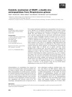
Fig. 2. Sequence alignment of PAF (top) and AFP (bottom) with three highly-conserved regions marked in yellow that are putatively assigned
to chitin binding (3–9), DNA binding (12–17) domains and cation channel forming (34–39) capabilities. Arrows indicate b-strands.
G. Batta et al. Structure and dynamics of an antifungal protein
FEBS Journal 276 (2009) 2875–2890 ª 2009 The Authors Journal compilation ª 2009 FEBS 2877
b-strand secondary structure. By contrast to AFP, no
additional minor signal set was observed in the NMR
spectra within the temperature range 280–320 °K.
However, similar to the structures reported for AFP
[14], the NMR data did not allow unambiguous
assignment of the disulfide pattern for PAF. The
assignment work was aided by the
15
N resolved 3D
TOCSY and NOESY spectra. Using the sparky [17]
spectrum visualization and assignment tool, many of
the NH(i)-HA(i-1) sequential NOE connectivities were
easily identified and often augmented with NH(i)-
HB(i-1) links. Although some lysine sidechain protons
remained unassigned, the completeness of
1
H assign-
ment finally reached 89%.
Secondary structure determination
Considering the secondary structure sensitive parame-
ters [a carbon atom (Ca) chemical shifts and
3
J
HN,HA
coupling constants] and the NOE constrained struc-
ture, we conclude that five antiparallel b-strands run
between residues: Lys2 to Thr8 (b1), Glu13 to Lys17
(b2), Asp23 to Ile26 (b3), Lys42 to Asp46 ( b4) and
Asn49 to Asp55 (b5) (Fig. 3). In addition, amide H-D
exchange rates (measured from HSQC spectra after
dissolving PAF in D
2
O) (Fig. 4) and relaxation experi-
ments also supported the presence of five b-strands in
PAF (Fig. 5). Measured deuteration rates clearly dem-
onstrated that amides in the proposed b-sheet regions
are protected from water access, whereas they are
more solvent exposed in loops and less structured
regions.
Tertiary structure determination
atnos ⁄ candid 1.1 software, in combination with
cyana 2.0, gave automatic NOE assignment [18–20]
L3
L1
β2
55
β3
β4
1
β1β5
L2
L4
Fig. 3. Super secondary structure of PAF.
0 10 20 30 40 50 60
−2
−1.5
−1
−0.5
0
0.5
1
1.5
2
2.5
Deuteration rates of 28 amide NH groups in PAF
Residue number
Rates
log10 scale, [1/h]
C7
C14
I26
C28
C36
C43
C54
D53
Fig. 4. Deuteration rates measured for amide NH groups in PAF
(note the logarithmic scale). Missing bars represent fast deuteration
rates (i.e. those NH signals that disappeared within 10 min); low
values mean high protection. Slow deuteration of amides protons
correlates with solvent protection in b-sheet regions. Note that all
six cysteines are well protected because they reside in the hydro-
phobic core.
0 10 20 30 40 50 60
0
0.2
0.4
0.6
0.8
1
Residue number
S
2
calculated from RCI
0 10 20 30 40 50 60
0
0.2
0.4
0.6
0.8
1
S
2
from
15
N relaxation
Fig. 5. S
2
order parameters reflecting internal mobility of the NH
residues obtained from the Lipari–Szabo analysis of
15
N T
1
, T
2
and
NOE relaxation parameters. Slightly enhanced mobility is clearly
detected at the N-terminus and in the loop regions, as indicated by
the dips in the bar plot. For comparison, S
2
values calculated from
the assigned chemical shifts are shown at the bottom using using
the random coil index (RCI) method. The average of
S
2
exp
=S
2
RCI
¼ 0:96 Æ 0:07. Residue 29 is proline and, consequently,
is not shown in the experimental data.
Structure and dynamics of an antifungal protein G. Batta et al.
2878 FEBS Journal 276 (2009) 2875–2890 ª 2009 The Authors Journal compilation ª 2009 FEBS
and gradual improvement of the PAF structure in
seven consecutive steps. Two probable conformational
families with different disulfide bridge patterns and
one in the absence of disulfide bonds were considered.
The goodness of the selected conformational families
along with their Ramachandran analysis are shown in
Table 1.
NMR relaxation combined with restrained
molecular dynamics calculations
The model-free analysis [21] of conventional
15
N relax-
ation experimental [22] data yielded a 3.0 ns global
correlation time for PAF (see Table S1), which is
appropriate for a monomeric globular protein of this
size [23]. The order parameters are shown in Fig. 5 in
comparison with those calculated from assigned chemi-
cal shifts using the random coil index method (http://
wishart.biolog y.ualbe rta.ca/rci/cgi-bin/rci_cgi_1_e .py).
On average, S
2
= 0.81 ± 0.05, with slight drops at
the N-terminus and loop regions, displaying enhanced
mobilities. The order parameters calculated straight
from chemical shifts agree well with those measured
from relaxation (S
2
exp
=S
2
RCI
¼ 0:96 Æ 0:07) and predict a
fairly compact structure. However, this type of relaxa-
tion is sensitive only to picosecond to nanosecond time
scales. In addition,
15
N and
1
H chemical shift aniso-
tropy (CSA) ⁄ dipolar–dipolar coupling (DD) cross-
correlated relaxation [24] has been measured (see
Table S1). The good correlation between secondary
structure and the
1
H transversal cross-correlated relax-
ation rates (Fig. 6) is a result of the extensive hydrogen
bonded networks, as well as the high CSA values of
these protons [25], in the b-sheet regions. Using the
15
N g
xy
and g
z
CSA ⁄ DD CCR rates according to the
method of Kroenke et al. [26], we separated exchange
contribution to R
2
relaxation rates, and found them to
be below 2 s
)1
for all NH groups. No outliers were
found; consequently, slow time scale conformational
exchange is unlikely in PAF.
The eighty final conformers of PAF obtained from
the minimal under restraining, minimal over restrain-
ing (MUMO) calculations [27] are shown in Fig.7.
These ensembles are assumed to be consistent with the
NOE-derived distance restraints, as well as with the
experimental S
2
order parameters. With respect to
NOE violations, the abbacc (7–36, 14–28, 43–54)
Table 1. Summary of CYANA calculations for 20 selected PAF struc-
tures and their respective
PROCHECK_NMR analyses.
S–S bond No disulfide abbacc abcabc
NOE upper distance limits 757 769 742
CYANA restraint violations 0 2 0
Target function 0.36 ± 0.16 0.42 ± 0.22 0.19 ± 0.07
rmsd from mean structure
Backbone (A
˚
) 0.65 ± 0.18 0.54 ± 0.14 0.60 ± 0.11
All heavy atoms (A
˚
) 1.15 ± 0.23 1.03 ± 0.11 1.09 ± 0.08
Ramachandran statistics
Most favoured 59.5% 46.3% 55.5%
Additionally allowed 32.7% 46.2% 42.9%
Generously allowed 5.3% 7.5% 1.6%
Disallowed 2.5% 0% 0%
0 10 20 30 40 50 60
0
1
2
3
4
5
6
7
15
N−
1
H CSA/DD cross−correlated relaxation rates
Hxy
Nzz
Nxy
Residue number
[1/s]
Fig. 6. Different CSA ⁄ DD relaxation interference rates displayed as
a pile-up bar graph. Instead of extracting site specific
15
N chemical
shift anisotropies from all the relaxation data, this type of straight
visualization of
1
H-
15
N CSA ⁄ DD transversal cross-correlated relaxa-
tion rates (Hxy) is sensitive to secondary structure elements.
abbacc
No disulfide bondabcabc
Fig. 7. Final MUMO ensembles (80 con-
formers) of PAF calculated with different
disulfide pairings labelled as abbacc, abcabc
and no disulfide bond. The average NMR
structure (red line) with no disulfide bonds
is overlaid on the ensembles.
G. Batta et al. Structure and dynamics of an antifungal protein
FEBS Journal 276 (2009) 2875–2890 ª 2009 The Authors Journal compilation ª 2009 FEBS 2879
ensemble appears to outperform the others (note that
perfect agreement with NOE data could not be easily
achieved because the parameterization is quite different
from those in ‘conventional’ structure calculation
methods that are designed to ensure this). Correlation
coefficients with S
2
values (see Figs S3 and S4) for the
‘no disulfide’ and abcabc (7–36, 14–43, 28–54) 80-mem-
bered ensembles are approximately 0.8, corresponding
to the performance of the restraining method [28]. The
abbacc ensemble exhibits relatively low correlation
(0.43), which, although the correlation was high for
each snapshot of the eight parallel replicas, can be
explained by the high structural divergence during the
simulation. The calculated ensembles show weak corre-
lation with the experimental
3
J
HN,HA
couplings, with
correlation coefficients in the range 0.42–0.5. Calculat-
ing J-value correlations using weighted averages of the
two pairings considered in the present study does not
improve the agreement. Ha chemical shifts back-calcu-
lated with shiftx [29] also do not favour any of the
ensembles. In all three MUMO ensembles, loop
regions on the surface of the protein (Lys17–Asp23,
Cys28–Lys35 and Asp46–Asn50) show increased
mobility coupled with structural heterogeneity. This
may indicate that one or more of these loops acts as
an interaction site with partner molecules. Lys9 resides
in loop 1, whereas Lys35 and Lys 38 are parts of the
large loop 3. They are surface and moderately solvent
exposed, and reside in conserved regions (Fig. 2). For
this reason, site-directed mutagenesis of these residues
was initiated.
Chitin binding function of PAF
We tested PAF for the ability to interact with oligo-
saccharides (oligosaccharide ⁄ oligonucleotide binding
domain; Fig. 2). This domain was suggested to con-
tribute to the cell wall [15] and ⁄ or nucleic acid [30,31]
binding activity of the homologous A. giganteus
protein AFP. Selective [32,33] and group selective [34]
saturation transfer difference NMR experiments with
chitobiose did not result in response signals in the
difference spectra (data not shown). The negative
results suggest that the chitobiose binding affinity (if it
persists) must be below the sub-milimolar regime. Sur-
face plasmon resonance testing of chitobiose binding
to immobilized PAF also provided no evidence for
strong binding. Furthermore, our attempts to colo-
calize the antifungal protein with nuclei in A. nidulans
hyphae failed (see Fig. S5). This indicates that PAF
does not interact with those cellular structures that
were suggested to be target molecules of the closely
related A. giganteus AFP protein [15,30,31].
Antifungal activity of mutated PAF versions
To investigate the impact of the highly-conserved,
lysine rich domain of PAF on antifungal activity, we
generated PAF mutants carrying amino acid
exchanges of distinct lysine residues, which originally
contributed to the high density of positive charges on
the same side of PAF. The antifungal potency of the
recombinant proteins was assessed by exposing
A. niger to mature PAF (mPAF), PAF
K9A
, PAF
K35A
,
PAF
K38A
and PAF
K9,35,38A
and carrying out a subse-
quent determination of growth rates (Fig. 8). Similar
growth inhibitory activity and morphological effects
could be observed between the native and the recom-
binant PAF (Fig. 8A). By contrast, a single exchange
of the lysine residues at positions 9, 35 or 38 reduced
the antifungal potential of PAF and increased the
proliferation of A. niger in a dose-dependent manner
(Fig. 8B). These results indicate that this conserved
lysine rich region behaves like a recognition motif for
the sensitive fungus. However, the triple mutation did
not further aggravate the loss of antifungal activity.
This allows the assumption that at least an additional
protein motif might contribute to the antifungal
activity.
Discussion
The structure of PAF
PAF from P. chrysogenum is a member of the posi-
tively-charged cysteine rich small proteins found in
other ascomycetes. PAF shares 43.6% amino acid
sequence identity and 71.3% sequence similarity with
AFP from A. giganteus [1]. Their homology is reflected
by their remarkable structural similarity presenting a
good alignment of their Ca traces (see Fig. S6).
Accordingly, the fold of PAF belongs to the structural
classification of proteins (.
ac.uk/scop/) fold class of AFP [14].
The 3D molecular structure of PAF consists of five
b-strands connected by three small loops involving
b-turn motifs (loops 1, 2 and 4) and the large loop 3
(Fig. 9). The b-strands create two orthogonally packed
b-sheets. Each b-sheet comprises three antiparallel
b-strands, which are ordered as 123 and 145, respec-
tively. The six conserved cysteines form three disulfide
bonds surrounded by the two orthogonal b-sheets,
creating a hidden central core.
The b1-strand running from Lys2 to Thr8 is highly
twisted as a result of the conserved flexible Gly5
followed by the bulky side chain of lysine and the
disulfide-paired Cys7, which pulls the strand towards
Structure and dynamics of an antifungal protein G. Batta et al.
2880 FEBS Journal 276 (2009) 2875–2890 ª 2009 The Authors Journal compilation ª 2009 FEBS
the core of the protein (Fig. 9). As a consequence of
the highly-twisted geometry, b1 is shared by both
sheets 1 and 2 providing a common interface.
The central strand of sheet 1 is b2 spread between
Glu13 and Lys17. The constituents of b2 are in an
extensive hydrogen bond network, with both b1 and
b3 contributing to the stabilization of sheet 1 (Table 2 ).
Loop 1 (9–12) and loop 2 (18–23) create a b-turn. A
characteristic of the PAF loop regions is the recurring
asparagine–aspartate or aspartate–asparagine (Asn18–
Asp19, Asp32–Asn33, Asp39–Asn40) sequence, either
preceding or following a lysine residue, resulting in a
preferential a-helical conformation [35,36]. This
sequence introduces a sharp turn geometry in the
loops. Strand b3, a stretch between Lys22-Ile26, and
the following first half of the large loop 3 (27–42),
spanning the segment from Lys27 to Phe31, is part of
the most extended hydrophobic region of the molecule
with low primary structure homology (Fig. 2) to AFP.
The single proline (Pro29) forms a trans-isomer, as in
AFP, and creates a bend in the large loop, which is
highly coiled as a result of two aspartate–asparagine
(Asp32 ⁄ Asn33; Asp39 ⁄ Asn40) turn preference motifs.
According to deuteration rates (Fig. 4). and S
2
order
parameters (Fig. 5), the most mobile region of loop 3
is between Lys30 and Lys34.
4.5
4.0
3.5
3.0
2.5
2.0
1.5
1.0
0.5
0
PA F
K9A
PA F
K35A
PA F
K38A
PA F
K9,35,38A
x-fold proliferation
A
B
a
b
c
C
A
B
Fig. 8. Analysis of the antifungal activity of mutated PAF protein versions on A. niger. (A) Microscopic analysis of A. niger exposed to
100 lgÆmL
)1
of PAF for 24 h. The recombinant mPAF protein (C) exhibited comparable growth inhibition potency with respect to the native
PAF (
B). Hyphae of the untreated control are shown in (A). (A–C) Microscopic overviews (· 20); (a–c) showing details of (A–C)(· 63). (B) The
increase in proliferation of A. niger when exposed to mutated PAF protein versions was correlated with the proliferation in the presence of
recombinant mPAF at the corresponding protein concentrations of 5 lgÆmL
)1
(light grey) and 100 lgÆmL
)1
(dark grey). The proliferation of
the PAF-unexposed A. niger control cells was 2.4 ± 0.2-fold and 7.4 ± 1.1-fold greater compared to the growth of the samples treated with
recombinant mPAF at the respective concentrations of 5 and 100 lgÆmL
)1
.
Fig. 9. Ribbon diagram of the mean PAF structure without disulfide
bond constraints.
G. Batta et al. Structure and dynamics of an antifungal protein
FEBS Journal 276 (2009) 2875–2890 ª 2009 The Authors Journal compilation ª 2009 FEBS 2881
The second half of loop 3 is one of the most highly-
conserved regions of PAF: three lysine side chains
(Lys34, Lys35 and Lys38) from this loop give rise to a
high density of positive charges in addition to the posi-
tively-charged side chains of Lys11 and Lys9 pointing
to the same region (Fig. 10). The importance of this
motif for the antifungal activity of PAF became
evident when replacing the Lys9, Lys35 and Lys38 by
alanines. Mutations within this motif resulted in a sig-
nificant reduction of antifungal activity. The slightly
solvent-exposed Asp39, which is a conserved residue in
the family, apart from its structural role of introducing
a perpendicular turn with the following Asn40, stabi-
lizes the positive charges of the adjacent Lys9 and
Lys39. Similarly, Asp23 stabilizes the juxtaposed
Lys15 and Lys17 side chains.
The hydrophobic strand b4 is located between Cys43
and Asp46 and, as a central strand of sheet 2, partici-
pates in an extensive hydrogen bonding network with
both strand b1 and b5 contributing to the sheet 2 stabil-
ization (Table 2). The b5 strand is the most negatively-
charged region of PAF and close to the C-terminus
starts at Ala51. Loop 4 (47–50) connects strands b4 and
b5 and creates a b-hairpin with a highly-exposed, con-
served Tyr48. All the three tyrosines of PAF with their
phenolato side chains can be found closely positioned in
the space between b4 and loop 2 and create a well-
defined aromatic region of the protein.
The topology of the disulfide pairs and the
function of the cysteines
According to the biochemical studies, no free thiol
groups can be detected in PAF. This is in good agree-
ment with the NMR measurements, which corroborate
that the six cysteines form three pairs of disulfide
bridges. These are essential for the inhibitory activity
on the growth of the sensitive fungi. Similar to the
AFP study [14], unambiguous assignment of the disul-
fide connectivity could not be obtained by NMR.
However, two sets of disulfide patterns are plausible
for PAF: abcabc and abbacc.
The safest assignment exists for the Cys7–Cys36
disulfide pair, which is supported by the approximately
400 pm Cb
i
–Cb
j
distance in the structures without any
SS bond constraint [37]. The well-defined Cys7–Cys36
disulfide pair cramps the large loop L3. Four cysteines
(Cys14, Cys28, Cys43 and Cys54) are in a close prox-
imity at the interface between sheet 1 and sheet 2,
Table 2. Hydrogen bonding pattern in the AFP fold proteins.
Secondary structural
elements PAF AFP
b1–b4 Tyr3HN–Val45O Ala1NH–Tyr45OH
Tyr3O–Val45HN Gly5O–Cys40HN
Ala1O–Thr47HN Cys7HN–Ala38O
Gly5HN–43CysO
b4–b5 Asp46O–Ala50HN Asp43O–Gly47HN
Asp46HN–Ala51O Tyr50HN–Glu41O
Thr44O–Asp53HN Tyr50O–Glu41HN
b1–b2 Lys6O–Lys15NH Ile13O–Tyr8HN
Lys6O–Lys15HN
Tyr8O–Ile13HN
b2–b3 Cys14HN–Ile26O Cys26HN–Cys14O
Asp18HN–Lys22O Cys28HN–Asn12O
Tyr16O–Thr24HN
b3–b5 Cys14N–Asp53O
in loop 2 (17–21) Gly21HN–Asn18O Ala18O–Gly21HN
b1 – large loop 3 Cys7O–Asn40HN Cys7O–Gly37NH
b1 – loop 1 Thr8NH–Lys11O
Fig. 10. The electrostatic surface potential of AFP (left) and PAF (right) structures representing the orientation of the side chain lysine resi-
dues. Lys9, Lys35 and Lys38 of PAF and Lys9, Lys32 and Arg35 of AFP are the corresponding conservative mutated lysines.
Structure and dynamics of an antifungal protein G. Batta et al.
2882 FEBS Journal 276 (2009) 2875–2890 ª 2009 The Authors Journal compilation ª 2009 FEBS
facing with their side chains to the core of the protein.
In the abcabc disulfide bond topology, the cross-link
between the two main sheets is proposed between
Cys14 (b2) from sheet 1 and Cys43 (b 4) from sheet 1,
as well as the bridge between Cys28 and Cys54, which
links b3 from sheet 1 to b5 from sheet 2. In the abbacc
disulfide pattern, connectivity inside the individual
sheets is favoured, where Cys14 makes a link with
Cys28 in sheet 1 and Cys43 of b4 connects Cys54 of
b5 in sheet 2. In the same structure for the abcabc pat-
tern, the 14–43 and 28–54 Cb
i
–Cb
j
distances are 442
and 525 pm, whereas, in the alternative abbacc pattern,
we obtain 455 pm for the 14–28 distance and 486 pm
for the 43–54 distance.
The question remains as to whether two intercon-
verting PAF species exist (i.e. one with each disulfide
topology) or whether only a single topology is present
but escapes identification. Relaxation-compensated
Carr–Purcell–Meiboom–Gill experiments [38] did not
indicate S–S bond rearrangement on the 0.5–5 ms time
scale (data not shown) in contrast to previously
reported isomerization of a disulfide bond in bovine
pancreatic trypsin inhibitor [39]. The extreme stability
of PAF is further evidence against a putative disulfide
bond rearrangement.
The disulfide bonds in PAF contribute to the overall
stability of the compact scaffold and stabilize the inter-
face between the two sheets. This feature helps to
maintain protein integrity in the extracellular environ-
ment, as well as to maintain stability at elevated tem-
peratures and extreme pH and to resist protease
digestion. Moreover, the disulfide bonds might play an
active role in the internalization process, as suggested
for diphtheria toxin or animal baculovirus gp64 [40].
In such cases, the rearrangement of the disulfide bonds
can be triggered by membrane-associated oxidoreduc-
tases, such as protein disulfide isomerases, and the
presumable conformational change could help with
the protein internalization [40–43].
It is not known whether PAF is subjected to struc-
tural changes and partial reduction in the cytoplasm,
providing some redox activity, as found for thiored-
oxins [44]. However, the disturbance of heterotrimeric
G-protein signalling alone may increase intracellular
reactive oxygen species concentrations in filamentous
fungi [45,46], which could occur in the absence of any
redox activity of PAF.
Differences and similarities between the
structures of PAF and AFP
The common geometrical arrangement of the two
proteins is the special Greek key fold, in which the
two orthogonally packed b-sheets are connected via
the b1 strand as a common interface. The similar
hydrogen bonding network might be the main deter-
minant of this scaffold formation (Table 2). The fold
is further stabilized by six conserved cysteines in
addition to three highly-conserved regions, and sev-
eral conserved residues with key locations in the two
proteins (Figs 2, 3 and 9). In the case of AFP, two
extra cysteines form the fourth disulfide bridge. In
the AFP structure, three disulfide bond topologies
were proposed: abbcadcd , aabccdbd and abcdabcd.
The latter correlates with the suggested abcabc disul-
fide pattern in PAF.
The three highly homologous regions are situated
in the sequence between Ala1 and Lys9 (region 1),
between Asn12 and Lys17 (region 2) and between
Lys34 and Asp39 (PAF) and Lys31 and Asp36
(AFP) (region 3). Two conserved GlyLys motifs are
repeated in the structure, the glycines (i.e. Gly5 from
b1 and Gly20 from loop 2) introduce flexibility into
the secondary structural elements. The side chains of
the two conserved tyrosines (Tyr3 and Tyr16) consti-
tute an aromatic patch in between loop 2 and b1,
which is stretched to the solvent-exposed Tyr48 of
PAF and Tyr45 of AFP from loop 4. In addition to
the three conserved tyrosines, AFP contains three
more tyrosines compared to PAF: two of them
(Tyr29 and Tyr50) are solvent exposed, and can pro-
vide target side chains for interactions with nucleic
acid bases (see Fig. S7). Indeed, DNA binding and
condensation activity was observed for AFP [31] and
also corroborated by a colocalization of AFP to the
nuclei [30]. An aromatic region was shown to repre-
sent a binding site for DNA in a structural homo-
logue cold shock protein from Bacillus caldolyticus
upon hexathymidine binding [47]. The function of
AFP was also associated with chitin binding activity
and interaction with the cell wall [15]. In carbohy-
drate binding modules, surface-exposed aromatic resi-
dues (e.g. Tyr and Trp) are in stacking interactions
with pyranose ⁄ furanose rings of oligosaccharides [48].
A few tyrosines of AFP are replaced by aspartates,
making the aspartate rich C-terminal negatively-
charged in PAF. However, we did not observe chito-
biose binding of PAF. PAF neither colocalizes to
the nuclei, nor binds exclusively to the cell wall. The
absence of surface-exposed tyrosines in PAF may
explain the difference in oligosaccharide binding
(Fig. 2).
Both PAF and AFP exhibit an amphipatic surface
alternating the positively- and negatively-charged
patches (Fig. 10). However, a well-defined positive
and an acidic region are formed only in PAF. The
G. Batta et al. Structure and dynamics of an antifungal protein
FEBS Journal 276 (2009) 2875–2890 ª 2009 The Authors Journal compilation ª 2009 FEBS 2883
positive charges concentrate at one side of the mole-
cule composed of Lys34, Lys35, Lys11 and Lys9. As
demonstrated by site-directed mutagenesis in the
present study, this positively-charged motif indeed
plays a central role with respect to the toxicity of
PAF on target organisms. A common characteristic
of the surface of both molecules, PAF and AFP,
comprises the numerous solvent-exposed positively-
charged lysine side chains, which could function in
disturbing the integrity of the plasma membrane or
determine the interaction with a target molecule that
is located in the plasma membrane. However, it
remains to be investigated in future studies whether
this motif mediates the binding of the protein to
structures of the outer layers of the target organism
or exerts its function intracellularly [6,12].
In conclusion, the solution structure of PAF has
been disclosed up to the extent of a disulfide pairing
ambiguity. No evidence for a putative disulfide bond
rearrangement on the millisecond time scale has been
found by NMR dynamics. With respect to the possible
mechanism behind the antifungal action of PAF, the
modulation of specific ion channels appears to be more
likely than chitin or DNA binding, in contrast to
AFP.
Experimental procedures
Production of PAF in P. chrysogenum
For PAF production, P. chrysogenum Q176 (ATCC 10002)
was cultivated in minimal medium (0.3% NaNO
3
, 0.05%
KCl, 0.05% MgSO
4
Æ7H
2
O, 0.005% FeSO
4
Æ7H
2
O, 2%
sucrose, 25 mm NaCl ⁄ P
i
, pH 5.8) at 25 °C (RT) as
described previously [5]. For preparation of
15
N-labelled
PAF for NMR analysis, 0.3% Na
15
NO
3
(Cambridge
Isotope Laboratories, Andover, MA, USA) was used as
nitrogen source in minimal medium.
Site-directed mutagenesis and heterologous
expression of mutated PAF protein variants in
Pichia pastoris
The nucleotide sequence coding for the mature PAF protein
version was PCR amplified from P. chrysogenum cDNA
using the primers with incorporated restrictions sites for
inframe cloning into the pPic9K expression vector (forward
5¢-AG
CTCGAGAAAAGAGCCAAATACACCGGAAAA
TG-3¢, XhoI site underlined; reverse 5¢-CT
GAATTCCTA
GTCACAATCGACAGCGTTG-3¢, EcoRI site underlined,
stop codon in bold). Amplification was performed in a two-
step PCR: three cycles of 1 min at 94 °C, 1 min at 50 °C
and 1 min at 72 °C; and then 30 cycles of 40 s at 94 °C
and 1 min at 72 °C; with a final extension for 7 min at
72 °C. Because an inefficient STE13 protease activity was
reported, we followed a previously described cloning strat-
egy [49] and eliminated the Kex2p and Ste13p signal cleav-
age sites. The vector for expression of the recombinant
wild-type version of PAF was named pPic9Kmpaf. Muta-
genesis of the PAF coding sequence (exchange of lysine into
alanine) was performed by two sequential PCR strategies,
essentially as described previously [50]. For the design of
the mismatch primers, P. pastoris preferential codon usage
was taken into account. Ten nanograms of pPic9Kmpaf
were used as a PCR template to generate the mutations:
PAF
K9A
(plasmid pPic9KpafK9A), PAF
K35A
(plasmid
pPic9KpafK35A) and PAF
K38A
(plasmid pPic9KpafK38A).
For generation of the triple mutant (PAF
K9,35,38A
), the
codon for lysine 35 was mutated in the plasmid pPic9K-
pafK38A to generate pPic9KpafK35,38, which served as
template for further mutagenesis using the appropriate
primers (Table 3). The amplified overlapping PCR products
containing the desired mutation were combined in a third
PCR where both fragments served as megaprimers for fur-
ther elongation of the PAF sequence in the first few PCR
cycles. The elongated fragments were then further amplified
using the primers 5¢AOX1 and 3¢AOX1 (33 cycles of 45 s
at 94 °C, 45 s at 54 °C and 1 min at 72 °C, with a final
Table 3. Oligonucleotides used for site-directed mutagenesis. Codons for amino acid exchanges are shown in bold-italic typeface.
Mutation Oligonucleotide Sequence (5¢-to3¢) PCR template
PAF
K9A
opafK9Ase GGAAAATGCACCGCTTCTAAGAACG pPic9Kmpaf
opafK9Arev CGTTCTTAGAAGCGGTGCATTTTCC
PAF
K35A
opafK35Ase GTTTGATAACAAGGCTTGCACCAAGG pPic9Kmpaf
opafK35Arev CCTTGGTGCAAGCCTTGTTATCAAAC
PAF
K38A
opafK38Ase GAAGTGCACCGCTGATAATAACAAATG pPic9Kmpaf
opafK38Arev CATTTGTTATTATCAGCGGTGCACTTC
PAF
K35,38A
opafK35,38Ase GTTTGATAACAAGGCTTGCACCGCTG pPic9KpafK38A
opafK35,38Arev CAGCGGTGCAAGCCTTGTTATCAAAC
PAF
K9,35,38A
opafK9Ase GGAAAATGCACCGCTTCTAAGAACG pPic9KpafK35,38A
opafK9Arev CGTTCTTAGAAGCGGTGCATTTTCC
Structure and dynamics of an antifungal protein G. Batta et al.
2884 FEBS Journal 276 (2009) 2875–2890 ª 2009 The Authors Journal compilation ª 2009 FEBS
extension of 7 min at 72 °C). The PCR product was
digested with BamHI ⁄ EcoRI and cloned into the Bam-
HI ⁄ EcoRI digested pPic9K vector. The occurrence of the
desired mutations was verified by nucleotide sequence deter-
mination using an automated 3100 abi prism DNA sequen-
cer (Applied Biosystems, Foster City, CA, USA).
Restriction enzymes and T4 DNA ligase were purchased
from Promega (Vienna, Austria), the multi-copy Pichia
expression kit was obtained from Invitrogen Life Technolo-
gies (Lofer, Austria) and primers were obtained from Euro-
fins MWG Operon (Ebersberg, Germany).
Manipulation of the P. pastoris KM71 strain was per-
formed using the Multi-copy Pichia expression kit version
F according to manufacturer’s instructions and as desribed
previously [49]. Protein purification was performed as
described below.
Purification of PAF
PAF was isolated by molecular mass filtration and ion-
exchange chromatography as described previously [3]. By
contrast to the wild-type PAF, the recombinant mutated pro-
teins did not bind to the CM-sepharose CL-6B column
(Amersham, Uppsala, Sweden), but were found to be pure in
the flow-through, which was subsequently concentrated in
Centriprep YM-3 filter devices (Millipore, Billerica, MA,
USA). The purity of PAF was checked by 16% SDS ⁄ PAGE
and silver staining before the protein solutions were filter-
sterilized (0.22 lm, Millipore) and stored at )20 °C. For
structural analysis, purified PAF was lyophilized.
Native PAF and PAF that had been exposed to various
test conditions in stability assays were subjected to RP-
HPLC, equipped with a 127 solvent module and a Model
166 UV-visible-region detector (Beckman Instruments, Palo
Alto, CA, USA). The separation of the samples was per-
formed on a Nucleosil 300-5 C
4
column (length, 125 mm;
inner diameter, 4 mm; particle pore size, 5 lm; pore size,
30 nm; end-capped; Machery-Nagel, Du
¨
ren, Germany).
Samples of approximately 5 lg of PAF were injected onto
the column and chromatographed within 20 min at a con-
stant flow of 0.5 mLÆmin
)1
with a linear acetonitrile gradi-
ent starting at solvent A: solvent B (15 : 85) (solvent A:
0.1% trifluoroacetic acid trifluoroacetic acid in H
2
O;
solvent B: 70% acetonitrile, 0.1% trifluoroacetic acid). The
concentration of solvent B was increased from 15% to 70%
over 20 min. The effluent was monitored at 210 nm and the
peaks were recorded using Beckman system gold software.
The PAF fraction was collected, lyophilized and stored at
)20 °C for further analysis.
Antifungal activity assays
In vitro assays were carried out in 96-well plates with the
test organism A. niger (CBS 120.49) in complete medium
(0.2% peptone, 0.1% yeast extract, 0.1% NZ-amine A, 2%
glucose, 0.05% KCl, 0.04% MgSO
4
Æ7H
2
O, 0.15% KH
2
PO
4
,
pH 6.5) at 30 °C. Antifungal activity of the protein samples
was determined by measuring the A
620
of A. niger cultures
at 24 h in a microtitre plate reader as described previously
[3]. The protein concentrations tested were in the range
1–100 lgÆmL
)1
.
Protein stability assays
Stability analysis of PAF was performed essentially as
described previously [51]. Thermal stability was investigated
by incubating 1 mgÆmL
)1
of PAF in 10 mm Na-phosphate
buffer, pH 6.6, 25 m m NaCl, 0.15 mm EDTA at 40–100 °C
for 10, 30 and 60 min. The pH stability of 1 mgÆmL
)1
of
PAF was tested within the pH range 1.5–11 at 25 °C for
24, 48 and 96 h. The buffers used (25 mm) comprised: gly-
cine-HCl, pH 1.5; sodium citrate-HCl, pH 3; citric acid-
Na
2
HPO
4
, pH 5; glycine-NaOH, pH 9 and 11. Stability
towards proteases was assayed by exposing 7 lg of PAF to
10 lg of pepsin, proteinase K or pronase (all from Sigma,
Vienna, Austria) in 0.1 m citric acid-Na
2
HPO
4
(at pH 4
and 5 for pepsin and at pH 7 for proteinase K and pron-
ase) for 3, 9 and 24 h at 30 °C.
To disclose the presence of the disulfide-bridges between
cysteine residues, PAF was treated with dithiothreitol (Fer-
mentas, St Leon-Rot, Germany), iodeacetamide (IAA;
Sigma) and cysteine (Sigma): 20 lg of PAF in 100 lLof
buffer A (100 mm NH
4
HCO
3
, pH 8) were mixed with
50 lL of dithiothreitol (10 mm in buffer A) and incubated
for 30 min at 56 °C. Next, 50 lL of IAA (55 mm in buffer
A) was added and the sample was further incubated at RT
for 20 min in the dark. Finally, excess IAA was blocked by
the addition of 50 lL of cysteine (55 mm in buffer A). As a
control, PAF was treated in the same way as described, but
without either dithiothreitol, IAA or cysteine, or by omit-
ting all three components. Instead, equivalent buffer
volumes were used. The sample was subsequently concen-
trated by reducing the volume to 50 lL using a Gyro Vap
centrifugal evaporator (Howe, Banbury, UK). For each
experiment, samples were taken for SDS ⁄ PAGE analysis,
an antifungal activity assay, HPLC analysis and MS.
Microscopic analysis
The intracellular localization of PAF was visualized in
A. nidulans hyphae by indirect immunofluorescence staining
with rabbit anti-PAF serum and fluorescein isothiocyanate-
conjugated anti- (rabbit IgG) (Sigma) as described previ-
ously [11]. After washing for 10 min in Tris ⁄ NaCl ⁄ P
i
,
nuclei were stained with the fluorescence stain 4¢,6¢-diamidi-
no-2-phenylindole (1 : 1.000 in Tris ⁄ NaCl ⁄ P
i
; Sigma) for
10 min. The samples were washed three times for 10 min in
Tris ⁄ NaCl ⁄ P
i
and mounted with Vectashield
Ò
mounting
medium (Vector Laboratories, Inc., Burlingame, CA, USA)
before visualization with a Zeiss Axioplan fluorescence
G. Batta et al. Structure and dynamics of an antifungal protein
FEBS Journal 276 (2009) 2875–2890 ª 2009 The Authors Journal compilation ª 2009 FEBS 2885
microscope, equipped with an AxioCam MRC camera
(Zeiss, Vienna, Austria). The samples were observed with
the appropriate filters: excitation ⁄ emission at 488 ⁄ 520 nm
for green fluorescence and 356 ⁄ 420 nm for blue fluores-
cence. Picture editing was performed using Adobe Photo-
shop CS3, version 10.0 (Adobe Systems Inc., San Jose, CA,
USA).
SDS ⁄ PAGE separation
Pre-treated proteins (1 lg per lane) were separated by
SDS ⁄ PAGE on 16% polyacrylamide precast gels in the
Tris–glycine buffer system (NOVEX; Invitrogen, Lofer,
Austria) under denaturing and reducing conditions (sample
buffer: 0.1 m Tris, 0.8% SDS, 5% glycerine, 2% b-mercap-
toethanol, 0.002% bromphenolblue, pH 6.8) or under
denaturating, nonreducing conditions (nonreducing sample
buffer: 0.1 m Tris, 10% glycerine, 0.002% bromphenolblue,
pH 8.8; without heat-denaturation). In all experiments,
untreated PAF served as loading control. Proteins were
visualized by Coomassie blue staining or silver staining.
MS analysis
Determination of the molecular mass of the samples (native
PAF, protease-treated PAF, reduced PAF) obtained by
RP-HPLC was carried out using an LCQ ion trap instru-
ment (ThermoFinnigan, San Jose, CA, USA) equipped with
an electrospray source (ESI-MS). The electrospray voltage
was set at 4.5 kV, and the heated capillary was held at
200 °C. Protein samples ($ 1 lg) were dissolved in 50%
aqueous methanol containing 0.1% formic acid, and
injected into ion source.
NMR spectroscopy
Two 1.4 mg
15
N-labelled PAF samples were dialyzed from
10 mm Na
3
PO
4
⁄ 20 mm NaCl solution at pH 5.0. Then,
2.8 mg of protein was dissolved in a volume of 275 lLof
95 : 5% H
2
O ⁄ D
2
O to yield an approximate PAF concen-
tration of 1.6 mmÆL
)1
. The protein solution in a shigemi
NMR tube contained 40 mmÆL
)1
NaCl and 0.04% NaN
3
.
The PAF NMR spectra did not exhibit sample decomposi-
tion for more than 1 year (the sample was stored at 4 °C).
All NMR spectra were acquired at 304 °K, except when
temperature-dependent
15
N HSQC spectra were measured
in the range 275–310 °K. Proton chemical shift scales are
referenced to sodium 2,2-dimethyl-2-silapentane-5-sulfo-
nate = 0 p.p.m. and heteronuclear shifts are referenced
indirectly from the gyromagnetic ratios for
15
N and
13
C,
which gives 67.1 p.p.m. for the dioxane
13
C signal.
For signal assignment and structure determination, the
NMR spectra were recorded on a DRX-700 (Bruker,
Rheinstetten, Germany) spectrometer. Water signal sup-
pression was achieved using the watergate5 sequence [52].
2D
1
H-
15
N HSQC spectrum was the seed for the assign-
ments at 700 MHz, and also allowed the straightforward
measurement of
3
J
HN,HA
couplings from signal splitting
(1400 · 256 time domain points transformed in a
8192 · 512 Fourier data table). Gradient echo-anti-echo
phase discrimination in both indirect dimensions was
applied in sensitivity enhanced 3D
15
N HSQC-TOCSY
(62 ms DIPSI mixing time) and 3D
15
N HSQC-NOESY
(130 ms mixing time) experiments. The double echo-
anti-echo technique provides better sensitivity and water
suppression quality than standard TOCSY-HSQC or
NOESY-HSQC methods. In the proton dimension, 12 or
5 p.p.m. (amides only) was used, whereas, in the
15
N
dimension, the spectral window was reduced to 19 p.p.m.,
which resulted in folding of Lys15 and Gly21 peaks. In 3D
experiments 2048 · 256 · 46 points were acquired and
transformed in 2048 · 512 · 128 points. In general, suitably
shifted squared cosine or Gaussian window functions were
applied. Sequence specific resonance assignments [53–55]
were determined from the 3D spectra using the sparky
software [17,56]. Distance restraints were obtained from
15
N decoupled and radiation damping suppressed 2D
NOESY (130 ms mixing time) spectrum. The 2D spectrum
was acquired in 2048 · 529 points and transformed to
4096 · 2048 points. Natural abundance
13
C-
1
H HSQC
spectra lent more support to assignments, and provided
invaluable Ca chemical shifts to aid secondary structure
determination. When the assignment was finished with
sparky, the automatic NOE assignment and structure cal-
culation was carried out using the atnos ⁄ candid 1.1
[19,20] in combination with cyana 2.0 [18]. The basis of
automatic structure determination was a single 2D NOESY
spectrum (130 ms mixing time, 4096 · 4096 Fourier size,
2.1 Hz ⁄ point digital resolution). Because unambiguous
assignment of the disulfide pattern could not be achieved,
several conformational families were explored with different
disulfide pairings, including the simplest case with no disul-
fides at all (e.g. six free thiol groups). The final mean struc-
tures were energy minimized with gromacs molecular
dynamics software using the optimized potential for liquid
simulations – all atom force field in vacuum to the force
field limit of 250 kJÆmol
)1
Ænm
)1
. For NMR dynamics,
besides the conventional
15
N relaxation [T
1
, T
2
and
15
N-
(
1
H) NOE] [22,57,58], the longitudinal and transverse
15
N
and
1
H CSA ⁄ DD cross-correlated relaxation rates g
zz
(
15
N),
g
xy
(
15
N) and g
xy
(
1
H) [59], and NH deuteration rates were
measured at 500 MHz. The PAF sample lyophilized from
H
2
O was dissolved in D
2
O, and then the progress of amide
deuteration at 304 °K at pH 5.0 was monitored in a series
of
15
N HSQC spectra using the peak volume integrals.
Sixty-nine time points were measured in the 0.17–48 h
interval. The decays were fitted with single exponential
functions, yielding the rates. For relaxation, a series of 2D
Structure and dynamics of an antifungal protein G. Batta et al.
2886 FEBS Journal 276 (2009) 2875–2890 ª 2009 The Authors Journal compilation ª 2009 FEBS
heteronuclear correlated spectra using sensitivity enhanced
gradient pulse schemes [25] were recorded. Regarding the
typical experimental parameters: the
1
H carrier frequency
was switched between the water resonance and the centre at
8.15 p.p.m. of the 5.2 p.p.m.
1
H spectral window, whereas
the
15
N window was 27 p.p.m. centred at 118.5 p.p.m. The
relaxation delay times were set as: T
1
, 11.2, 101.2, 201.2,
401.2, 601.2, 801.2, 1001.2 and 1201.2 ms; T
2
, Carr–Pur-
cell–Meiboom–Gill pulse trains of 0.03, 30.4, 60.8, 91.2,
121.6, 182.4, 243.2, 302.4, 360 and 417.6 ms in duration
were used; for the measurement of cross-correlated relaxa-
tion rates, g
zz
and g
xy
, the relaxation interference was
allowed to be active for 20 or 21.6 ms in pairs of experi-
ments, including the reference (D = 0 ms) experiment. The
number of transients collected per t
1
increment were 8 for
T
1
, 16 for T
2
, 32 for NOE, and 80 for g
zz
and g
xy
measure-
ments. A spin-lock field of 3400 Hz was used for the
15
N
transverse cross-correlation experiment. Two-parameter
exponential fits of the measured volume intensities of cross
peaks were applied to extract the relaxation times T
1
and
T
2
. The cross-correlation rate constants were determined
using the initial linear build-up rate approach. The theoreti-
cal expressions for the autorelaxation (R
1
, R
2
) and cross-
correlation rate constants (g
xy
, g
zz
) and for the steady-state
heteronuclear NOE in terms of the spectral density func-
tions [J
a
(x) auto- and J
c
(x) cross-correlation] are used as
described previously [60]. The simplifying assumption of
isotropic overall tumbling and the axial symmetry of con-
stant (Dr = )160 p.p.m.)
15
N chemical shielding tensors
were applied. A bond length of r
NH
= 0.102 nm was used
in all calculations. The model-free [21] analysis of T
1
, T
2
and heteronuclear NOE yielded the S
2
order parameters
and local correlation times for all NH and the global corre-
lation time. The calculated ‘theoretical’ relaxation data
derived from these parameters agreed with experimental
values within a range of 2.3–2.8% (one standard deviation).
For the detection of chitobiose binding selective [32,33]
and group selective [34] saturation, transfer difference
experiments were run using solutions of 0.1 mm
15
N-
labelled PAF and 5 mm chitobiose and a 3 s total satura-
tion time. Selective irradiation at 0.0 p.p.m. was carried
out using a pulse train of 50 ms 270° Gaussian pulses,
whereas simultaneous pre-irradiation of all amides was
aided with repeated (30 ms) bilinear rotational decoupling
pulse trains.
The MUMO combined molecular mechanics and
NMR dynamics
To obtain realistic conformational ensembles reflecting the
dynamical features of PAF, structure calculations using the
MUMO approach [27] were applied. Half-harmonic S
2
restraints [28] and pairwise treatment of NOE restraints
between replicas were implemented in gromacs 3.3.1 [61].
A simulated annealing protocol using ten 230 ns cycles
similar to that described previously [27] was used, except
that the force constants were not modified during the
cycles. A simulation with eight replicas using the optimized
potential for liquid simulations – all atom force field [62] in
explicit water (SPC model) was run after a short round of
energy minimization and solvent equilibration. The starting
NMR structure was the output of the atnos ⁄ candid ⁄ cya-
na calculations. Disulfide bridges were introduced followed
by short energy minimization to ensure realistic geometry
using sybyl (Tripos, St Louis, MO, USA) molecular mod-
elling software. Three ensembles were generated: one with-
out disulfide bridges, one with the pairing abbacc (7–36,
14–28, 43–54) and one with that of abcabc (7–36, 14–43,
28–54) (see Fig. S8). To validate the obtained structures,
3
J
HN-HA
couplings were back-calculated from the ensembles
(Table 4) (parameters for the Karplus equation were taken
from a previous NMR ⁄ X-ray data set) [63] and correlated
with the measured values. Chemical shifts estimated with
shiftx [29] and averaged for the different ensembles were
also used for the evaluation of different ensembles.
Acknowledgements
Financial support was provided by the Hungarian
Scientific Research Fund OTKA NK 68578 CK 77515
and F 68079, and by the Austrian Science Foundation
(FWF P19970-B11) and the D. Swarowski Foerde-
rungsfonds (FB2 ⁄ 06). The EU-NMR – Contract no.
RII3-026145 ⁄ CERM13 project Grant for access to
700 MHz NMR facilities in Florence (CERM) is grate-
fully acknowledged. We thank Professors Ivano Bertini
and Isabella Felli, as well as Mr Massimo Lucci, for
providing excellent support in Florence. We also thank
Renate Weiler-Goerz for technical assistance. Z. G.
acknowledges the support of a FEBS Short-Term Fel-
lowhsip and a Ja
´
nos Bolyai Postdoctoral Fellowship.
This work was also supported by the Hungarian
National Office for Research and Technology (Grant
reference numbers: OMFB 01501 ⁄ 2006 and 01528 ⁄
2006) and by the GENOMNANOTECH-DEBRET
Table 4. Analysis of the MUMO ensembles (80 members) with
respect to experimental NMR parameters, as calculated using r
)6
averaging on the ensembles.
No disulfide abbacc abcabc
Number of violated NOEs 15 9 22
Average violation (A
˚
) 1.12 0.33 1.65
Maximum violation (A
˚
) 4.22 1.01 4.38
S
2
(correlation coefficient) 0.81 0.44 0.79
3
J
NH-HA
(correlation coefficient) 0.43 0.50 0.42
HAÆchemical shifts
(correlation coefficient)
0.60 0.64 0.67
G. Batta et al. Structure and dynamics of an antifungal protein
FEBS Journal 276 (2009) 2875–2890 ª 2009 The Authors Journal compilation ª 2009 FEBS 2887
(RET-06 ⁄ 2004). A. K. C. wishes to thank D. B. T.
(Government of India).
References
1 Marx F (2004) Small, basic antifungal proteins secreted
from filamentous ascomycetes: a comparative study
regarding expression, structure, function and potential
application. Appl Microbiol Biotechnol 65, 133–142.
2 Geisen R (2000) P-nalgiovense carries a gene which is
homologous to the paf gene of P. chrysogenum which
codes for an antifungal peptide. Int J Food Microbiol
62, 95–101.
3 Kaiserer L, Oberparleiter C, Weiler-Gorz R, Burgstaller
W, Leiter E & Marx F (2003) Characterization of the
Penicillium chrysogenum antifungal protein PAF. Arch
Microbiol 180, 204–210.
4 Lee DG, Shin SY, Maeng CY, Jin ZZ, Kim KL &
Hahm KS (1999) Isolation and characterization of a
novel antifungal peptide from Aspergillus niger. Biochem
Biophys Res Commun 263, 646–651.
5 Marx F, Haas H, Reindl M, Stoffler G, Lottspeich F &
Redl B (1995) Cloning, structural organization and
regulation of expression of the Penicillium chrysogenum
paf gene encoding an abundantly secreted protein with
antifungal activity. Gene 167, 167–171.
6 Marx F, Binder U, Leiter E & Po
´
csi I (2008) The Peni-
cillium chrysogenum antifungal protein PAF, a promis-
ing tool for the development of new antifungal
therapies and fungal cell biology studies. Cell Mol Life
Sci 65, 445–454.
7 Theis T, Wedde M, Meyer V & Stahl U (2003) The
antifungal protein from Aspergillus giganteus causes
membrane permeabilization. Antimicrob Agents Chemo-
ther 47, 588–593.
8 Wnendt S, Ulbrich N & Stahl U (1994) Molecular-clon-
ing, sequence-analysis and expression of the gene encod-
ing an antifungal-protein from Aspergillus giganteus.
Curr Genet 25, 519–523.
9 Galgo
´
czy L, Papp T, Leiter T, Marx F, Po
´
csi I &
Va
´
gvo
¨
lgyi C (2005) Sensitivity of different zygomycetes
to the Penicillium chrysogenum antifungal protein
(PAF). J Basic Microbiol 45, 136–141.
10 Meyer V (2008) A small protein that fights fungi: AFP
as a new promising antifungal agent of biotechnological
value. Appl Microbiol Biotechnol 78 , 17–28.
11 Oberparleiter C, Kaiserer L, Haas H, Ladurner P,
Andratsch M & Marx F (2003) Active internalization
of the Penicillium chrysogenum antifungal protein PAF
in sensitive aspergilli. Antimicrob Agents Chemother 47,
3598–3601.
12 Leiter E, Szappanos H, Oberparleiter C, Kaiserer L,
Csernoch L, Pusztahelyi T, Emri T, Po
´
csi I, Salv-
enmoser W & Marx F (2005) Antifungal protein PAF
severely affects the integrity of the plasma membrane
of Aspergillus nidulans
and induces an apoptosis-like
phenotype. Antimicrob Agents Chemother 49, 2445–
2453.
13 Szappanos H, Szigeti GW, Pa
´
l B, Ruszna
´
k Z, Szu
¨
cs G,
Rajnavo
¨
lgyi E, Balla J, Balla G, Nagy E, Leiter T et al.
(2005) The Penicillium chrysogenum-derived antifungal
peptide shows no toxic effects on mammalian cells in
the intended therapeutic concentration. Naunyn Schmi-
edebergs Arch Pharmacol 371, 122–132.
14 Campos-Olivas R, Bruix M, Santoro J, Lacadena J,
Delpozo AM, Gavilanes JG & Rico M (1995) NMR
solution structure of the antifungal protein from Asper-
gillus giganteus – evidence for cysteine pairing isomer-
ism. Biochemistry 34, 3009–3021.
15 Hagen S, Marx F, Ram AF & Meyer V (2007) The
antifungal protein AFP from Aspergillus giganteus
inhibits chitin synthesis in sensitive fungi. Appl Environ
Microbiol 73, 2128–2134.
16 Marx F, Salvenmoser W, Kaiserer L, Graessle S,
Weiler-Gorz R, Zadra I & Oberparleiter C (2005)
Proper folding of the antifungal protein PAF is required
for optimal activity. Res Microbiol 156, 35–46.
17 Goddard TD & Kneller DG (2001) SPARKY. Univer-
sity of California, San Francisco, CA.
18 Gu
¨
ntert P (2003) Automated NMR protein structure
calculation. Progr Nucl Magn Reson Spectros 43, 105–
125.
19 Herrmann T, Gu
¨
ntert P & Wu
¨
thrich K (2002) Protein
NMR structure determination with automated NOE-
identification in the NOESY spectra using the new soft-
ware ATNOS. J Biomol NMR 24, 171–189.
20 Herrmann T, Gu
¨
ntert P & Wu
¨
thrich K (2002) Protein
NMR structure determination with automated NOE
assignment using the new software CANDID and the
torsion angle dynamics algorithm DYANA. J Mol Biol
319, 209–227.
21 Lipari G & Szabo A (1982) Model-free approach to the
interpretation of nuclear magnetic-resonance relaxation
in macromolecules .1. Theory and range of validity.
J Am Chem Soc 104, 4546–4559.
22 Farrow NA, Muhandiram R, Singer AU, Pascal SM,
Kay CM, Gish G, Shoelson SE, Pawson T, Formankay
JD & Kay LE (1994) Backbone dynamics of a free and
a phosphopeptide-complexed Src homology-2 domain
studied by N-15 NMR relaxation. Biochemistry 33,
5984–6003.
23 Daragan VA & Mayo KH (1997) Motional model anal-
yses of protein and peptide dynamics using C-13 and
N-15 NMR relaxation. Progr Nucl Magn Reson Spec-
tros 31, 63–105.
24 Tessari M, Mulder FAA, Boelens R & Vuister GW
(1997) Determination of amide proton CSA in N-15-
labeled proteins using H-1 CSA ⁄ N-15-H-1 dipolar and
N-15 CSA ⁄
N-15-H-1 dipolar cross-correlation rates.
J Magn Reson 127, 128–133.
Structure and dynamics of an antifungal protein G. Batta et al.
2888 FEBS Journal 276 (2009) 2875–2890 ª 2009 The Authors Journal compilation ª 2009 FEBS
25 Tessari M, Vis H, Boelens R, Kaptein R & Vuister GW
(1997) Quantitative measurement of relaxation interfer-
ence effects between H-1(N) CSA and H-1-N-15 dipolar
interaction: correlation with secondary structure. JAm
Chem Soc 119, 8985–8990.
26 Kroenke CD, Loria JP, Lee LK, Rance M & Palmer
AG (1998) Longitudinal and transverse H-1-N-15 dipo-
lar N-15 chemical shift anisotropy relaxation interfer-
ence: unambiguous determination of rotational
diffusion tensors and chemical exchange effects in
biological macromolecules. J Am Chem Soc 120,
7905–7915.
27 Richter B, Gsponer J, Varnai P, Salvatella X &
Vendruscolo M (2007) The MUMO (minimal under-
restraining minimal over-restraining) method for the
determination of native state ensembles of proteins.
J Biomol NMR 37, 117–135.
28 Best RB & Vendruscolo M (2004) Determination of
protein structures consistent with NMR order parame-
ters. J Am Chem Soc 126, 8090–8091.
29 Neal S, Nip AM, Zhang HY & Wishart DS (2003)
Rapid and accurate calculation of protein H-1, C-13
and N-15 chemical shifts. J Biomol NMR 26 , 215–
240.
30 Moreno AB, del Pozo AM & Segundo BS (2006) Bio-
technologically relevant enzymes and proteins – antifun-
gal mechanism of the Aspergillus giganteus AFP against
the rice blast fungus Magnaporthe grisea. Appl Micro-
biol Biotechnol 72, 883–895.
31 del Pozo AM, Lacadena V, Mancheno JM, Olmo N,
Onaderra M & Gavilanes JG (2002) The antifungal
protein AFP of Aspergillus giganteus is an oligonucleo-
tide ⁄ oligosaccharide binding (OB) fold-containing pro-
tein that produces condensation of DNA. J Biol Chem
277, 46179–46183.
32 Mayer M & Meyer B (1999) Characterization of ligand
binding by saturation transfer difference NMR spec-
troscopy. Angew Chem Int Edit 38, 1784–1788.
33 Mayer M & Meyer B (2001) Group epitope mapping by
saturation transfer difference NMR to identify segments
of a ligand in direct contact with a protein receptor.
J Am Chem Soc 123, 6108–6117.
34 Ko
¨
ve
´
r KE, Groves P, Jime
´
nez-Barbero J & Batta G
(2007) Molecular recognition and screening using a
N-15 group selective STD NMR method. J Am Chem
Soc 129, 11579–11582.
35 Richardson JS (1981) The anatomy and taxonomy of
protein structure. Adv Protein Chem 34, 167–218.
36 Wilmot CM & Thornton JM (1988) Analysis and pre-
diction of the different types of beta-turn in proteins.
J Mol Biol 203, 221–232.
37 Klaus W, Broger C, Gerber P & Senn H (1993) Determi-
nation of the disulfide bonding pattern in proteins by
local and global analysis of nuclear-magnetic-resonance
data – application to flavoridin. J Mol Biol 232, 897–906.
38 Loria JP, Rance M & Palmer AG (1999) A relaxation-
compensated Carr–Purcell–Meiboom–Gill sequence for
characterizing chemical exchange by NMR spectros-
copy. J Am Chem Soc 121, 2331–2332.
39 Wang CY, Grey MJ & Palmer AG (2001) CPMG
sequences with enhanced sensitivity to chemical
exchange. J Biomol NMR 21, 361–366.
40 Wouters MA, Lau KK & Hogg PJ (2004) Cross-strand
disulphides in cell entry proteins: poised to act. BioEs-
says 26, 73–79.
41 Hogg PJ (2003) Disulfide bonds as switches for protein
function. Trends Biochem Sci 28, 210–214.
42 Turano C, Coppari S, Altieri F & Ferraro A (2002)
Proteins of the PDI family: unpredicted non-ER
locations and functions. J Cell Physiol 193, 154–
163.
43 Markovic I, Pulyaeva H, Sokoloff A & Chernomordik
LV (1998) Membrane fusion mediated by baculovirus
gp64 involves assembly of stable gp64 trimers into mul-
tiprotein aggregates. J Cell Biol 143, 1155–1166.
44 Po
´
csi I, Prade RA & Penninckx MJ (2004) Glutathione,
altruistic metabolite in fungi. Adv Microb Physiol 49,
1–76.
45 Han KH, Seo JA & Yu JH (2004) Regulators of G-pro-
tein signalling in Aspergillus nidulans: RgsA downregu-
lates stress response and stimulates asexual sporulation
through attenuation of GanB (G alpha) signalling. Mol
Microbiol 53, 529–540.
46 Molna
´
r Z, Emri T, Zavaczki E, Pusztahelyi T & Po
´
csi I
(2006) Effects of mutations in the GanB ⁄ RgsA G pro-
tein mediated signalling on the autolysis of Aspergil-
lus nidulans. J Basic Microbiol 46, 495–503.
47 Max KEA, Zeeb M, Bienert R, Balbach J & Heine-
mann U (2007) Common mode of DNA binding to cold
shock domains – crystal structure of hexathymidine
bound to the domain-swapped form of a major cold
shock protein from Bacillus caldolyticus. FEBS J 274,
1265–1279.
48 Hashimoto H (2006) Recent structural studies of carbo-
hydrate-binding modules. Cell Mol Life Sci 63, 2954–
2967.
49 Cabral KMS, Almeida MS, Valente AP, Almeida FCL
& Kurtenbach E (2003) Production of the active anti-
fungal Pisum sativum defensin 1 (Psd1) in Pichia pas-
toris: overcoming the inefficiency of the STE13
protease. Protein Expr Purif 31, 115–122.
50 DeSamblanx GW, Goderis IJ, Thevissen K, Raemae-
kers R, Fant F, Borremans F, Acland DP, Osborn RW,
Patel S & Broekaert WF (1997) Mutational analysis of
a plant defensin from radish (Raphanus sativus L)
reveals two adjacent sites important for antifungal
activity. J Biol Chem 272 , 1171–1179.
51 Bormann C, Baier D, Horr I, Raps C, Berger J, Jung G
& Schwarz H (1999) Characterization of a novel, anti-
fungal, chitin-binding protein from Streptomyces tendae
G. Batta et al. Structure and dynamics of an antifungal protein
FEBS Journal 276 (2009) 2875–2890 ª 2009 The Authors Journal compilation ª 2009 FEBS 2889
Tu901 that interferes with growth polarity. J Bacteriol
181, 7421–7429.
52 Liu ML, Mao XA, Ye CH, Huang H, Nicholson JK &
Lindon JC (1998) Improved WATERGATE pulse
sequences for solvent suppression in NMR spectros-
copy. J Magn Reson 132, 125–129.
53 Wu
¨
thrich K, Billeter M & Braun W (1984) Polypeptide
secondary structure determination by nuclear magnetic-
resonance observation of short proton proton distances.
J Mol Biol 180, 715–740.
54 Wu
¨
thrich K, Strop P, Ebina S & Williamson MP (1984)
A globular protein with slower amide proton-exchange
from an alpha-helix than from antiparallel beta-sheets.
Biochem Biophys Res Commun 122, 1174–1178.
55 Wu
¨
thrich K (1986) NMR of Proteins and Nucleic Acids.
Wiley, New York, NY.
56 Kneller DG & Kuntz ID (1993) Ucsf Sparky – an
NMR display, annotation and assignment tool. J Cell
Biochem Suppl. 17C, 254.
57 Barbato G, Ikura M, Kay LE, Pastor RW & Bax A
(1992) Backbone dynamics of calmodulin studied by
N-15 relaxation using inverse detected 2-dimensional
NMR-spectroscopy – the central helix is flexible.
Biochemistry 31, 5269–5278.
58 Kay LE, Torchia DA & Bax A (1989) Backbone
dynamics of proteins as studied by N-15 inverse
detected heteronuclear NMR-spectroscopy – application
to staphylococcal nuclease. Biochemistry 28, 8972–8979.
59 Ko
¨
ve
´
r KE, Bruix M, Santoro J, Batta G, Laurents
DV & Rico M (2008) The solution structure and
dynamics of human pancreatic ribonuclease deter-
mined by NMR spectroscopy provide insight into its
remarkable biological activities and inhibition. J Mol
Biol 379, 953–965.
60 Goldman M (1984) Interference effects in the relaxation
of a pair of unlike spin-1 ⁄ 2 nuclei. J Magn Reson 60,
437–452.
61 Van der Spoel D, Lindahl E, Hess B, Groenhof G,
Mark AE & Berendsen HJC (2005) GROMACS: Fast,
flexible, and free. J Comput Chem 26, 1701–1718.
62 Kaminski GA, Friesner RA, Tirado-Rives J & Jorgensen
WL (2001) Evaluation and reparametrization of the
OPLS-AA force field for proteins via comparison with
accurate quantum chemical calculations on peptides.
J Phys Chem B 105, 6474–6487.
63 Wang AC & Bax A (1996) Determination of the back-
bone dihedral angles phi in human ubiquitin from
reparametrized empirical Karplus equations. JAm
Chem Soc 118, 2483–2494.
Supporting information
The following supplementary material is available:
Fig. S1. MS analysis of native PAF.
Fig. S2. Assigned
1
H-
15
N HSQC spectrum of PAF.
Fig. S3. Disulfide bond patterns in PAF and AFP.
Fig. S4. Calculated S
2
values in the course of the
MUMO dynamics simulations.
Fig. S5. Fluorescent micrographs of an A. nidulans
germling.
Fig. S6. Overlayed structures of PAF and AFP.
Fig. S7. Comparison of PAF and AFP with surface
tyrosines.
Fig. S8. The hydrophobic core of PAF blown up with
cysteines.
Table S1. Selected NMR relaxation data of PAF.
This supplementary material can be found in the
online version of this article.
Please note: Wiley-Blackwell is not responsible for
the content or functionality of any supplementary
materials supplied by the authors. Any queries (other
than missing material) should be directed to the corre-
sponding author for the article.
Structure and dynamics of an antifungal protein G. Batta et al.
2890 FEBS Journal 276 (2009) 2875–2890 ª 2009 The Authors Journal compilation ª 2009 FEBS