Báo cáo khoa học: Membrane compartments and purinergic signalling: the role of plasma membrane microdomains in the modulation of P2XR-mediated signalling pot
Bạn đang xem bản rút gọn của tài liệu. Xem và tải ngay bản đầy đủ của tài liệu tại đây (246.86 KB, 11 trang )
MINIREVIEW
Membrane compartments and purinergic signalling: the
role of plasma membrane microdomains in the modulation
of P2XR-mediated signalling
Mikel Garcia-Marcos
1
, Jean-Paul Dehaye
2
and Aida Marino
3
1 Department of Cellular and Molecular Medicine, University of California, San Diego, La Jolla, CA, USA
2 Laboratoire de Biochimie et de Biologie Cellulaire, Institut de Pharmacie C.P. 205 ⁄ 3, Universite
´
libre de Bruxelles, Belgium
3 Departamento de Bioquimica y Biologia Molecular, Universidad del Pais Vasco, Bilbao, Spain
ATP is the major energy reserve within cells, where its
concentration is in the millimolar range. Most of the
energy needed by the cell is obtained through hydroly-
sis of the anhydride bond between the b and the
c phosphate of the nucleotide. This canonical feature
of ATP in cellular function was probably the cause of
the scientific community’s resistance to the ‘purinergic
hypothesis’ proposed by Geoffrey Burnstock in the
early 1970s [1,2]. The regulation of cellular functions
by extracellular purines had been reported as early as
1929 [3], but the idea of ATP (and its derivatives)
working as a neurotransmitter was conceived as an
attack on the rational conservation of energy by the
cell. First, why would a cell release ATP, and second,
what would be the targets of its action? These ques-
tions have been answered to some extent by the uncov-
ering of a myriad of roles for extracellular nucleosides
and nucleotides in the control of multiple cellular and
physiological functions. It is now clearly established
that adenine nucleotides can be released into the med-
ium via a number of mechanisms, including release
from dying cells or leakage from large pores or trans-
port mechanisms in intact cells [4], and that cellular
responses to these extracellular nucleotides are medi-
Keywords
caveolae; compartmentalization; detergent-
resistant membrances; ectonucleotidases;
lipid rafts; membrane fractionation;
microdomains; P2X; purinergic receptors;
purinoceptors
Correspondence
M. Garcia-Marcos, 9500 Gilman Dr.,
Mailcode 0651, La Jolla, CA 92093-0651,
USA
Fax: +1 858 534 8649
Tel: +1 858 534 7713
E-mail:
(Received 15 July 2008, accepted 29
September 2008)
doi:10.1111/j.1742-4658.2008.06794.x
Purinergic signalling is implicated in virtually any cellular and physiological
function. These functions are mediated through the activation of different
receptor subfamilies, among which P2X receptors (P2XRs) are ligand-gated
ion channels that respond mostly to ATP. In addition to forming a nonse-
lective cation channel, these receptors engage with a complex network of
signalling pathways, including protein kinase cascades, lipid signal media-
tors and proteases. It is poorly understood how P2XR stimulation couples
to such a variety of intracellular pathways and how the outcome from this
complex signalling network is tuned. In this context, segregation of recep-
tors and other signalling components at the plasma membrane is an attrac-
tive explanation. Lipid rafts are microdomains of biological membranes
with unique physicochemical properties that make them segregate from the
bulk of the membrane, provoking the differential partition of receptors and
signalling molecules among different domains of the plasma membrane.
Here we give an overview of the properties of lipid rafts and how they are
studied, along with recent advances in the understanding of their role in
modulating P2XR-mediated signalling.
Abbreviations
ENaC, epithelial sodium channel; ERK 1 ⁄ 2, extracellular signal-regulated kinase 1 ⁄ 2; MAP, mitogen-activated protein; N-SMase, neutral
sphingomyelinase; PKC, protein kinase C; PKD, protein kinase D; PLA
2
, phospholipase A
2
; PLC, phospholipase C; PLD, phospholipase D;
RTK, receptor tyrosine kinase.
330 FEBS Journal 276 (2009) 330–340 ª 2008 The Authors Journal compilation ª 2008 FEBS
ated through the activation of plasma membrane
receptors. Many of these receptors have been cloned
and characterized both functionally and pharmacologi-
cally [5]. Two main classes of purinergic receptors are
well characterized, i.e. P1 receptors which bind adeno-
sine, and P2 receptors which are responsive to phos-
phorylated nucleosides as ATP, ADP and other related
nucleotides [5,6]. P2 receptors have been further subdi-
vided into the P2Y and P2X subfamilies [6]. P2Y
receptors, like P1 receptors, are members of the super-
family of G protein-coupled receptors (GPCR) and
P2X receptors are ligand-gated ion channel receptors.
The overall structure of the seven P2X receptors
cloned to date indicates a number of conserved fea-
tures [4,7,8]: each subunit possesses two transmem-
brane-spanning regions and a large extracellular loop.
This extracellular domain contains conserved
sequences for nucleotide binding as well as for possible
modulation of receptor function by cations; it also
contains 10 conserved cysteine residues for disulfide
bond bridging, and several glycosylation sites. Regard-
ing the intracellular extremities, conserved phosphory-
lation sites for protein kinases have been reported to
play a role in receptor desensitization and in limited
cases, such as for the ‘atypical’ P2X
7
receptor which
contains a larger C-terminal tail [9], defined motifs for
binding to signalling adaptor proteins have been
described. The protein sequence identity between the
different P2X receptors ranges from 30% to 50%.
The stoichiometry of a functional P2X receptor is
believed to involve three identical or different subunits
[7]. Activation of a functional receptor by ligand bind-
ing leads to the opening of a channel pore, which
behaves as a non-selective cation channel (typically for
sodium, calcium and potassium ions as well as pro-
tons). P2X receptors are widely distributed across
different tissues and organs [10]. They are expressed in
both excitable cells (such as neurons) and non-excit-
able cells (such as epithelial and immune cells). Some
remarkable functions of P2XR include regulation of
exocrine secretion, pain transmission, ion transport in
the kidney, inflammatory response, homeostasis of the
central nervous system and tumour development. A
detailed description of the tissue distribution and func-
tion of the P2X receptor in a subtype-specific way is
given in more comprehensive reviews [10,11]. The
(physio)pathological implications of this subfamily of
receptors in cellular signalling make them attractive
therapeutic targets.
The coupling between stimulation of the P2X recep-
tor and the generation of intracellular signalling
cascades is still poorly understood. Although P2XR
activation can trigger the rapid elevation of intracellu-
lar calcium ions as a second messenger, it is also cou-
pled to a number of signalling molecules (which in
many cases are not directly regulated by intracellular
calcium). It has been reported that P2XR can activate
several Ser ⁄ Thr kinases [such as protein kinase C
(PKC), Akt ⁄ protein kinase B (PKB), protein kinase D
(PKD), extracellular-signall regulated kinase 1 ⁄ 2 (ERK
1 ⁄ 2), mitogen-activated protein kinase (MAPK) p38]
[12–17], caspases [18], lipid kinases (such as phosphoi-
nositide 3-kinase) [14,17] and phospholipases (such as
PLA
2
, PLD and SMase) [19–24]. This variety of signal-
ling pathways that can be activated by the different
P2XR members raises the question about how the cor-
rect coupling and fine tuning of the signalling in
response to extracellular stimuli is achieved. An attrac-
tive explanation is presented by the concept of plasma
membrane compartmentation brought up by the ‘lipid
raft’ hypothesis.
What is a lipid raft?
The fluid–mosaic model proposed by Singer and
Nicholson in 1972 depicted biological membranes as a
homogeneous sea of lipids where proteins floated as
icebergs [25]. In this scenario, lipids formed a 2D
milieu having little effect on protein function. How-
ever, seminal biophysical studies on model lipid mem-
branes indicated that biomembranes may be
heterogeneous and that lipids could coexist in differ-
ent physical phases [26]. According to this hypothesis,
cholesterol and phospholipids with saturated alkyl
chains would form a tightly packed liquid-ordered
phase, where lipids would have restrained mobility
[26]. Translating this concept to the cellular mem-
branes was first attempted through formulation of the
‘lipid raft’ hypothesis in the context of membrane
transport in polarized epithelial cells [27]. It was pos-
tulated that domains rich in cholesterol and sphingoli-
pids are preformed in the trans-Golgi network giving
rise to the differential composition between apical and
basolateral membranes of polarized epithelial cells.
The idea evolved, not without controversy, in the
1990s to be crystallized in the proposal of ‘lipid rafts’
as functional entities in the cellular membranes
involved in both trafficking and cellular signalling
processes [27–30]. Recently, a definition of lipid rafts
was proposed during a Keynote Symposium on Lipid
Rafts and Cell Function held in Steamboat Springs,
CO: ‘Membrane rafts are small (10–200 nm), hetero-
geneous, highly dynamic, sterol- and sphingolipid-
enriched domains that compartmentalize cellular
processes. Small rafts can sometimes be stabilized to
form larger platforms through protein–protein and
M. Garcia-Marcos et al. P2X receptors and membrane microdomains
FEBS Journal 276 (2009) 330–340 ª 2008 The Authors Journal compilation ª 2008 FEBS 331
protein–lipid interactions’ [31]. By extension, lipid
rafts could be defined as localized rigid regions within
the bulk of fluid membrane and which are enriched in
cholesterol and (glycero-)sphingolipids. In addition the
fatty acid chains of the phospholipids composing
these rigid domains are generally more saturated than
those in the lipids of the surrounding membrane. In
fact, this fatty acid composition favors a tighter pack-
ing of phospholipids and cholesterol, increasing the
rigidity of these domains [32–35]. This composition
accounts for their insolubility in non-ionic detergents,
a hallmark that has been used as an ‘operational defi-
nition’ of these domains and which has been exploited
to study them from a biochemical point of view (see
below) [29,35,36]. Probably the most relevant reason
for the implication of lipid rafts in the signalling pro-
cess is that they can contain or exclude proteins such
as receptors, transducer ⁄ adaptor proteins and effec-
tors, which are directly involved in signal transduction
[35,37,38]. The clustering of signalling molecules
within rafts provides a rational explanation for the
high efficiency and specificity observed in signal trans-
duction processes which otherwise would be difficult
to explain in a model in which the different signalling
components localize and interact randomly across the
plane of a fluid plasma membrane. Interestingly,
caveolae are also considered to be plasma membrane
domains with the property of organizing signalling
molecules [35,37]. Caveolae were originally described
by Palade in 1953 as flask-shaped plasma membrane
invaginations [39] and were later found to be enriched
in the structural protein caveolin-1 [40,41]. Caveolae
are often studied as a subset of lipid rafts because
they are also enriched in cholesterol and in (glycero-
)sphingolipids with saturated fatty acids and are less
fluid than the surrounding bulk of membranes. In
fact, many methods designed to isolate lipid rafts
(such as resistance to solubilization in non-ionic deter-
gents) cannot discriminate for caveolae.
How are lipid rafts studied?
One of the biggest challenges in the field of lipid raft
research has been the transposition from model mem-
brane systems to cell membranes. It was originally
reported that lipids can coexist in model membranes
in liquid-disordered and liquid-ordered states and that
the latter have the property to resist solubilization by
non-ionic detergents (i.e. Triton X-100) [26,42]. These
results were later used to explain how a subset of
membranes acquired resistance to solubilization by
detergents during its transport from the Golgi to the
plasma membrane [27,29]. Thus, the proposal that
membranes in a cell could be segregated into domains
with different properties and resistance to solubiliza-
tion by non-ionic detergents was initially established
as an operational definition of lipid rafts. The use of
this general biochemical approach has not been with-
out controversy [43]. The existence of lipid rafts was
questioned and some naysayers pretended they were
merely an artefactual consequence of the methodology
used to isolate them. The existence of lipid rafts
in vivo is now supported by a number of studies. For
example, it has been shown by direct live cell micros-
copy that ‘raft-resident’ clusters of proteins segregate
from ‘non-raft’ proteins in the cell membrane [44–46].
The same technique combined with the use of Laur-
dan as a fluorescent probe has also demonstrated that
the lipid components of the membrane can segregate
into domains with different physical properties (i.e.
more versus less ordered) [47]. The use of immuno-
electron microscopy has been another interesting
approach to quantify the degree of clustering of mole-
cules in ‘ripped-off’ plasma membrane sheets [48–50].
Using this technique, several protein and lipid markers
have been studied for their ability to cluster in
response to extracellular stimuli and the size of the
domains containing those clusters has been estimated.
In summary, multiple lines of evidence have helped to
argue in favour of the existence of lipid rafts in cell
membranes and the use of detergent-resistance and
other alternatives (see below) as a biochemical
approach to their study are considered to be valid if
performed carefully.
Isolation of lipid rafts ⁄ caveolae is very often the first
step in the biochemical analysis of the role of these
domains in cellular signalling. After disruption of cells,
lipid rafts ⁄ caveolae can be extracted by virtue of their
resistance to solubilization in non-ionic detergents or
even using non-detergent methods. These micro-
domains can then be isolated by ultracentrifugation in
density gradients because they are highly buoyant due
to a relatively high lipid ⁄ protein ratio and can be
recovered from the lower density fractions [29,35]. The
‘canonical’ detergent-based method consists of solubili-
zation of cellular membranes with 1% Triton at 4 °C
and recovery of the buoyant membrane fractions from
the interface of a sucrose density gradient (typically
from the 5–35% sucrose interface) [29]. Similar meth-
ods have been described using lower detergent concen-
trations and other non-ionic detergents such as NP-40,
Chaps, Lubrol, Brij-98 or octyl-glucoside [36,51]. The
fact that the fractions isolated using different methods
contain not only a substantial overlap but also major
differences has been interpreted as pre-existing hetero-
geneity in the population of lipid rafts. Detergent-free
P2X receptors and membrane microdomains M. Garcia-Marcos et al.
332 FEBS Journal 276 (2009) 330–340 ª 2008 The Authors Journal compilation ª 2008 FEBS
methods have also been described, where the critical
step is a fine disruption of the membranes by extensive
sonication. A popular version of this method utilizes a
high pH buffer to strip peripheral proteins [52];
another common method is performed in neutral pH
buffers but includes several density-gradient steps to
obtain the desired purified fraction [53]. More recently,
a simplified version of the latter protocol was
described by MacDonald and Pike, which yields a
membrane fraction enriched in lipid raft markers by a
one-step density gradient ultracentrifugation [54].
Methods developed to specifically isolate caveolae and
not other membrane microdomains have also been
developed. Oh and Schnitzer used the silica-coating
technique originally described for the isolation of
endothelial membranes to subsequently disrupt them
and obtain a caveolar fraction by floatation in a den-
sity gradient [55,56]. Immunoisolation of the caveolar
fraction from a detergent-resistant preparation of
membranes using caveolin Ig has also been successful
[57].
None of the methods described above is flawless and
the best way to achieve a meaningful result is by per-
forming rigorous controls and using complementary
approaches to validate the results. For example,
detergent-based methods can abolish the interaction of
proteins weakly associated with lipid rafts or provoke
the loss of raft proteins that also tightly bind to cyto-
skeletal components. The detergent extraction condi-
tions can also lead to the artefactual formation of
membrane domains and promote lipid mixing between
different membrane fractions [42,51]. ‘Detergent-free’
methods might seem to interfere less with the native
properties of the membranes, but are less reproducible
and more likely to contain contaminants because any
lipid-rich membranous fraction can potentially float in
a density gradient [35,54,58]. Normally, the lipid raft
fractions should contain < 5% of the total protein;
they should be relatively enriched in cholesterol, con-
tain the majority of lipid raft ⁄ caveolar markers (caveo-
lin-1, flotillins, etc.) and, importantly, be devoid of
protein markers for other organelles (Golgi, endoplas-
mic reticulum, nucleus) or for non-raft membrane
domains (adaptins, transferrin receptor).
Another way to study the role of lipid rafts is to
analyze the impact on cell functions of the manipula-
tion of their constituents. Methyl-b-cyclodextrins,
which sequester cholesterol from membranes, or filipin,
a cholesterol-binding antibiotic, are used extensively to
interfere with the ability of cholesterol to maintain the
structure of lipid rafts [35,37]. The role of lipid rafts
can be first tested by analysing isolated raft fractions
from pre-treated cells and subsequently use these
agents to uncover the effect of lipid raft disassembly in
signalling functions of intact cells.
Lipid rafts in cellular signalling
One crucial role attributed to lipid rafts is their ability
to organize signalling molecules in an environment
proper for efficient and fine-tuned signal transduction
[37]. The ability of lipid rafts to recruit some molecules
and exclude others can help, for example, to couple
receptor ⁄ transducer ⁄ adaptor ⁄ effectors and exclude
enzymes that contribute to turn off the signal. The
dynamic nature of lipid rafts might also contribute to
regulate the duration of a response. Lipid rafts ⁄ caveo-
lae have been shown to be implicated in a myriad of
signalling pathways. For example, receptor tyrosine
kinase (RTK) for epidermal growth factor, insulin,
nerve growth factor or platelet-derived growth factor
have been shown to localize to lipid rafts [59–61].
However, upon stimulation with the respective agon-
ists, the localization of these receptors with regard to
raft versus non-raft domains varies from case to case,
implying different mechanisms of regulation. A sub-
stantial number of studies have investigated Ras sig-
nalling in the context of lipid rafts. Ras is a small
GTPase that is activated downstream of many RTKs
and mediates signalling to MAPK and phosphatidyl-
inositol 3-kinase from the plasma membrane. Specifi-
cally, the H-ras isoform is recruited to lipid rafts upon
activation, which triggers intracellular signalling
[49,62]. Another molecule that mediates signalling
from RTKs and is found in lipid rafts is the lipid
phosphatidylinositol 4,5-bisphosphate [63,64]. This is a
substrate for both PLCc and phosphatidylinositol
3-kinase, two enzymes usually coupled to RTKs. The
intermediate phosphatidylinositol 4,5-bisphosphate is
also shared with certain signalling pathways coupled
to GPCRs. Many members of this family of receptors
(b-adrenergic receptors, muscarinic receptors, endo-
thelin receptors, rhodopsin) are located in lipid rafts
[65–70] where they are in close proximity to their
transducing GTP-binding protein (Gs, Gi, Go, Gq and
transducin alpha subunits) [53,69,71–73] and to some
effectors like adenylyl cyclases or the guanosine 3¢,5¢-
cyclic monophosphate-dependent phosphodiesterase in
retinal cells [65,66,69,72]. In addition to all the compo-
nents listed above some regulators of G-protein signal-
ling (RGS proteins), responsible for turning off the Ga
subunit, have also been found in lipid rafts [74,75].
Another major signalling pathways found to operate
in lipid rafts include immune system-related receptors,
such as immunoglobulin E receptors (FceRI) [76] or
T-cell antigen receptors [77].
M. Garcia-Marcos et al. P2X receptors and membrane microdomains
FEBS Journal 276 (2009) 330–340 ª 2008 The Authors Journal compilation ª 2008 FEBS 333
Lipid rafts and P2X receptors
Purinergic receptors have also been found to localize
to lipid rafts ⁄ caveolae. Considering that many compo-
nents of the signalling machinery coupled to GPCRs
are targeted to lipid rafts, it is not surprising that P1
and P2Y receptors have been found to compartmen-
talize in the plasma membrane. One of the earliest
findings in this regard was the enrichment of the aden-
osine 1 receptor in caveolar fractions of cardiomyo-
cytes [78]. This receptor was found to translocate out
of caveolae upon stimulation, contrary to other cardiac
receptors such as the M2 muscarinic receptor [68]. This
observation could be interpreted as a different mecha-
nism for the regulation of coupling to effectors and ⁄ or
desensitization. It was also shown that signalling
through P2Y subfamily receptors in endothelial cells
was compartmentalized within cholesterol-rich
domains [79] and P2Y
2
receptors have also been mor-
phologically localized to caveolae at least in placenta
[80]. In platelets, the P2Y
12
-mediated decrease in
cAMP levels is sensitive to lipid raft disruption [81,82].
This receptor forms functional homo-oligomers in
platelet membrane rafts; clopidogrel (an antithrom-
botic drug) and its active compound derivative prob-
ably block the P2Y
12
receptor by disassembling these
oligomers and displacing them to non-raft domains
[82]. In contrast to these results, the depletion of
cholesterol by methyl-b-cyclodextrins does not affect
the increase of calcium levels in response to the activa-
tion of platelet P2Y
1
receptors [83].
As previously mentioned, P2X receptors are not
coupled to G proteins but form non-selective cation
channels with structural similarities to the sodium
channels encoded by the ENaC ⁄ degenerins gene [84].
Some voltage-regulated ion channels have been
reported to function via lipid rafts [85], a property
shared with some ligand-gated channels like nicotinic,
AMPA, NMDA, GABA and ATP receptors [86].
However, localization in membrane microdomains is
not a general property of P2X receptors and the exper-
imental demonstration of their localization depends on
the methodology to isolate rafts (see Table 1 for a
summary of the different P2XR found in lipid rafts
and the methodology used in each case). The first evi-
dence of a P2X receptor in lipid rafts was provided by
Vacca et al. who reported that the P2X
3
receptor
(endogenous or exogenously expressed) localized to
lipid rafts in neuronal cells regardless of the isolation
method used (detergent-based or detergent-free) and
the activation status of the receptor [87]. In the same
study, these authors also reported that other P2X
receptors (P2X
1
, P2X
2
, P2X
4
, P2X
7
) did not localize in
lipid rafts prepared using a Triton X-100 extraction
method. The presence of these receptors was not inves-
tigated in lipid rafts prepared by a detergent-free pro-
tocol. Yet further studies confirmed that this method
was the most appropriate considering that the localiza-
tion of P2X receptors in lipid rafts was sensitive to
detergent extraction. For example, the P2X
1
receptor
could be isolated in raft fraction prepared by a deter-
gent-free protocol but increasing concentrations of
Triton X-100 (0.1–1%) led to a shift of the protein to
high-density detergent-soluble fractions [88]. This
receptor was found in lipid rafts when heterologously
expressed in HEK 293 cells or when investigated in
smooth muscle cells and platelets that constitutively
express the receptor. Disruption of lipid rafts by
Table 1. P2XR localization in lipid rafts ⁄ caveolae. ND, not determined.
Receptor
Method used to isolate lipid
rafts ⁄ caveolae
Also found
in the non-raft
fraction?
a
Effect on P2XR function
observed upon lipid raft
disruption Ref.Detergent-based Detergent-free
P2X
1
No
b
Yes No Blockade of receptor-mediated
currents and artery contraction
[83,88]
P2X
2
No ND – –
P2X
3
Yes Yes Yes ND [87]
P2X
4
Yes
c
ND No ND [89]
P2X
5
ND ND – –
P2X
6
ND ND – –
P2X
7
No
a
Yes Yes Inhibition of receptor-mediated
activation of PLA
2
and SMase
[21,89–92]
a
Defined only for those cases where a significant population of receptors is found in lipid rafts ⁄ caveolae.
b
This is considered negative when
the amount of receptors is considerably lower when compared to detergent-free methods.
c
However, it is only found when mild detergent
conditions (Brij 95 instead of Triton X-100) are used.
P2X receptors and membrane microdomains M. Garcia-Marcos et al.
334 FEBS Journal 276 (2009) 330–340 ª 2008 The Authors Journal compilation ª 2008 FEBS
cholesterol depletion delocalized P2X
1
receptors out of
lipid rafts and greatly impaired the increase in intra-
cellular calcium concentrations and the muscle con-
traction in response to receptor occupancy [88]. As for
the P2X
3
receptor, no translocation upon receptor acti-
vation could be observed. More recently, Barth and
colleagues reported that only a minor fraction of the
P2X
4
receptors of lung epithelial cells were located in
rafts isolated with 1% Triton X-100 but that these
receptors were prominently in lipid rafts prepared with
Brij-95, a less stringent detergent. Interestingly, in
these cells the P2X
4
receptor expression and recruit-
ment to raft fractions were promoted upon ATP stim-
ulation [89]. However the role of P2X receptor
partition between different membrane domains in the
coupling to specific downstream signalling pathways is
still poorly understood. In this regard, some informa-
tion has been made available for the P2X
7
receptor
which has been reported to be localized, at least in
part, to lipid rafts by three different groups [21,89–92].
Working on lymphoma cells, submandibular gland
cells or lung epithelial cells these authors concluded
that the P2X
7
receptor could be found in raft-like
membranes isolated in detergent-free or in mild deter-
gent conditions (such as 0.05% Triton X-100) but not
in the traditional 1% Triton X-100 conditions. The
fact that the raft fractions obtained by the detergent-
free method preserved all the biochemical and biophys-
ical properties argues in favour of an effect of the
detergent on the interaction that maintains the recep-
tor associated to these fractions rather than a conse-
quence of a methodological artefact [91]. Importantly,
in the work published by Barth et al. the P2X
7
recep-
tor was morphologically localized to caveolae by
immunoelectron microscopy [92]. The integrity of
caveolae was shown to be critical for the normal
expression of P2X
7
receptors, because cells either
depleted from caveolin-1 by siRNA or obtained from
caveolin-1 knock-out mice expressed lower levels of
the receptor. This result suggests that the localization
of the P2X
7
receptor to caveolae is critical for its nor-
mal turnover in the cell [92]. In addition, the same
group has recently observed that the P2X
7
receptor
can form a protein complex with caveolin-1 [89]. This
supports the idea that the P2X
7
receptor localizes to
lipid rafts ⁄ caveole via a protein–protein interaction
that might be destabilized by detergent extraction dur-
ing lipid raft isolation. Moreover, these studies have
provided some insights into the significance of the
localization of the P2X
7
receptors in lipid rafts regard-
ing the regulation of intracellular signalling pathways.
Interestingly, the P2X
7
receptor is a substrate for
ART 2.2, a glycosylphosphatidylinositol-anchored ADP-
ribosyltransferase that is also enriched in lipid rafts
[90]. The fact that the P2X
7
can be activated by ADP-
ribosylation as an alternative to ligand binding [93,94],
strongly suggests that the receptors localized in lipid
rafts could be biased to this alternative way of activa-
tion which in turn could activate a specific downstream
signalling pathway. In fact, the distribution of the
P2X
7
among raft and non-raft fractions seems to
dictate the signalling pathway to which the receptor
couples. Disruption of lipid rafts by cholesterol seques-
tering agents shifts the receptor from raft to non-raft
fractions and abolishes its ability to activate lipid sig-
nalling pathways such as ceramide production upon
N-SMase activation or downstream PLA
2
activation; it
does not affect its ability to form a non-selective cation
channel [21]. These observations indicated that the
specific signalling pathway activated by the P2X
7
greatly depended on the topology of the receptor at
the cell surface. The P2X
7
receptor is much longer
than the other P2X receptors. It has a long intracellu-
lar C-terminal tail (150 amino acids versus 30 for the
other receptors) which contributes to some additional
features unique to the P2X
7
receptor (ability to
increase plasma membrane permeability > 900 Da
molecules, to induce apoptosis, etc.) [7]. The group of
Surprenant clearly established that this receptor inter-
acted with several intracellular proteins via its C-termi-
nal end [95,96]. These features raise the possibility that
the localization of the P2X
7
receptor to lipid rafts pro-
motes its interaction with proteins coupled to specific
signalling pathways.
Given that both P2X and other purinergic receptors
(i.e. P1 and P2Y) can localize differentially in microdo-
mains of the plasma membrane, the topology of puri-
nergic receptors is important for modulating signalling
triggered by P2X receptors, and also for its integration
with other purinergic signalling events (Fig. 1). Extra-
cellular levels of nucleotides are regulated by ectonu-
cleotidases [97]. Considering the exquisite specificity of
different purinergic receptors for different purinergic
compounds [6], this extracellular processing of nucleo-
tides is crucial in determining the final cellular
response. The ATP released to the media could act on
P2X receptors but once degraded to ADP the signal-
ling would shift to P2Y receptors and in a similar fash-
ion to P1 receptors once adenosine is generated by
subsequent enzymatic processing. Nucleotides can be
hydrolysed by enzymes with a broad spectrum of sub-
strates, such as ecto-alkaline phosphatases (hydrolysis
of nucleoside-5¢-tri-, di- and monophosphates) or ecto-
5¢-nucleotidases (hydrolysis of nucleoside-5¢-mono-
phosphates) [97]. Interestingly, these enzymes are not
localized randomly at the plasma membrane but are
M. Garcia-Marcos et al. P2X receptors and membrane microdomains
FEBS Journal 276 (2009) 330–340 ª 2008 The Authors Journal compilation ª 2008 FEBS 335
glycosylphosphatidylinositol-anchored proteins, which
are commonly found in lipid rafts. In fact, glycosyl-
phosphatidylinositol-anchored proteins are archetypi-
cal markers for lipid rafts and have historically been
used as bona fide markers of lipid rafts [29,45,55,98].
Nucleotides can also be hydrolysed by enzymes of
the ecto-nucleoside triphosphate diphosphohydrolase
family, which more specifically hydrolyse nuleotides
tri- and diphosphate (ATP, ADP, UTP, UDP) [97,99].
Among this family the CD39 ectonucleotidase has
been extensively reported to regulate purinergic signal-
ling [97] and to localize to lipid rafts⁄ caveolae via
palmitoylation [80,100–102]. In this scenario, local con-
centrations of nucleotides surrounding lipid rafts are
more tightly controlled than in the adjacent membrane
and would promote a differential pattern of purinergic
signalling in different microdomains. For example, one
can imagine that if starting from a homogeneous con-
centration of ATP in the media, the P2XR localized
within lipid rafts would signal for a shorter time frame
than the P2XR out of these microdomains given the
relative enrichment of ectonucleotidases within
rafts ⁄ caveolae. At the same time, P2Y and P1 recep-
tors localized within lipid rafts would start to receive
input signal in the form of ADP and adenosine as
stimulation of P2XR fades.
Conclusions
Signal transduction is a complex process by which
membrane receptors couple to a variety of downstream
effectors. The idea of the plasma membrane as a com-
partmentalized entity that organizes the signal trans-
duction machinery serves as a hypothesis to explain
part of this complex process. In the case of P2XR, this
plasma membrane compartmentalization seems to
determine coupling to different signalling pathways. In
addition, it also seems to contribute to the fine-tuning
of P2XR-mediated signalling by controlling the local
kinetics of extracellular agonist degradation and the
integration with different purinergic signal inputs (via
other purinoceptors such as P2Y or P1) to generate
the final cellular response. Further investigation of this
topic might shed light on some controversial or unre-
solved issues regarding purinergic signalling [103], such
as the functional interaction of multiple receptors in
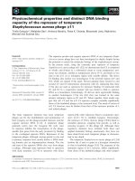
Fig. 1. Schematic diagram of how the topological distribution of purinergic signalling components might regulate the final cellular response.
P2XR have been described as being localized in both raft and non-raft membrane fractions and to couple to different downstream signalling
pathways depending on their location once activated by ATP. The enrichment of ecto-nucleotidases in lipid rafts would promote the acceler-
ated degradation of ATP to ADP and adenosine in the periphery of these microdomains. ADP and adenosine would activate respectively P2Y
and P1 receptors which are localized in lipid rafts and would engage their respective signalling pathways through G proteins. The differential
localization of receptors and ecto-nucleotidases would modulate the input signals in the form of ATP or its degradation products, as well as
the specific intracellular signalling outputs from each subclass of receptors. Finally, all these different intracellular signalling outputs would
be integrated to provoke the cellular response.
P2X receptors and membrane microdomains M. Garcia-Marcos et al.
336 FEBS Journal 276 (2009) 330–340 ª 2008 The Authors Journal compilation ª 2008 FEBS
single cells or the complex responses associated to
some receptors like the P2X
7
.
Acknowledgements
This work was supported by grant no. 3.4.528.07.F
from the Fonds National de la Recherche Scientifique
to JPD and by grant BFU2007-62728 Direccio
´
n
General de Investigacio
´
n. Ministerio de Educacio
´
ny
Ciencia (MEC) to AM.
References
1 Burnstock G (1972) Purinergic nerves. Pharmacol Rev
24, 509–581.
2 Burnstock G (2006) Historical review: ATP as a neuro-
transmitter. Trends Pharmacol Sci 27, 166–176.
3 Drury AN & Szent-Gyorgyi A (1929) The physiologi-
cal activity of adenine compounds with especial refer-
ence to their action upon the mammalian heart.
J Physiol 68, 213–237.
4 Schwiebert EM & Zsembery A (2003) Extracellular
ATP as a signaling molecule for epithelial cells.
Biochim Biophys Acta 1615, 7–32.
5 Burnstock G (2006) Purinergic signalling. Br J Pharma-
col 147(Suppl. 1), S172–S181.
6 Abbracchio MP & Burnstock G (1994) Purinoceptors:
are there families of P2X and P2Y purinoceptors?
Pharmacol Ther 64, 445–475.
7 North RA (2002) Molecular physiology of P2X recep-
tors. Physiol Rev 82, 1013–1067.
8 Khakh BS & North RA (2006) P2X receptors as cell-
surface ATP sensors in health and disease. Nature 442,
527–532.
9 Denlinger LC, Fisette PL, Sommer JA, Watters JJ,
Prabhu U, Dubyak GR, Proctor RA & Bertics PJ
(2001) Cutting edge: the nucleotide receptor P2X7 con-
tains multiple protein- and lipid-interaction motifs
including a potential binding site for bacterial lipopoly-
saccharide. J Immunol 167, 1871–1876.
10 Burnstock G & Knight GE (2004) Cellular distribution
and functions of P2 receptor subtypes in different
systems. Int Rev Cytol 240, 31–304.
11 Koles L, Furst S & Illes P (2007) Purine ionotropic
(P2X) receptors. Curr Pharm Des 13, 2368–2384.
12 Heo JS & Han HJ (2006) ATP stimulates mouse
embryonic stem cell proliferation via protein kinase C,
phosphatidylinositol 3-kinase ⁄ Akt, and mitogen-acti-
vated protein kinase signaling pathways. Stem Cells 24,
2637–2648.
13 da Cruz CM, Ventura AL, Schachter J, Costa-Junior
HM, da Silva Souza HA, Gomes FR, Coutinho-Silva
R, Ojcius DM & Persechini PM (2006) Activation of
ERK1 ⁄ 2 by extracellular nucleotides in macrophages is
mediated by multiple P2 receptors independently of
P2X7-associated pore or channel formation. Br J Phar-
macol 147, 324–334.
14 Cruz CM, Rinna A, Forman HJ, Ventura AL, Perse-
chini PM & Ojcius DM (2007) ATP activates a reactive
oxygen species-dependent oxidative stress response and
secretion of proinflammatory cytokines in macrophag-
es. J Biol Chem 282, 2871–2879.
15 Amstrup J & Novak I (2003) P2X7 receptor activates
extracellular signal-regulated kinases ERK1 and ERK2
independently of Ca
2+
influx. Biochem J 374, 51–61.
16 Bradford MD & Soltoff SP (2002) P2X7 receptors acti-
vate protein kinase D and p42 ⁄ p44 mitogen-activated
protein kinase (MAPK) downstream of protein kinase
C. Biochem J 366, 745–755.
17 Jacques-Silva MC, Rodnight R, Lenz G, Liao Z, Kong
Q, Tran M, Kang Y, Gonzalez FA, Weisman GA & Ne-
ary JT (2004) P2X7 receptors stimulate AKT phosphor-
ylation in astrocytes. Br J Pharmacol 141, 1106–1117.
18 Bulanova E, Budagian V, Orinska Z, Hein M, Petersen
F, Thon L, Adam D & Bulfone-Paus S (2005) Extra-
cellular ATP induces cytokine expression and apoptosis
through P2X7 receptor in murine mast cells. J Immunol
174, 3880–3890.
19 Perez-Andres E, Fernandez-Rodriguez M, Gonzalez
M, Zubiaga A, Vallejo A, Garcia I, Matute C, Pochet
S, Dehaye JP, Trueba M et al. (2002) Activation of
phospholipase D-2 by P2X(7) agonists in rat subman-
dibular gland acini. J Lipid Res 43, 1244–1255.
20 Pochet S, Garcia-Marcos M, Seil M, Otto A, Marino
A & Dehaye JP (2007) Contribution of two ionotropic
purinergic receptors to ATP responses in submandibu-
lar gland ductal cells. Cell Signal 19, 2155–2164.
21 Garcia-Marcos M, Perez-Andres E, Tandel S, Fontan-
ils U, Kumps A, Kabre E, Gomez-Munoz A, Marino
A, Dehaye JP & Pochet S (2006) Coupling of two
pools of P2X7 receptors to distinct intracellular signal-
ing pathways in rat submandibular gland. J Lipid Res
47, 705–714.
22 Alzola E, Perez-Etxebarria A, Kabre E, Fogarty DJ,
Metioui M, Chaib N, Macarulla JM, Matute C,
Dehaye JP & Marino A (1998) Activation by P2X7
agonists of two phospholipases A
2
(PLA
2
) in ductal
cells of rat submandibular gland. Coupling of the
calcium-independent PLA2 with kallikrein secretion.
J Biol Chem 273, 30208–30217.
23 Andrei C, Margiocco P, Poggi A, Lotti LV, Torrisi
MR & Rubartelli A (2004) Phospholipases C and A
2
control lysosome-mediated IL-1 beta secretion: implica-
tions for inflammatory processes. Proc Natl Acad Sci
USA 101, 9745–9750.
24 Lee YH, Lee SJ, Seo MH, Kim CJ & Sim SS (2001)
ATP-induced histamine release is in part related to
phospholipase A
2
-mediated arachidonic acid metabo-
M. Garcia-Marcos et al. P2X receptors and membrane microdomains
FEBS Journal 276 (2009) 330–340 ª 2008 The Authors Journal compilation ª 2008 FEBS 337
lism in rat peritoneal mast cells. Arch Pharm Res 24,
552–556.
25 Singer SJ & Nicolson GL (1972) The fluid mosaic
model of the structure of cell membranes. Science 175,
720–731.
26 Simons K & Vaz WL (2004) Model systems, lipid rafts,
and cell membranes. Annu Rev Biophys Biomol Struct
33, 269–295.
27 Simons K & van Meer G (1988) Lipid sorting in epi-
thelial cells. Biochemistry 27, 6197–6202.
28 Jacobson K, Sheets ED & Simson R (1995) Revisiting
the fluid mosaic model of membranes. Science 268,
1441–1442.
29 Brown DA & Rose JK (1992) Sorting of GPI-anchored
proteins to glycolipid-enriched membrane subdomains
during transport to the apical cell surface. Cell 68,
533–544.
30 Simons K & Ikonen E (1997) Functional rafts in cell
membranes. Nature 387, 569–572.
31 Pike LJ (2006) Rafts defined: a report on the Keystone
Symposium on Lipid Rafts and Cell Function. J Lipid
Res 47, 1597–1598.
32 Brown RE (1998) Sphingolipid organization in bio-
membranes: what physical studies of model membranes
reveal. J Cell Sci 111(Pt 1), 1–9.
33 Ramstedt B & Slotte JP (2002) Membrane properties
of sphingomyelins. FEBS Lett 531, 33–37.
34 Ramstedt B & Slotte JP (2006) Sphingolipids and the
formation of sterol-enriched ordered membrane
domains. Biochim Biophys Acta 1758, 1945–1956.
35 Pike LJ (2003) Lipid rafts: bringing order to chaos.
J Lipid Res 44, 655–667.
36 Pike LJ (2004) Lipid rafts: heterogeneity on the high
seas. Biochem J 378, 281–292.
37 Simons K & Toomre D (2000) Lipid rafts and signal
transduction. Nat Rev Mol Cell Biol 1, 31–39.
38 Insel PA, Head BP, Ostrom RS, Patel HH, Swaney
JS, Tang CM & Roth DM (2005) Caveolae and lipid
rafts: G protein-coupled receptor signaling microdo-
mains in cardiac myocytes. Ann NY Acad Sci 1047,
166–172.
39 Palade GE (1953) The fine structure of blood capillar-
ies. J Appl Phys 24, 1424.
40 Glenney JR Jr & Soppet D (1992) Sequence and
expression of caveolin, a protein component of caveo-
lae plasma membrane domains phosphorylated on
tyrosine in Rous sarcoma virus-transformed fibroblasts.
Proc Natl Acad Sci USA 89, 10517–10521.
41 Monier S, Parton RG, Vogel F, Behlke J, Henske A &
Kurzchalia TV (1995) VIP21-caveolin, a membrane
protein constituent of the caveolar coat, oligomerizes
in vivo and in vitro. Mol Biol Cell 6, 911–927.
42 Lichtenberg D, Goni FM & Heerklotz H (2005) Deter-
gent-resistant membranes should not be identified with
membrane rafts. Trends Biochem Sci 30, 430–436.
43 Munro S (2003) Lipid rafts: elusive or illusive? Cell
115, 377–388.
44 Varma R & Mayor S (1998) GPI-anchored proteins
are organized in submicron domains at the cell surface.
Nature 394, 798–801.
45 Harder T, Scheiffele P, Verkade P & Simons K (1998)
Lipid domain structure of the plasma membrane
revealed by patching of membrane components. J Cell
Biol 141, 929–942.
46 Meder D, Moreno MJ, Verkade P, Vaz WL & Simons
K (2006) Phase coexistence and connectivity in the api-
cal membrane of polarized epithelial cells. Proc Natl
Acad Sci USA 103, 329–334.
47 Gaus K, Gratton E, Kable EP, Jones AS, Gelissen I,
Kritharides L & Jessup W (2003) Visualizing lipid
structure and raft domains in living cells with two-pho-
ton microscopy. Proc Natl Acad Sci USA 100, 15554–
15559.
48 Prior IA & Hancock JF (2001) Compartmentalization
of Ras proteins. J Cell Sci 114, 1603–1608.
49 Prior IA, Parton RG & Hancock JF (2003) Observing
cell surface signaling domains using electron micros-
copy. Sci STKE 2003, PL9.
50 Wilson BS, Steinberg SL, Liederman K, Pfeiffer JR,
Surviladze Z, Zhang J, Samelson LE, Yang LH, Kotu-
la PG & Oliver JM (2004) Markers for detergent-resis-
tant lipid rafts occupy distinct and dynamic domains in
native membranes. Mol Biol Cell 15, 2580–2592.
51 Chamberlain LH (2004) Detergents as tools for the
purification and classification of lipid rafts. FEBS Lett
559, 1–5.
52 Song KS, Li S, Okamoto T, Quilliam LA, Sargiacomo
M & Lisanti MP (1996) Co-purification and direct
interaction of Ras with caveolin, an integral membrane
protein of caveolae microdomains. Detergent-free puri-
fication of caveolae microdomains. J Biol Chem 271,
9690–9697.
53 Smart EJ, Ying YS, Mineo C & Anderson RG (1995)
A detergent-free method for purifying caveolae mem-
brane from tissue culture cells. Proc Natl Acad Sci
USA 92, 10104–10108.
54 Macdonald JL & Pike LJ (2005) A simplified method
for the preparation of detergent-free lipid rafts. J Lipid
Res 46, 1061–1067.
55 Schnitzer JE, McIntosh DP, Dvorak AM, Liu J & Oh
P (1995) Separation of caveolae from associated micr-
odomains of GPI-anchored proteins. Science 269,
1435–1439.
56 Liu J, Oh P, Horner T, Rogers RA & Schnitzer JE
(1997) Organized endothelial cell surface signal trans-
duction in caveolae distinct from glycosylphosphat-
idylinositol-anchored protein microdomains. J Biol
Chem 272, 7211–7222.
57 Oh P & Schnitzer JE (1999) Immunoisolation of caveo-
lae with high affinity antibody binding to the oligo-
P2X receptors and membrane microdomains M. Garcia-Marcos et al.
338 FEBS Journal 276 (2009) 330–340 ª 2008 The Authors Journal compilation ª 2008 FEBS
meric caveolin cage. Toward understanding the basis
of purification. J Biol Chem 274, 23144–23154.
58 Foster LJ, De Hoog CL & Mann M (2003) Unbiased
quantitative proteomics of lipid rafts reveals high speci-
ficity for signaling factors. Proc Natl Acad Sci USA
100, 5813–5818.
59 Mineo C, Gill GN & Anderson RG (1999) Regulated
migration of epidermal growth factor receptor from
caveolae. J Biol Chem 274, 30636–30643.
60 Huang CS, Zhou J, Feng AK, Lynch CC, Klumper-
man J, DeArmond SJ & Mobley WC (1999) Nerve
growth factor signaling in caveolae-like domains at the
plasma membrane. J Biol Chem 274, 36707–36714.
61 Liu P, Ying Y, Ko YG & Anderson RG (1996) Locali-
zation of platelet-derived growth factor-stimulated
phosphorylation cascade to caveolae. J Biol Chem 271,
10299–10303.
62 Prior IA, Harding A, Yan J, Sluimer J, Parton RG &
Hancock JF (2001) GTP-dependent segregation of
H-ras from lipid rafts is required for biological activity.
Nat Cell Biol 3, 368–375.
63 Liu Y, Casey L & Pike LJ (1998) Compartmentaliza-
tion of phosphatidylinositol 4,5-bisphosphate in low-
density membrane domains in the absence of caveolin.
Biochem Biophys Res Commun 245, 684–690.
64 Pike LJ & Casey L (1996) Localization and turnover
of phosphatidylinositol 4,5-bisphosphate in caveolin-
enriched membrane domains. J Biol Chem 271, 26453–
26456.
65 Ostrom RS, Gregorian C, Drenan RM, Xiang Y, Regan
JW & Insel PA (2001) Receptor number and caveolar
co-localization determine receptor coupling efficiency to
adenylyl cyclase. J Biol Chem 276, 42063–42069.
66 Rybin VO, Xu X, Lisanti MP & Steinberg SF (2000)
Differential targeting of beta -adrenergic receptor sub-
types and adenylyl cyclase to cardiomyocyte caveolae.
A mechanism to functionally regulate the cAMP sig-
naling pathway. J Biol Chem 275, 41447–41457.
67 Xiang Y, Rybin VO, Steinberg SF & Kobilka B (2002)
Caveolar localization dictates physiologic signaling of
beta 2-adrenoceptors in neonatal cardiac myocytes.
J Biol Chem 277, 34280–34286.
68 Feron O, Smith TW, Michel T & Kelly RA (1997)
Dynamic targeting of the agonist-stimulated m2 musca-
rinic acetylcholine receptor to caveolae in cardiac
myocytes. J Biol Chem 272, 17744–17748.
69 Seno K, Kishimoto M, Abe M, Higuchi Y, Mieda M,
Owada Y, Yoshiyama W, Liu H & Hayashi F (2001)
Light- and guanosine 5¢-3-O-(thio)triphosphate-sensi-
tive localization of a G protein and its effector on
detergent-resistant membrane rafts in rod photorecep-
tor outer segments. J Biol Chem 276, 20813–20816.
70 Chun M, Liyanage UK, Lisanti MP & Lodish HF
(1994) Signal transduction of a G protein-coupled
receptor in caveolae: colocalization of endothelin and
its receptor with caveolin. Proc Natl Acad Sci USA 91,
11728–11732.
71 Sargiacomo M, Sudol M, Tang Z & Lisanti MP
(1993) Signal transducing molecules and glycosyl-
phosphatidylinositol-linked proteins form a caveolin-
rich insoluble complex in MDCK cells. J Cell Biol
122, 789–807.
72 Huang C, Hepler JR, Chen LT, Gilman AG, Anderson
RG & Mumby SM (1997) Organization of G proteins
and adenylyl cyclase at the plasma membrane. Mol
Biol Cell 8, 2365–2378.
73 Oh P & Schnitzer JE (2001) Segregation of heterotri-
meric G proteins in cell surface microdomains. G(q)
binds caveolin to concentrate in caveolae, whereas G(i)
and G(s) target lipid rafts by default. Mol Biol Cell 12,
685–698.
74 Hiol A, Davey PC, Osterhout JL, Waheed AA, Fischer
ER, Chen CK, Milligan G, Druey KM & Jones TL
(2003) Palmitoylation regulates regulators of G-protein
signaling (RGS) 16 function. I. Mutation of amino-ter-
minal cysteine residues on RGS16 prevents its targeting
to lipid rafts and palmitoylation of an internal cysteine
residue. J Biol Chem 278, 19301–19308.
75 Nini L, Waheed AA, Panicker LM, Czapiga M, Zhang
JH & Simonds WF (2007) R7-binding protein targets
the G protein beta 5 ⁄ R7-regulator of G protein signal-
ing complex to lipid rafts in neuronal cells and brain.
BMC Biochem 8, 18.
76 Baird B, Sheets ED & Holowka D (1999) How does
the plasma membrane participate in cellular signaling
by receptors for immunoglobulin E? Biophys Chem 82,
109–119.
77 Langlet C, Bernard AM, Drevot P & He HT (2000)
Membrane rafts and signaling by the multichain
immune recognition receptors. Curr Opin Immunol 12,
250–255.
78 Lasley RD, Narayan P, Uittenbogaard A & Smart EJ
(2000) Activated cardiac adenosine A(1) receptors
translocate out of caveolae. J Biol Chem 275, 4417–
4421.
79 Kaiser RA, Oxhorn BC, Andrews G & Buxton IL
(2002) Functional compartmentation of endothelial
P2Y receptor signaling. Circ Res 91, 292–299.
80 Kittel A, Csapo ZS, Csizmadia E, Jackson SW & Rob-
son SC (2004) Co-localization of P2Y1 receptor and
NTPDase1 ⁄ CD39 within caveolae in human placenta.
Eur J Histochem 48, 253–259.
81 Quinton TM, Kim S, Jin J & Kunapuli SP (2005)
Lipid rafts are required in Galpha(i) signaling down-
stream of the P2Y12 receptor during ADP-mediated
platelet activation. J Thromb Haemost 3, 1036–1041.
82 Savi P, Zachayus JL, Delesque-Touchard N, Labouret
C, Herve C, Uzabiaga MF, Pereillo JM, Culouscou
JM, Bono F, Ferrara P et al. (2006) The active metab-
olite of Clopidogrel disrupts P2Y12 receptor oligomers
M. Garcia-Marcos et al. P2X receptors and membrane microdomains
FEBS Journal 276 (2009) 330–340 ª 2008 The Authors Journal compilation ª 2008 FEBS 339
and partitions them out of lipid rafts. Proc Natl Acad
Sci USA 103, 11069–71104.
83 Vial C, Fung CY, Goodall AH, Mahaut-Smith MP &
Evans RJ (2006) Differential sensitivity of human
platelet P2X1 and P2Y1 receptors to disruption of lipid
rafts. Biochem Biophys Res Commun 343, 415–419.
84 Kellenberger S & Schild L (2002) Epithelial sodium
channel ⁄ degenerin family of ion channels: a variety of
functions for a shared structure. Physiol Rev 82, 735–
767.
85 Szabo I, Adams C & Gulbins E (2004) Ion channels
and membrane rafts in apoptosis. Pflugers Arch 448,
304–312.
86 Allen JA, Halverson-Tamboli RA & Rasenick MM
(2007) Lipid raft microdomains and neurotransmitter
signalling. Nat Rev Neurosci 8, 128–140.
87 Vacca F, Amadio S, Sancesario G, Bernardi G &
Volonte C (2004) P2X3 receptor localizes into lipid
rafts in neuronal cells. J Neurosci Res 76, 653–661.
88 Vial C & Evans RJ (2005) Disruption of lipid rafts
inhibits P2X1 receptor-mediated currents and arterial
vasoconstriction. J Biol Chem 280, 30705–30711.
89 Barth K, Weinhold K, Guenther A, Linge A, Gereke
M & Kasper M (2008) Characterization of the molecu-
lar interaction between caveolin-1 and the P2X recep-
tors 4 and 7 in E10 mouse lung alveolar epithelial cells.
Int J Biochem Cell Biol 40, 2230–2239.
90 Bannas P, Adriouch S, Kahl S, Braasch F, Haag F &
Koch-Nolte F (2005) Activity and specificity of toxin-
related mouse T cell ecto-ADP-ribosyltransferase
ART2.2 depends on its association with lipid rafts.
Blood 105, 3663–3670.
91 Garcia-Marcos M, Pochet S, Tandel S, Fontanils U,
Astigarraga E, Fernandez-Gonzalez JA, Kumps A,
Marino A & Dehaye JP (2006) Characterization and
comparison of raft-like membranes isolated by two
different methods from rat submandibular gland cells.
Biochim Biophys Acta 1758, 796–806.
92 Barth K, Weinhold K, Guenther A, Young MT,
Schnittler H & Kasper M (2007) Caveolin-1 influences
P2X7 receptor expression and localization in mouse
lung alveolar epithelial cells. FEBS J 274, 3021–3033.
93 Adriouch S, Bannas P, Schwarz N, Fliegert R, Guse
AH, Seman M, Haag F & Koch-Nolte F (2008) ADP-
ribosylation at R125 gates the P2X7 ion channel by
presenting a covalent ligand to its nucleotide binding
site. FASEB J 22, 861–869.
94 Seman M, Adriouch S, Haag F & Koch-Nolte F
(2004) Ecto-ADP-ribosyltransferases (ARTs): emerging
actors in cell communication and signaling. Curr Med
Chem 11, 857–872.
95 Kim M, Jiang LH, Wilson HL, North RA & Surpre-
nant A (2001) Proteomic and functional evidence for a
P2X7 receptor signalling complex. EMBO J 20, 6347–
6358.
96 Wilson HL, Wilson SA, Surprenant A & North RA
(2002) Epithelial membrane proteins induce membrane
blebbing and interact with the P2X7 receptor C termi-
nus. J Biol Chem 277, 34017–34023.
97 Zimmermann H (2000) Extracellular metabolism of
ATP and other nucleotides. Naunyn Schmiedebergs
Arch Pharmacol 362, 299–309.
98 Kenworthy AK & Edidin M (1998) Distribution of a
glycosylphosphatidylinositol-anchored protein at the
apical surface of MDCK cells examined at a resolution
of < 100 A
˚
using imaging fluorescence resonance
energy transfer. J Cell Biol 142, 69–84.
99 Kukulski F, Levesque SA, Lavoie EG, Lecka J, Bigon-
nesse F, Knowles AF, Robson SC, Kirley TL & Sev-
igny J (2005) Comparative hydrolysis of P2 receptor
agonists by NTPDases 1, 2, 3 and 8. Purinergic Signal
1, 193–204.
100 Kittel A, Kaczmarek E, Sevigny J, Lengyel K, Csizm-
adia E & Robson SC (1999) CD39 as a caveolar-asso-
ciated ectonucleotidase. Biochem Biophys Res Commun
262, 596–599.
101 Koziak K, Kaczmarek E, Kittel A, Sevigny J, Blus-
ztajn JK, Schulte Am Esch J II, Imai M, Guckelberger
O, Goepfert C, Qawi I et al. (2000) Palmitoylation tar-
gets CD39 ⁄ endothelial ATP diphosphohydrolase to
caveolae. J Biol Chem 275, 2057–2062.
102 Papanikolaou A, Papafotika A, Murphy C, Papamar-
caki T, Tsolas O, Drab M, Kurzchalia TV, Kasper M
& Christoforidis S (2005) Cholesterol-dependent lipid
assemblies regulate the activity of the ecto-nucleotidase
CD39. J Biol Chem 280, 26406–26414.
103 Burnstock G (2008) Unresolved issues and controver-
sies in purinergic signalling. J Physiol 586, 3307–3312.
P2X receptors and membrane microdomains M. Garcia-Marcos et al.
340 FEBS Journal 276 (2009) 330–340 ª 2008 The Authors Journal compilation ª 2008 FEBS