Báo cáo khoa học: What determines the degree of compactness of a calcium-binding protein? pdf
Bạn đang xem bản rút gọn của tài liệu. Xem và tải ngay bản đầy đủ của tài liệu tại đây (357.42 KB, 12 trang )
What determines the degree of compactness of a
calcium-binding protein?
Liliane Mouawad
1
, Adriana Isvoran
2
, Eric Quiniou
1
and Constantin T. Craescu
1
1 Inserm U759 ⁄ Institut Curie-Recherche, Centre Universitaire Paris-Sud, Orsay, France
2 Department of Chemistry, West University of Timisoara, Romania
Calcium transport and ⁄ or regulation are important
events for the normal morphology and metabolism of
the cell and play significant roles in the mechanisms of
many disease processes [1]. The proteins that interact
with the calcium ions involved in these events are
called calcium-binding proteins (CaBPs). They form
two main subfamilies: the EF-hand CaBPs and the
non-EF-hand CaBPs. EF-hand CaBPs, whose proto-
type is calmodulin [2], are characterized by the pres-
ence of structural motifs called ‘EF-hands’. Non
EF-hand CaBPs do not use this structural motif to
bind calcium; they may be found in the cytoplasm
(similar to C2 domain proteins) [3], in the extracellular
medium [4] or associated with the membrane (similar
to annexins) [5].
For the EF-hand CaBPs, each EF-hand motif con-
tains two helices connected by the calcium-binding
loop, a highly conserved region that binds the metal
ion. Many CaBPs exhibit two domains, each contain-
ing two EF-hand motifs; the N-terminal (helices A, B,
C and D) and C-terminal (helices E, F, G and H)
domains are connected by a linker region (Fig. 1).
Keywords
calcium-binding proteins; centrin; EF-hand;
hydrophobicity; predicted form
Correspondence
L. Mouawad, Inserm U759 ⁄ Institut Curie-
Recherche, Centre Universitaire Paris-Sud,
Ba
ˆ
timent 112, 91405 Orsay Cedex, France
Fax: +33 1 69 07 53 27
Tel: +33 1 69 86 71 51
E-mail:
(Received 8 September 2008, revised 8
December 2008, accepted 10 December
2008)
doi:10.1111/j.1742-4658.2008.06851.x
The EF-hand calcium-binding proteins may exist either in an extended or a
compact conformation. This conformation is sometimes correlated with the
function of the calcium-binding protein. For those proteins whose structure
and function are known, calcium sensors are usually extended and calcium
buffers compact; hence, there is interest in predicting the form of the pro-
tein starting from its sequence. In the present study, we used two different
procedures: one that already exists in the literature, the sosuidumbbell
algorithm, mainly based on the charges of the two EF-hand domains, and
the other comprising a novel procedure that is based on linker average
hydrophilicity. The linker consists of the residues that connect the domains.
The two procedures were tested on 17 known-structure calcium-binding
proteins and then applied to 59 unknown-structure centrins. The sosui-
dumbbell algorithm yielded the correct conformations for only 15 of the
known-structure proteins and predicted that all centrins should be in a
closed form. The linker average hydrophilicity procedure discriminated well
between all the extended and non-extended forms of the known-structure
calcium-binding proteins, and its prediction concerning centrins reflected
well their phylogenetic classification. The linker average hydrophilicity cri-
terion is a simple and powerful means to discriminate between extended
and non-extended forms of calcium-binding proteins. What is remarkable
is that only a few residues that constitute the linker (between 2 and 20 in
our tested sample of proteins) are responsible for the form of the calcium-
binding protein, showing that this form is mainly governed by short-range
interactions.
Abbreviations
CaBP, calcium-binding protein; LAH, linker average hydrophilicity; PDB, Protein Data Bank.
1082 FEBS Journal 276 (2009) 1082–1093 Journal compilation ª 2009 FEBS. No claim to original French government works
EF-hand CaBPs are divided into two broad classes [6]:
those that bind calcium to regulate its concentration
(calcium-buffering and calcium-transporting proteins)
and those that bind calcium to decode its signal (cal-
cium-sensor proteins). The two functional classes also
have different structural features: calcium-buffering
and calcium-transporting proteins, such as parvalbu-
min [7] or the Nereis diversicolor sarcoplasmic calcium-
binding protein [8], usually have a compact tertiary
structure and are not conformationally sensitive to cal-
cium-binding, whereas calcium sensor proteins, such as
calmodulin [2] and troponin C [9], have extended ter-
tiary structures and show important conformational
changes upon calcium-binding. In the extended form,
the linker between the two domains may be structured
in a straight helix, whereas, in the non-extended form,
the linker is unstructured leading to either a floppy
conformation or a very compact one (Fig. 2) [10]. It is
important to understand the physical reasons for these
differences. This would provide tools to predict the
form of the CaBPs from their sequences, and therefore
indicate their biological function.
Recently, a protein classification tool, sosuidumb-
bell [11], was developed to predict the degree of com-
pactness of proteins starting from their amino acid
sequences. This tool is based on studies undertaken on
all the monomers of the Protein Data Bank (PDB)
[12], and not just CaBPs, indicating that the electro-
static repulsion between the domains is a dominant
factor in the stabilization of the extended structures, in
addition to the amphiphilic character of the central
flexible region. By contrast, globular proteins are pre-
dicted to be stabilized by a hydrophobic core built by
residues from the two domains. Using the sosuidumb-
bell algorithm, we have analyzed 17 CaBPs with
known 3D structures (Table 1). Fifteen of them were
predicted in the correct form but, unfortunately, two
structures were incorrectly predicted. Indeed, human
calmodulin-like protein (1GGZ) [13] and human cen-
trin 2 (2GGM) [14], which are extended proteins, were
predicted to be compact. These exceptions represent a
non-negligible percentage (12%) and they emphasize
the need for a more detailed analysis of the sequence–
structure relationship in the case of CaBPs.
In the present study, we have developed a novel pro-
cedure based on the linker average hydrophilicity
(LAH), which we applied to our sample of 17 known-
structure CaBPs and to unknown structures of cent-
rins. Centrins, a subfamily of CaBPs, are essential
components of microtubule-organizing centers in
organisms ranging from algae and yeast to humans
[15,16]. They are EF-hand calcium-binding proteins
with a sequence similarity to calmodulin but distinct
calcium-binding properties [15]. They were shown to
be involved in centrosome duplication [17] and the
contraction of centrin-based fiber systems [18] and to
play a functional role in nuclear export pathways [19].
The Ca
2+
dependence of the centrin interactions with
their targets suggests that centrins play a regulatory
role by activating or changing the conformation of
various target proteins. Analyses of amino acid
sequences of centrins from different organisms reveal
at least four phylogenetic families and several phyloge-
netic subfamilies [20,21]. The centrins that we consider
in the present study are listed in Table 2: (a) the Chla-
mydomonas reinhardtii-like family (CrCen-like), which
contains centrins from the subfamilies of green algae
and vertebrate isoforms Cen1 and Cen2; (b) the higher
plants Arabidopsis-like family (AtCen-like); (c) the
yeast Saccharomyces cerevisiae-like family (Cdc31-like),
which contains mainly two subfamilies, fungal centrins
and the vertebrate isoform Cen3; and (d) the Parame-
cium tetraurelia infraciliary lattice family (PtICL1-like),
A
BC
D
E
FGH
N-domain C-domain
Loop I Loop II Loop III
Loop IV
Linker
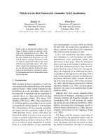
Fig. 1. The EF-hand protein schematic representation. Each EF-
hand motif consists of two helices linked by a calcium loop (black
dots represent calcium ions). Two motifs constitute one EF-hand
domain. The N- and C-domains are bound by a linker (bold line).
AB
Fig. 2. View of the 3D structures of two CaBPs: (A) the extended
form of calmodulin (PDB code: 1CLL) and (B) the non-extended
form of guanylate cyclase activating protein 2 (PDB code: 1JBA).
The helices are in cyan, the b-sheets are in yellow and the linker is
in red. The linker in 1CLL is structured, whereas it is a loop in
1JBA. The view was drawn using
VMD software [10].
L. Mouawad et al. Compactness of calcium-binding proteins
FEBS Journal 276 (2009) 1082–1093 Journal compilation ª 2009 FEBS. No claim to original French government works 1083
organized in ten subfamilies that contain 35 identified
isoforms [22]. The 3D structure of the entire protein in
complex with its target polypeptide is known for only
two centrins: the human centrin: HsCen2 (2GGM) [14]
and the Saccaromyces cerevisiae centrin, ScCdc31
(2DOQ) [23].
The functional diversity of centrins should depend
on their sequence and their Ca
2+
binding properties.
However, we may ask whether the global conforma-
tion or the conformational preference of individual
centrin molecules also play a role in the target recogni-
tion and the plasticity of heteromolecular complexes.
This idea is supported by the recent observation that
yeast ScCdc31 bound to a ScSfi1 fragment shows a
bent conformation [23], whereas human HsCen2 in
complex with an XPC peptide is completely extended
[14]. In the present study, we present a new and simple
theoretical procedure for the global shape prediction
of EF-hand proteins that allows us to analyze the pos-
sible shape diversity of centrins presented in Table 2.
Results and Discussion
Utilization of the SOSUIDUMBBELL algorithm
We first applied the sosuidumbbell algorithm (http://
bp.nuap.nagoya-u.ac.jp/sosui/sosuidumbbell/dumbbell_
submit.html) to all the CaBPs with known 3D struc-
tures (Table 1). In this algorithm, a structure is pre-
dicted to be extended if it obeys four criteria: (a) the
absolute value of the net charge of the entire protein is
higher than 20 (|Q
prot
| > 20); (b) the absolute net
charge density (|Q
prot
| ⁄ N, where N is the total number
of residues) is higher than 0.14 (d
Q
> 0.14); (c) there
is a charge balance between the two domains
(|Q
N
Q
C
| > 100); and (d) there is a high amphiphilicity
at the center of the linker region and a high hydropa-
thy at its termini [11]. Based on these four criteria, the
results yielded 15 well-predicted structures and two
incorrectly predicted ones. The latter are human cal-
modulin-like protein (1GGZ) and human centrin 2
(2GGM), the structures of which are extended but pre-
dicted as non-extended. Therefore the question
remained as to which of the four criteria described
above is responsible for this misprediction. To address
this question, we verified initially the first two criteria.
For this purpose, we calculated the absolute net charge
and the charge density of the entire protein for all the
investigated CaBPs (Table 3), with known and
unknown structures (Tables 1 and 2). First, we fol-
lowed exactly the procedure described by Uchikoga
et al. [11], namely that histidine residues were consid-
ered as positively charged (although at the pH values
corresponding to the great majority of the experiments,
they are deprotonated) and the calcium ions that might
bind to the protein were omitted. The results
Table 1. Features of the known-structure CaBPs used in the present study, showing the name of the protein, its code in the PDB, its code
in the SwissProt data bank, its form as determined experimentally and its form as predicted by the
SOSUIDUMBBELL algorithm (http://
bp.nuap.nagoya-u.ac.jp/sosui/sosuidumbbell/dumbbell_submit.html). CIB, calcium-and-integrin-binding protein; SCBP, sarcoplasmic calcium-
binding protein.
Protein
PDB
code
SwissProt
code
Experimental
structure
Structure predicted by
the
SOSUIDUMBBELL algorithm
Chicken troponin C 4TNC P02588 Extended Extended
Rabbit troponin C 1TN4 P02586 Extended Extended
Human calmodulin 1CLL P62158 Extended Extended
Paramecium calmodulin 1OSA P07463 Extended Extended
Potato calmodulin 1RFJ Q42478 Extended Extended
Human calmodulin-like protein 1GGZ P27482 Extended Non-extended
Human centrin 2 2GGM P41208 Extended Non-extended
Yeast centrin 2DOQ P06704 Non-extended Non-extended
Yeast myosin light chain 1GGW
a
Q09196 Non-extended Non-extended
Calcineurin B homologous protein 1 2CT9 P61023 Non-extended Non-extended
Bovine recoverin 1REC P21457 Non-extended Non-extended
Guanylate cyclase activating protein 2 1JBA
a
P51177 Non-extended Non-extended
Bovine neurocalcin d 1BJF P61602 Non-extended Non-extended
Amphioxus SCBP 2SAS P04570 Non-extended Non-extended
Sandworm SCBP 2SCP P04571 Non-extended Non-extended
Bacterial calerythrin 1NYA
a
P06495 Non-extended Non-extended
Human CIB 1DGU
a
Q99828 Non-extended Non-extended
a
Structure determined by NMR.
Compactness of calcium-binding proteins L. Mouawad et al.
1084 FEBS Journal 276 (2009) 1082–1093 Journal compilation ª 2009 FEBS. No claim to original French government works
Table 2. Phylogenetic classification of centrins. All centrins considered in the present study (with known and unknown structures) are classi-
fied by families and subfamilies. The PDB codes of the known structures of fragments (*) or the entire protein are given.
Phylogenetic family Subfamily Protein name Abbreviation SwissProt code ⁄ PDB code
CrCen Cen1 Human centrin 1 HsCen1 Q12798
Mouse centrin 1 MmCen1 P41209
Bovine centrin 1 BtCetn1 Q32LE3
Cen2 Human centrin 2 HsCen2 P41208 ⁄ 2GGM ⁄ 2OBH ⁄
1M39
*
⁄ 1ZMZ
*
⁄ 2A4J
*
Mouse centrin 2 MmCen2 Q9R1K9
Pig centrin 2 SsCen2 Q4U4N2
Xenopus laevis centrin 2 XlCen2 Q7SYA4
Xenopus tropicalis centrin 2 XtCen2 Q28HC5
Algae centrins Dunaliella salina centrin DsCen P54213
Chlamydomonas reinhardtii centrin CrCen P05434 ⁄ 1OQP
*
⁄ 2AMI
*
Tetraselmis striata centrin TsCen P43646
Scherffelia dubia centrin SdCen Q06827
Micromonas pusilla centrin MpCen Q40303
Marsilea vestita centrin MvCen O49999
Spermatozopsis similis centrin SsCen P43645
Pterosperma cristatum centrin PcCen Q40791
AtCen Higher plant centrins Arabidopsis thaliana centrin AtCen O82659
Nicotiana tabacum centrin 1 NtCen Q9SQI5
Atriplex nummularia centrin AnCen P41210
Cdc31 Cen3 Human centrin 3 HsCen3 O15182
Rat centrin 3 RnCen3 Q91ZZ8
Mouse centrin 3 MmCen3 O35648
Xenopus laevis centrin 3 XlCen3 Q9DEZ4
Euplotes octocarinatus centrin EoCen Q9XZV2 ⁄ 2JOJ
*
Yeast centrin ScCdc31 P06704 ⁄ 2DOQ ⁄ 2GV5
Xenopus tropicalis centrin 3 XtCen3 Q28GW2
PtICLs ICL1a PtICL1a Q27177
PtICL1b Q 27179
PtICL1c Q 27178
PtICL1d Q 94726
PtICL1f Q3SEK2
ICL1e PtICL1e Q3SEK0
PtICL1g Q3SEJ9
PtCen8 Q3SEJ6
PtCen10 Q3SEJ7
PtCen12 Q6BFB6
PtCen15 Q3SEJ0
PtCen18 A0CTY5
ICL3a PtICL3a Q3SDB8
PtICL3c Q3SDA6
PtICL3d Q3SEI1
PtICL3e Q3SEI3
PtICL3f Q3SEI4
ICL3b PtICL3b Q3SEI0
PtICL3g A0BUT1
ICL5 PtICL5a Q3SEH8
PtICL5b Q3SEH7
PtICL6a Q3SEH9
PtICL6b Q3SCX3
ICL7 PtICL7a A0DZH6
PtICL7b A0DZH
ICL8 PtICL8a A0BTY0
PtICL8b A0C3G3
ICL9 PtICL9a Q3SEI2
L. Mouawad et al. Compactness of calcium-binding proteins
FEBS Journal 276 (2009) 1082–1093 Journal compilation ª 2009 FEBS. No claim to original French government works 1085
(Fig. 3A,B and Table 3) show that, as indicated above,
only five known-structure proteins are predicted to be
extended instead of the seven expected (1GGZ and
2GGM are mispredicted) and all centrins with
unknown structures are predicted in a non-extended
form. In a second step, the histidines were considered
neutral (CaBPs usually contain very little His) and the
Ca
+2
ions were added, but the results were even worse
(data not shown) because the net charge was dimin-
ished and therefore the structures were predicted to be
even more compact. The first two criteria appear to be
responsible for the misprediction of the form of 1GGZ
and 2GGM. Moreover, concerning centrins with
unknown structures, some experimental results (C. T.
Craescu & S. Miron, unpublished data) in addition to
the phylogenetic classification indicate that at least the
CrCen family proteins should be in an extended form,
which is not the case in the prediction based on the
first two criteria.
The last two criteria in the sosuidumbbell algo-
rithm are strongly dependent on the definition of the
domains and the inter-domain linker. The delimita-
tion of this linker is not always obvious: in the
extended structures, it forms a helix in the continuity
of helices D and E, whereas, in some compact con-
formations, it is a very short unstructured region
(Fig. 2). In the sosuidumbbell algorithm, the linker
considered may be too long and, consequently, the
domains too short, as for calmodulin, where helices
D and E, which belong to the N- and C-domains,
respectively, are considered as parts of the linker
[11]. In the present study, to determine the linker,
we identified first the calcium-binding loops (Fig. 1),
then we counted ten residues after loop II (corre-
sponding to helix D) and ten residues before loop III
(corresponding to helix E), and the remaining resi-
dues inbetween were considered as the inter-domain
linker. Ten residues were considered for helices D
and E because the experimental structural data show
that a helix belonging to an EF-hand motif contains
ten residues on average. Consequently, in the pro-
teins investigated in the present study, the linker was
between two and 20 residues long (Table 3), corre-
sponding to 0.96% and 10.26%, respectively, of the
protein sequence length.
Based on this definition of the linker, the charges of
the N- and C-domains were calculated without consid-
ering the calcium ions. In Fig. 3C, we report the abso-
lute value of the product of these charges, |Q
N
Q
C
|,
which represents the charge balance between the
domains. With the exception of troponins, all the
investigated proteins are characterized by products
|Q
N
Q
C
| lower than 100, and therefore are predicted to
be non-extended.
From these results, it is clear that, for CaBPs, the
charges of the entire protein or of the separated
domains are not responsible for the extended or com-
pact form of the protein. This assertion is obvious in
the case of human centrin 2 (HsCen2). In this protein,
the first 25 amino acids, corresponding to a disordered
region, are highly charged [24,25], with the net charge
of this peptide being equal to 6 (it contains seven basic
and one acidic residues). The X-ray structure of this
protein was obtained in the presence [14] and in the
absence [25] of these residues (PDB codes 2GGM and
2OBH, respectively). In both cases, HsCen2 adopts an
extended conformation, showing that the charge bal-
ance of the domains does not play an important role
for this protein. Nevertheless, in both cases, the sosui-
dumbbell algorithm predicts a non-extended form,
which is not correct. Moreover, the structure of all the
extended forms of the CaBPs considered in the present
study was determined experimentally in the presence of
calcium ions. Knowing that these ions reduce signifi-
cantly the charges of the domains and therefore their
electrostatic repulsions, calcium-binding should favor
the compact structure of CaBPs, which is not the case.
The fourth criterion of the sosuidumbbell tool
refers to the hydrophobicity of the central linker
region, which is calculated using the Kyte & Doolittle
Scale [26]. Ushikoga et al. [11] described the linker
region of an extended protein as having an important
negative hydrophobicity in its center (i.e. to be signifi-
cantly hydrophilic), whereas its edges (helices D and
Table 2. Continued.
Phylogenetic family Subfamily Protein name Abbreviation SwissProt code ⁄ PDB code
PtICL9b A0BE66
PtICL9c A0D3D5
PtICL9d A0D6A4
ICL10 PtICL10a A0DZD2
PtICL10b A0BJD5
ICL11 PtICL11a A0BI27
PtICL11b A0BQH1
Compactness of calcium-binding proteins L. Mouawad et al.
1086 FEBS Journal 276 (2009) 1082–1093 Journal compilation ª 2009 FEBS. No claim to original French government works
Table 3. Results of all our calculations on CaBPs, showing the PDB code or the abbreviation of the protein name, the number of the protein as used in Figs 3 and 4 , the net charge of
the N- and C-domains (Q
N
, Q
C
), the net charge of the linker (Q
link
) and of the entire protein (Q
prot
), the charge balance of the domains (|Q
N
Q
C
|), the absolute charge density (d
Q
), the value
of the LAH, the number of Pro, Gly, Trp and Phe in the linker region plus three residues from each side of the sequence (length equals n+6), the residues belonging to the linker as
defined in the text, the total number of residues (N), the linker length (n) and, finally, the percentage of linker length with respect to the protein length.
Protein No. Q
N
Q
c
Q
link
Q
prot
|Q
N
Q
C
| d
Q
¼
Q
prot
jj
N
LAH
No. Pro
residues
No. Gly
residues
No. Trp
residues
No. Phe
residues
Linker region
]J + 10, K – 10[
Total no.
of residues (N)
Linker
length (n)
Percent linker
length
n
N
 100
ÀÁ
Extended structures
4TNC 1 )14 )13 )1 )28 182 0.172 1.662 0 1 0 0 89–96 163 8 4.91
1TN4 2 )14 )13 )1 )28 182 0.175 1.662 0 1 0 0 86–93 160 8 5.00
1CLL 3 )10 )10 )3 )23 100 0.154 1.822 0 0 0 0 79–83 149 5 3.36
1OSA 4 )10 )9 )3 )22 90 0.147 1.782 0 0 0 0 79–83 149 5 3.36
1RFJ 5 )10 )9 )3 )22 90 0.147 1.822 0 0 0 0 79–83 149 5 3.36
1GGZ 6 )8 )7 )3 )18 56 0.121 1.811 0 0 0 0 79–83 149 5 3.36
2GGM 7 1 )90 )8 9 0.046 1.713 0 0 0 0 99–103 172 5 2.91
Non-extended structures
2DOQ 8 )7 )10 1 )16 70 0.099 1.174 1 0 0 0 91–95 161 5 3.11
1GGW 9 )3 )5 )2 )10 15 0.071 0.516 3 2 0 2 70–76 141 7 4.96
2CT9 10 0 )4 )2 )6 0 0.031 0.965 3 1 0 1 93–112 195 20 10.26
1REC 11 ) 2 )21 )3 4 0.015 0.477 0 1 0 0 96–99 202 4 1.98
1JBA 12 )90)1 )10 0 0.049 0.317 0 1 0 0 91–94 204 4 1.96
1BJF 13 )2 )10 )3 2 0.015 1.016 0 1 0 0 95–98 193 4 2.07
2SAS 14 )8 )20)10 16 0.054 )0.202 1 0 1 0 92–104 185 13 7.03
2SCP 15 )2 )10 )1 )13 20 0.075 0.532 2 1 0 0 86–93 174 8 4.60
1NYA 16 )5 )7 )1 )13 35 0.073 0.003 1 2 0 2 90–102 177 13 7.34
1DGU 17 0 )9 )1 )10 0 0.052 )0.404 0 0 0 2 93–97 191 5 2.62
Unknown structures
HsCen1 18 1 )11 0 )10 11 0.058 1.713 0 0 0 0 99–103 172 5 2.91
MmCen1 19 1 )11 0 )10 11 0.058 1.642 0 0 0 0 99–103 172 5 2.91
BtCen1 20 1 )10 0 )9 10 0.052 1.642 0 0 0 0 99–103 172 5 2.91
MmCen2 21 1 )90 )8 9 0.046 1.713 0 0 0 0 99–103 172 5 2.91
SsCen2 22 )2 )90)11 18 0.079 1.713 0 0 0 0 66–70 139 5 3.60
XlCen2 23 1 )90 )8 9 0.046 1.731 0 0 0 0 99–103 172 5 2.91
XtCen2 24 1 )90 )8 9 0.046 1.731 0 0 0 0 99–103 172 5 2.91
DsCen 25 0 )90 )9 0 0.053 1.767 0 1 0 0 96–100 169 5 2.96
CrCen 26 ) 1 )11 0 )12 11 0.071 1.749 0 1 0 0 96–100 169 5 2.96
TsCen 27 )7 )10 0 )17 70 0.115 1.760 0 1 0 0 75–79 148 5 3.38
SdCen 28
)1 )10 0 )11 10 0.065 1.760 0 1 0 0 95–99 168 5 2.98
MpCen 29 )6 )10 0 )16 60 0.108 1.760 0 1 0 0 75–79 148 5 3.38
MvCen 30 ) 1 )10 0 )11 10 0.065 1.762 0 1 0 0 97–101 170 5 2.94
SsCen 31 )9 )90)18 81 0.121 1.760 0 1 0 0 75–79 148 5 3.38
PcCen 32 )5 )12 0 )17 60 0.128 1.760 0 1 0 0 67–71 133 5 3.76
AtCen 33 0 )70 )7 0 0.041 1.669 0 1 0 0 94–98 169 5 2.96
NtCen 34 )5 )70)12 35 0.068 1.698 0 1 0 1 103–107 177 5 2.82
AnCen 35 )1 )70 )8 7 0.048 1.649 0 1 0 0 93–97 167 5 2.99
L. Mouawad et al. Compactness of calcium-binding proteins
FEBS Journal 276 (2009) 1082–1093 Journal compilation ª 2009 FEBS. No claim to original French government works 1087
Table 3. Continued.
Protein No. Q
N
Q
c
Q
link
Q
prot
|Q
N
Q
C
| d
Q
¼
Q
prot
jj
N
LAH
No. Pro
residues
No. Gly
residues
No. Trp
residues
No. Phe
residues
Linker region
]J + 10, K – 10]
Total no.
of residues (N)
Linker
length (n)
Percent linker
length
n
N
 100
ÀÁ
HsCen3 36 )3 )11 0 )14 33 0.084 0.927 1 0 1 0 96–100 167 5 2.99
RnCen3 37 )2 )11 0 )13 22 0.082 0.927 1 0 1 0 88–92 159 5 3.14
MmCen3 38 )2 )11 0 )13 22 0.078 0.927 1 0 1 0 96–100 167 5 2.99
XlCen3 39 )2 )11 )1 )14 22 0.084 1.076 1 0 0 0 96–100 167 5 2.99
EoCen 40 )3 )90)12 27 0.071 1.215 1 0 0 0 95–99 168 5 2.98
XtCen3 41 )2 )11 ) 1 )14 22 0.084 1.076 1 0 0 0 96–100 167 5 2.99
PtICL1a 42 )6 )70)13 42 0.072 1.476 0 0 0 0 108–112 181 5 2.76
PtICL1b 43 )6 )70)13 42 0.071 1.476 0 0 0 0 109–113 182 5 2.75
PtICL1c 44 )6 )70)13 42 0.071 1.476 0 0 0 0 110–114 183 5 2.73
PtICL1d 45 )6 )70)13 42 0.072 1.476 0 0 0 0 108–112 181 5 2.76
PtICL1f 46 )6 )70)13 42 0.071 1.476 0 0 0 0 110–114 183 5 2.73
PtICL1e 47 )6 )51)10 30 0.057 1.196 0 2 0 0 102–107 174 6 3.45
PtICL1g 48 )6 )51)10 30 0.054 1.196 0 2 0 0 110–115 182 6 3.30
PtCen8 49 )6
)52 )9 30 0.051 1.107 0 2 0 0 104–109 176 6 3.41
PtCen10 50 )6 )51)10 30 0.057 1.196 0 2 0 0 102–107 174 6 3.45
PtCen12 51 )6 )51)10 30 0.057 1.196 0 2 0 0 102–107 174 6 3.45
PtCen15 52 )6 )51)10 30 0.057 1.185 0 2 0 0 104–109 176 6 3.41
PtCen18 53 )6 )51)10 30 0.057 1.196 0 2 0 0 102–107 174 6 3.45
PtICL3a 54 )6 )80)14 48 0.072 1.542 0 0 0 0 117–121 192 5 2.60
PtICL3c 55 11 )8 0 3 88 0.015 1.522 0 0 0 0 117–121 192 5 2.60
PtICL3d 56 )3 )80)11 24 0.057 1.542 0 0 0 0 117–121 192 5 2.60
PtICL3e 57 )3 )80)11 24 0.057 1.542 0 0 0 0 115–119 190 5 2.63
PtICL3f 58 )2 )80)10 16 0.050 1.542 0 0 0 0 122–126 197 5 2.54
PtICL3b 59 )3 )71 )9 21 0.046 0.820 0 1 0 0 119–123 193 5 2.59
PtICL3g 60 )3 )71 )9 21 0.047 0.820 0 1 0 0 118–122 192 5 2.60
PtICL5a 61 )5 )42 )7 20 0.038 0.918 0 2 0 0 104–109 182 6 3.30
PtICL5b 62 )5 )42 )7 20 0.038 0.918 0 2 0 0 104–109 182 6 3.30
PtICL6a 63 )4 )42 )6 16 0.032 0.865 0 2 0 0 106–111 184 6 3.26
PtICL6b 64 )4
)42 )6 16 0.033 0.865 0 2 0 0 106–111 184 6 3.26
PtICL7a 65 0 )2 )1 )3 0 0.016 0.527 0 2 0 1 100–104 184 5 2.72
PtICL7b 66 0 )1 )1 )2 0 0.011 0.527 0 2 0 1 100–104 184 5 2.72
PtICL8a 67 )1 )2 )1 ) 4 2 0.022 0.409 1 2 0 1 99–104 184 6 3.26
PtICL8b 68 )1 )20 )3 2 0.016 0.332 1 2 0 1 99–104 184 6 3.26
PtICL9a 69 )6 )10 0 )16 60 0.077 0.206 0 2 0 1 132–133 208 2 0.96
PtICL9b 70 )6 )10 0 )16 60 0.077 0.206 0 2 0 1 132–133 208 2 0.96
PtICL9c 71 )6 )10 0 )16 60 0.078 0.206 0 2 0 1 130–131 206 2 0.97
PtICL9d 72 )6 )10 0 )16 60 0.077 0.206 0 2 0 1 132–133 208 2 0.96
PtICL10a 73 3 )7 ) 1 )5 21 0.024 1.218 1 1 0 0 129–133 205 5 2.44
PtICL10b 74 2 )6 )1 )5 12 0.024 1.280 1 1 0 0 129–133 205 5 2.44
PtICL11a 75 )3 1 2 0 3 0 1.878 0 0 0 0 164–167 240 4 1.67
PtICL11b 76 )2 1 2 1 2 0.004 1.878 0 0 0 0 164–167 240 4 1.67
Compactness of calcium-binding proteins L. Mouawad et al.
1088 FEBS Journal 276 (2009) 1082–1093 Journal compilation ª 2009 FEBS. No claim to original French government works
E) are hydrophobic. In the present study, the same
calculations were applied to all known-structure pro-
teins, and it was observed that, in some cases, non-
extended proteins (e.g. recoverin; 1REC) present the
same hydropathy profile around the linker as extended
proteins, such as calmodulin or troponin C (1OSA and
4TNC; Fig. 3D). Therefore, none of the criteria
retained in the sosuidumbbell algorithm are com-
pletely reliable to predict the form of the CaBPs. This
motivated our search for other criteria.
Utilization of other criteria
Contact area
We analyzed the contact area between the domains of
known-structure non-extended CaBPs. As expected,
most of the residues at the interface were found to be
hydrophobic. In most compact structures, a trypto-
phan (or less frequently a phenylalanine) located in
one domain was buried in a hydrophobic cavity in the
other domain, which would stabilize the compact
structure. Unfortunately, this observation cannot be
used as a predictive tool starting from the sequence
because the aromatic residue is not located in a specific
part of it. Indeed, the sequence of the linker and its
close vicinity (three more residues from each side of
the linker) does not always contain tryptophan or
phenylalanine residues for compact forms (see 1REC,
1JBA, 1BJF and 2SCP in Table 3).
The presence of helix breakers
Prolines and, to a lesser extent, glycines, are well-
known helix breakers. We investigated the presence of
0
5
10
15
20
25
30
AB
CD
0 1020304050607080
Q
prot
Protein number
0 1020304050607080
Protein number
Protein number
–0.05
0
0.05
0.1
0.15
0.2
0 1020304050607080
d
Q
150
200
1
2
3
4
Linker
Helix E
Helix D
0
50
100
|Q
N
Q
C
|
–4
–3
–2
–1
0
–30 –20 –10 0 10 20 30
Relative residue number
Hydrophobicity
Fig. 3. Test of the four criteria used in the SOSUIDUMBBELL algorithm. (A) The absolute net charge (|Q
prot
|) of investigated proteins without cal-
cium ions versus the protein number from Table 3. The horizontal line corresponds to the limit of net charge between extended (|Q
prot
| > 20)
and non-extended structures (|Q
prot
| £ 20) as considered by Uchikoga et al. [11]. Vertical lines delimit between the known extended struc-
tures (filled circles), the known non-extended structures (open diamonds) and the unknown structures of centrins (filled triangles). It can be
seen that two extended structures are mispredicted (1GGZ and 2GGM) and that all the unknown-structure centrins are predicted to be non-
extended. (B) The absolute net charge density (d
Q
) with a horizontal line limit at 0.14. (C) The absolute value of the product of the two
domain charges (|Q
N
Q
C
|) in the absence of calcium ions with a horizontal line limit at 100. In this case, only tropnin C molecules are pre-
dicted to be extended. (D) The hydrophobicity profile of the linker region and its surroundings using the Kyte & Doolitle Scale for two
extended structures (dotted lines, 1OSA and dashed line, 4TNC) and for a non-extended one (solid line, 1REC). For convenience of compari-
son, the three sequences were renumbered and centered on the linker. The zero point corresponds to residue number 92 in 4TNC, 81 in
1OSA and 98 in 1REC, which represents the center of the linker in each case.
L. Mouawad et al. Compactness of calcium-binding proteins
FEBS Journal 276 (2009) 1082–1093 Journal compilation ª 2009 FEBS. No claim to original French government works 1089
such residues in the linker or its vicinity (i.e. plus three
residues from each side of the linker). The results pre-
sented in Table 3 show that, as expected, the presence
of a Pro yields a non-extended form by breaking the
central helix that constitutes the linker, but the reverse
is not true because all the compact CaBPs do not con-
tain a Pro in the linker. Therefore, this criterion can-
not constitute a predictive rule. Moreover, concerning
glycines, it was observed that, in both troponin C pro-
teins (4TNC and 1TN4), which are extended, there is
one Gly in the linker, as in bovine recoverin (1REC),
guanylate cyclase activating protein 2 (1JBA) and
bovine neurocalcin d (1BJF), which present very com-
pact structures.
Net electric charge of the linker
It might be assumed that the net electric charge of the
linker plays a role if there is repulsion between this lin-
ker and the adjacent domains. Thus, this property was
investigated (Table 3) but did not yield a good discrim-
inating criterion because, in HsCen2 (2GGM), which
is extended, the linker is neutral as in bovine neurocal-
cin d (1BJF) or amphioxus sarcoplasmic calcium-bind-
ing protein (2SAS), which are non-extended structures.
Hydrophilicity of the linker
The criterion that yielded the best results was based on
the hydrophilicity of the linker. It was obtained by the
procedure detailed below. First, the hydrophilicity (h
i
)
of each residue i of the protein was calculated using
the Hopp & Woods Scale [27] with a nine-residue slid-
ing window. In this scale, positive values correspond
to hydrophilic positions.
Second, the linker was determined as described
above: if the last residue of the calcium-binding loop II
is denoted J and the first residue of the calcium-bind-
ing loop III is denoted K, the linker consists of all resi-
dues comprised in the interval ]J + 10, K ) 10[.
Finally, the LAH was calculated:
LAH ¼
X
i2 Jþ10;KÀ10½
h
i
n
where n is the number of residues in the linker and h
i
is the hydrophilicity at position i of the linker.
This procedure was applied to all proteins in
Tables 1 and 2. The results are presented in Fig. 4.
Remarkably, the LAH values discriminated well
between the extended and non-extended forms of the
known structures of the CaBPs, with two distinct sets
of points, where LAH was greater than 1.6 for the
extended forms and < 1.2 for the others. Therefore,
an average value of 1.4 was considered as the thresh-
old above which a two-domain EF-hand protein is
extended. Moreover, one of the reviewers of the pres-
ent study suggested the case of calcineurin B-like pro-
tein 2 from Arabidopsis (SwissProt code: Q8LAS7,
PDB code: 1UHN), which we omitted to consider in
our sample. The protein consists of 226 residues and
the linker of five residues (residues 117–121). The cal-
culated LAH value is 0.2978, predicting a compact
structure in good agreement with the 3D structure of
the protein. Considering centrins with unknown struc-
tures, it can be seen that the LAH values reflect well
the phylogenetic classification, although this classifica-
tion is based on the entire sequence, whereas LAH is
based on only few residues in the linker region.
To determine whether the discrimination potency of
the linker average hydrophilicity is fortuitous or not,
LAH values were reported versus the radius of gyra-
tion of the known structures in Fig. 5. A clear correla-
tion is demonstrated between these two features, with
a correlation coefficient equal to 0.82 and a Student
coefficient of 36.98 (for 16 degrees of freedom that cor-
respond to 17 points), indicating that the probability
of this correlation to be random is < 0.001. The LAH
algorithm is available at: />modelisation/LAH.
The predictive potency of the present method
depends on the determination of the linker limits,
which must be defined objectively. To find such a defi-
nition, several delimitations were tested, including the
0 102030405060708
0
–0.5
0
0.5
1
1.5
2
2.5
LAH
Protein number
Cen2
Cen1
Algae
AtCen
ICL1a
ICL3a
ICL11
ICL1e
ICL3b
ICL7
ICL5
ICL10
Cdc31
ICL8
ICL9
Fig. 4. The LAH for the investigated proteins. The horizontal line
delimits between the predicted extended structures (LAH > 1.4)
and the predicted non-extended ones (LAH £ 1.4). Vertical lines
delimit between the known extended structures (filled circles), the
known non-extended structures (open diamonds) and the unknown
structures of centrins (filled triangles). For the unknown-structure
centrins, we indicate the phylogenetic subfamilies.
Compactness of calcium-binding proteins L. Mouawad et al.
1090 FEBS Journal 276 (2009) 1082–1093 Journal compilation ª 2009 FEBS. No claim to original French government works
one used in the sosuidumbbell tool. We have
observed that considering long linkers, which overlap
adjacent helices, does not allow us to discriminate
between the different forms of CaBPs because the
results were polluted by the nature of the extra resi-
dues, whereas the shortest possible linkers provided
the most reliable way to discriminate between the
extended and compact forms. However, it must be
noted that the influence of four neighboring residues
at both ends of the linker are taken indirectly into
account because of the nine-residue window used in
the calculations of hydrophilicity. Raw hydrophilicity
data (equivalent to a one-residue window) were also
tested to check the importance of this influence. The
results were qualitatively similar to those obtained with
the nine-residue window with respect to the prediction
of the form of the protein, but the correlation between
LAH and the radius of gyration was less evident.
Moreover, this discrimination was possible when calcu-
lating LAH with the Hopp & Woods Scale for hydrop-
athy. Three other scales were tested (Kyte & Doolittle
[26], Miyazawa & Jernigen [28] and Janin [29]) but did
not provide satisfactory results. This is mainly due to
the scores attributed to the Asn, Gln and Trp residues,
which are considered to be much more hydrophilic in
these scales than in the Hopp & Woods Scale.
Applying the LAH method to centrins showed that
the CrCen-like proteins are predicted to be extended,
which is in good agreement with the known structure
of one member of this family, HsCen2 [14,25]. The
Cdc31-like family is predicted to be in the non-
extended form, which is also in good agreement with
the known structure of ScCdc31 [23]. There are no
experimental information about the other centrins, but
we predict that members of the AtCen family are in an
extended form, similar to the CrCen family, and that
the PtICL family is divided into two sets: the extended
proteins (ICL1a, ICL3a and ICL11 subfamilies) and
the non-extended ones (ICL1e, ICL3b, ICL5, ICL7,
ICL8, ICL9 and ICL10 subfamilies).
Conclusions
The results obtained in the present study indicate that
the extended and compact forms of EF-hand proteins
do not necessarily depend on the electric charge of the
domains, but they are mainly determined by the hydro-
philicity (as determined by the Hopp & Woods Scale)
of the residues that link the two domains. The definition
of the linker is very important and should not include
residues from the adjacent helices. What is remarkable
is that, once the linker is defined objectively, the nature
of its residues appears to determine the form of the
CaBP, whatever the length of this linker; it can be as
long as 20 residues, as in calcineurin B homologous
protein 1, 2CT9 (representing approximately 10% of
the protein length; Table 3), or as short as two residues,
as in P. tetraurelia infraciliary lattice centrins 9, PtICL9
(< 1% of the protein length). However, the length of
the linker in the set of proteins considered in the present
study is approximately five residues on average, which
is rather short. This indicates that the form of CaBPs is
likely governed by short-distance interactions.
Experimental procedures
Seventeen CABPs with known structures, two of them com-
prising centrins, in addition to 59 centrins with unknown
structures, were considered in the present study.
Choice of the proteins
CaBPs with known structures were taken from the PDB
[12]. Only proteins containing four EF-hand motifs were
considered. The chosen structures had to obey to several
criteria.
First, the proteins had to be in their unbound state (i.e.
not in complex with their target peptides because peptide
binding may cause conformational changes of the entire
protein). There were, however, two exceptions: human cen-
trin 2 (2GGM) and yeast centrin (2DOQ), in which the
peptide interacts with only one domain (C-domain) and
therefore does not modify the relative position of the two
domains. In addition, these two structures were the only
ones available in the PDB for this family of proteins.
Second, the EF-hand proteins, which had an extended
structure resolved by NMR, were discarded because they
did not provide enough information concerning the relative
positions of their domains.
16
18
20
22
–0.5 0 0.5 1 1.5 2
LAH
Radius of gyration (
Å
)
Fig. 5. The radius of gyration of the known-structure CaBPs versus
their LAH. The straight line shows the linear fit of the points. The
correlation coefficient is 0.82.
L. Mouawad et al. Compactness of calcium-binding proteins
FEBS Journal 276 (2009) 1082–1093 Journal compilation ª 2009 FEBS. No claim to original French government works 1091
Third, only three families of known extended structures of
CaBPs were found in the PDB: troponin C , calmodulin and
human centrin 2. The chosen structures in each family had to
share the least possible sequence identity. The most divergent
ones shared between 78% and 90% identity. However, the
sequence identity b etween the three f amilies did not exceed
50%.
Fourth, all the non-extended structures constituted of two
domains, with a linker containing more than one residue,
were kept. They share between 5% and 50% sequence iden-
tity.
This left a set of 17 CaBPs with known structures: seven
extended and ten non-extended forms. It should be noted
that one of these structures is a mutant protein, the rabbit
troponin C (1TN4) [30], where Cys98 was replaced by Leu.
This residue is located in helix E and does not modify the
extended structure of the protein.
Concerning the unknown CaBP structures, we considered
the three well-characterized phylogenetic families of centrin,
in addition to all the PtICLs. Inside each family (or sub-
family for the PtICL), the sequence identity was in the
range 60–98%, whereas, between different families, it was
in the range 11–50%.
Sequence alignments
Sequence alignments were used to identify the calcium
loops and therefore to delimit the linker as described
in the Results and Discussion. They were performed
with clustalw [31] ( />index.html). Known structures were aligned together using
the default settings. Unknown structures were aligned sepa-
rately by families (four distinct families and four distinct
alignments). In each alignment, there were all possible sub-
families in addition to calmodulin to help identify calcium
loops. No structural alignments were taken into account.
Form prediction
To predict the form of the structures (extended or not), all
the sequences of our CaBP sample were introduced in the
sosuidumbbell algorithm [11]. Then, to analyze the rea-
sons for the misprediction of some structures, we used
bespoke software that was based on the same criteria as the
sosuidumbbell algorithm. Similar to the latter, only the
electric charge of basic and acidic residues (in addition to
His) was taken into account, but not the charge of the
N- and C-termini of the protein.
Calculation of the hydrophilicity
The hydrophilicity of each protein was calculated using the
Hopp & Woods Scale [27] available on the ExPASy server
[32] ( The default
parameters were conserved (i.e. the window size was equal
to nine residues, with a relative weight of the window edges
compared to its center equal to 100%). The scale was not
normalized. In more detail, the ‘smoothed’ hydrophilicity h
i
was calculated for each residue i of the protein by averag-
ing the raw hydrophilicities over the residues of the sliding
window (here i ) 4toi+4). Then, to obtain LAH, only
the values for the linker residues were averaged again and
taken into account.
Several other hydrophobicity scales were also used
(which are available on the ExPASy server) either for com-
parison with the Hopp & Woods Scale or for verification
of the sosuidumbbell criteria. Kyte & Doolittle [26],
Miyazawa & Jernigen [28] and Janin [29] scales were used
with the same default parameters.
Acknowledgements
This work was supported by the Institut National
de la Sante
´
et de la Recherche Me
´
dicale (INSERM)
and the Institut Curie. We acknowledge the finan-
cial support of the EGIDE (ECO-NET project
16342RH) and a FEBS short-term fellowship for
A. Isvoran.
References
1 Carafoli E (2002) Calcium signaling: a tale for all
seasons. PNAS 99, 1115–1122.
2 Babu YS, Bugg CE & Cook WJ (1988) Structure of
calmodulin refined at 2.2 A resolution. J Mol Biol 204,
191–204.
3 Nalefski EA & Falke JJ (1996) The C2 domain
calcium-binding motif: structural and functional
diversity. Protein Sci 5, 2375–2390.
4 Krebs J & Heizmann CW (2007) Calcium binding
proteins and the EF-hand principles. In Calcium: A
Matter of Life or Death (Krebs J & Michalak M, eds),
pp. 49–132. Elsevier BV, Oxford.
5 Raynald P & Pollard HB (1994) Annexins: the problem
of assessing the biological role for a gene family of mul-
tifunctional calcium- and phospholipid-binding proteins.
Biochim Biophys Acta 1197, 63–93.
6 Carafoli E (2003) The calcium signaling saga: tap water
and protein crystals. Nature 4, 326–332.
7 Chard PS, Bleakman D, Christakos S, Fullmer CS &
Miller RJ (1993) Calcium buffering properties of calbin-
din D28k and parvalbumin in rat sensory neurones.
J Physiol 472, 341–357.
8 Christova P, Cox JA & Craescu CT (2000) Ion-induced
conformational and stability changes in Nereis sarco-
plasmic calcium binding protein: evidence that the APO
state is a molten globule. Proteins Struct Funct Bioinfor-
matics 40, 177–184.
Compactness of calcium-binding proteins L. Mouawad et al.
1092 FEBS Journal 276 (2009) 1082–1093 Journal compilation ª 2009 FEBS. No claim to original French government works
9 Satyshur KA, Rao ST, Pyzalska D, Drendel W, Greaser
M & Sundaralingam M (1988) Refined structure of
chicken skeletal muscle troponin C in the two-calcium
state at 2A
˚
resolution. J Biol Chem 263, 1628–1647.
10 Humphrey W, Dalke A & Schulten K (1996) VMD –
visual molecular dynamics. J Mol Graph 14, 33–38.
11 Uchikoga N, Takahashi SY, Ke R, Sonoyama M &
Mitaku S (2005) Electric charge balance mechanism
of extended soluble proteins. Protein Sci 14, 74–80.
12 Berman HM, Westbrook J, Feng Z, Gilliland G, Bhat
TN, Weissig H, Shindyalov IN & Bourne PE (2000)
The Protein Data Bank. Nucleic Acids Res 28, 235–
242.
13 Han BG, Han M, Sui H, Yaswen P, Walian PJ & Jap
BK (2002) Crystal structure of human calmodulin-like
protein: insights into its functional role. FEBS Lett 521,
24–30.
14 Thompson JR, Ryan ZC, Salisbury JL & Kumar R
(2006) The structure of the human centrin 2-xeroderma
pigmentosum group C protein complex. J Biol Chem
281, 18746–18752.
15 Schiebel E & Bornens M (1995) In search of a function
for centrins. Trends Cell Biol 5, 197–201.
16 Zamora I & Marchall FW (2005) A mutation in the
centriole-associated protein centrin causes genomic
instability via increased chromosome lost in Chlamydo-
monas reinhardtii. BMC Biol 3, 15–22.
17 Salisbury JL, Suino KM, Busby R & Springett M
(2002) Centrin-2 is required for centriole duplication in
mammalian cells. Curr Biol 12, 1287–1292.
18 Wiech H, Geier BM, Paschke T, Spang A, Grein K,
Steinkotter J, Melkonian M & Schiebel E (1996) Char-
acterization of green algae, yeast and human centrins.
J Biol Chem 271, 22453–22461.
19 Resendes KK, Rasala BA & Forbes DJ (2008) Centrin
2 localizes to the vertebrate nuclear pore and plays a
role in mRNA and protein export. Mol Cell Biol 28,
1755–1769.
20 Wolfrum U, Giebl A & Pulvermuller A (2002) Centrins,
a novel group of Ca
2+
-binding proteins in vertebrate
photoreceptor cells. In Photoreceptors and Calcium
(Baehr W & Palczewski K, eds), pp. 155–178, Kluwer
Academic ⁄ Plenum Publishers, New York, NY.
21 Azimzadeh J & Bornens M (2004) The centrosome in
evolution. In Centrosome in Development and Disease
(Nigg EA, ed.), pp. 93–112. Wiley-VCH Verlag
GmbH&Co KGaA, Weinheim.
22 Gogendeau D, Beisson J, Garreau de Loubresse N, Le
Caer JP, Ruiz F, Cohen J, Sperling L, Koll F & Klotz
C (2007) A sfi1p-like centrin-binding protein mediates
centrin based Ca
2+
dependent contractility in Parame-
cium. Eucaryot Cell 6, 1992–2000.
23 Li S, Sandercock AM, Conduit PT, Robinson CV,
Williams RL & Kilmartin JV (2006) Structural role of
Sfi1p-centrin filaments in budding yeast spindle pole
body duplication. J Cell Biol 73, 867–877.
24 Yang A, Miron S, Duchambon P, Assairi L, Blouquit
Y & Craescu CT (2006) The N-terminal domain of
human centrin 2 has a closed structure, binds calcium
with a very low affinity and plays a role in the protein
self-assembly. Biochemistry 45, 880–889.
25 Charbonnier JB, Renaud E, Miron S, Le Du MH,
Blouquit Y, Duchambon P, Christova P, Shosheva A,
Rose T, Angulo JF et al. (2007) Structural, thermody-
namic, and cellular characterization of human centrin 2
interaction with xeroderma pigmentosum group C
protein. J Mol Biol 373, 1032–1046.
26 Kyte J & Doolittle RF (1982) A simple method for
displaying the hydropathic character of a protein.
J Mol Biol 157, 105–132.
27 Hopp TP & Woods KR (1981) Prediction of protein
antigenic determinants from amino acid sequences.
PNAS 78, 3824–3828.
28 Miyazawa S & Jernigen RL (1985) Estimation of effec-
tive inter-residue contact energies from protein crystal
structures: quasi-chemical approximation. Macromole-
cules 18, 534–552.
29 Janin J (1979) Surface and inside volumes in globular
proteins. Nature 277, 491–492.
30 Houdusse A, Love ML, Dominguez R, Grabarek Z &
Cohen C (1997) Structures of four Ca
2+
-bound tropo-
nin C at 2.0 A
˚
resolution: further insights into the
Ca
2+
-switch in the calmodulin superfamily. Structure 5,
1695–1711.
31 Thompson JD, Higgins DG & Gibson TJ (1994)
CLUSTAL W: improving the sensitivity of progressive
multiple sequence alignment through sequence weight-
ing, positions-specific gap penalties and weight matrix
choice. Nucleic Acids Res 22, 4673–4680.
32 Gasteiger E, Hoogland C, Gattiker A, Duvaud S, Wil-
kins MR, Appel RD & Bairoch A (2005) Protein identi-
fication and analysis tools on the ExPASy server. In
The Proteomics Protocols Handbook (Walker JM, ed),
pp. 571–607. Humana Press, Totowa, NJ.
L. Mouawad et al. Compactness of calcium-binding proteins
FEBS Journal 276 (2009) 1082–1093 Journal compilation ª 2009 FEBS. No claim to original French government works 1093