Báo cáo khoa học: Expression level and agonist-binding affect the turnover, ubiquitination and complex formation of peroxisome proliferator activated receptor b pptx
Bạn đang xem bản rút gọn của tài liệu. Xem và tải ngay bản đầy đủ của tài liệu tại đây (411.6 KB, 9 trang )
Expression level and agonist-binding affect the turnover,
ubiquitination and complex formation of peroxisome
proliferator activated receptor b
Markus Rieck*, Lena Wedeken*, Sabine Mu
¨
ller-Bru
¨
sselbach, Wolfgang Meissner and Rolf Mu
¨
ller
Institute of Molecular Biology and Tumor Research (IMT), Philipps University, Marburg, Germany
The peroxisome proliferator-activated receptors
(PPARs) are ligand-activated transcription factors
that belong to the nuclear hormone receptor super-
family [1–6]. Although the DNA binding domains of
the three subtypes PPARa, PPAR b ⁄ d and PPARc
are 80% identical, their ligand-binding domains exhi-
bit a higher degree of divergence (approximate 65%
identity), which likely accounts for the differential
activation of PPARs by fatty acid derivatives and
synthetic compounds [6–9]. All PPARs bind to spe-
cific DNA elements, the peroxisome proliferator
responsive elements, as heterodimers with the reti-
noid X receptor. Peroxisome proliferator responsive
elements are found in many PPAR target genes
involved in, for example, lipid metabolism and
energy homeostasis [6].
PPARa is expressed at high levels in the liver, kid-
ney, heart and muscle, where it plays a pivotal role in
fatty acid catabolism, energy homeostasis and gluco-
neogenesis [6,9–11]. PPARb is expressed ubiquitously,
and is implicated in fatty acid oxidation and glucose
homeostasis [6,12–14], but also in inflammation, pla-
cental development, wound healing and keratinocyte
differentiation and proliferation [15–20]. There are two
tissue-specific PPARc isoforms generated by alterna-
tive splicing [21,22]. PPARc1 is expressed in the liver
and other tissues, whereas PPARc2 is expressed exclu-
sively in adipose tissue, where it has essential functions
Keywords
GW501516; polyubiquitination; PPARb;
ubiquitin
Correspondence
R. Mu
¨
ller, Institute of Molecular Biology and
Tumor Research (IMT), Philipps University,
Emil-Mannkopff-Strasse 2, 35032 Marburg,
Germany
E-mail:
*These authors contributed equally to this
work
(Received 4 July 2007, revised 30 July
2007, accepted 1 August 2007)
doi:10.1111/j.1742-4658.2007.06037.x
Peroxisome proliferator-activated receptors (PPARs) are members of the
nuclear hormone receptor superfamily that modulate target gene expression
in response to fatty acid ligands. Their regulation by post-translational
modifications has been reported but is poorly understood. In the present
study, we investigated whether ligand binding affects the turnover and
ubiquitination of the PPARb subtype (also known as PPARd). Our data
show that the ubiquitination and degradation of PPAR b is not significantly
influenced by the synthetic agonist GW501516 under conditions of moder-
ate PPARb expression. By contrast, the overexpression of PPARb dramati-
cally enhanced its degradation concomitant with its polyubiquitination and
the formation of high molecular mass complexes containing multiple,
presumably oligomerized PPARb molecules that lacked stoichiometical
amounts of the obligatory PPARb dimerization partner, retinoid X recep-
tor. The formation of these apparently aberrant complexes, as well as the
ubiquitination and destabilization of PPARb, were strongly inhibited by
GW501516. Our findings suggest that PPARb is subject to complex post-
translational regulatory mechanisms that partly may serve to safeguard the
cell against deregulated PPARb expression. Furthermore, our data have
important implications regarding the widespread use of overexpression sys-
tems to evaluate the function and regulation of PPARs.
Abbreviations
PPAR, peroxisome proliferator-activated receptor; PSL, photo-stimulated luminescence.
5068 FEBS Journal 274 (2007) 5068–5076 ª 2007 The Authors Journal compilation ª 2007 FEBS
in adipocyte differentiation, lipid storage and energy
dissipation [6,9,23]. All three PPAR subtypes have also
been implicated in macrophage activation, immune
modulation, atherosclerosis and other metabolic dis-
eases, and cancer [3,11,13,24–28].
The activity of PPARs is regulated not only by the
binding of ligands, but also appears to be influenced
by post-translational modifications. For example,
PPARc activity is regulated by sumoylation at differ-
ent sites [29–31], and there is evidence that phosphory-
lation may regulate PPARc and PPARa activity
[32,33]. Furthermore, both PPARa and PPARc have
been reported to be ubiquitinated in a ligand-regulated
fashion [34,35]. However, although the agonist-medi-
ated activation of PPARa resulted in decreased ubiqui-
tination and increased stability [35], the opposite was
reported for PPARc [34]. To date, no post-transla-
tional modifications have been described for PPARb.
Likewise, the effect of PPARb ligands on protein turn-
over has not been analyzed. We addressed these ques-
tions in the present study. We show that: (a) the
turnover of PPARb is not affected by its synthetic ago-
nist GW501516 under conditions of moderate PPARb
expression; (b) the overexpression of PPARb dramati-
cally enhances its degradation, which is inhibited by
GW501516; and (c) this increased turnover correlates
with the ubiquitination of PPARb and the formation
of apparently aberrant high molecular mass complexes.
Our results point to a new regulatory mechanism
impinging on PPARb that could be relevant, for exam-
ple, in protecting the cell against the overexpression
of PPARb in pathophysiological conditions. Further-
more, our findings indicate that the experimental data
obtained by the overexpression of PPARs have to be
considered with great caution, and suggest that previ-
ously published studies making use of overexpressed
PPARs may have to be re-evaluated.
Results and Discussion
Agonist and protein level influence PPARb
turnover
The stability of PPARb protein was determined by
pulse-chase labeling under different experimental con-
ditions. First, we measured PPARb turnover in tran-
siently transfected cells (i.e. an approach previously
used with other PPAR subtypes). The expression vec-
tor pCMX-mPPARb was transfected into HEK293
cells and, after 24 h in either the presence or absence
of 1 lm GW501516, the cells were metabolically
labeled for 2 h with [
35
S]methionine and [
35
S]cysteine.
Cell extracts were analyzed by immunoprecipitaton
after different times of chase in normal growth med-
ium containing unlabeled methionine and cysteine. The
autoradiographs in Fig. 1 show long-term kinetics
0481216A
B
C
20 24 36 48
no GW
+GW
*
5.3 2.2 1.6 1.6 1.3 0.6 0.6 0.3 0.01
100
41
31 30 25 11 11 4.6 1.6
signal intensity
% of t=0
signal intensity
% of t=0
11 15 8.6 7.4 4.3 2.7 2.8 1.3 0.4
100 136 78 67 39 24 25 11 3.4
x10
3
x10
3
0
60
80
100
0246
40
20
120
untreated
GW501516
untreated
GW501516
Time (h)
Time (h)
IntensityIntensity
0
60
80
100
40
20
120
140
0 5 10 15 20 25
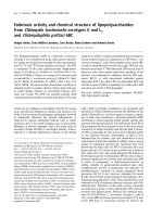
Fig. 1. Ligand-dependent turnover of overexpressed PPARb in
transiently transfected cells. HEK293 cells were transfected with
pCMX-mPPARb and either treated with 1 l
M GW501516 for 24 h
post-transfection or left untreated. The cells were metabolically
pulse-labeled with [
35
S]methionine and [
35
S]cysteine for the final 2 h
in methionine- and cysteine-free medium. The medium was
exchanged with normal growth medium and cells were harvested
after different times (chase). PPARb protein was immunoprecipitat-
ed and analyzed by PAGE followed by phosphorimaging. (A) Auto-
radiograph showing a 48 h chase (*nonspecific band). The amount
of labeled PPARb in the GW501516-treated cells is higher due its
greater stability under these conditions. (B) Quantitative evaluation
by phosphorimaging of pulse chase experiments (24 h chase) per-
formed as in (A). (C) Short-term pulse chase experiment (6 h chase)
performed as in (A). Exposure times were 48 h for autoradiography
and 26 h for phosphorimaging. Signal intensities represent phospho-
stimulated luminescence (PSL) ⁄ mm
2
⁄ 1000 (PSL values generated
by a Phospoimager; Fuji, Du
¨
sseldorf, Germany). Values represent
the mean of three independent experiments; error bars indicate SD.
M. Rieck et al. Regulation of PPARb turnover
FEBS Journal 274 (2007) 5068–5076 ª 2007 The Authors Journal compilation ª 2007 FEBS 5069
(48 h and 24 h) (Fig. 1A,B) and a short-term chase
(6 h) (Fig. 1C). The quantitative evaluation by phos-
phorimaging revealed clear differences between
untreated and GW501516-treated cells with respect to
both protein levels (upper rows: signal intensity) and
degradation (bottom rows: percent of t ¼ 0). Thus, the
levels of labeled PPARb protein were approximately
two-fold higher already at the beginning of the chase
period (t ¼ 0), and remained higher throughout the
time course. Differences in protein stability were, how-
ever, only evident during the initial chase period: in
untreated cells, PPARb protein levels dropped to less
than 50% at 4 h whereas, in the presence of
GW501516, no decrease was detectable. Both these
observations are consistent with a drastically increased
stability of PPAR b in the presence of GW501516. Very
similar results were obtained with transiently transfect-
ed NIH3T3 cells (data not shown), indicating that the
observed effects are not cell line specific.
At later time points of the chase, differences in
stability between untreated and GW501516-treated
cells became basically undetectable, indicating that the
PPARb protein levels may have an impact on the
kinetics of degradation. To address this question,
we established a cell line (3Fb1) stably expressing
3xFLAG-tagged PPARb in a PPARb null background
at less than 1% of the PPARb level observed in tran-
siently transfected cells. These cells were analyzed in a
pulse-chase experiment similar to the one described
above (Fig. 2A,B). In addition, we used a FLAG-
1 2 4 8 12 24 31 48
*
no GW
+GW
*
0
27 20 31 30 15 16 12 8.1 4.7
29 29 32 15 13 10 12 6.1 4.2
0
Con
signal intensity
%oft=0 100
signal intensity
%oft=0
74 114 111 56 61 44 30 18
100 100 112 51 45 36 42 21 15
chase (h)
FLAG-PPAR
β
A
B
C
D
FLAG-PPAR
β
0
5
10
15
20
25
30
35
01020304050
Chase (h)
no GW
+GW
481220
untreated
+GW
0
rel. signal intensity 100 42 16 15 11 11
chase (h) 24
rel. signal intensity 100 80 71 56 30 36
U
D
no GW L no GW L
- MG132 + MG132
Fig. 2. Turnover of FLAG-PPARb expressed at moderate levels in retrovirally transduced mouse fibroblasts. (A) Pparb null cells were infected
with a 3xFLAG-PPARb expressing retrovirus and a stable clone (3Fb1 cells) was analyzed in a pulse-chase experiment as in Fig. 1A, except
that the cells were labeled for 30 min. The experiment was also repeated with cells labeled for 2 h with basically results (the 4 h value
shown for GW501516-treated cells is an outlier). The autoradiograph exposed for 6 days. Signal intensities represent PSL ⁄ mm
2
and indicate
a more than 100-fold lower expression of PPARb compared to Fig. 1 (*nonspecific band). (B) Quantitative evaluation by phosphorimaging
(exposure time 24 h) of the experiment shown in (A). (C) Pulse-chase experiment as in Fig. 1A, except that the same expression vector for
3xFLAG-PPARb as in (A) was used for transient transfection (high expression). (D) Immunoblot analysis of 3xFLAG-PPARb in 3Fb1 cells;
moderate expression, see (A). Cells were either untreated, or treated with the PPARb agonists GW501516 (GW) or L165 041 (L) either alone
or in combination with the proteasome inhibitor MG132. Agonist treatment was for 48 h. MG132 was included during the final 6 h of the
experiment. U, presumably polyubiquitinated high-molecular mass 3xFLAG-PPARb forms; D, presumably a 3xFLAG-PPARb protein fragment
stabilized by MG132. The agonist function of GW501516 and L165 041 was verified in transient reporter gene assays performed in parallel
(not shown).
Regulation of PPARb turnover M. Rieck et al.
5070 FEBS Journal 274 (2007) 5068–5076 ª 2007 The Authors Journal compilation ª 2007 FEBS
specific antibody because none of the available
PPARb-specific antibodies are suitable for a quantifi-
able detection of PPARb at low expression levels. In
these experiments, no significant differences were
detectable between untreated and GW501516-treated
cells with respect to either the initial level of labeled
FLAG-PPARb or the turnover FLAG-PPARb. This
turnover of FLAG-PPARb is similar to that of
PPARb in transiently transfected GW501516-treated
cells (Fig. 1), indicating that overexpressed PPARb
protein is subject to an enhanced degradation that is
prevented by GW501516. To exclude the possibility
that the FLAG tag influenced the results obtained with
the 3Fb1 cells, we also analyzed 3xFLAG-tagged
PPARb in transiently transfected cells with virtually
identical results compared to untagged PPARb
(Figs 1B and 2C).
Finally, we analyzed steady-state 3xFLAG-PPARb
levels in 3Fb1 cells by immunoblotting either untreated,
treated with the PPARb agonists GW501516 or
L165 041 and in combination with the proteasome
inhibitor MG132 (Fig. 2D). In agreement with the
pulse-chase experiments, the immunoblot data clearly
show that, in 3Fb1 cells expressing PPARb at moderate
levels, neither agonist had any detectable effect on pro-
tein levels in spite of a clear stabilization by MG132
(visible as strongly increased protein levels and the
presence of presumably polyubiquitinated 3xFLAG-
PPARb).
Formation of high M
r
complexes in PPARb
overexpressing cells
We next sought to elucidate the biochemical basis of
the enhanced degradation of overexpressed PPARb
protein. Expression plasmids for normal PPARb
(pCMX-PPARb) and FLAG-tagged PPARb (3xFlag-
PPARb) were cotransfected into HEK293 cells, and
cell extracts were investigated by immunoblot analysis
of immunoprecipitated PPARb (Fig. 3A). Three dif-
ferent antibodies were used for immunoprecipitation:
polyclonal-antibody directed against the subtype-
specific N-terminus of PPARb (lane 2), polyclonal
antibody against FLAG (lane 3) and monoclonal
antibody against FLAG (M2, lane 4). PPARb pro-
teins were visualized on immunoblots with either the
PPARb-specific antibody (upper panel) or the M2
antibody (lower panel). This experiment clearly
showed that FLAG-PPARb was precipitated by the
PPARb-specific antibody (lane 2), and vice versa, that
PPARb was coprecipitated by both FLAG-directed
antibodies (lanes 3 and 4), suggesting the formation
of PPARb oligomers. This conclusion was confirmed
Fig. 3. Effect of PPARb protein levels and GW501516 on oligo-
merization of PPARb. (A) Co-immunoprecipitation of FLAP-PPARb
and PPARb. HEK293 cells were cotransfected with pCDNA3.1-
zeo-3xFlag-mPPARb and pCMX-mPPARb. Cells were harvested
after 24 h and RIPA extracts were immunoprecipitated using
anti-mPPARb serum (lane 2), polyclonal (pc) antibody against
FLAG (lane 3), monoclonal antibody against FLAG M2 (lane 4) or
no antibody (mock, lane 5). One third of the immunoprecipitate
was analyzed by immunoblotting using antibodies specific for
PPARb (upper panel) and FLAG (lower panel), respectively
(*immunoglobulin heavy chain). The indicated molecular masses
are based on a calibration curve using molecular mass standards.
The 3xFlag-mPPARb protein shows a higher M
r
as calculated
due the highly charged nature of the tag (DYKDDDDK). (B) Effect
of PPARb protein levels on oligomerization. Decreasing amounts
of pCMX-mPPARb and pCDNA3.1zeo-3xFlag-mPPARb were trans-
fected into HEK293 cells as in (A). All samples contained a total
amount of 10 lg plasmid DNA. RIPA extracts were immunopre-
cipitated and analyzed by immunoblotting using antibodies spe-
cific for PPARb as in (A). (C) Reduction of PPARb oligomerization
by GW501516. HEK293 cells were transfected as in (A), and
subsequently cultured in the presence of different concentrations
of GW501516 for 24 h. RIPA extracts were immunoprecipitated
with antibody against FLAG M2. One third of the immunoprecipi-
tate was analyzed by immunoblotting using PPARb specific
antibodies.
M. Rieck et al. Regulation of PPARb turnover
FEBS Journal 274 (2007) 5068–5076 ª 2007 The Authors Journal compilation ª 2007 FEBS 5071
by superose 6 size exclusion chromatography followed
by immunoblot analysis of the collected fractions
(Fig. 4A). As expected, RxRa specific antibodies
detected proteins that presumably represent mono-
meric RxRa (55 kDa) and, to a lesser extent, higher
order RxR a complexes. By contrast, PPARb occurred
mainly in protein complexes of approximately 2 MDa
(fraction 16). The same fraction contained only very
low levels of RxRa in comparison to PPAR b, indicat-
ing that these complexes are not composed of stoi-
chiometric amounts of PPARb and its obligatory
RxR heterodimerization partner.
Agonist and protein level influence the degree of
high M
r
complex formation
To investigate the nature of the high M
r
PPARb com-
plexes, we analyzed the effects of PPARb protein
concentration and binding of GW501516. For this pur-
pose, we performed the same analyses as above, but
after transfection of different amounts of plasmid
DNA into HEK293 cells. As can be seen in Fig. 3B,
there was a clear reduction on the coprecipitation of
PPARb by the FLAG-specific M2 antibody. Quantita-
tion of the data showed a coprecipitation of PPARb
of 98% relative to FLAG-PPARb after transfection of
2 lg of plasmid DNA, which was reduced to 82%,
52% and 14% when the amounts of transfected plas-
mids were decreased to 0.2 lg, 0.05 lg and 0.02 lg,
respectively. A clear reduction of coprecipitated
PPARb was also seen when the transfected cells
were treated with GW501516 (Fig. 3C). Although, in
untreated cells (lane 1), coprecipitation of PPARb rela-
tive to FLAG-PPARb was 87%, this was decreased to
55%, 37% and 35% in the presence of 0.5 lm,1lm
and 2 lm GW501516, respectively. Likewise, the incu-
bation with 0.1 lm GW501516 of a PPARb immuno-
precipitate from untreated transfected cells resulted in
the release of PPARb protein (data not shown). Con-
sistent with these results, we observed a strong increase
in the relative levels of lower M
r
complexes (frac-
tions 22–30; corresponding to a molecular mass of
approximately 800–100 kDa) after transfection of
reduced amounts of plasmids or treatment with 1 lm
GW501516 (Fig. 4B,C). Taken together, these findings
clear suggest that the high M
r
complexes form selec-
tively under conditions of PPARb overexpression.
Ligand-inhibitable polyubiquitination of PPARb
The results described above suggest that overexpres-
sion of PPARb leads to the formation of aberrant
complexes that are subject to an enhanced degrada-
tion. We therefore investigated whether this would
correlate with an enhanced ubiquitination of PPARb.
HEK293 cells were transiently transfected with
pCMX-PPARb or cotransfected with pCMX-PPARb
and an expression vector for histidine-tagged ubiquitin
(Ubi-His) [36]. The immunoblot in Fig. 5 clearly shows
the presence of high M
r
PPARb forms in pCMX-
PPARb transfected cells (lane 1). These occur at
increased levels in the cotransfected cells (lane 3),
strongly suggesting that these proteins represent poly-
ubiquitinated PPARb. In both cases, ubiquitination
was strongly inhibited by GW501516 (lanes 2 and 4).
In spite of the readily detectable agonist effect on
10ng plasmid
40ng plasmid
no GW501516
no GW50151
6
+ GW501516
+ GW501516
fraction 14 16 18 20 22 24 26 28 30 32 34 36 38
2 MDa
1MDa
60 kDa
fraction
A
B
C
14 16 18 20 22 24 26 28 30 32 34 36 38
2MDa
1 MDa
60 kDa
1
10
100
14 16 18 20 22 24 26 28 30 32 34
Fraction
Relative units
10ng DNA, no GW
10ng DNA +GW
40ng DNA, no GW
40ng DNA +GW
Fig. 4. Effect of GW501516 and protein levels on the native molec-
ular mass of PPARb complexes. (A) High M
r
complexes in PPARb
overexpressing cells. RIPA extract from HEK293 cells transiently
transfected with pCMX-mPPARb (as in Fig. 1) was loaded on a su-
perose 6 column. Forty-five 500 lL fractions were collected. Frac-
tions were analyzed by immunoblotting using PPARb and RxRa
specific antibodies. Cells were transfected with 4 lg of pCMXmP-
PARb per 10 cm dish. (B) Effect of GW501516 and protein levels.
The experiment was performed as in (A), except that cells were
transfected with 10 ng and 40 ng of expression plasmid, respec-
tively, in the presence or absence of 1 l
M GW501516. (C) Quantita-
tion by densitometric analysis of the gels shown in (B). Data are
expressed as arbitrary units normalized to 1.0 for fraction 16.
Regulation of PPARb turnover M. Rieck et al.
5072 FEBS Journal 274 (2007) 5068–5076 ª 2007 The Authors Journal compilation ª 2007 FEBS
polyubiquitination, no significant differences in protein
levels are visible between untreated and GW501516-
treated cells, although the pulse-chase experiments in
Fig. 1 showed a clear effect of the agonist on protein
stability ⁄ degradation. We attribute this difference to
the fact that the experiment in Fig. 5 analyzes steady-
state levels, where the high rate of de novo synthesis
presumably outweighs protein degradation. Consistent
with this interpretation, we did not observe any change
in protein levels in the PPARb overexpressing cells
after treatment with the proteasome inhibitor MG132
(data not shown), in contrast to 3Fb1 cells expressing
moderate levels of PPARb (Fig. 2D).
Conclusions
Our data show that the PPARb is a relatively stable
protein when expressed at moderate levels in fibroblasts
and that, under these conditions, its turnover is not sig-
nificantly affected by the synthetic agonist GW501516.
Transient transfection, on the other hand, leads to a
more than 100-fold increased expression concomitant
with a clearly accelerated degradation, which in turn
can be prevented by GW501516. This influence of pro-
tein levels and agonist binding on PPARb stability
correlate with the formation of high M
r
PPARb
complexes that consist predominantly of PPARb, and
may even represent homooligomers. Such complexes
have never been observed, and are unlikely to exist
under physiological conditions. The correlation of their
formation with high expression levels indeed strongly
suggests that they occur specifically under conditions of
overexpression. It is likely that overexpressed PPARb
forms high M
r
complexes consisting at least in part of
oligomerized PPARb, and that these complexes are
polyubiquitinated and rapidly degraded. This possibly
serves as a safeguard mechanism protecting the cell
from deregulated PPARb expression that could poten-
tially occur under certain pathological conditions. Such
a safeguard mechanisms may be of particular impor-
tance in view of the fact that, unlike steroid hormone
receptors, PPARs do not require the interaction with a
specific ligand for transcriptional activity [37,38] and
figure in cancer-associated biological processes [26–28].
Our observations are also relevant in view of the fact
that the modification, regulation and function of
PPARs are commonly studied in transiently transfected
cells (i.e. under conditions of PPAR overexpression), as
is the case, for example, for the ligand-regulated turn-
over and ubiquitination of PPARa [35] and PPARc
[34]. Agonist-regulated PPARb ubiquitination and
turnover has also been described in a recent study [39]
published after the submission of this manuscript.
However, because most experiments were performed
with overexpressed tagged PPARb, the physiological
relevance of these findings remains to be seen. In light
of our results, it may be important to revaluate any
conclusions derived from transient PPAR transfection
and overexpression experiments.
Experimental procedures
Chemicals and antibodies
GW501516 was purchased from Axxora (Lo
¨
rrach, Ger-
many), MG132 was obtained from Sigma (Taufkirchen, Ger-
many) and the protease inhibitor cocktail (PIC) was from
Roche (Mannheim, Germany). The following sera were used
in this study: polyclonal goat-anti-PPARb (sc-1987; Santa
Cruz, Heidelberg, Germany), monoclonal anti-FLAG (M2,
Sigma), polyclonal rabbit-anti-FLAG (sc-807; Santa Cruz)
and polyclonal rabbit-anti-RxRa (sc-553; Santa Cruz). Ben-
zonase was obtained from Merck (Darmstadt, Germany).
Cell culture
HEK293, NIH3T3 (provided by D. Lowy, NIH, Bethesda,
MD, USA) and 3Fb1 cells (see below) were cultured in
PPARβ
β
GW501516
-
+
-
+
PPAR
β
β
+Ubi-His
1234
Ubi-PPAR
β
Fig. 5. Ligand-regulated ubiquitination of overexpressed PPARb
HEK293 cells were transfected with pCMX-mPPARb plus either an
empty vector (lanes 1 and 2) or an expression vector for histidine-
tagged ubiquitin (Ubi-His; lanes 3 and 4). The cells were either trea-
ted with GW501516 (lanes 2 and 4) or left untreated (lanes 1 and
3). Cells were harvested after 24 h and analyzed by immunoblotting
using PPARb specific antibodies. The picture shows an overexpo-
sure to visualize the ubiquitinated high M
r
PPARb forms.
M. Rieck et al. Regulation of PPARb turnover
FEBS Journal 274 (2007) 5068–5076 ª 2007 The Authors Journal compilation ª 2007 FEBS 5073
DMEM supplemented with 10% fetal bovine serum,
100 UÆmL
)1
penicillin and 100 lgÆmL
)1
streptomycin in a
humidified incubator at 37 °C and 5% CO
2
.
Plasmids pCMX-mPPARaˆ [7] was kindly provided by Dr
R. Evans (The Salk Institute, La Jolla, CA, USA). 3xFlag-
PPARb (pCDNA
3.1
zeo) was generated by cloning the
coding sequence of mPPARb N-terminally fused to a triple
FLAG tag (Sigma) into pcDNA3.1(+) zeo (Invitrogen,
Karlsruhe, Germany). pCMX-empty has been described
previously [40]. The Ubi-His expression vector [36] was a
gift from Dr M. Eilers (Marburg, Germany).
Transfections
Transfections were performed with polyethylenimine (aver-
age molecular mass ¼ 25 000 kDa; Sigma-Aldrich, Munich,
Germany). Cells were transfected on 60 mm dishes at 70–
80% confluence in DMEM plus 2% fetal bovine serum
with 10 lg of plasmid DNA and 10 lL of polyethylenimine
(1 : 1000 dilution, adjusted to pH 7.0 and preincubated for
15 min in 200 lL of NaCl ⁄ P
i
for complex formation). Four
hours after transfection, the medium was changed and cells
were incubated in normal growth medium for 24–48 h.
Retrovirally transduced cells expressing
FLAG-PPARb
3xFLAG-PPARb was cloned into the retroviral vector
pLPCX (Clontech, Heidelberg, Germany). Phoenix cells
expressing ecotropic env were transfected with 3xFLAG-
mPPARb-pLPCX ( roup/nolan/
retroviral_systems/retsys .html). Culture supernatant was
used to infect PPARb null fetal mouse lung fibroblasts that
had previously been established from PPARb knockout
mice by standard procedures. Cells were selected with puro-
mycin (2 lgÆmL
)1
; Sigma), and a clone expressing
3xFLAG-mPPARb (3Fb1 cells) at moderate levels, compa-
rable to endogenous PPARb in mouse fibroblasts, was used
in the present study.
Preparation of denatured whole cell extract
Cells (60 mm dishes) were lysed with 400 lL of SDS sample
buffer containing 125 U benzonaseÆmL
)1
for 5 min at room
temperature. The lysed cells were scraped with a rubber
policeman and transferred to a 1.5 mL tube. After boiling
for 5 min, the lysate was centrifuged for 10 min at 13 000 g
with a Pico Biofuge (Heraeus, Osterode, Germany), and the
supernatant was used for immunoblot analysis.
Preparation of native whole cell extract
Cells were lysed on 60 mm dishes with 400 lL of RIPA
buffer containing 10 mm Tris (pH 7.5), 150 mm NaCl,
1% NP-40, 0.25% SDS, 1% sodium desoxycholate, 5 mm
dithiothreitol, 0.2 mm phenylmethanesulfonyl fluoride,
0.5 · PIC and 125 U benzonaseÆmL
)1
. Cells were scraped
with a rubber policeman, and the lysate was incubated for
20 min on ice. Samples were centrifuged for 10 min at
13 000 g and 4 °C with a Pico Biofuge. The supernatant
was transferred to a fresh 1.5 mL tube; 100 lL were used
for size exclusion chromatography (see below) and 150 lL
for immunoprecipitation.
Size exclusion chromatography
One hundred microlitres of native whole cell extract were
loaded onto a HR10 ⁄ 30 column containing superose 6
(Amersham-Biosciences, Freiburg, Germany) using an
A
¨
kta-purifier (Amersham-Biosciences). The running buffer
consisted of 20 mm Tris ⁄ HCl pH 7.9, 5% (v ⁄ v) glycerol,
150 mm NaCl, 3 mm dithiothreitol and 0.2 phenylmethane-
sulfonyl fluoride. Five-hundred microliter fractions were
collected and 160 lL of each fraction were analyzed by
immunoblotting.
Immunoprecipitation
150 lL of the native whole cell extract were precleared with
20 lL of a 50% Protein A ⁄ G Plus agarose (Santa Cruz) for
3 h. The lysate was centrifuged for 1 min at 13 000 g and
4 °C with a Pico Biofuge, and the supernatant was subse-
quently incubated overnight with 1 lg antibody. After the
addition of 50 lL of protein A ⁄ G Plus agarose (preblocked
with 50 lgÆmL
)1
bovine serum albumin) the incubation was
continued for another 4 h. The precipitate was washed
three times with RIPA buffer, bound proteins were eluted
with 100 lL of SDS sample buffer and analyzed by immu-
noblotting as described below.
Pulse-chase experiments
Pulse chase experiments were carried out according to the
Tansey Laboratory Protocol ( />protocols.html). After transfection, cells were starved for
45 min in methionine ⁄ cysteine-free DMEM (Invitrogen,
Karlsruhe, Germany) containing 1% glutamine and 5%
dialyzed fetal bovine serum, and incubated with 430 lCi of
Redivue ProMix (14.3 lCiÆlL
)1
; Amersham-Biosciences,
Freiburg, Germany). After labeling for 2 h or 30 min, cells
were washed and subsequently incubated with standard
growth medium (DMEM plus 10% fetal bovine serum).
Cells were collected at different time points in ice-cold
NaCl ⁄ P
i
with a rubber policeman and centrifuged at
13 000 g for 1 min with a Pico Biofuge. For storage, the
cell pellet was frozen in liquid nitrogen. Prior to immuno-
precipitation, the frozen cells were lysed in 400 lL of ice-
cold RIPA buffer for 20 min, centrifuged at 13 000 g for
Regulation of PPARb turnover M. Rieck et al.
5074 FEBS Journal 274 (2007) 5068–5076 ª 2007 The Authors Journal compilation ª 2007 FEBS
10 min with a Pico Biofuge and transferred to a 1.5 mL
tube. Immunoprecipitation was carried out with 150 lLof
the lysate, as described above. Kinetics were performed
with the same pool of transfected cells to avoid the problem
of variable transfection efficiencies.
Immunoblot analysis
Protein samples were separated by 12.5% SDS ⁄ PAGE, and
proteins were transferred by semidry blotting to a
poly(vinylidene difluoride) membrane (Millipore, Schwal-
bach, Germany), stained with Ponceau S solution,
destained and blocked with 5% skimmed milk in NaCl ⁄ P
i
-
Tween. The membrane was incubated with the first anti-
body (1 : 2000–1 : 4000) overnight at 4 °C. Membranes
were washed three times for 10 min in NaCl ⁄ P
i
-Tween and
then incubated with an peroxidase-coupled second antibody
(1 : 4000) for 2 h at room temperature. Membranes were
washed and bands were visualized on X-ray film (Fuji, Du
¨
s-
seldorf, Germany) using the enhanced chemiluminescent
method (Amersham-Biosciences, Freiburg, Germany).
Acknowledgements
We are grateful to Drs Ronald Evans (Salk Institute,
La Jolla, CA, USA) and Martin Eilers (IMT Marburg,
Germany) for plasmid vectors, and to Margitta Alt
and Bernard Wilke for their excellent technical assis-
tance. This work was supported by grants from the
Deutsche Forschungsgemeinschaft (SFB-TR17) and
the Deutsche Krebshilfe.
References
1 Desvergne B & Wahli W (1999) Peroxisome prolifera-
tor-activated receptors: nuclear control of metabolism.
Endocr Rev 20, 649–688.
2 Berger J & Moller DE (2002) The mechanisms of action
of PPARs. Annu Rev Med 53, 409–435.
3 Lee CH, Olson P & Evans RM (2003) Minireview: lipid
metabolism, metabolic diseases, and peroxisome prolifer-
ator-activated receptors. Endocrinology 144, 2201–2207.
4 Evans RM, Barish GD & Wang YX (2004) PPARs and
the complex journey to obesity. Nat Med 10 , 355–361.
5 Feige JN, Gelman L, Michalik L, Desvergne B & Wahli
W (2006) From molecular action to physiological out-
puts: peroxisome proliferator-activated receptors are
nuclear receptors at the crossroads of key cellular func-
tions. Prog Lipid Res 45, 120–159.
6 Desvergne B, Michalik L & Wahli W (2006) Transcrip-
tional regulation of metabolism. Physiol Rev 86, 465–514.
7 Forman BM, Chen J & Evans RM (1997) Hypolipidem-
ic drugs, polyunsaturated fatty acids, and eicosanoids
are ligands for peroxisome proliferator-activated
receptors alpha and delta. Proc Natl Acad Sci USA 94,
4312–4317.
8 Peraza MA, Burdick AD, Marin HE, Gonzalez FJ &
Peters JM (2006) The toxicology of ligands for peroxi-
some proliferator-activated receptors (PPAR). Toxicol
Sci 90, 269–295.
9 Michalik L, Auwerx J, Berger JP, Chatterjee VK, Glass
CK, Gonzalez FJ, Grimaldi PA, Kadowaki T, Lazar
MA, O’Rahilly S et al. (2006) International Union of
Pharmacology. LXI. Peroxisome proliferator-activated
receptors. Pharmacol Rev 58, 726–741.
10 Rosen ED (2003) Energy balance: a new role for PPAR-
alpha. Curr Biol 13, R961–R963.
11 Gouni-Berthold I & Krone W (2005) Peroxisome prolif-
erator-activated receptor alpha (PPARalpha) and ath-
erosclerosis. Curr Drug Targets Cardiovasc Haematol
Disord 5, 513–523.
12 Lee CH & Evans RM (2002) Peroxisome proliferator-
activated receptor-gamma in macrophage lipid homeo-
stasis. Trends Endocrinol Metab 13, 331–335.
13 Barish GD, Narkar VA & Evans RM (2006) PPAR
delta: a dagger in the heart of the metabolic syndrome.
J Clin Invest 116, 590–597.
14 Lee CH, Olson P, Hevener A, Mehl I, Chong LW, Olef-
sky JM, Gonzalez FJ, Ham J, Kang H, Peters JM et al.
(2006) PPARdelta regulates glucose metabolism and
insulin sensitivity. Proc Natl Acad Sci USA 103, 3444–
3449.
15 Barak Y, Nelson MC, Ong ES, Jones YZ, Ruiz-Lozano
P, Chien KR, Koder A & Evans RM (1999) PPAR
gamma is required for placental, cardiac, and adipose
tissue development. Mol Cell 4, 585–595.
16 Tan NS, Michalik L, Noy N, Yasmin R, Pacot C, Heim
M, Fluhmann B, Desvergne B & Wahli W (2001) Criti-
cal roles of PPAR beta ⁄ delta in keratinocyte response
to inflammation. Genes Dev 15, 3263–3277.
17 Schmuth M, Haqq CM, Cairns WJ, Holder JC, Dorsam
S, Chang S, Lau P, Fowler AJ, Chuang G, Moser AH
et al. (2004) Peroxisome proliferator-activated receptor
(PPAR)-beta ⁄ delta stimulates differentiation and lipid
accumulation in keratinocytes. J Invest Dermatol 122,
971–983.
18 Kim DJ, Bility MT, Billin AN, Willson TM, Gonzalez
FJ & Peters JM (2006) PPARbeta ⁄ delta selectively
induces differentiation and inhibits cell proliferation.
Cell Death Differ 13, 53–60.
19 Nadra K, Anghel SI, Joye E, Tan NS, Basu-Modak S,
Trono D, Wahli W & Desvergne B (2006) Differentiation
of trophoblast giant cells and their metabolic functions
are dependent on peroxisome proliferator-activated
receptor beta ⁄ delta. Mol Cell Biol 26, 3266–3281.
20 Varnat F, Heggeler BB, Grisel P, Boucard N, Corthesy-
Theulaz I, Wahli W & Desvergne B (2006) PPARbeta ⁄
delta regulates paneth cell differentiation via controlling
M. Rieck et al. Regulation of PPARb turnover
FEBS Journal 274 (2007) 5068–5076 ª 2007 The Authors Journal compilation ª 2007 FEBS 5075
the hedgehog signaling pathway. Gastroenterology 131,
538–553.
21 Fajas L, Auboeuf D, Raspe E, Schoonjans K, Lefebvre
AM, Saladin R, Najib J, Laville M, Fruchart JC, Deeb
S et al. (1997) The organization, promoter analysis, and
expression of the human PPARgamma gene. J Biol
Chem 272, 18779–18789.
22 Ren D, Collingwood TN, Rebar EJ, Wolffe AP &
Camp HS (2002) PPARgamma knockdown by engi-
neered transcription factors: exogenous PPARgamma2
but not PPARgamma1 reactivates adipogenesis. Genes
Dev 16, 27–32.
23 Rosen ED & Spiegelman BM (2001) PPARgamma: a
nuclear regulator of metabolism, differentiation, and cell
growth. J Biol Chem 276, 37731–37734.
24 Marx N, Duez H, Fruchart JC & Staels B (2004) Per-
oxisome proliferator-activated receptors and atherogene-
sis: regulators of gene expression in vascular cells. Circ
Res 94, 1168–1178.
25 Zhang L & Chawla A (2004) Role of PPARgamma in
macrophage biology and atherosclerosis. Trends Endo-
crinol Metab 15, 500–505.
26 Burdick AD, Kim DJ, Peraza MA, Gonzalez FJ &
Peters JM (2006) The role of peroxisome proliferator-
activated receptor-beta ⁄ delta in epithelial cell growth
and differentiation. Cell Signal 18 , 9–20.
27 Michalik L, Desvergne B & Wahli W (2004) Peroxi-
some-proliferator-activated receptors and cancers: com-
plex stories. Nat Rev Cancer 4, 61–70.
28 Mu
¨
ller R (2004) Crosstalk of oncogenic and prostanoid
signaling pathways. J Cancer Res Clin Oncol 130, 429–
444.
29 Yamashita D, Yamaguchi T, Shimizu M, Nakata N,
Hirose F & Osumi T (2004) The transactivating func-
tion of peroxisome proliferator-activated receptor
gamma is negatively regulated by SUMO conjugation in
the amino-terminal domain. Genes Cells 9, 1017–1029.
30 Ohshima T, Koga H & Shimotohno K (2004) Tran-
scriptional activity of peroxisome proliferator-activated
receptor gamma is modulated by SUMO-1 modification.
J Biol Chem 279, 29551–29557.
31 Pascual G, Fong AL, Ogawa S, Gamliel A, Li AC,
Perissi V, Rose DW, Willson TM, Rosenfeld MG &
Glass CK (2005) A SUMOylation-dependent pathway
mediates transrepression of inflammatory response genes
by PPAR-gamma. Nature 437, 759–763.
32 Hu E, Kim JB, Sarraf P & Spiegelman BM (1996) Inhi-
bition of adipogenesis through MAP kinase-mediated
phosphorylation of PPARgamma. Science 274, 2100–
2103.
33 Lazennec G, Canaple L, Saugy D & Wahli W (2000)
Activation of peroxisome proliferator-activated recep-
tors (PPARs) by their ligands and protein kinase A acti-
vators. Mol Endocrinol 14, 1962–1975.
34 Hauser S, Adelmant G, Sarraf P, Wright HM, Mueller
E & Spiegelman BM (2000) Degradation of the peroxi-
some proliferator-activated receptor gamma is linked to
ligand-dependent activation. J Biol Chem 275, 18527–
18533.
35 Blanquart C, Barbier O, Fruchart JC, Staels B & Gli-
neur C (2002) Peroxisome proliferator-activated receptor
alpha (PPARalpha) turnover by the ubiquitin-protea-
some system controls the ligand-induced expression level
of its target genes. J Biol Chem 277, 37254–37259.
36 Adhikary S, Marinoni F, Hock A, Hulleman E, Popov
N, Beier R, Bernard S, Quarto M, Capra M, Goettig S
et al. (2005) The ubiquitin ligase HectH9 regulates tran-
scriptional activation by Myc and is essential for tumor
cell proliferation. Cell 123, 409–421.
37 Molnar F, Matilainen M & Carlberg C (2005) Struc-
tural determinants of the agonist-independent associa-
tion of human peroxisome proliferator-activated
receptors with coactivators. J Biol Chem 280, 26543–
26556.
38 Michalik L, Zoete V, Krey G, Grosdidier A, Gelman L,
Chodanowski P, Feige JN, Desvergne B, Wahli W &
Michielin O (2007) Combined simulation and mutagene-
sis analyses reveal the involvement of key residues for
peroxisome proliferator-activated receptor alpha helix
12 dynamic behavior. J Biol Chem 282, 9666–9677.
39 Genini D & Catapano CV (2007) Block of nuclear
receptor ubiquitination. A mechanism of ligand-depen-
dent control of peroxisome proliferator-activated recep-
tor delta activity. J Biol Chem 282, 11776–11785.
40 Umesono K, Murakami KK, Thompson CC & Evans
RM (1991) Direct repeats as selective response elements
for the thyroid hormone, retinoic acid, and vitamin D3
receptors. Cell 65, 1255–1266.
Regulation of PPARb turnover M. Rieck et al.
5076 FEBS Journal 274 (2007) 5068–5076 ª 2007 The Authors Journal compilation ª 2007 FEBS