Báo cáo khoa học: Functional reconstitution of mammalian ‘chloride intracellular channels’ CLIC1, CLIC4 and CLIC5 reveals differential regulation by cytoskeletal actin docx
Bạn đang xem bản rút gọn của tài liệu. Xem và tải ngay bản đầy đủ của tài liệu tại đây (1.67 MB, 11 trang )
Functional reconstitution of mammalian ‘chloride
intracellular channels’ CLIC1, CLIC4 and CLIC5 reveals
differential regulation by cytoskeletal actin
H. Singh,* M. A. Cousin and R. H. Ashley
Centre for Integrative Physiology, University of Edinburgh Medical School, UK
Chloride intracellular channel (CLIC) proteins are
‘structural homologues’ of W-class glutathione S-trans-
ferases (W-GSTs) [1]. They are widely expressed in
multicellular organisms, and can coexist in both solu-
ble and integral membrane forms, because some solu-
ble, signal peptide-less CLICs bypass the classical
secretory pathway, and autoinsert directly into mem-
branes [2,3]. Soluble CLIC2 displays minor GST-
related enzymatic activities, although it does not seem
to be a classical thiol transferase [4], and soluble
CLIC4 (p64H1) is associated with apoptosis [5], but in
general the cellular roles of the soluble proteins are
poorly understood, and more interest has centred on
membrane CLICs. The regulatory mechanisms of
membranous CLICs in vivo and in vitro have not been
elucidated.
The Caenorhabditis elegans CLIC-like protein exc-4
appears to form a charge-compensating ion channel to
facilitate the fusion of intracellular vesicles, explaining
its essential role to generate a hollow tubule from a
single cell [6,7]. Mammalian CLIC1 and CLIC4 are
transmembrane components [8,9] of poorly selective
intracellular and plasma membrane ion channels, and
form similar channels in vitro in the absence of any
other protein [10,11]. Although the roles of the mam-
malian channels remain obscure, and CLIC proteins
can also modify the behaviour [12] of other, well-estab-
lished ion channels, the channel activity of CLIC1 does
Keywords
chloride intracellular channels; cytoskeleton;
ion channel; multiconductance channel;
planar bilayer
Correspondence
H. Singh, Department of Physiology,
UCLA, David Geffen School of Medicine,
10833 LeConte Avenue, 53-373 CHS,
Los Angeles, CA 90095-1751, USA
Fax: +1 310 206 5661
Tel: +1 310 825 7185
Email:
*Present address
Department of Physiology, David Geffen
School of Medicine, UCLA, CA, USA
(Received 10 September 2007, revised 10
October 2007, accepted 16 October 2007)
doi:10.1111/j.1742-4658.2007.06145.x
Chloride intracellular channels (CLICs) are soluble, signal peptide-less pro-
teins that are distantly related to W-type glutathione-S-transferases.
Although some CLICs bypass the classical secretory pathway and auto-
insert into cell membranes to form ion channels, their cellular roles remain
unclear. Many CLICs are strongly associated with cytoskeletal proteins,
but the role of these associations is not known. In this study, we incorpo-
rated purified, recombinant mammalian CLIC1, CLIC4 and (for the first
time) CLIC5 into planar lipid bilayers, and tested the hypothesis that the
channels are regulated by actin. CLIC5 formed multiconductance channels
that were almost equally permeable to Na
+
,K
+
and Cl
–
, suggesting that
the ‘CLIC’ nomenclature may need to be revised. CLIC1 and CLIC5, but
not CLIC4, were strongly and reversibly inhibited (or inactivated) by ‘cyto-
solic’ F-actin in the absence of any other protein. This inhibition effect on
channels could be reversed by using cytochalasin to disrupt the F-actin.
We suggest that actin-regulated membrane CLICs could modify solute
transport at key stages during cellular events such as apoptosis, cell and
organelle division and fusion, cell-volume regulation, and cell movement.
Abbreviations
CLIC, chloride intracellular channel; GE, gel exclusion; GHK, Goldman–Hodgkin–Katz equation; GSH, reduced glutathione; GST, glutathione
S-transferase; GST, glutathione S-transferase; IMAC, immobilized metal affinity chromatography; I ⁄ V, current–voltage; TMD, transmembrane
domain.
6306 FEBS Journal 274 (2007) 6306–6316 ª 2007 The Authors Journal compilation ª 2007 FEBS
not appear to be an artifact of artificial protein over-
expression, because the activity of endogenous CLIC1
increases in dividing, nontransfected Chinese hamster
ovary cells [13], and in brain microglia exposed to
Alzheimer’s Ab protein [14].
Mammalian CLICs interact extensively with compo-
nents of the cytoskeleton. For example, rat brain
CLIC4 (p64H1) is associated with actin in a multi-
protein complex [15], and human CLIC5 (strictly
CLIC5A, which is expressed from the same gene as
CLIC5B, or p64) was identified in an actin-rich com-
plex from placental microvilli [16]. The proposed
topology and membrane organization of CLIC1,
CLIC4 and CLIC5 [17] suggest that these interactions
could be retained in the membrane forms of the pro-
teins, and the association of membrane CLICs with
the actin cytoskeleton may be functionally important.
For example, rearrangement of the cytoskeleton and
concurrent activation or inhibition of plasma mem-
brane solute transporters are often prominent features
of cell-volume regulation, and specific ion channels are
known to be functionally associated with the cortical
actin cytoskeleton, especially in epithelial cells [18].
We began our studies by reconstitution of recombi-
nant CLIC5 in the planar bilayers, and set out to test
the hypothesis that membrane CLICs are regulated by
cytoskeletal actin, after functionally reconstituting
recombinant human CLIC1, CLIC4 and CLIC5 in pla-
nar lipid bilayers. CLIC1 and CLIC4 have previously
been characterized at the single-channel level, and, in
this study, we confirmed for the first time that mem-
brane CLIC5 also forms ion channels. The ion chan-
nels formed by CLIC1 and CLIC5 were directly and
reversibly regulated by F-actin, without an intermedi-
ate molecule or adaptor protein. In contrast, the ion
channels formed by CLIC4 were not regulated by actin
under the same conditions.
Results
Bilayer reconstitution of CLIC5
Unlike CLIC1 and CLIC4, CLIC5 has not been recon-
stituted previously at the single-channel level, but
purified recombinant CLIC5 (Fig. 1) gave rise
to characteristic ion channel activity (Fig. 2) within
5–10 min of adding 25 ng mL
)1
protein to bilayers
formed from palmitoyl-oleoyl phosphatidylethanol-
amine, palmitoyl-oleoyl phosphatidylserine and choles-
terol (4 : 1 : 1, mol ⁄ mol) in the presence of 5 mm
reduced glutathione (GSH). No channels were observed
using control of immobilized metal affinity chromato-
graphy (IMAC) eluates from non-CLIC5-expressing
bacteria (five independent preparations), confirming
that the activity did not arise from endogenous bacte-
rial proteins.
The channels were more complex than previous
recordings from CLIC1 and CLIC4, and almost always
showed multiple conductance levels (Fig. 2A,B), even
after adding reduced amounts of protein (1–5 ng) to
minimize channel incorporation. Following recordings
at +100 mV with 500 mm KCl in the cis chamber and
50 mm KCl in the trans chamber, to maximize the
single-channel currents (Fig. 2C), CLIC5 amplitudes
could be grouped into seven well-defined, nonoverlap-
ping distributions (Fig. 2D). The amplitudes extended
from 0.21 ± 0.15 pA (mean ± SD, n ¼ 7), close to the
minimum amplitude measurable in our system, to a
maximum level of 12.5 ± 7.5 pA (mean ± SD, n ¼ 7).
However, the maximum level was only seen in approxi-
mately 10% of our experiments.
Transitions between the various open levels of
CLIC5 appeared to be strongly cooperative. For
example, we occasionally observed ‘direct’ transitions
between large-amplitude channels and the closed (zero
current) level, with no apparent intermediate levels
(examples are noted in Fig. 3A). Most of the ampli-
tudes in Fig. 2D (which may represent a combination
of substates and cooperative gating) could be fitted to
a simple exponential distribution, consistent with an
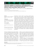
Fig. 1. FPLC and SDS ⁄ PAGE analysis of reduced, recombinant
CLIC5. The FPLC elution peaks correspond to molecular mass of
125, 63 and 38 kDa (main peak), respectively, calculated from the
inset semi-log calibration curve. The protein standards were BSA
(67 kDa), ovalbumin (43 kDa), pGEX vector GST (27 kDa) and ribo-
nuclease A (13.7 kDa). The void volume V
0
and the total column
volume V
t
were 4.5 and 70 mL, respectively, measured using
bromophenol blue and blue dextran. The inset Coomassie-stained
SDS ⁄ PAGE analysis shows a major band at approximately 35 kDa,
with no evidence of multimers under denaturing conditions, unlike
the properly folded protein during FPLC.
H. Singh et al. Chloride intracellular channel regulation by F-actin
FEBS Journal 274 (2007) 6306–6316 ª 2007 The Authors Journal compilation ª 2007 FEBS 6307
initial model in which individual CLIC5 channels con-
tain various numbers of CLIC5 subunits, such that
linear increases in circumference result in squared
increases in cross-sectional area and a corresponding
exponential increase in conductance. This simple model
was testable, because it predicted reduced inter-ionic
selectivities in ‘large-diameter’ pores compared with
‘small-diameter’ pores.
Ionic selectivity of CLIC5
We initially assessed the cation versus anion (K
+
ver-
sus Cl
–
) selectivities of multilevel CLIC5 channels
reconstituted in 500 versus 50 mm KCl based on the
‘macroscopic’ reversal potential, i.e. the voltage-clamp
potential at which the net transbilayer current was
zero. Surprisingly, for a putative anion-selective
(CLIC) channel, the reversal potential was negative (at
least )15 mV in four successive experiments). We then
measured channel amplitudes at a range of holding
potentials in experiments for which at least two spe-
cific, individual channel amplitudes could be clearly
distinguished from each other by amplitude histogram
analysis (Fig. 3A, compare with Fig. 2B,C), and plot-
ted the corresponding current ⁄ voltage (I ⁄ V) relation-
ships (Fig. 3B).
A
B
CD
Fig. 2. Bilayer reconstitution of CLIC5 reveals multiconductance channels. CLIC5 was reconstituted in 5 mM GSH with 500 mM KCl in the
cis chamber and 50 m
M KCl in the trans chamber, and channel activity was recorded at a holding potential of +100 mV. (A) Contiguous
3 min recording illustrating a range of amplitudes up to approximately 2 pA. The solid line shows the zero-current level. The central portion
of the trace is expanded, with an encircled arrow to indicate the smallest measurable current transition. (B) A record from another experi-
ment under the same conditions showing an additional open level at approximately 12 pA. The solid line shows the zero-current level. (C)
All-points amplitude histogram of the data in trace (B) (0.4 pA per bin), fitted to two Gaussian distributions (the arrow shows the zero-current
level). (D) All the observed CLIC5 amplitudes grouped into seven discreet levels (bars represent mean ± SD, n ¼ 7 independent experi-
ments). The curve fits the equation: current (pA) ¼ [5 · 10
)5
· exp(1.77 · level number)] +0.62.
Chloride intracellular channel regulation by F-actin H. Singh et al.
6308 FEBS Journal 274 (2007) 6306–6316 ª 2007 The Authors Journal compilation ª 2007 FEBS
The I ⁄ V relationships in Fig. 3B were assembled
from six independent experiments showing the
same general pattern of channel activity. Under
asymmetrical ionic conditions, positive single-channel
currents at a holding potential of 0 mV (i.e. with no
electrical driving force) can only be explained by net
diffusion of K
+
from the cis chamber to the trans
chamber. The reversal potentials of the two clearly
identified conductance levels were indistinguishable, at
)20.5 ± 1.5 and )20.6 ± 6.7 mV for the high- and
intermediate-amplitude currents, respectively (means ±
SD, n ¼ 6), suggesting that their selectivities (and their
conduction pathways) were identical. This argues
against the hypothesis that different conductances
result from pores with different diameters, or indeed
different proteins, and instead suggests that collections
of basically similar CLIC5 channels open and close
together in a cooperative manner, and intermediate
conductance levels are too brief to be resolved [19,20].
After starting experiments with 50 mm KCl in both
chambers (with a corresponding reversal potential of
0 mV), addition of 150 mm KCl to the cis chamber
shifted the reversal potential to )8.7 ± 1.6 mV (mean
± SD, n ¼ 3), again indicating a slight preference for
cations. Using Eqn (1), the value for P
K
⁄ P
Cl
was
2.0 ± 0.2 (mean ± SD, n ¼ 6) or 2.1 ± 0.4 (mean ±
SD, n ¼ 3) for the two conditions (high- and low-salt
gradients), respectively. Under bi-ionic conditions
(Fig. 4), with KCl in the cis chamber and NaCl in the
trans chamber, the mean reversal potential averaged
over a wide range of salt activities was
+7.7 ± 0.3 mV (mean ± SD, n ¼ 8 activity ratios).
Given that the cis versus trans activity of Cl
–
was
equal in each case, regardless of the total ionic activity,
these experiments directly compare the relative selectiv-
ity of CLIC5 for the two cations. Using Eqn (2), the
relative cation permeability ratio P
Na
⁄ P
K
is 1.3 ± 0.04
(mean ± SD, n ¼ 8).
In vitro regulation of CLIC channels by F-actin
Given the original association of native CLIC5 with
actin [16], we first tested the effect of adding purified
platelet actin (Cytoskeleton Inc., Denver, CO, USA) to
CLIC5 channels reconstituted in 5 mm GSH with
500 mm KCl in the cis chamber and 50 mm KCl in the
trans chamber, after adding 100 lgmL
)1
BSA to
block nonspecific protein binding sites. G-actin was
stirred into each chamber in turn to a final concentra-
tion of 250 nm (10 lgmL
)1
), then polymerized by
adding 5 mm MgCl
2
and 0.5 mm ATP. The trans KCl
concentration was also increased to 100 mm to pro-
mote polymerization. The critical concentration of
actin is comfortably exceeded under these conditions
[21]. Finally, 10 lm cytochalasin B was added to the
cis chamber to disrupt the F-actin. Mean currents were
A
B
Fig. 3. CLIC5 current ⁄ voltage (I ⁄ V) relationships show mild selectiv-
ity for cations versus anions. Channel activity similar to Fig. 2B was
analysed by amplitude histogram analysis (as in Fig. 1C) to return
two well-defined main open levels (corresponding to levels 6 and 7
in Fig. 1D) under asymmetric ionic conditions (500 m
M KCl cis,
50 m
M KCl trans). (A) Example traces from a single experiment at a
range of holding potentials, for which the solid lines indicate the
closed (zero-current) levels. The arrow below the +75 mV trace
indicates a direct transition between the maximum open level and
the closed level, similar to various transitions at )100 mV. (B) I ⁄ V
relationships of the large (filled circle) and small (open circle) con-
ductances, shown as means ± SD (n ¼ 7 independent experi-
ments). The smooth curves are fitted by linear interpolation (three
orders), and the common reversal (equilibrium) potential is marked
by an arrow.
H. Singh et al. Chloride intracellular channel regulation by F-actin
FEBS Journal 274 (2007) 6306–6316 ª 2007 The Authors Journal compilation ª 2007 FEBS 6309
calculated from contiguous 60 s recordings for each
condition, and typical recordings from a single experi-
ment are illustrated, in the order in which they were
obtained, in Fig. 5A–F. Figure 5G summarizes the
results obtained from seven independent experiments.
Single-channel currents through CLIC5 were almost
completely abolished by polymerizing the cis actin
(Fig. 5E), and the effect was reversed by disassembling
the F-actin with cytochalasin B (Fig. 5F). Addition of
G-actin to the cis or trans chambers in the absence of
actin-polymerizing agents did not affect channel activ-
ity, nor did polymerization to F-actin on the trans
side. None of the additions affected CLIC5 in the
absence of actin, and neither G-actin nor F-actin mod-
ified control bilayers in the absence of CLIC proteins.
In additional control experiments carried out in the
presence of 10 lm latrunculin B (to inhibit actin poly-
merization [22]), the mean current of 99 ± 12% with
cis G-actin (mean ± SD, n ¼ 3) remained essentially
unchanged even under ‘actin-polymerizing’ conditions,
at 105 ± 16% (mean ± SD, n ¼ 3).
We next determined whether CLIC1 and CLIC4
showed similar sensitivities to cis F-actin, by reconsti-
tuting them in the presence of 5 mm GSH and repeat-
ing the experiments (and analysis) carried out on
CLIC5. The ion channels formed by CLIC1 were also
inhibited or inactivated by cis F-actin, and the effect
was reversed by cytochalasin B (Fig. 6A,C). Like
CLIC5, 10 lm latrunculin B prevented inhibition, with
a mean current of 114 ± 27% (mean ± SD, n ¼ 3)
with cis G-actin, compared to 118 ± 22% (mean ± SD,
n ¼ 3) under ‘actin-polymerizing’ conditions. In con-
trast, CLIC4 was unaffected by F-actin (Fig. 6B,D). It
should be stressed that, apart from using a different
CLIC isoform in each case, our experiments on
CLIC1, CLIC4 and CLIC5 were in every other respect
identical, so CLIC4 contributes a useful negative con-
trol. Thus F-actin regulates CLIC channel activity in
an isoform-specific manner.
Discussion
CLIC5 forms poorly selective ion channels
CLIC1 and CLIC4 autoinsert into membranes to form
ion channels [3]; however, this property has not been
investigated for CLIC5. CLIC5 also associates with
cytoskeletal filaments [16], but the functional conse-
quences of such interactions are unknown. We set out
to express and purify mammalian CLIC1, CLIC4 and
CLIC5, and incorporate the proteins into planar bi-
layers to test the hypothesis that they form actin-
regulated ion channels. CLIC5 channels, like those
corresponding to CLIC1 [17], and especially CLIC4
[11], were poorly selective rather than chloride-selec-
tive, reinforcing the suggestion that the CLIC nomen-
clature may need to be revised as more information
becomes available. Strikingly, both CLIC1 and CLIC5
are directly and very strongly inhibited by cytoskeletal
F-actin, in the complete absence of any other accessory
or adapter protein, while CLIC4 is unaffected under
similar conditions.
Given that human CLIC5 is 63% identical to
human CLIC1, and 75% identical to human (and rat)
CLIC4, we anticipated that CLIC5 would also insert
spontaneously into planar lipid bilayers to form
B
A
Fig. 4. Bi-ionic reversal potential measurements show poor selec-
tivity between Na
+
and K
+
. (A) Mean reversal potentials ± SD (n ¼
3–6 independent experiments) with KCl cis and NaCl trans, at the
(matching) activities (a[XCl]) indicated. Note the breaks in the plot
after a[XCl) exceeds 300 m
M. The line (fitted by linear regression)
has a gradient of zero, and corresponds to a mean E
r
of +7.7 mV,
averaged over a range of eight activities. (B) I ⁄ V relationships for
matching activities as described in (A) for 150 m
M (open circles)
and 250 m
M (filled circles); each point is the mean (± SD) of 3–6
experiments. The smooth lines are best least-squares fits to the
GHK current equation (Eqn 3). The fits returned P
Na
⁄ P
K
ratios of
0.70 and 0.74, respectively, and the corresponding reversal poten-
tials (+9.5 and +7.8 mV) are indicated.
Chloride intracellular channel regulation by F-actin H. Singh et al.
6310 FEBS Journal 274 (2007) 6306–6316 ª 2007 The Authors Journal compilation ª 2007 FEBS
broadly similar ion channels. The channels were mildly
cation- (not anion-) selective, like CLIC4 under similar
recording conditions [11], and the value for P
K
⁄ P
Cl
was approximately 2.0 under both high- (500 mm ver-
sus 50 mm, Fig. 3) and low- (150 mm versus 50 mm)
salt gradients. The multiple conductance levels of
CLIC5 followed an approximately exponential distri-
bution (Fig. 2D), consistent with highly cooperative
gating of individual unit conductances. We suggest
that this reflects arrays of channels, rather than com-
plex substate behaviour in a single large channel,
because most incorporations lacked the largest-ampli-
tude openings. This behaviour recalls the tendency of
the purified protein to form multimolecular complexes
A
B
C
D
E
F
G
Fig. 5. cis F-actin reversibly inhibits CLIC5 channels. (A–F) Successive CLIC5 recordings from a single experiment at a holding potential of
+100 mV under asymmetric ionic conditions (500 m
M KCl in the cis chamber, 50 mM KCl in the trans chamber), with the additions shown to
the left of each trace. The closed levels are shown as solid lines, and in this experiment the channels correspond mainly to level 4 in
Fig. 1D. Each contiguous 60 s recording is accompanied by its corresponding all-points amplitude histogram [binwidth 0.1 pA, note the fre-
quency scale change in (E)]. Actin and cytochalasin B were added at concentrations of 250 n
M and 10 lM, respectively. (G) Combined results
from seven independent experiments (bars represent means ± SD, n ¼ 7). *P < 0.001 for the reduction in mean current with cis F-actin.
H. Singh et al. Chloride intracellular channel regulation by F-actin
FEBS Journal 274 (2007) 6306–6316 ª 2007 The Authors Journal compilation ª 2007 FEBS 6311
(Fig. 1). Unfortunately, the common multiconductance
activity of CLIC5 prevented a detailed investigation of
channel gating behaviour, and precluded detailed
examination of the channel’s conductance ⁄ activity rela-
tionship.
We measured a consistent bi-ionic reversal potential
(Fig. 4) over a wide range of salt activities, and con-
firmed that CLIC5 discriminates poorly between K
+
and Na
+
(P
Na
⁄ P
K
approximately 1.3). Overall, the rel-
ative selectivity of CLIC5 for physiologically important
monovalent ions is: P
Na
> P
K
> P
Cl
, with a ratio of
about 1.0: 0.75: 0.37. This indicates minimal inter-ion
selectivity. Interestingly, the selectivity ratios were not
concentration- (or, more correctly, activity-) depen-
dent, even though, like CLIC1 and CLIC4, CLIC5
was expected to form a multi-ion channel. However,
we could only examine relative selectivities at activity
ratios extending over less than a single order of magni-
tude, and not at all at very low activities, so we may
have been unable to detect important evidence for
nonindependent ion permeation [23].
Recent in vivo [7] and in vitro [11] experiments iden-
tified a single putative transmembrane domain (TMD)
near the N-terminus of invertebrate and vertebrate
A
C
B
D
Fig. 6. cis F-actin inhibits single-channel currents through CLIC1 but not CLIC4. Examples of recordings from CLIC1 (A) and CLIC4 (B) in
experiments carried out under exactly the same conditions used for CLIC5 (Fig. 4). The solid lines show the closed levels, and arrows indi-
cate previously reported substates of approximately 25% and approximately 45% (double arrows for CLIC1). (C,D) Mean CLIC1 and CLIC4
currents measured from 60 s recordings under various conditions. The bars show means ± SD; n ¼ 8 for CLIC1 in (C) and n ¼ 7 for CLIC4
in (D). *P < 0.001 for the reduction in mean current with cis F-actin for CLIC1.
Chloride intracellular channel regulation by F-actin H. Singh et al.
6312 FEBS Journal 274 (2007) 6306–6316 ª 2007 The Authors Journal compilation ª 2007 FEBS
CLICs. The TMD appears to be both necessary and
sufficient for membrane targeting and membrane pro-
tein function, and, provided it remains intact, the
‘cytosolic’ regions of the proteins, from the TMD to
the C-terminus, are interchangeable between CLICs
[7]. Both approaches (in vitro and in vivo) implied that
the ion channels formed by membrane CLICs must be
oligomers, but sequence comparisons suggested that
the slightly different ionic selectivity of individual
channels does not depend on specific residues in the
identified TMD. This led to the suggestion that other
parts of the protein, including the channel vestibules,
modulate selectivity [11], and key molecular determi-
nants of specific CLIC properties may not become test-
able until the structures of the membrane proteins are
available.
CLICs exist as soluble and membranous proteins [3],
and they interact with various other cytosolic proteins
[15]. As CLIC function was investigated by their
reconstitution in planar bilayers, it is possible that
modulation of either channel permeability or selectivity
by cytoplasmic and membranous components present
inside an intact cell may have been overlooked. How-
ever, as the channel properties of CLIC5 have not
been reported to date, this reduced system has the
advantage of characterizing CLIC5 function in the
absence of modulatory factors. Furthermore, the role
of actin in regulating channel function is best observed
in such a reduced system considering the multiple and
contrasting roles that the cytoskeleton performs in
intact cells.
Ion channel regulation by F-actin
Every conductance level in multiconductance CLIC5
bilayer recordings was inhibited by F-actin, and the
effect was reversed by F-actin disassembly. Direct
F-actin regulation was specific to CLIC1 and CLIC5,
and did not extend to the very similar protein CLIC4,
which served as an excellent negative control for non-
specific binding. Actin modulation of CLIC1 and
CLIC5 was specific from the cytosolic side, implying a
role of the C-terminus of CLICs in channel regulation.
Recently, CLIC1 was shown to be regulated by the
cystic fibrosis transmembrane conductance regulator
[24], which, in turn, is known to be directly regulated
by actin [25]; similarly, CLIC5 is known to colocalize
with actin and ezrin [16], indicating a possible in vivo
modulation of CLIC1 by actin filaments and func-
tional interaction between CLICs and other ion chan-
nels.
Ion channels are always tightly regulated in cells,
and membrane CLICs appear to be controlled in at
least three ways. Firstly, the proteins preferentially
assemble into functional ion channels in specific lipid
environments [17]. Secondly, mammalian membrane
CLICs contain a critical cysteine residue immediately
before the putative TMD, and we have suggested that
channel complexes could be functionally regulated by
the trans (extracellular or luminal) redox potential via
glutathione-dependent trans-thiolation [17]. Finally, as
we show here, CLIC1 and CLIC5 are regulated in situ
by cytoplasmic F-actin.
Potential roles of CLIC5 and other membrane
CLICs
Our results add to the growing list of diverse ion chan-
nels regulated by actin [18,25–27]. Further work will be
required to determine how F-actin interacts with the
proteins, and how it mediates channel inhibition. Direct
regulation of cystic fibrosis transmembrane conduc-
tance regulator channels by cytoskeletal actin has been
attributed to putative actin-binding regions [25], but,
apart from noting some suggestive charge differences
when flexible [1] ‘hinge’ regions in the soluble structures
are aligned, we could find no structural evidence to
support this hypothesis for the CLIC proteins. This
does not exclude the possibility of additional structural
roles for the proteins. For example, CLIC5 appears to
be an important component of many actin-rich struc-
tures in cells [28], including the inner ear stereocilia that
were found to be defective in CLIC5-deficient mice
with impaired hearing and balance [29]. However, it is
tempting to speculate that selected membrane CLICs
could be specifically activated when cells or organelles
undergo specific physiological changes.
Actin is one of the most abundant proteins in the
cell, and plays a significant role in many physiological
functions. It has numerous binding partners and a high
tendency for specific and nonspecific interactions in the
cell, including the nucleus [30], where CLICs have been
localized and shown to participate in physiological
processes such as regulation of the cell cycle [13]. It is
known that ion channels do not operate as randomly
diffusing moieties in the plasma membrane of cells.
They interact with the cytoplasmic proteins, which in
turn link them to cytoskeleton or intracellular signal-
ling pathways. Direct or indirect interaction of CLICs
with cytoskeletal elements such as actin or dynamin
[15,16] is likely to result in their immobilization and
clustering in membranes, and in the targeting of these
proteins to an appropriate site where they may partici-
pate in various physiological processes. The direct
interaction of actin with CLIC1 and CLIC5 (but not
CLIC4) actin implicate them in diverse CLIC-specific
H. Singh et al. Chloride intracellular channel regulation by F-actin
FEBS Journal 274 (2007) 6306–6316 ª 2007 The Authors Journal compilation ª 2007 FEBS 6313
functions, which could include movement, swelling or
division of the cell, endocytosis and exocytosis, intra-
cellular vesicle fusion, and apoptosis. The major chal-
lenge in future is to understand the functional
significance of these protein interactions in various
physiological processes.
A number of key questions remain. Although p64
and other CLICs contain multiple protein interaction
sites [3], we could not identify a putative actin-binding
site, e.g. a site similar to the actin-binding site in the
a subunit of amiloride-sensitive epithelial Na
+
chan-
nels [31], nor could we identify (from alignments of the
three CLIC proteins) speculative actin-binding residues
in CLIC1 and CLIC5 that are absent in CLIC4. With
respect to the three-dimensional structure, we do not
of course know whether (or to what extent) the CLIC
‘cytoplasmic domain’ refolds in the membrane forms
of the proteins, or indeed how the proteins assemble
into subunits. However, the tendency of CLIC5 to
form multimolecular assemblies may be encouraging
from the perspective of future structural studies of this
membrane protein.
Experimental procedures
Preparation of CLIC proteins
Selected CLICs were expressed as His-tagged proteins in
Escherichia coli and purified by a combination of IMAC
and gel-exclusion (GE) chromatography, as detailed previ-
ously for rat CLIC4 [11] and human CLIC1 [17]. In this
study, we also expressed a cDNA encoding human CLIC5
(CLIC5A, MGC:53405, IMAGE:4611102, MRC Geneser-
vice, Cambridge, UK). Like CLIC1 and CLIC4, the rele-
vant cDNA was cloned by PCR into pHIS-8, a modified
pET-28a(+) vector encoding an N-terminal octahistidine
tag and a thrombin cleavage site. The insert was verified by
DNA sequencing (MWG Biotech, Ebersberg, Germany),
and soluble CLIC5 was recovered from transformed E. coli
BL21 (DE3) cells by Ni
2+
-NTA affinity chromatography
after isopropyl thio-b-d-galactoside-induced overexpression.
The tag was cleaved by thrombin, and the enzyme and the
free tag were scavenged using benzamidine–Sepharose 4B
beads and Ni
2+
-NTA resin (Amersham, Chalfont St Giles,
UK). CLIC5 aggregated and precipitated in buffers con-
taining 5 mm dithiothreitol, but not in those containing
5mm b-mercaptoethanol or 5 mm GSH when the protein
was diluted, e.g. when added to bilayers. Further purifica-
tion by GE FPLC using Superdex 200 (Pharmacia,
Uppsala, Sweden) in the presence of 5 mm b-mercaptoetha-
nol showed a major peak consistent with the monomeric
protein, and additional peaks suggestive of dimers and tet-
ramers of the soluble protein (shown in Fig. 1, along with
an example of Coomassie-stained SDS ⁄ PAGE carried out
under reducing conditions in a 10% w ⁄ v acrylamide gel).
The yield of (monomeric) CLIC5 was 5.0 ± 0.80 mg L
)1
(mean ± SD, n ¼ 5), and the protein was stored in aliquots
at )70 °C in buffer containing 5 mm b-mercaptoethanol.
Ion channel reconstitution
CLIC proteins were incorporated into voltage-clamped
planar lipid bilayers formed from palmitoyl-oleoyl phos-
phatidylethanolamine, palmitoyl-oleoyl phosphatidylserine
and cholesterol (4 : 1 : 1, mol ⁄ mol), as previously
described for CLIC1 [11] and CLIC4 [17]. Briefly, the lip-
ids were dispersed in n-decane, and membranes were cast
across a 0.3 mm hole separating two solution-filled cham-
bers designated cis (the side of subsequent protein addi-
tion, which corresponds to the cell cytosol) and trans (the
external side, which corresponds to the luminal side of
intracellular organelles). The cis chamber was voltage-
clamped at selected holding potentials relative to the trans
chamber, which was grounded, using agar salt bridges and
Ag ⁄ AgCl
2
wires connected to an Axopatch 200B patch-
clamp amplifier (Axon Instruments, Foster City, CA,
USA). Liquid junction potentials were routinely offset to
0 mV. Transmembrane currents were low-pass-filtered at
25–250 Hz (8-pole Bessel response) and digitally recorded
(pclamp software, Axon Instruments). The contents of the
chambers were adjusted to provide 500 mm KCl in the cis
chamber and 50 mm KCl in the trans chamber, each con-
taining 10 mm Tris ⁄ HCl (pH 7.4) and 5 mm GSH. Purified,
soluble CLIC proteins were stirred into the cis chamber at
up to 25 ng mL
)1
, and, following the appearance of chan-
nels (within 5–10 min), the solution was replaced by per-
fusion (10 volumes) to limit further incorporation. Test
bilayers had a capacitance of 310 ± 20 pF (mean ± SD,
n ¼ 20) and remained stable for at least 45 min, with no
channel-like activity in the absence of added protein.
Single-channel analysis
We adopted the standard electrophysiological convention
(i.e. upgoing currents represent net cation flux from cis to
trans in bilayers, and outward positive currents in voltage-
clamped cells). The data were analysed using pclamp (Axon
Instruments) and sigmaplot (SPSS, Chicago, IL, USA).
We measured unit or mean channel currents, and generated
all-points amplitude histograms and I ⁄ V relationships. Rel-
ative ionic permeabilities were analysed using appropriate
forms of the Nernst equation or the Goldman–Hodgkin–
Katz (GHK) voltage equation. The permeability ratio of
anions to cations (P
A
⁄ P
C
) was determined from:
P
A
=P
C
¼½n Á expðE
r
=kÞÀ1=½n À expðE
r
=kÞ ð1Þ
where n is the cis:trans salt activity ratio, E
r
is the reversal
(equilibrium) potential, and k ¼ RT ⁄ zF (26 mV under our
Chloride intracellular channel regulation by F-actin H. Singh et al.
6314 FEBS Journal 274 (2007) 6306–6316 ª 2007 The Authors Journal compilation ª 2007 FEBS
conditions). Cation permeabilities relative to K
+
(P
C
⁄ P
K
)
were determined under bi-ionic conditions from:
ðP
C
=P
K
Þ¼a½K
þ
cis
=a½C
þ
trans
ÁexpðÀzFE
r
=RTÞð2Þ
where a is the activity coefficient of the relevant salt. E
r
was estimated by regression analysis (up to three compo-
nents) from I ⁄ V plots. Selected I ⁄ V relationships were
refitted to the GHK current equation by calculating the
transmembrane currents carried by specific ions (I
s
):
I
s
¼
P
s
Áz
2
s
ÁE
r
F
2
=RTÁf½S
i
À½S
o
ÁexpðÀz
s
FE
r
=RTÞg
f1 À expðÀz
s
FE
r
=RTÞg
ð3Þ
where P
s
is the permeability of ion s. Differences between
means were taken to be significant if P < 0.05.
Acknowledgements
HS was supported by a University of Edinburgh Col-
lege of Medicine & Veterinary Medicine Scholarship,
and by the Overseas Research Students award scheme.
We thank Sutherland Maciver (Centre for Integrative
Physiology, University of Edinburgh, UK) for helpful
discussions.
References
1 Harrop SJ, DeMaere MZ, Fairlie WD, Reztsova T, Val-
enzuela SM, Mazzanti M, Tonini R, Qiu MR, Jankova
L, Warton K, et al. (2001) Crystal structure of a soluble
form of the intracellular chloride ion channel CLIC1
(NCC27) at 1.4-A
˚
resolution. J Biol Chem 276, 44993–
45000.
2 Cromer BA, Morton CJ, Board PG & Parker MW
(2002) From glutathione transferase to pore in a CLIC.
Eur Biophys J 31, 356–364.
3 Ashley RH (2003) Challenging accepted ion channel
biology: p64 and the CLIC family of putative intracellu-
lar anion channel proteins. Mol Membr Biol 20, 1–11.
4 Board PG, Coggan M, Watson S, Gage PW & Dulhunty
AF (2004) CLIC-2 modulates cardiac ryanodine receptor
Ca
2+
release channels. Int J Biochem Cell Biol 36, 1599–
1612.
5 Fernandez-Salas F, Suh KS, Speransky VV, Bowers
WL, Levy JM, Adams T, Pathak KR, Edwards LE,
Hayes DD, Cheng C, et al. (2002) mtCLIC ⁄ CLIC4, an
organellular chloride channel protein, is increased by
DNA damage and participates in the apoptotic response
to p53. Mol Cell Biol 22, 3610–3620.
6 Berry KL, Bulow HE, Hall DH & Hobert O (2003) A C.
elegans CLIC-like protein required for intracellular tube
formation and maintenance. Science 302, 2134–2137.
7 Berry KL & Hobert O (2006) Mapping functional
domains of chloride intracellular channel (CLIC) pro-
teins in vivo. J Mol Biol 359, 1316–1333.
8 Tonini R, Ferroni A, Valenzuela SM, Warton K,
Campbell TJ, Breit SN & Mazzanti M (2000) Func-
tional characterization of the NCC27 nuclear protein in
stable transfected CHO-K1 cells. FASEB J 14, 1171–
1178.
9 Proutski I, Karoulias N & Ashley RH (2002) Overex-
pressed chloride intracellular channel protein CLIC4
(p64H1) is an essential component of novel plasma
membrane anion channels. Biochem Biophys Res
Commun 297, 317–322.
10 Warton K, Tonini R, Fairlie WD, Matthews JM, Valen-
zuela SM, Qiu MR, Wu WM, Pankhurst S, Bauskin
AR, Harrop SJ et al. (2002) Recombinant CLIC1
(NCC27) assembles in lipid bilayers via a pH-dependent
two-state process to form chloride ion channels with
identical characteristics to those observed in Chinese
hamster ovary cells expressing CLIC1. J Biol Chem 277,
26003–26011.
11 Singh H & Ashley RH (2007) CLIC4 (p64H1) and its
putative transmembrane domain form poorly-selective,
redox-regulated ion channels. Mol Membr Biol 24,
41–52.
12 Dulhunty AF, Pouliquin P, Coggan M, Gage PW &
Board PG (2005) A recently identified member of the
glutathione transferase structural family modifies car-
diac RyR2 substate activity, coupled gating and activa-
tion by Ca
2+
and ATP. Biochem J 390, 333–343.
13 Valenzuela SM, Mazzanti M, Tonini R, Qiu MR, War-
ton K, Musgrove EA, Campbell TJ & Breit SN (2000)
The nuclear chloride ion channel NCC27 is involved in
regulation of the cell cycle. J Physiol 529, 541–552.
14 Novarino G, Fabrizi C, Tonini R, Denti MA, Malchi-
odi-Albedi F, Lauro GM, Sacchetti B, Paradisi S,
Ferroni A, Curmi PM et al. (2004) Involvement of the
intracellular ion channel CLIC1 in microglia-mediated
beta-amyloid-induced neurotoxicity. J Neurosci 24,
5322–5330.
15 Suginta W, Karoulias N, Aitken A & Ashley RH (2001)
Chloride intracellular channel protein CLIC4 (p64H1)
binds directly to brain dynamin I in a complex containing
actin, tubulin and 14-3-3 isoforms. Biochem J 359, 55–64.
16 Berryman M & Bretscher A (2000) Identification of a
novel member of the chloride intracellular channel gene
family (CLIC5) that associates with the actin cytoskele-
ton of placental microvilli. Mol Biol Cell 11, 1509–1521.
17 Singh H & Ashley RH (2006) Redox regulation of
CLIC1 by cysteine residues associated with the putative
channel pore. Biophys J 90, 1628–1638.
18 Mazzochi C, Benos DJ & Smith PR (2006) Interaction
of epithelial ion channels with the actin-based cytoskele-
ton. Am J Physiol 291, F1113–F1122.
19 Hayman KA & Ashley RH (1993) Structural features of
a multisubstate cardiac mitoplast anion channel: infer-
ences from single-channel recording. J Membr Biol 136,
191–197.
H. Singh et al. Chloride intracellular channel regulation by F-actin
FEBS Journal 274 (2007) 6306–6316 ª 2007 The Authors Journal compilation ª 2007 FEBS 6315
20 Clark AG, Murray D & Ashley RH (1997) Single-chan-
nel properties of a rat brain endoplasmic reticulum
anion channel. Biophy J 73, 168–178.
21 Lal AA, Korn ED & Brenner SL (1984) Rate constants
for actin polymerization in ATP determined using cross-
linked actin trimers as nuclei. J Biol Chem 259, 8794–
8800.
22 Morton WM, Ayscough KR & McLaughlin PJ
(2000) Latrunculin alters the actin-monomer subunit
interface to prevent polymerization. Nat Cell Biol 2,
376–378.
23 Hille B (1992) Ionic Channels of Excitable Membranes,
2nd edn. Sinauer, Sunderland, MA.
24 Edwards JC (2006) The CLIC1 chloride channel is regu-
lated by the cystic fibrosis transmembrane conductance
regulator when expressed in Xenopus oocytes. J Membr
Biol 213, 39–46.
25 Cantiello HF (1996) Role of the actin cytoskeleton in
the regulation of the cystic fibrosis transmembrane con-
ductance regulator. Exp Physiol 81, 505–514.
26 Jovov B, Tousson A, Ji HL, Keeton D, Shlyonsky V,
Ripoll PJ, Fuller CM & Benos DJ (1999) Regulation of
epithelial Na
+
channels by actin in planar lipid bilayers.
J Biol Chem 274, 37845–37854.
27 Ahmed N, Ramjeesingh M, Wong S, Varga A, Garami
E & Bear CE (2000) Chloride channel activity of ClC-2
is modified by the actin cytoskeleton. Biochem J 352,
789–794.
28 Berryman M, Bruno J, Price J & Edwards JC (2004)
CLIC-5A functions as a chloride channel in vitro and
associates with the cortical actin cytoskeleton in vitro
and in vivo. J Biol Chem 279, 34794–34801.
29 Gagnon LH, Longo-Guess CM, Berryman M, Shin JB,
Saylor KWYuH, Gillespie PG & Johnson KR (2006)
The chloride intracellular channel protein CLIC5 is
expressed at high levels in hair cell stereocilia and is
essential for normal inner ear function. J Neurosci 26,
10188–10198.
30 Bettinger BT, Gilbert DM & Amberg DC (2004) Actin
up in the nucleus. Nat Rev Mol Cell Biol 5, 410–415.
31 Mazzochi C, Bubien JK, Smith PR & Benos DJ (2006)
The carboxyl terminus of the alpha-subunit of the
amiloride-sensitive epithelial sodium channel binds to
F-actin. J Biol Chem 281, 6528–6538.
Chloride intracellular channel regulation by F-actin H. Singh et al.
6316 FEBS Journal 274 (2007) 6306–6316 ª 2007 The Authors Journal compilation ª 2007 FEBS