Báo cáo khoa học: Biosynthesis of D-arabinose in mycobacteria – a novel bacterial pathway with implications for antimycobacterial therapy pdf
Bạn đang xem bản rút gọn của tài liệu. Xem và tải ngay bản đầy đủ của tài liệu tại đây (669.75 KB, 21 trang )
REVIEW ARTICLE
Biosynthesis of D-arabinose in mycobacteria – a novel
bacterial pathway with implications for antimycobacterial
therapy
Beata A. Wolucka
Laboratory of Mycobacterial Biochemistry, Institute of Public Health, Brussels, Belgium
Keywords
cell wall biosynthesis;
D-ribose; ethambutol;
Mycobacterium tuberculosis; mycolic acid;
polyisoprenoid glycolipid; review
Correspondence
B. A. Wolucka, Laboratory of Mycobacterial
Biochemistry, Institute of Public Health, 642
Engeland Street, B-1180 Brussels, Belgium
Fax: +32 2 373 3282
Tel: +32 2 373 3100
E-mail:
(Received 8 February 2008, revised 6 March
2008, accepted 12 March 2008)
doi:10.1111/j.1742-4658.2008.06395.x
Decaprenyl-phospho-arabinose (b-d-arabinofuranosyl-1-O-monophospho-
decaprenol), the only known donor of d-arabinose in bacteria, and its
precursor, decaprenyl-phospho-ribose (b-d-ribofuranosyl-1-O-monophospho-
decaprenol), were first described in 1992. En route to d-arabinofuranose, the
decaprenyl-phospho-ribose 2¢-epimerase converts decaprenyl-phospho-ribose
to decaprenyl-phospho-arabinose, which is a substrate for arabinosyltransfe-
rases in the synthesis of the cell-wall arabinogalactan and lipoarabinomannan
polysaccharides of mycobacteria. The first step of the proposed decaprenyl-
phospho-arabinose biosynthesis pathway in Mycobacterium tuberculosis and
related actinobacteria is the formation of d-ribose 5-phosphate from sedohep-
tulose 7-phosphate, catalysed by the Rv1449 transketolase, and ⁄ or the isom-
erization of d-ribulose 5-phosphate, catalysed by the Rv2465 d-ribose
5-phosphate isomerase. d-Ribose 5-phosphate is a substrate for the Rv1017
phosphoribosyl pyrophosphate synthetase which forms 5-phosphoribosyl
1-pyrophosphate (PRPP). The activated 5-phosphoribofuranosyl residue of
PRPP is transferred by the Rv3806 5-phosphoribosyltransferase to decaprenyl
phosphate, thus forming 5¢-phosphoribosyl-monophospho-decaprenol. The
dephosphorylation of 5¢-phosphoribosyl-monophospho-decaprenol to deca-
prenyl-phospho-ribose by the putative Rv3807 phospholipid phosphatase is
the committed step of the pathway. A subsequent 2¢-epimerization of decapre-
nyl-phospho-ribose by the heteromeric Rv3790 ⁄ Rv3791 2¢-epimerase leads to
the formation of the decaprenyl-phospho-arabinose precursor for the synthe-
sis of the cell-wall arabinans in Actinomycetales. The mycobacterial 2¢-epimer-
ase Rv3790 subunit is similar to the fungal d-arabinono-1,4-lactone oxidase,
the last enzyme in the biosynthesis of d-erythroascorbic acid, thus pointing to
an evolutionary link between the d-arabinofuranose- and l-ascorbic acid-
related pathways. Decaprenyl-phospho-arabinose has been a lead compound
for the chemical synthesis of substrates for mycobacterial arabinosyltransfe-
rases and of new inhibitors and potential antituberculosis drugs. The peculiar
(x,mono-E,octa-Z) configuration of decaprenol has yielded insights into lipid
biosynthesis, and has led to the identification of the novel Z-polyprenyl
diphosphate synthases of mycobacteria. Mass spectrometric methods were
developed for the analysis of anomeric linkages and of dolichol phosphate-
related lipids. In the field of immunology, the renaissance in mycobacterial
polyisoprenoid research has led to the identification of mimetic mannosyl-b-
1-phosphomycoketides of pathogenic mycobacteria as potent lipid antigens
presented by CD1c proteins to human T cells.
Abbreviations
ALO,
D-arabinono-1,4-lactone oxidase; Araf, D-arabinofuranose; GLO, L-gulono-1,4-lactone oxidase; PRPP, 5-phosphoribosyl 1-pyrophosphate.
FEBS Journal 275 (2008) 2691–2711 ª 2008 The Author Journal compilation ª 2008 FEBS 2691
The family of mycobacteria comprises about 100
species, several of which are pathogens of humans
and ⁄ or animals, including Mycobacterium tuberculosis,
M. bovis, M. leprae, M. avium-intracellulare, M. ulcerans
and M. marinum. The pathogenic mycobacteria are
inherently resistant to many antibacterial drugs and
can persist for years inside infected cells. Mycobacte-
rium tuberculosis, the aetiological agent of tuberculosis,
kills about 1.7 million people per year [1] and, accord-
ing to World Health Organization estimations, is pres-
ent in a latent form in about one-third of the world’s
population ( A combination
of several factors, such as the requirement of long-term
multidrug therapy for the treatment of tuberculosis,
the synergy between M. tuberculosis and human immu-
nodeficiency virus infections [2], the emergence of mul-
tidrug-resistant strains and, in particular, the recent
outbreaks of extensively drug-resistant tuberculosis
[3,4], has contributed to the persistence of tuberculosis
as a global public health problem.
Several existing antituberculosis drugs, including the
first-line drugs isoniazid and ethambutol, act at the
level of the cell wall. This vital structure plays a crucial
role in the virulence and pathogenicity of M. tuberculo-
sis. Mycobacteria possess a thick, highly impermeable
hydrophobic cell wall composed of a thin layer of pep-
tidoglycan, d-arabinofuranose (Araf)-containing arabi-
nogalactan and arabinomannan polysaccharides,
mannans, glucans, long-chain (C
70
–C
90
) a-branched,
b-hydroxy fatty acids (mycolic acids) and other lipids,
glycolipids, poly-l-glutamate–glutamine polymers,
enzymes and other proteins. Like teichoic acids in
other Gram-positive bacteria [5], arabinogalactan is
covalently attached to peptidoglycan by a phosphodi-
ester linkage. The arabinan part of arabinogalactan is,
in turn, esterified to mycolic acids, thus forming a pep-
tidoglycan–arabinogalactan–mycolic acid skeleton
(reviewed in [6]). This rigid model of the mycobacterial
cell wall is now being replaced by a more dynamic pic-
ture, in which the cell wall undergoes substantial modi-
fications in response to changing growth conditions, as
may occur in host cells, for example, after the
proposed transfer from phagosomal to cytosolic com-
partments [7]. The plasma membrane-anchored lipo-
arabinomannans and lipomannans, reminiscent of
lipoteichoic acid, are probably translocated to the
outer layer of the cell wall and processed to lipid-free
arabinomannans and mannans [8]. The presence, at
least transient, of different proteins and enzymes in the
M. tuberculosis cell wall, such as the porins that are
involved in the transport of hydrophilic molecules
[9,10], the catalase-peroxidase katG [11,12], the heat
shock protein 60 chaperones (GroEL1) that assist lipid
traffic [13,14], the antigen 85 mycolyltransferases [15]
complexed with a histone-like protein [16], the gluta-
mine synthetase involved in the synthesis of poly-l-
glutamate–glutamine polymers [17], serine ⁄ threonine
protein kinases [18–20] and other virulence factors
[21,22], points to the dynamic structure, and suggests
an active role of the organelle in host–pathogen inter-
actions. Indeed, profound alterations of the cell-wall
composition are thought to occur that could lead to
antigenic variation [23] and isoniazid resistance [24] of
non-replicating, dormant M. tuberculosis found in per-
sistent infections. Moreover, during human infection,
the pathogen elaborates new macromolecular struc-
tures at the cell surface: pili, putative host colonization
factors [25].
d-Arabinose occurs rarely in nature. In contrast with
d-arabinopyranose, which is found in some eukaryotes,
such as trypanosomatids and plants, Araf is confined
to the prokaryotic world, where it is a constituent of
cell-surface polymers and glycolipids. In mycobacteria
and related Actinomycetales species, Araf is a compo-
nent of the arabinan parts of the arabinogalactan and
(lipo)arabinomannan polymers of the cell wall and of
some glycerol-based glycolipids [26]. The branched
arabinan chains of the arabinogalactan are attached to
the linear galactan backbone. The arabinan consists of
an inner linear region of Araf-(1 fi 5)-a-Araf and of
branched non-reducing terminal Ara6 motifs: Arafb1
fi 2Arafa1 fi 5(A rafb1 fi 2Arafa1 fi 3)Arafa1 fi
5Arafa1. About two-thirds of the terminal b-Araf
and the penultimate 2-a-Araf serve as attachment
sites for mycolic acids (reviewed in [6]).
The arabinan part of the M. tuberculosis lipoara-
binomannan consists of linear segments of Araf-
(1 fi 5)-a-Ara
f with some a(1 fi 3) branching. The
non- reducing termini are composed of two distinct
motifs: the Ara6 motif similar to that present in arabi-
nogalactan, and a simplified linear Ara4 motif: Ara-
fb fi 2Arafa1 fi 5Arafa1 fi 5Arafa1. Some of the
non-reducing arabinofuranose termini are capped with
short chains of a(1 fi 2) d-mannose [27].
The physiological role of arabinans was thought to
be exclusively structural and of similar importance
within Corynebacterineae (the mycobacteria ⁄ nocar-
dia ⁄ corynebacteria group); however, recent studies have
challenged this simplistic view. For example, arabinan-
devoid mutants of corynebacteria can be obtained
[28,29], whereas abrogation of arabinan synthesis is
lethal in mycobacteria. In addition, the complex regula-
tion [30] and functions [31] of arabinan-assembling
Emb proteins suggest that this polymer could play a
role in sensing mechanisms and possibly other pro-
cesses, in particular in pathogenic mycobacteria.
A role for the D-arabinose lipid carrier B. A. Wolucka
2692 FEBS Journal 275 (2008) 2691–2711 ª 2008 The Author Journal compilation ª 2008 FEBS
Despite the efforts of many research groups, the bio-
synthesis of d-arabinose in mycobacteria was an
enigma for many years until the isolation of decaprenyl
-phospho-arabinose and its decaprenyl-phospho-ribose
precursor in 1990, and the proposal of the last step of
d-arabinose synthesis catalysed by a 2¢-epimerase
(Scheme 1) [32]. The subsequent structural charac-
terization of both the b-d-arabinofuranosyl-1-
monophosphodecaprenol (Fig. 1B) [33] and the
b-d-ribofuranosyl-1-monophosphodecaprenol (Fig. 1C)
[34] allowed the biological origins of bacterial Araf to
be deciphered, and a new era in the study of cell-wall
biosynthesis in mycobacteria to be started.
The discovery: decaprenyl-phospho-
arabinose, decaprenyl-phospho-ribose
and other endogenous lipid-linked
sugars of mycobacteria
In spite of several claims of the existence of activated
nucleotide and 1-phosphate derivatives of d-arabinose
[35–37], water-soluble activated forms of d-arabinose,
Scheme 1. The original scheme of biosynthesis of D-arabinofuranosyl residues of the cell-wall arabinogalactan and lipoarabinomannan in
mycobacteria, including a feedback mechanism and possible sites of action of ethambutol, an antituberculosis drug [32]. Two possible sites
of ethambutol are indicated: 1, inhibition of arabinosyltransferase activity; 2, inhibition of certain step(s) in the biosynthesis of the acceptor X,
where X may be a polyprenyl-pyrophosphoryl-oligosaccharide or a growing polymer chain. Note that option 2, namely the inhibition of arab-
inan synthase activity (Emb), was demonstrated later by others (see text and Fig. 2). Ara
f
, D-arabinofuranose; Rib
f
, D-ribofuranose.
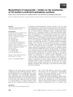
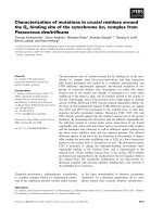
Fig. 1. Decaprenyl phosphate and decapre-
nyl-phospho-monosaccharides of myco-
bacteria. (A) The mycobacterial lipid carrier
C
50
-decaprenyl phosphate has a unique
stereoconfiguration and contains only one
trans (E)-isoprene residue at its x-end [33]
(see Fig. 3). (B) Decaprenyl-phospho-arabi-
nose, the only known
D-arabinose donor for
the synthesis of the cell-wall arabinogalactan
and lipoarabinomannan in mycobacteria
[32,33]. (C) Decaprenyl-phospho-ribose, the
direct precursor of the b-
D-arabinofuranosyl-
monophosphodecaprenol donor (B) and the
major form of the naturally occurring deca-
prenyl-phospho-sugars of mycobacteria
[32,34]. (D) The mycobacterial decaprenyl-
phospho-mannose, a minor component
[107].
B. A. Wolucka A role for the
D-arabinose lipid carrier
FEBS Journal 275 (2008) 2691–2711 ª 2008 The Author Journal compilation ª 2008 FEBS 2693
such as d-arabinose phosphates and d-arabinose nucle-
otides, have never been demonstrated in mycobacteria.
Exogenously added d-arabinose is catabolized by a
spontaneous M. smegmatis mutant via an inducible,
fungal-like pathway [32,38,39] that converts an aldo-
pentose into a ketopentose [40] (Fig. 2). In the myco-
bacterial pathway, d-arabinose is reduced by a
NADPH-dependent d-arabinose dehydrogenase to
d-arabinitol, and the latter compound is oxidized to
d-xylulose by a NAD-dependent d-arabinitol dehydro-
genase. d-Xylulose can then be phosphorylated to
d-xylulose 5-phosphate and enter the pentose phos-
phate cycle [32,39]. In contrast with mycobacteria, the
majority of bacteria use either isomerase ⁄ kinase or
oxidation pathways for the utilization of pentoses
[41,42]. Interestingly, the oxidation of d-arabinose to
A role for the D-arabinose lipid carrier B. A. Wolucka
2694 FEBS Journal 275 (2008) 2691–2711 ª 2008 The Author Journal compilation ª 2008 FEBS
d-arabinono-1,4-lactone does not occur in mycobacte-
ria [32,39], but in fungi, where it has been believed, at
least until recently [43], to be involved in the biosyn-
thesis of d-erythroascorbic acid [44].
After a fruitless search for water-soluble intermedi-
ates of d-arabinose, we looked for lipid-linked pyro-
phospho-oligosaccharides similar to the dolichol-linked
oligosaccharides of archaebacteria [45]. Indeed, gradi-
ent-eluted DEAE-cellulose fractions of organic extracts
from M. smegmatis contained lipid-linked galactose-
oligosaccharides, but also large amounts of mono-
charged, acid-labile arabinose, ribose and mannose
linked to phosphorylated isoprenoid lipids, although
some mycolic acids could be detected as well. Subse-
quent analysis of the monocharged glycolipids by fast-
atom bombardment mass spectrometry demonstrated
the presence of decaprenyl-phospho-pentoses and deca-
prenyl phosphate ions at m ⁄ z 909 and m ⁄ z 777, respec-
tively [32]. This was the beginning of a fruitful search
that has led to the identification of the d-arabinose
pathway, and to a better understanding of cell-wall bio-
synthesis and of the mechanism of action of ethambutol
in mycobacteria. In particular, we discovered that eth-
ambutol does not interfere with decaprenyl-phospho-
arabinose synthesis, and that the site of action of the
drug is downstream in the arabinan pathway [32].
Accordingly, it was proposed that: (a) decaprenyl-phos-
pho-arabinose is synthesized via a 2¢-epimerization of
decaprenyl-phospho-ribose, and serves as the donor of
d-arabinofuranosyl residues in the biosynthesis of the
cell-wall arabinogalactan and (lipo)arabinomann; (b)
ethambutol inhibits an arabinosyltransferase or an
arabinan-forming enzyme, and this inhibition results in
the accumulation of decaprenyl-phospho-arabinose in
mycobacteria; (c) the synthesis of the decaprenyl-phos-
pho-ribose precursor is controlled by a feedback mecha-
nism (Scheme 1) [32]. These conclusions have proven to
be correct and have served as the basis for further
research.
The details of the decaprenyl-phospho-arabinose
structure, including the determination of the absolute
configuration, anomeric linkage and ring form of the
d-arabinosyl residue, were solved later using combined
proton-NMR spectroscopy, gas chromatography and
mass spectrometry (Fig. 1B) [33]. NMR analysis also
allowed the determination of the particular structure
of the mycobacterial decaprenol with important impli-
cations regarding its biosynthesis (Figs 1A and 3). It
was a big surprise for us to find that what is lacking in
the 10 isoprene unit-containing C
50
-decaprenol of
mycobacteria is not a cis (Z)-unit, but one of the two
trans (E)-isoprene units that are localized at the x-end
of the known polyisoprenyl lipid carriers, including the
common bacterial undecaprenol. The proposed
x,mono-E,octa-Z configuration of the mycobacterial
decaprenol [33] was, in fact, the first hint of the exis-
tence of unusual Z-prenyl diphosphate synthases in
mycobacteria: a Z-farnesyl diphosphate synthase that
would provide an x,E,Z-farnesyl diphosphate for a
subsequent specific enzyme, a Z -decaprenyl diphos-
phate synthase. These unusual enzymes have been
identified recently (see below).
The structure of the endogenous b-d-arabinofurano-
syl-1-monophosphodecaprenol of mycobacteria was
solved (Fig. 1B). This was unprecedented because,
until that time, no other natural lipid-linked sugar iso-
lated from an organism had been fully structurally
characterized [46,47].
The next step was the structural elucidation of deca-
prenyl-phospho-ribose (Fig. 1C) [34]. The presence of
substantial amounts of decaprenyl-phospho-ribose was
puzzling because no ribose-containing polymers have
ever been described in mycobacteria. We proposed that
decaprenyl-phospho-ribose is converted to decaprenyl-
phospho-d-arabinose by a novel 2¢-epimerase of
mycobacteria (Scheme 1) [32]. The decaprenyl-
phospho-ribose 2¢-epimerase has been identified
recently.
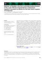
Fig. 2. The metabolism of D-arabinose in mycobacteria. The fungal-like assimilation pathway for D-arabinose of Mycobacterium smegmatis
[32,39] is shown (top reactions). Decaprenyl-phospho-
D-arabinose, the only known D-arabinofuranose donor, and decaprenyl-phospho-ribose
(in rectangles), were isolated from M. smegmatis [32] and structurally characterized (see Fig. 1). Decaprenyl-phospho-arabinose was pro-
posed to be synthesized via a 2¢-epimerization of decaprenyl-phospho-ribose, and to control the synthesis of the latter compound by a feed-
back mechanism. The heteromeric decaprenyl-phospho-ribose 2¢-epimerase (Rv3790 ⁄ Rv3791) was identified recently. Ethambutol, a first-line
drug for the treatment of tuberculosis, inhibits the utilization of decaprenyl-phospho-arabinose [32,33] at the level of the Emb proteins that
are involved in the formation of arabinans [75,88]. The enzymatic steps leading from the well-known 5-phosphoribosyl 1-pyrophosphate
(PRPP) intermediate to the formation of decaprenyl-phospho-ribose were identified later by in vitro assays.
D-Ribose 5-phosphate, the direct
precursor of PRPP, is proposed to be synthesized mainly by an essential transketolase (Rv1449) of the non-oxidative pentose phosphate
pathway. A possible involvement of a non-essential ribose 5-phosphate isomerase (Rv2465) and of the oxidative pentose phosphate pathway
enzymes is also shown. Intermediates of the fungal-like catabolic pathway are shown in green; the non-oxidative and oxidative parts of the
pentose phosphate pathway are shown in blue and violet, respectively; the decaprenyl-phospho-arabinose pathway is shown in red. Essen-
tial genes of M. tuberculosis, as determined by Himar1-based transposon mutagenesis [52,133], are indicated in bold, and cloned genes are
underlined.
B. A. Wolucka A role for the
D-arabinose lipid carrier
FEBS Journal 275 (2008) 2691–2711 ª 2008 The Author Journal compilation ª 2008 FEBS 2695
The discovery of decaprenyl-phospho-ribose pointed
to the involvement of activated ribose derivatives in
the biosynthesis pathway to d-arabinose. This observa-
tion was crucial for the identification of the precursor
of decaprenyl-phospho-ribose. An obvious candidate
to test as a donor of the activated d-ribofuranosyl resi-
due was the well-known, high-energy bond-containing
intermediate for nucleotide synthesis: 5-phosphoribosyl
1-pyrophosphate (PRPP). In vitro assays of crude
membranes of M. smegmatis incubated with [
14
C]-
labelled PRPP and synthetic decaprenyl phosphate as
substrates demonstrated the synthesis of decaprenyl-
phospho-ribose 5¢-phosphate, which, on dephosphory-
lation, produces decaprenyl-phospho-ribose [37]. The
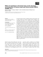
Fig. 3. The biosynthesis of C
50
-decaprenyl
pyrophosphate in Mycobacterium tuberculo-
sis. The particular structure of the mycobac-
terial decaprenol (see Fig. 1A) implies the
existence in mycobacteria of unique Z-prenyl
diphosphate synthases that use x,E-geranyl
pyrophosphate as a substrate. The non-
essential Rv1086 Z-farnesyl diphosphate
synthase and the essential Rv2361 Z-deca-
prenyl diphosphate synthase have been
identified [71,72].
A role for the
D-arabinose lipid carrier B. A. Wolucka
2696 FEBS Journal 275 (2008) 2691–2711 ª 2008 The Author Journal compilation ª 2008 FEBS
progress of the mycobacterial genome sequencing pro-
jects [48,49] has allowed a comparative genomics
approach that has led to the identification of the
mycobacterial decaprenyl-phospho-ribose 2¢-epimerase
and the phosphoribosyl transferase, involved in the
biosynthesis of decaprenyl-phospho-arabinose [50] and
decaprenyl-phospho-ribose [51], respectively.
The proposed pathway for the
biosynthesis of b-
D-arabinofuranosyl-
1-O-monophosphodecaprenol in
mycobacteria
Synthesis of D-ribose 5-phosphate
The first step in the biosynthesis of the b-d-arabino-
furanosyl-1-O-monophosphodecaprenol (decaprenyl-
phospho-arabinose) in mycobacteria (Fig. 2) is the
synthesis of d-ribose 5-phosphate. d-Ribose 5-phos-
phate could be synthesized by an amphibolic, thiamine
(vitamin B
1
) diphosphate-dependent transketolase
(sedoheptulose 7-phosphate:d-glyceraldehyde 3-phos-
phate glycolaldehydetransferase; EC 2.2.1.1), which
reversibly transfers a keto group from sedoheptulose
7-phosphate to d-glyceraldehyde 3-phosphate, and
produces d-ribose 5-phosphate and d -xylulose 5-phos-
phate, according to reaction (1):
sedoheptulose 7-phosphateþ
D-glyceraldehyde 3-phosphate
,
D-ribose 5-phosphateþD-xylulose5-phosphate ð1Þ
The transketolase is a ubiquitous enzyme that links the
glycolytic and pentose phosphate pathways, but has
never been studied in mycobacteria. The M. tuberculo-
sis genome contains one sequence encoding a putative
transketolase (Rv1449), and the gene is essential [52].
Otherwise, d-ribose 5-phosphate could be formed
from another intermediate of the pentose phosphate
pathway, d-ribulose 5-phosphate, by the ribose 5-phos-
phate isomerase (Rv2465) (Fig. 2). Surprisingly, the
ribose 5-phosphate isomerase, and also several other
pentose phosphate pathway genes, such as d-xylulose
5-phosphate 3-epimerase (Rv1408) and the 6-phos-
phoglucono-1,5-lactone lactonase (Rv1445), are app-
arently not essential in M. tuberculosis [52].
Consequently, the reaction catalysed by the ribose
5-phosphate isomerase probably plays a minor role in
the synthesis of the vital arabinans in mycobacteria.
Formation of 5-phosphoribosyl-a-1-pyrophosphate
The second step in the biosynthesis of decaprenyl-
phospho-arabinose (Fig. 2) is the reaction of ribose
5-phosphate with ATP to yield 5-phosphoribosyl-a-1-
pyrophosphate and AMP, catalysed by a PRPP
synthetase (ribose 5-phosphate diphosphokinase;
EC 2.7.6.1) (reaction 2):
ribose 5-phosphate þ ATP , 5-phospho-a-
D
-ribose 1-pyrophosphate þ AMP ð2Þ
PRPP is a key metabolite in the purine and pyrimidine
nucleotide de novo and salvage pathways, the biosynthe-
sis of pyridine nucleotide coenzymes and the synthesis
of histidine and tryptophan. By analogy with the
decaprenyl-phospho-arabinose biosynthesis of myco-
bacteria, PRPP is proposed to be a precursor of b-d-
ribofuranosyl residues of lipopolysaccharides and
capsular polysaccharides of Gram-negative bacteria,
such as Pseudomonas aeruginosa, Salmonella sp., Shigella
sp., Escherichia coli, Proteus sp., Haemophilus influenzae
and, perhaps, of eukaryotic trypanosomatids [34].
Mycobacterium tuberculosis contains one PRPP syn-
thetase protein (Rv1017) that shares at least 43% iden-
tity with its human, plant and bacterial homologues.
The mycobacterial PRPP synthetase sequence contains
a conserved PRK03092 domain from Val227 to
Ala240 (VLIDDMIDTGGTIA) that corresponds to
the PRPP binding motif. The PRPP synthetases are
known to undergo a complex regulation, and both
ADP and inorganic phosphate (P
i
) are the known allo-
steric regulators of the enzyme [53]. In spite of its cen-
tral role in cell-wall, nucleic acid and protein
biosynthesis, the mycobacterial PRPP synthetase has
not yet been characterized.
In Fig. 2, we propose that the inhibition of arabinan
synthesis by ethambutol, and the resulting accumula-
tion of decaprenyl-phospho-arabinose [32,33], could
have further repercussions via a feedback mechanism,
and inhibit, directly or indirectly, the PRPP synthetase
activity in mycobacteria. This would result in
decreased amounts of the PRPP precursor and, in
agreement with the observed complex effects of the
drug, lead to the inhibition of the synthesis of decapre-
nyl-phospho-ribose [32,33], but also of nucleic acids
and other compounds [54].
Synthesis of b-
D-5¢-phosphoribosyl-1-monophos-
phodecaprenol
The next step of the decaprenyl-phospho-arabinose
pathway (Fig. 2) is the reversible transfer of the
5-phosphoribosyl residue from the activated PRPP
donor to the decaprenyl phosphate acceptor, catalysed
by a 5-phospho-a-d-ribose 1-pyrophosphate:decaprenyl
phosphate 5-phosphoribosyltransferase (reaction 3):
B. A. Wolucka A role for the D-arabinose lipid carrier
FEBS Journal 275 (2008) 2691–2711 ª 2008 The Author Journal compilation ª 2008 FEBS 2697
5-phospho-a-D-ribose 1-pyrophosphate
þdecaprenyl phosphate , 5
0
-phosphoribosyl
-b-1-monophospho-decaprenol þ PP
i
ð3Þ
On the basis of the determined chemical structure of
b-d-ribosyl-1-monophosphodecaprenol [34], it can be
predicted that the product of the ribosyltransferase
reaction is b-d-5¢-phosphoribosyl-1-monophosphodeca-
prenol. Thus, the reaction would occur with an
inversion of the anomeric configuration of the 5-phos-
phoribosyl residue, although direct evidence is lacking.
The decaprenyl phosphate-dependent phosphoribosyl-
transferase activity was demonstrated in vitro using
crude membranes from M. smegmatis and a [
14
C]-
labelled PRPP substrate [37]. It was claimed that poly-
prenylphosphate-5-phosphoarabinose was one of the
reaction products and the direct precursor of polypre-
nyl-phospho-arabinose in mycobacteria, and it was
concluded that the epimerization at the C2 position of
the ribosyl residue takes place at the level of either
phosphoribose pyrophosphate or polyprenylphosphate-
5-phosphoribose [36,37].
The M. tuberculosis genes encoding 5-phospho-a-d-
ribose 1-pyrophosphate:decaprenyl phosphate 5-phos-
phoribosyltransferase (Rv3806) and the downstream
enzyme decaprenyl-phos pho-ribose 2 ¢-epimerase (Rv3790 ⁄
Rv3791) were identified only recently using a compara-
tive genomics strategy, as suggested earlier [34], namely
by searching M. tuberculosis orthologues of the
Azorhizobium genes that are involved in the d-arabi-
nosylation of nodulation factor glycolipids [50,51]. It is
worth noting that the sequences of the Nod-factor
genes for d-arabinosylation have never been published,
and the gene functions are, in fact, unknown [51]. In
addition, the early work reported the presence of
d-arabinose in the Azorhizobium Nod factor glycolipids
in the pyranose rather than furanose form [55], and
convincing evidence for the presence of Araf is lacking.
In contrast, the advent of the M. tuberculosis and
M. leprae genome data [48,49] has played an indisput-
able role in the identification of genes for the myco-
bacterial arabinogalactan ⁄ arabinomannan synthesis,
and led to the proposed d-arabinose pathway in myco-
bacteria.
Homologues of the Rv3806 protein (annotated as
UbiA prenyltransferases) are present in some Archaea
and in many eubacteria, such as mycobacteria, coryne-
bacteria and nocardia that share a similar composition
of their cell walls, certain species of cyanobacteria,
gamma-proteobacteria, clostridia and others. The
Rv3806 phosphoribosyltransferase (302 amino acids) is
an integral membrane protein that requires Mg
2+
for
its activity. The unpurified recombinant enzyme pres-
ent in the membrane of the E. coli host had apparent
K
m
values for PRPP and the decaprenyl phosphate of
plant origin substrates of 120 and 22 lm, respectively
[51]. The enzyme had a preference for medium-chain
polyprenyl phosphates (C
50
–C
55
) and showed no activ-
ity with a short-chain C
20
-polyprenyl phosphate. The
pH optimum for the phosphoribosyltransferase reac-
tion was pH 7.5–8. Contrary to the authors’ claim
[51], the reaction catalysed by the 5-phospho-a-d-
ribose 1-pyrophosphate:decaprenyl phosphate 5-phos-
phoribosyltransferase is probably not the committed
step of decaprenyl-phospho-arabinose biosynthesis,
because it is reversible in the absence of pyrophospha-
tase activity.
Synthesis of b-
D-ribosyl-1-monophosphodecapre-
nol (decaprenyl-phospho-ribose)
Decaprenyl-phospho-ribose is the major form of the
lipid-linked pentoses in mycobacteria [34] (Fig. 1C). It
is formed by the removal of a 5¢-phosphate group of
the b-d-5¢-phosphoribosyl-1-monophosphodecaprenol
precursor, catalysed by a phosphatase (reaction 4):
5
0
-phosphoribosyl-b-1-monophospho-decaprenol
! b-
D-ribosyl-1-monophospho-decaprenol þ P
i
ð4Þ
The phosphatase reaction is expected to be irreversible,
and thus it would represent the committed step in the
biosynthesis of decaprenyl-phospho-arabinose in myco-
bacteria. Inspection of the M. tuberculosis operons
involved in the biosynthesis of the arabinan and galac-
tan polymers has revealed the presence of an unknown
PAP2-family phospholipid phosphatase (Rv3807),
which is located next to the phosphoribosyltransferase
(Rv3806) discussed above. The Rv3807 orthologues are
present in all Corynebacterineae. The Rv3807 protein
is therefore a good candidate for a specific decaprenyl-
phospho-ribose-5¢-phosphate phosphatase. Surpris-
ingly, the Rv3807 gene is apparently not essential [52],
whereas all the other genes related to decaprenyl-phos-
pho-arabinose synthesis are annotated as essential
genes. Further studies are necessary to elucidate the
biological function of the Rv3807 gene product.
2¢-Epimerization of decaprenyl-phospho-ribose to
decaprenyl-phospho-arabinose
The last step of the biosynthetic pathway of decapre-
nyl-phospho-arabinose (b-d-arabinofuranosyl-1-mono-
phosphodecaprenol) is the 2¢-epimerization of
d-ribofuranosyl to d-arabinofuranosyl at the level of
decaprenyl-phospho-pentoses, as originally proposed
A role for the D-arabinose lipid carrier B. A. Wolucka
2698 FEBS Journal 275 (2008) 2691–2711 ª 2008 The Author Journal compilation ª 2008 FEBS
[32,33] (Fig. 2). This conversion proceeds via a
decaprenyl-phospho-2¢-keto-d-arabinose intermediate,
which is probably not released from the mycobacterial
enzyme under physiological conditions (reaction 5):
b-
D-ribofuranosyl-1-monophosphodecaprenol
!½2
0
-keto-b-D-arabinofuranosyl-
1-monophosphodecaprenol!b-
D-arabinofuranosyl-
1-monophosphodecaprenol ð5Þ
The decaprenyl-phospho-ribose 2¢-epimerase is a het-
eromeric enzyme composed of two types of polypep-
tide that are annotated as an oxidoreductase and a
short-chain dehydrogenase ⁄ reductase, and encoded by
the Rv3790 and Rv3791 genes, respectively, of the
M. tuberculosis genome [50]. The exact composition of
the enzyme is unknown. However, simultaneous
expression of both polypeptides is required for epimer-
ase activity. Close homologues of the Rv3790 and
Rv3791 proteins are present in arabinan-synthesizing
mycobacteria, corynebacteria, nocardia and related
actinobacteria, but also in other bacteria, many of
which are pathogens and symbionts of animals and
plants: for example, Pseudomonas aeruginosa, Burk-
holderia sp., Legionella pneumophila, Leptospira interro-
gans and Rhizobium etli. Interestingly, species that are
known to contain Araf as a component of their lipo-
polysaccharide, such as the opportunistic pathogen
Pseudomonas aeruginosa and the legume symbiont
Sinorhizobium meliloti, possess sequences that are simi-
lar (35% identity) to the Rv3790 and Rv3791 subunits
of the heteromeric 2¢-epimerase of M. tuberculosis.
The Rv3790 oxidoreductase protein (461 amino
acids) contains a FAD-binding N-terminal domain and
a C-terminal d-arabinono-1,4-lactone oxidase (ALO)
signature from T423 to L458. The ALO domain is
characteristic for l-gulono-1,4-lactone oxidase (GLO)-
like enzymes that catalyse the last step in the biosyn-
thesis of l-ascorbic acid (or its 5-carbon homologue
d-erythroascorbic acid) in plants, animals, fungi and
some microbes [56,57]. The Rv3790 protein shares
22% identical residues with d-arabinono-1,4-lactone
oxidase of Sacccharomyces cerevisiae (ALO1) [58]. The
protein also shows a limited identity at both the
N- and C-termini (26% and 38% identity, respectively)
with the recently identified l-gulono-1,4-lactone dehy-
drogenase (Rv1771) of M. tuberculosis [59]. The yeast
ALO1 enzyme catalyses the last step of oxidation of
d-arabinono-1,4-lactone to d-erythroascorbic acid, and
uses molecular oxygen as electron acceptor. The
Rv1771 dehydrogenase is probably involved in the syn-
thesis of l-ascorbic acid (vitamin C) in M. tuberculosis;
the enzyme is specific for l-gulono-1,4-lactone, and
can use both cytochrome c and a phenazine derivative
as electron acceptors [59].
The d-arabinono-1,4-lactone substrate of the yeast
ALO1 enzyme has a furan-based ring structure that is
similar to the d-arabinofuranosyl moiety of the epim-
erase reaction product (Fig. 5). Although the mecha-
nism of GLO and other GLO-like enzymes is not well
understood, the GLO-catalysed reaction is thought to
proceed via oxidation of the 2-hydroxyl group to a
2-keto derivative, which subsequently undergoes an
enolization to form l-ascorbic acid (or d-erythroascor-
bic acid). It is probable therefore that the Rv3790 sub-
unit(s) is directly responsible for the conversion of
decaprenyl-phospho-ribose to the corresponding
2
¢-keto-b-d-erythropentofuranose derivative (Figs 2
and 5).
In conclusion, little is known about the Rv3790 ⁄
Rv3791 decaprenyl-phospho-ribose 2¢-epimerase of
M. tuberculosis. In particular, the nature of the flavin
cofactor of the Rv3790 subunit and of the electron ac-
ceptors has not been elucidated.
As discussed above, an evolutionary link exists
between Araf and l-ascorbic acid ⁄ d-erythroascorbic
acid biosynthesis pathways. An ancestor GLO-like
gene of Actinomycetales or an unrelated gene that has
acquired an ALO-like domain by convergent evolution
could evolve into an Araf synthesizing enzyme
(Rv3790) by recruiting an ancient short-chain dehydro-
genase ⁄ reductase (Rv3791) that reduces the 2¢-keto
d-arabinofuranose ring to a d-arabinofuranosyl resi-
due. Interestingly, pathogenic actinobacteria, including
M. tuberculosis, M. bovis, M. ulcerans and M. mari-
num, have acquired, via gene duplication ⁄ divergent
evolution or horizontal gene transfer, another GLO
gene (Rv1771 in M. tuberculosis) for the synthesis of
l-ascorbic acid (or a related compound). The product
of the Rv1771-catalysed reaction might interfere with
l-ascorbic acid-dependent signal transduction path-
ways of animal hosts; however, its functions in
M. tuberculosis are still unknown [59].
Other activated forms of D-arabinose
Decaprenyl-phospho-arabinose (b-d-arabinofuranosyl-
1-monophosphodecaprenol) is the only known donor
of d-arabinofuranosyl units in the synthesis of
arabinans of Actinomycetales. Disruption of the gene
encoding the 5-phospho-a-d-ribose 1-pyrophos-
phate:decaprenyl phosphate 5-phosphoribosyltransfer-
ase (UbiA) produces a d-arabinose-deficient mutant of
Corynebacterium glutamicum that is devoid of the cell-
wall arabinan–corynomycolic acid complex [28]. This
surprising result indicates that both the arabinan part
B. A. Wolucka A role for the D-arabinose lipid carrier
FEBS Journal 275 (2008) 2691–2711 ª 2008 The Author Journal compilation ª 2008 FEBS 2699
and the bound corynomycolic acids of the cell-wall
peptidoglycan–arabinogalactan–corynomycolate core
are not essential for the survival of C. glutamicum. In
contrast, the arabinan part of the peptidoglycan–arabi-
nogalactan–mycolate core is essential in mycobacteria,
because disruption of the priming arabinosyltransferase
AftA (Rv3792), which adds the first d-arabinofurano-
syl residue to the galactan core, or of the Rv3806
phosphoribosyl transferase, is lethal in M. tuberculosis
[28,60].
Mycobacterium smegmatis synthesizes an additional
compound containing an activated d-arabinose resi-
due, namely a partially saturated b-d-arabinosyl-1-
monophospho-octahydroheptaprenol (Fig. 4B) [61].
The biosynthesis of the C
35
-isoprenyl-phospho deriva-
tive of d-arabinose is unknown, although it is possible
that the compound is synthesized via the decaprenyl-
phospho-arabinose pathway because of the low speci-
ficity of the decaprenyl-phosphate-dependent enzymes.
Otherwise, the C
35
-octahydroheptaprenyl-phospho-
arabinose could be synthesized by a direct transfer of
the d-arabinofuranosyl unit from decaprenyl-phospho-
arabinose or another donor to the C
35
-octahydrohep-
taprenyl phosphate acceptor. In agreement with the
latter proposal, ribosylated derivatives of C
35
-octahy-
droheptaprenyl phosphate have never been described.
The biological function of b-d-arabinosyl-1-mono-
phospho-octahydroheptaprenol of M. smegmatis is
unknown. Moreover, it is not clear whether other
mycobacteria synthesize C
35
-octahydroheptaprenyl-
phosphate derivatives.
Interestingly, single terminal d-arabinofuranosyl
residues of short lipoarabinomannans of C. glutami-
cum are apparently not derived from decaprenyl-phos-
pho-arabinose, but rather from another, still unknown,
donor [62].
In contrast with Araf, which is present exclusively in
bacteria, d-arabinopyranose is found in polysaccharides
Fig. 4. The partially and fully saturated gly-
cosylated phospholipids of mycobacteria. (A)
The major form of the lipid-linked mannose
in Mycobacterium smegmatis, the partially
saturated short-chain C
35
-octahydrohepta-
prenyl-phospho-mannose [107]. (B) A minor
form of the lipid-linked
D-arabinose of
M. smegmatis, the partially saturated short-
chain C
35
-octahydroheptaprenyl-phospho-
arabinose [61]. (C) The mycolylated isopren-
oid phospholipid of M. smegmatis [120]. (D)
The C
30
-mannosyl-b-1-phosphomycoketide
of M. avium. (E) A similar C
34
derivative of
M. tuberculosis [121]. The compounds in (D)
and (E) are not related to polyisoprenoids,
and are synthesized in pathogenic mycobac-
teria by a polyketide synthase [108].
A role for the
D-arabinose lipid carrier B. A. Wolucka
2700 FEBS Journal 275 (2008) 2691–2711 ª 2008 The Author Journal compilation ª 2008 FEBS
of some eukaryotic microorganisms, such as
trypanosomes, but also in plants. Sequences similar to
the mycobacterial enzymes of the decaprenyl-phospho-
arabinose biosynthesis are apparently absent from
eukaryotes, thus pointing to the existence of a totally
different pathway(s) for the synthesis of d-arabinopyr-
anosyl residues. In agreement, GDP-d-arabinopyra-
nose is the precursor of d-arabinopyranose residues
present in the glycoconjugates of some trypanosomatid
parasites. In Leishmania major and Crithidia fascicula-
ta, GDP-d-arabinose for the synthesis of lipophospho-
glycan is synthesized from d-glucose via an undefined
pathway that involves the loss of carbon C1 [63].
Decaprenyl phosphate: structure and
biosynthesis
x,mono-E,octa-Z C
50
-decaprenyl phosphate (Fig. 1A)
[33] is the lipid carrier of mycobacteria that plays a
crucial role in the biosynthesis of all three polymers of
the cell wall: peptidoglycan, arabinogalactan and
(lipo)arabinomannans.
As in many other eubacteria, plant chloroplasts,
algae and apicomplexan parasites, mycobacteria syn-
thesize isopentenyl diphosphate, the precursor for the
biosynthesis of polyisoprenols and other isoprenoids,
via the non-mevalonate (or 1-deoxy-d-xylulose-5-phos-
phate) route [64,65]. Like the decaprenyl-phos-
pho-arabinose pathway, non-mevalonate isoprenoid
biosynthesis is a potential target for new antimycobac-
terial drugs [66,67].
Polyisoprenyl phosphates are synthesized by sequen-
tial condensation of isopentenyl diphosphate with
allylic diphosphates in a reaction catalysed by unre-
lated E- and Z-prenyl diphosphate synthases that
introduce an E- and Z-isoprene unit, respectively, in
the reaction product (Fig. 3) (for a review, see [68]). In
contrast with most bacteria that use C
55
-undecaprenol
phosphate, consisting of 11 isoprene units in the x,
di-E,octa-Z configuration, mycobacteria employ a
shorter derivative: C
50
-decaprenyl phosphate [69]. The
mycobacterial decaprenol has a unique x,mono-
E,octa-Z stereoconfiguration of the polyisoprene chain
(Fig. 1A) [33]. Such a structure implies the existence in
mycobacteria of an unusual Z-prenyl diphosphate
synthase that uses x,E-geranyl pyrophosphate (C
10
)
and ⁄ or x,E,Z-farnesyl pyrophosphate (C
15
) as allylic
substrate, instead of the common x,E ,E-farnesyl
pyrophosphate (Fig. 3).
The first Z-prenyl diphosphate synthase (Z-undeca-
prenyl diphosphate synthase) was identified in Micro-
coccus luteus [70]. Mycobacterium tuberculosis contains
two homologues of the M. luteus Z-prenyl diphosphate
synthase: the Rv1086 and Rv2361 proteins. The
Rv1086 gene is apparently not essential and encodes a
specific, short-chain Z-farnesyl diphosphate synthase
which synthesizes x,E,Z-farnesyl diphosphate and
x,Z,Z-farnesyl diphosphate [71] (Fig. 3). Another gene
(Rv2361) encodes a Z-decaprenyl diphosphate synthase
that preferentially uses x,E,Z-farnesyl diphosphate as
a substrate [72]. x,E-Geranyl diphosphate also serves
as a substrate for the mycobacterial Z-decaprenyl
Fig. 5. The last step of the biosynthesis of b-D-arabinofuranosyl-1-monophosphodecaprenol in mycobacteria (A) and D-erythroascorbic acid in
yeasts (B). The oxidoreductase subunit (Rv3790) of the decaprenyl-phospho-ribose 2¢-epimerase of Mycobacterium tuberculosis shares 22%
identity with the
D-arabinono-1,4-lactone oxidase of Saccharomyces cerevisiae (ALO1); the enzymes catalyse similar reactions and employ
structurally similar sugar intermediates.
B. A. Wolucka A role for the
D-arabinose lipid carrier
FEBS Journal 275 (2008) 2691–2711 ª 2008 The Author Journal compilation ª 2008 FEBS 2701
diphosphate synthase, albeit with lower efficiency. This
suggests that the Rv2361 Z-decaprenyl diphosphate
synthase could compensate for the lack of Z-farnesyl
diphosphate synthase activity in the Rv1086-deficient
mutants of M. tuberculosis (Fig. 3).
The mycobacterial Z-diphosphate synthases Rv1086
and Rv2361 do not use dimethylallyl diphosphate as
substrate. Therefore, another, still unidentified, enzyme
must exist that synthesizes either x,E-geranyl diphos-
phate or x,Z-neryl diphosphate in mycobacteria. In
addition, a novel, essential x,E,E-farnesyl diphosphate
synthase (Rv3398) has been identified in M. tuberculosis
[73]. The Rv3398 enzyme may be involved in the synthe-
sis of compounds other than decaprenol isoprenoids,
such as menaquinones, carotenoids or hopanoids, but
its physiological function in mycobacteria is not known.
Interestingly, a non-essential bacA decaprenyl pyro-
phosphate phosphatase of M. smegmatis (erroneously
named ‘undecaprenyl phosphokinase’), a homologue of
the Rv2136 protein of M. tuberculosis, has been shown
to be involved in biofilm formation and, not surpris-
ingly, bacitracin resistance in M. smegmatis [74]. The
bacA gene product participates in the recycling of
polyprenyl pyrophosphates for cell-wall synthesis in
bacteria. The bacA deletion mutant of M. smegmatis
was viable, thus pointing to the existence of alternative
pathways for the regeneration of decaprenyl phosphate
in mycobacteria.
Chemically synthesized D-arabinose
donors and analogues
[
14
C]-Labelled b-d-arabinofuranosyl-1-monophospho-
decaprenol was first obtained by a semi-in vivo micro-
method [33], and served for the development of a basic
assay for the mycobacterial arabinosyltransferases
[75,76]. The stereoselective chemical synthesis of
b-d-[1-
14
C]arabinofuranosyl-1-monophosphodecaprenol
was achieved using phosphoramidite coupling of deca-
prenol (of plant origin) and a protected 2,3,5-tri-
O-tert-butyl dimethylsilyl derivative of Araf, followed
by final deprotection with ammonium fluoride under
mild conditions [77].
A similar approach was used for the efficient syn-
thesis of b-d-ribofuranosyl-1-monophosphodecaprenol
and shorter chain C
10
-neryl- and C
15
-farnesyl-mono-
phospho-b-d-ribofuranose derivatives [78]. The b-d-
ribofuranosyl-1-monophosphodecaprenol [34], a direct
precursor of decaprenyl-phospho-arabinose, is neces-
sary to study the decaprenyl-phospho-ribose 2¢-epimer-
ase of mycobacteria.
Recently, it has been shown that the classical poly-
prenyl trichloroacetimidate-based methodology is
particularly suitable for the stereoselective synthesis of
polyprenyl-phospho-b-d-arabinofuranoses by coupling
a polyprenyl trichloroacetimidate intermediate with a
protected b-d-arabinofuranose 1-phosphate derivative
[79].
Studies of mycobacterial arabinosyltransferases
require specific oligosaccharide acceptors, in addition
to the b-d-arabinofuranosyl-1-monophosphodecapre-
nol donor. A variety of O- and S-alkyl arabinosides
were synthesized and tested as substrates in arabinosyl-
transferase assays [80–82]. The chemically synthesized
trisaccharide acceptors and the O-alkyl disaccharide
acceptors with a C
8
alkyl chain were good substrates
for the mycobacterial arabinosyltransylferases, whereas
monosaccharides did not serve as acceptors [80].
Modified oligosaccharide analogues can inhibit poly-
saccharide synthesis, and may represent lead com-
pounds for the synthesis of new drugs [81]. Recently,
fluorescent dansyl derivatives of Ara
fur
(a1 fi 5)Ara
fur
disaccharides were prepared as photoaffinity probes to
study mycobacterial arabinosyltransferases and to
screen drug candidates [83,84].
Arabinosyltransferases, arabinan
biosynthesis and the mode of action
of ethambutol
Recent studies have confirmed that decaprenyl-phos-
pho-arabinose is the only donor of the arabinofurano-
syl residues for the synthesis of the cell-wall
arabinogalactans of Corynebacterineae [29,60]. The
synthetic b-d-arabinofuranosyl-1-monophosphodeca-
prenol supported the in vitro formation of a(1 fi 5)
and b(1 fi 2) linkages of arabinans by a crude prepa-
ration of mycobacterial arabinosyltransferases [80].
Further studies led to the identification of specific
arabinosyltransferases, such as the priming AftA arabi-
nosyltransferase (Rv3792), which adds the first arabi-
nofuranosyl residue to the preformed linear galactan
[60], and the terminal b(1 fi 2) AftB arabinosyltrans-
ferase (Rv3805), which is involved in the synthesis of
mycolylation sites [85]. Another arabinosyltransferase
activity involved in the elaboration of arabinan chains
of lipoarabinomannans has been detected recently, but
not identified [86].
Disruption of the Rv3806 orthologue gene encoding
the decaprenyl-phospho-ribose-5¢-phosphate synthase
[28], or of the priming AftA arabinosyltransferase [60],
resulted in viable C. glutamicum mutants that lacked
both the arabinan part and the esterified corynomyco-
lates of the cell-wall peptidoglycan–arabinogalactan–
corynomycolate core. These astonishing results
demonstrate that, unlike mycobacteria, corynebacteria
A role for the D-arabinose lipid carrier B. A. Wolucka
2702 FEBS Journal 275 (2008) 2691–2711 ª 2008 The Author Journal compilation ª 2008 FEBS
do not require arabinans for survival. It might be
envisaged, therefore, that arabinans play a distinct,
although still unknown, role in mycobacteria, in addi-
tion to their structural functions. Consequently, the
regulation of arabinan synthesis in mycobacteria is
expected to differ from that in corynebacteria. In
agreement, decaprenyl-phospho-arabinose does not
accumulate in the disruption Cg-Emb mutant of
C. glutamicum [60], although it is accumulated in eth-
ambutol-treated M. smegmatis cells [32,33].
Ethambutol, dextro-2,2¢-(ethylenediimino)-di-1-buta-
nol, is a first-line antituberculosis drug with pleiotropic
effects. One of the early effects of ethambutol is the
inhibition of arabinan biosynthesis in mycobacteria
[87]. Although ethambutol does not block the synthesis
of decaprenyl-phospho-arabinose, it interferes with the
utilization of the arabinose donor by inhibiting either
arabinosyltransferase activity or the formation of an
arabinose acceptor in mycobacteria [32,33] (Scheme 1,
Fig. 2).
Studies of the mechanisms of resistance to ethambu-
tol in M. tuberculosis led to the identification of the
embCAB operon [88]. Structural mutations in embB
and embC genes are found in clinical isolates of
M. tuberculosis [89], and the emb region is thought to
determine intrinsic and acquired resistance to etham-
butol in mycobacteria [90], or even broad drug resis-
tance [91]. The emb region of M. tuberculosis encodes
large (about 1000 amino acids) integral membrane pro-
teins (EmbB, EmbA and EmbC) that share 61–68%
sequence identity. The Emb protein sequences are
unique to Corynebacterineae and closely related spe-
cies, and are involved in the formation of arabinan
chains of the mycobacterial arabinogalactan (EmbB
and EmbA) [92] and lipoarabinomannan (EmbC) [93].
In contrast with M. smegmatis, the embA gene is
apparently essential in M. tuberculosis, and expressed
independently of the embC gene [94]. Truncated forms
of lipoarabinomannan were found in clinical isolates
of ethambutol-resistant M. tuberculosis [95]. An early
claim that the EmbAB proteins act as simple arabi-
nosyltransferases [75] is now being revised. Indeed,
Emb proteins show little homology with known glyco-
syltransferases [96], and the arabinosyltransferase
activity of Emb proteins has not been demonstrated in
an unequivocal manner. Recent studies have shown
that ethambutol does not inhibit any of the identified
arabinosyltransferases [60,85]. Significantly, disruption
of the emb gene results in l-glutamate efflux in C. glu-
tamicum [31]. In the same line of evidence, ethambutol
treatment results not only in a block of arabinan syn-
thesis, but also in the loss of the previously formed
arabinan from the cell wall in M. smegmatis [97].
However, the underlying mechanism is not under-
stood.
The expression of embCAB genes in M. tuberculosis
undergoes a complex control process that involves the
EmbR transcriptional regulator, and PknH [30] and
other serine–threonine protein kinases ⁄ phosphatase
systems [98]. The serine–threonine protein kinase
enzymes are absent from the M. smegmatis saprophyte.
Therefore, significant differences in the regulation of
Emb-dependent arabinan synthesis probably exist
between the pathogenic and non-pathogenic myco-
bacteria.
In conclusion, a working hypothesis is that Emb
proteins might act as arabinan-forming ‘polymerases’
or arabinan synthases that assemble larger blocks of
oligosaccharide nature, but also function in substrate
channelling and, perhaps, in species-specific signal
transduction. Clearly, the biological functions of Emb
proteins and the actual target of ethambutol still await
elucidation.
New drugs: decaprenyl-phospho-
arabinose as a lead compound
An amazing number of compounds with antimycobac-
terial activity have been designed, but only a few can-
didates, such as nitroimidazole PA-824 prodrug [99]
and diarylquinolines [100], are currently undergoing
clinical trials as antituberculosis drugs (for a review,
see [101]).
b-d-Arabinofuranosyl-1-monophosphodecaprenol [33]
has served as a model molecule for the rational design
of new antituberculosis drugs. Of the tested phospho-
nate, phosphinic and sulfone analogues of decaprenyl-
phospho-arabinose, a C-phosphonate analogue
[102,103] is active against M. tuberculosis and is cur-
rently undergoing trials in a mice model of
tuberculosis. Recently, new 2-deoxy-2-fluoro deriva-
tives were obtained [104], as well as an aza-ribose
analogue with promising antimycobacterial activity
[105].
Interestingly, an ethambutol-like diaminated com-
pound SQ109, which contains two isoprene units, is a
very efficient antimycobacterial, effective against multi-
drug-resistant strains (see [101]).
Decaprenyl-phospho-D-mannose and
related compounds
Early studies on the mycobacterial decaprenyl-phos-
pho-mannose played an important role in the discov-
ery of d-arabinose-containing polyisoprenoid lipids
and in a better understanding of arabinogalactan and
B. A. Wolucka A role for the D-arabinose lipid carrier
FEBS Journal 275 (2008) 2691–2711 ª 2008 The Author Journal compilation ª 2008 FEBS 2703
arabinomannan biosynthesis. Two forms of polypre-
nyl-phospho-mannose were synthesized from GDP-
[
14
C]mannose by membrane fractions of M. smegmatis:
aC
50
-decaprenyl-phospho-mannose and a C
35
-octa-
hydroheptaprenyl-phospho-mannose [69,106]. Similar
endogenously synthesized activated mannose deriva-
tives were isolated from M. smegmatis and structurally
characterized as b-d-mannopyranosyl-1-monophospho-
decaprenol (Fig. 1D) and b-d-mannopyranosyl-1-
monophospho-C
35
-octahydroheptaprenol (Fig. 4A) [107].
Surprisingly, the short-chain C
35
-octahydroheptapre-
nyl-phospho-mannose was the predominant form,
whereas decaprenyl-phospho-mannose represented only
5% of the total polyprenyl-phospho-monosaccharides
of M. smegmatis.
Although decaprenyl phosphate is a lipid carrier in
M. tuberculosis and other mycobacteria, the partially
saturated C
35
-octahydroheptaprenol was found only in
the saprophytic M. smegmatis species, and its synthesis
is totally unknown. The saturated isoprenoid-like
x-end of the lipid chain is similar to the recently iden-
tified mycoketides of M. tuberculosis [108]. However,
the presence of unsaturated isoprene residues next to
the a-hydroxyl group points to an isoprenoid route of
lipid synthesis. Perhaps, the C
35
-octahydroheptaprenol
is synthesized via a convergent isoprene and polyketide
biosynthetic machinery similar to that described in
Bacillus subtilis [109]. Otherwise, the C
35
-octahydro-
heptaprenol might be synthesized from an unsaturated
heptaprenyl diphosphate intermediate [110]. The
enzymes involved in the synthesis and biological func-
tions of C
35
-octahydroheptaprenyl-phospho-mannose
in M. smegmatis are still unknown.
b-d-Mannosyl-1-phosphodecaprenol is synthesized
from decaprenyl phosphate and GDP-mannose by a
GDP-mannose-dependent mannosyltransferase Ppm1
(Rv2051) [111,112]. Decaprenyl-phospho-mannose is a
substrate for mannosyltransferases that are involved in
the synthesis of phosphatidylinositolmannosides, lipo-
mannans, lipoarabinomannans and glycoproteins in
mycobacteria. A decaprenyl-phospho-mannose-depen-
dent mannosyltransferase PimE (Rv1159) has been
shown to synthesize the PIM5 phosphatidylinositolm-
annoside [113]. Another enzyme, a branching a1,
2-mannosyltransferase (Rv2181), is necessary for the
synthesis of lipomannan [114]. Inactivation of the
Rv2181 orthologue resulted in the lack of lipomannan
and the formation of a truncated lipoarabinomannan
in M. smegmatis, thus suggesting that the two poly-
mers are synthesized via at least partially independent
routes. In addition, mannosyltransferases involved in
the extension of the lipomannan⁄ lipoarabinomannan
core precursor (Rv2174) [115], the synthesis of man-
nose caps of lipoarabinomannan (Rv1635) [116] and
protein O-mannosylation (Rv1002) [117] have been
identified as putative decaprenyl-phospho-mannose-
dependent enzymes.
Decaprenyl phosphate is also a carrier of building
blocks for peptidoglycan synthesis [118], and
participates in the synthesis of lipid-linked pyro-
phosphoryloligosaccharides in the assembly of the
peptidoglycan–galactan part of the mycobacterial cell
wall [119].
The ‘apparent carrier’ for mycolic acids
of M. smegmatis
From the lipid extracts of M. smegmatis, Besra et al.
[120] isolated a mycolic acid ester of C
35
-octahydro-
heptaprenyl-phospho-mannose (Myc-PL): 6-O-mycol-
yl-b-d-mannopyranosyl-1-monophosphoryl-3,7,11,15,
19,23,27-heptamethyl-(2Z,6E,10E)-octacosatrien-1-o l
(Fig. 4C). It is worth noting that the stereoconfigura-
tion of the isoprene units (E,E,Z-a) in the structure
proposed in [120] differs from that determined for the
b-d-mannos yl-1-monophospho-octahydroheptaprenol
of M. smegmatis (Z,Z,Z-a) (Fig. 4A) [107]. Moreover,
the b-d-mannosyl-1-monophospho-octahydroheptapre-
nol was shown to be the major form of lipid-linked
mannose, and present in a non-esterified state in
M. smegmatis [107]. It is possible that the latter dis-
crepancy is a result of the use of different procedures
for lipid isolation. In particular, spontaneous or
enzyme-mediated acyl migration might occur as a
result of the alkaline conditions applied in [120].
Another possibility is that the esterification step does
not occur immediately after b-d-mannosyl-1-mono-
phospho-octahydroheptaprenol synthesis, but only
later during ageing of M. smegmatis cells.
C
35
-Octahydroheptaprenol phosphate derivatives
have never been found in M. tuberculosis and other
pathogenic mycobacteria [106,121]. Therefore, the
mycoloylated mannophospholipid of M. smegmatis
(Fig. 4C), described in [120], is obviously not a com-
mon carrier for mycolic acids in mycobacteria.
Mycobacterial ‘polyisoprenoid
glycolipids’, phosphoantigens and
immune response
Human T cells of the immune system recognize peptide
antigens, but also respond to lipid and glycolipid anti-
gens displayed by CD1 proteins, and to some
ill-defined phosphoantigens. This recognition plays an
important role in both innate and acquired immunity
during tuberculosis infection.
A role for the D-arabinose lipid carrier B. A. Wolucka
2704 FEBS Journal 275 (2008) 2691–2711 ª 2008 The Author Journal compilation ª 2008 FEBS
Human CD1c proteins recognize fully saturated
(C
30
–C
34
) mannosyl-b-1-phospholipids, erroneously
called ‘polyisoprenoid glycolipids’, of pathogenic
mycobacteria (Fig. 4D,E), in addition to short-chain
(C
35
) a-saturated mannosyl-b-1-phosphoryldolichols
[121]. The branched alkyl chain of the antigenic
mannosyl-b-1-phospholipid of M. tuberculosis is syn-
thesized from malonyl (C
2
) and methylmalonyl (C
3
)
units by the PKS12 polyketide synthase [108]. Thus, in
spite of some structural resemblance to polyisoprenols,
the antigenic phosphoglycolipids of mycobacterial
pathogens belong to a new class of secondary metabo-
lites: phosphorylated and mannosylated polyketides
(mannosyl-b-1-phosphomycoketides). Interestingly, the
partially saturated (C
35
) b-d-mannosyl-1-phospho-octa-
hydroheptaprenol of M. smegmatis [107], but not
a similar mycoloylated derivative Myc-PL [120], was
recognized by human CD8 cells [121].
Independent of CD1c-mediated recognition, myco-
bacterial non-peptide phosphoantigens, including
isoprenoid products of the non-mevalonate pathway,
are recognized by Vc2V d2 T cells via a mechanism
that does not require antigen processing or presenta-
tion by major histocompatibility complex I and II or
CD1 molecules [122]. However, the structures and
mechanism of action of mycobacterial phosphoanti-
gens are still a matter of debate [123].
New methods for the analysis of
polyisoprenoid glycolipids
The application of tandem mass spectrometric methods
for the analysis of polyisoprenoid glycolipids was first
demonstrated using preparations of polyprenyl-phos-
pho-sugars isolated from M. smegmatis [61]. Desorp-
tion chemical ionization tandem mass spectrometry
proved to be suitable for the structural determination
of polyisoprenyl phosphates and, in particular, allowed
a facile discrimination between unsaturated polypre-
nols and a-saturated (dolichol) derivatives [124].
Recent developments in related desorption electrospray
ionization methods for ambient analysis of complex
solid-state samples [125] will probably find new appli-
cations in the field of lipidomics and metabolomics of
isoprenoid compounds.
Fast-atom bombardment and, later, electrospray-
ionization tandem mass spectrometric techniques have
been shown to be useful for the determination of the
anomeric configuration of the glycosyl residue of poly-
isoprenyl-phospho-sugars, sugar nucleotides and sugar
1-phosphates [126,127]. Collision-induced dissociation
of glycosyl 1-phosphate derivatives produced different
fragmentation patterns depending on the cis ⁄ trans
configuration of their 2-hydroxyl and phosphate
groups. Further studies have shown that stereochemis-
try at the 2-position of the non-reducing sugar ring
affects the fragmentation of disaccharides [128] and
acetyl glycosides [129], thus allowing anomeric distinc-
tion of these non-phosphorylated derivatives. The
method was successfully applied for the determination
of the anomeric linkage of the d-mannosyl residue of
the scarce, antigenic C
30
–C
34
mannosyl-b-1-phospho-
mycoketides (erroneously called ‘polyisoprenoid glycol-
ipids’) isolated from M. tuberculosis and M. avium
(Fig. 4D,E) [121,130].
A similar method was applied to screen for galactose
1-phosphate levels in neonatal galactosaemia [131],
and for the identification of a novel sugar nucleotide
precursor of pseudaminic acid of the Campylobacter
jejuni pathogen [132].
Conclusion
The discovery of decaprenyl-phospho-d-arabinose and
decaprenyl-phospho-ribose in 1990, and the subsequent
proposal of the last steps of the Araf pathway in 1992
(Scheme 1) [32], marked the beginning of a new era in
the study of cell-wall synthesis in mycobacteria. The
complete pathway to d-arabinose proposed here is
unique in bacteria and represents a good target for
new drugs. The enzymes for d-arabinose biosynthesis
have not been studied and await thorough biochemical
characterization. The novel decaprenyl-phospho-ribose
2¢-epimerase and, in particular, its relatedness to
l-ascorbic acid (vitamin C) biosynthetic enzymes,
including the recently evoked GDP-d-mannose
2¢-epimerase [57], deserve special attention. In addition,
similar 2¢-epimerases may be involved in the biosynthe-
sis of still unknown, water-soluble d-arabinofuranosyl
donors for glycoconjugates, whose synthesis does not
require long-chain polyisoprenyl carriers.
Ethambutol, a first-line antituberculosis drug, does
not interfere with the synthesis of d-arabinose, but
rather inhibits the incorporation of d-arabinofurano-
syl residues of decaprenyl-phospho-arabinose into the
arabinogalactan and (lipo)arabinomannan of the
mycobacterial cell wall at the level of the Emb pro-
teins – the putative arabinan synthases. Specific muta-
tions in the Emb proteins confer resistance to
ethambutol in M. tuberculosis [88]. However, in spite
of almost 15 years of intensive research all over the
world, and our better knowledge of the structure,
functions and biosynthesis of the mycobacterial cell
wall, the mode of action of ethambutol and the
molecular mechanisms of resistance to the drug are
not understood.
B. A. Wolucka A role for the D-arabinose lipid carrier
FEBS Journal 275 (2008) 2691–2711 ª 2008 The Author Journal compilation ª 2008 FEBS 2705
However, synthetic isoprenoid derivatives, including
those based on the structure of b-d-arabinofuranosyl-
monophospho-decaprenol and b-d-ribofuranosyl-mono-
phospho-decaprenol, have promising antituberculosis
activity, and some may show immunomodulatory
effects.
Further research by new generations of enthusiastic
and devoted scientists will be needed to take up the
challenge.
References
1 Dye C (2006) Global epidemiology of tuberculosis.
Lancet 367, 938–940.
2 Nunn P, Williams B, Floyd K, Dye C, Elzinga G &
Raviglione M (2005) Tuberculosis control in the era of
HIV. Nat Rev Immunol 5, 819–826.
3 Gandhi NR, Moll A, Sturm AW, Pawinski R, Gov-
ender T, Lalloo U, Zeller K, Andrews J & Friedland
G (2006) Extensively drug-resistant tuberculosis as a
cause of death in patients co-infected with tuberculosis
and HIV in a rural area of South Africa. Lancet 368,
1575–1580.
4 Raviglione MC & Smith IM (2007) XDR tuberculosis
– implications for global public health. N Engl J Med
356, 656–659.
5 Coley J, Tarelli E, Archibald AR & Baddiley J (1978)
The linkage between teichoic acid and peptidoglycan in
bacterial cell walls. FEBS Lett 88, 1–9.
6 Brennan PJ (2003) Structure, function, and biogenesis
of the cell wall of Mycobacterium tuberculosis .
Tuberculosis (Edinb) 83, 91–97.
7 van der Wel N, Hava D, Houben D, Fluitsma D, van
Zon M, Pierson J, Brenner M & Peters PJ (2007)
M. tuberculosis and M. leprae translocate from the
phagolysosome to the cytosol in myeloid cells. Cell
129, 1287–1298.
8 Ortalo-Magne A, Dupont MA, Lemassu A, Andersen
AB, Gounon P & Daffe M (1995) Molecular composi-
tion of the outermost capsular material of the tubercle
bacillus. Microbiology 141, 1609–1620.
9 Trias J, Jarlier V & Benz R (1992) Porins in the cell
wall of mycobacteria. Science 258, 1479–1481.
10 Niederweis M (2003) Mycobacterial porins – new
channel proteins in unique outer membranes. Mol
Microbiol 49, 1167–1177.
11 Zhang Y, Heym B, Allen B, Young D & Cole S
(1992) The catalase-peroxidase gene and isoniazid
resistance of Mycobacterium tuberculosis. Nature 358,
591–593.
12 Rosenkrands I, Weldingh K, Jacobsen S, Hansen CV,
Florio W, Gianetri I & Andersen P (2000) Mapping
and identification of Mycobacterium tuberculosis pro-
teins by two-dimensional gel electrophoresis, microse-
quencing and immunodetection. Electrophoresis 21,
935–948.
13 De Bruyn J, Soetaert K, Buyssens P, Calonne I, De
Coene JL, Gallet X, Brasseur R, Wattiez R, Falmagne
P, Montrozier H et al. (2000) Evidence for specific and
non-covalent binding of lipids to natural and recombi-
nant Mycobacterium bovis BCG hsp60 proteins, and to
the Escherichia coli homologue GroEL. Microbiology
146, 1513–1524.
14 Ojha A, Anand M, Bhatt A, Kremer L, Jacobs WR &
Hatfull GF (2005) GroEL1: a dedicated chaperone
involved in mycolic acid biosynthesis during biofilm
formation in mycobacteria. Cell 123, 861–873.
15 Belisle JT, Vissa VD, Sievert T, Takayama K, Brennan
PJ & Besra GS (1997) Role of the major antigen of
Mycobacterium tuberculosis in cell wall biogenesis.
Science
276, 1420–1422.
16 Katsube T, Matsumoto S, Takatsuka M, Okuyama M,
Ozeki Y, Naito M, Nishiuchi Y, Fujiwara N, Yoshim-
ura M, Tsuboi T et al. (2007) Control of cell wall
assembly by a histone-like protein in mycobacteria.
J Bacteriol 189, 8241–8249.
17 Harth G, Zamecnik PC, Tang JY, Tabatadze D &
Horwitz MA (2000) Treatment of Mycobacterium
tuberculosis with antisense oligonucleotides to gluta-
mine synthetase mRNA inhibits glutamine synthetase
activity, formation of the poly-l-glutamate ⁄ glutamine
cell wall structure, and bacterial replication. Proc Natl
Acad Sci USA 97, 418–423.
18 Walburger A, Koul A, Ferrari G, Nguyen L,
Prescianotto-Baschong C, Huygen K, Klebl B,
Thompson C, Bacher G & Pieters J (2004) Protein
kinase G from pathogenic mycobacteria promotes
survival within macrophages. Science 304, 1800–1804.
19 Jones G & Dyson P (2006) Evolution of transmem-
brane protein kinases implicated in coordinating
remodeling of gram-positive peptidoglycan: inside
versus outside. J Bacteriol 188, 7470–7476.
20 Narayan A, Sachdeva P, Sharma K, Saini AK, Tyagi
AK & Singh Y (2007) Serine threonine protein kinases
of mycobacterial genus: phylogeny to function. Physiol
Genomics 29, 66–75.
21 Brodin P, Rosenkrands I, Andersen P, Cole ST &
Brosch R (2004) ESAT-6 proteins: protective anti-
gens and virulence factors? Trends Microbiol 12,
500–508.
22 Basu SK, Kumar D, Singh DK, Ganguly N, Siddiqui
Z, Rao KV & Sharma P (2006) Mycobacterium
tuberculosis secreted antigen (MTSA-10) modulates
macrophage function by redox regulation of phospha-
tases. Febs J 273, 5517–5534.
23 Seiler P, Ulrichs T, Bandermann S, Pradl L, Jorg S,
Krenn V, Morawietz L, Kaufmann SH & Aichele P
(2003) Cell-wall alterations as an attribute of
A role for the D-arabinose lipid carrier B. A. Wolucka
2706 FEBS Journal 275 (2008) 2691–2711 ª 2008 The Author Journal compilation ª 2008 FEBS
Mycobacterium tuberculosis in latent infection. J Infect
Dis 188, 1326–1331.
24 Neyrolles O, Hernandez-Pando R, Pietri-Rouxel F,
Fornes P, Tailleux L, Barrios Payan JA, Pivert E,
Bordat Y, Aguilar D, Prevost MC et al. (2006) Is
adipose tissue a place for Mycobacterium tuberculosis
persistence? PLoS ONE 1, e43.
25 Alteri CJ, Xicohtencatl-Cortes J, Hess S, Caballero-
Olin G, Giron JA & Friedman RL (2007) Mycobacte-
rium tuberculosis produces pili during human infection.
Proc Natl Acad Sci USA 104, 5145–5150.
26 Watanabe M, Ohta A, Sasaki SI & Minnikin D (1999)
Structure of a new glycolipid from the Mycobacterium
avium–Mycobacterium intracellulare complex. J Bacte-
riol 181, 2293–2297.
27 Chatterjee D & Khoo KH (1998) Mycobacterial lipo-
arabinomannan: an extraordinary lipoheteroglycan
with profound physiological effects. Glycobiology 8,
113–120.
28 Alderwick LJ, Radmacher E, Seidel M, Gande R,
Hitchen PG, Morris HR, Dell A, Sahm H, Eggeling
L & Besra GS (2005) Deletion of Cg-emb in
Corynebacterineae leads to a novel truncated cell
wall arabinogalactan, whereas inactivation of
Cg-ubiA results in an arabinan-deficient mutant with
a cell wall galactan core. J Biol Chem 280,
32362–32371.
29 Alderwick LJ, Dover LG, Seidel M, Gande R, Sahm
H, Eggeling L & Besra GS (2006) Arabinan-deficient
mutants of Corynebacterium glutamicum and the
consequent flux in decaprenylmonophosphoryl-
d-arabinose metabolism. Glycobiology 16, 1073–
1081.
30 Molle V, Kremer L, Girard-Blanc C, Besra GS,
Cozzone AJ & Prost JF (2003) An FHA phosphopro-
tein recognition domain mediates protein EmbR phos-
phorylation by PknH, a Ser ⁄ Thr protein kinase from
Mycobacterium tuberculosis . Biochemistry 42, 15300–
15309.
31 Radmacher E, Stansen KC, Besra GS, Alderwick LJ,
Maughan WN, Hollweg G, Sahm H, Wendisch VF &
Eggeling L (2005) Ethambutol, a cell wall inhibitor of
Mycobacterium tuberculosis , elicits l-glutamate efflux
of Corynebacterium glutamicum. Microbiology 151,
1359–1368.
32 Wolucka BA (1992)
D-Arabinose metabolism in myco-
bacteria. Isolation of two novel glycolipids from Myco-
bacterium smegmatis . PhD Thesis. University of
Louvain, Louvain-la-Neuve.
33 Wolucka BA, McNeil MR, de HoffmannE, Chojnacki
T & Brennan PJ (1994) Recognition of the lipid inter-
mediate for arabinogalactan ⁄ arabinomannan biosyn-
thesis and its relation to the mode of action of
ethambutol on mycobacteria. J Biol Chem 269, 23328–
23335.
34 Wolucka BA & de Hoffmann E (1995) The presence of
beta-d-ribosyl-1-monophosphodecaprenol in mycobac-
teria. J Biol Chem 270, 20151–20155.
35 McNeil MR & Brennan PJ (1991) Structure, function
and biogenesis of the cell envelope of mycobacteria in
relation to bacterial physiology, pathogenesis and drug
resistance; some thoughts and possibilities arising from
recent structural information. Res Microbiol 142, 451–
463.
36 Scherman M, Weston A, Duncan K, Whittington A,
Upton R, Deng L, Comber R, Friedrich JD &
McNeil M (1995) Biosynthetic origin of mycobacte-
rial cell wall arabinosyl residues. J Bacteriol 177,
7125–7130.
37 Scherman MS, Kalbe-Bournonville L, Bush D, Xin Y,
Deng L & McNeil M (1996) Polyprenylphosphate-pen-
toses in mycobacteria are synthesized from 5-phos-
phoribose pyrophosphate. J Biol Chem 271, 29652–
29658.
38 Izumori K, Watanabe Y & Sugimoto S (1980) Evolution
of d-arabinose, l-xylose and l -ribose utilization in
Mycobacterium smegmatis – mutants with a novel
enzyme pentose reductase. Agric Biol Chem 44, 1443–
1446.
39 Wolucka-Wojtkiewicz B, Szmidzinski R, Jezierska A &
Cocito C (1988) Identification of a salvage pathway
for d-arabinose in Mycobacterium smegmatis. Eur J
Biochem 172, 197–203.
40 Chiang C & Knight SG (1960) Metabolism of d-xylose
by moulds. Nature 188, 79–81.
41 Watanabe S, Kodaki T & Makino K (2006) Cloning,
expression, and characterization of bacterial l-arabi-
nose 1-dehydrogenase involved in an alternative path-
way of l-arabinose metabolism. J Biol Chem 281,
2612–2623.
42 Brouns SJ, Walther J, Snijders AP, van de Werken HJ,
Willemen HL, Worm P, de Vos MG, Andersson A,
Lundgren M, Mazon HF et al. (2006) Identification of
the missing links in prokaryotic pentose oxidation
pathways: evidence for enzyme recruitment. J Biol
Chem 281, 27378–27388.
43 Amako K, Fujita K, Iwamoto C, Sengee M, Fuchi-
gami K, Fukumoto J, Ogishi Y, Kishimoto R & Goda
K (2006) NADP(+)-dependent d-arabinose dehydro-
genase shows a limited contribution to erythroascorbic
acid biosynthesis and oxidative stress resistance in
Saccharomyces cerevisiae . Biosci Biotechnol Biochem
70, 3004–3012.
44 Kim ST, Huh WK, Lee BH & Kang SO (1998)
d-arabinose dehydrogenase and its gene from
Saccharomyces cerevisiae . Biochim Biophys Acta 1429,
29–39.
45 Lechner J, Wieland F & Sumper M (1985) Biosynthesis
of sulfated saccharides N-glycosidically linked to the
protein via glucose. Purification and identification of
B. A. Wolucka A role for the D-arabinose lipid carrier
FEBS Journal 275 (2008) 2691–2711 ª 2008 The Author Journal compilation ª 2008 FEBS 2707
sulfated dolichyl monophosphoryl tetrasaccharides
from halobacteria. J Biol Chem 260, 860–866.
46 Trent MS, Ribeiro AA, Doerrler WT, Lin S, Cotter
RJ & Raetz CR (2001) Accumulation of a polyiso-
prene-linked amino sugar in polymyxin-resistant
Salmonella typhimurium and Escherichia coli: structural
characterization and transfer to lipid A in the peri-
plasm. J Biol Chem 276, 43132–43144.
47 Kanjilal-Kolar S & Raetz CR (2006) Dodecaprenyl
phosphate-galacturonic acid as a donor substrate for
lipopolysaccharide core glycosylation in Rhizobium
leguminosarum. J Biol Chem 281, 12879–12887.
48 Cole ST, Brosch R, Parkhill J, Garnier T, Churcher C,
Harris D, Gordon SV, Eiglmeier K, Gas S, Barry CE
3rd et al. (1998) Deciphering the biology of Mycobac-
terium tuberculosis from the complete genome
sequence. Nature 393, 537–544.
49 Cole ST, Eiglmeier K, Parkhill J, James KD, Thomson
NR, Wheeler PR, Honore
´
N, Garnier T, Churcher C,
Harris D et al. (2001) Massive gene decay in the
leprosy bacillus. Nature 409 , 1007–1011.
50 Mikusova K, Huang H, Yagi T, Holsters M, Vereecke
D, D’Haeze W, Scherman MS, Brennan PJ, McNeil
MR & Crick DC (2005) Decaprenylphosphoryl arabin-
ofuranose, the donor of the d-arabinofuranosyl
residues of mycobacterial arabinan, is formed via a
two-step epimerization of decaprenylphosphoryl ribose.
J Bacteriol 187, 8020–8025.
51 Huang H, Scherman MS, D’Haeze W, Vereecke D,
Holsters M, Crick DC & McNeil MR (2005) Identifi-
cation and active expression of the Mycobacterium
tuberculosis gene encoding 5-phospho-{alpha}-d-ribose-
1-diphosphate:decaprenyl-phosphate 5-phosphoribos-
yltransferase, the first enzyme committed to
decaprenylphosphoryl-d-arabinose synthesis. J Biol
Chem 280, 24539–24543.
52 Sassetti CM, Boyd DH & Rubin EJ (2003) Genes
required for mycobacterial growth defined by high
density mutagenesis. Mol Microbiol 48, 77–84.
53 Eriksen TA, Kadziola A, Bentsen AK, Harlow KW &
Larsen S (2000) Structural basis for the function of
Bacillus subtilis phosphoribosyl-pyrophosphate
synthetase. Nat Struct Biol 7, 303–308.
54 Forbes M, Kuck NA & Peets EA (1965) Effect of eth-
ambutol on nucleic acid metabolism in Mycobacterium
smegmatis and its reversal by polyamines and divalent
cations. J Bacteriol 89, 1299–1305.
55 Mergaert P, Van Montagu M, Prome JC & Holsters
M (1993) Three unusual modifications, a d-arabino-
syl, an N-methyl, and a carbamoyl group, are pres-
ent on the Nod factors of Azorhizobium caulinodans
strain ORS571. Proc Natl Acad Sci USA 90, 1551–
1555.
56 Wolucka BA & van Montagu M (2003) GDP-mannose
3
¢,5¢-epimerase forms GDP-l-gulose, a putative
intermediate for the de novo biosynthesis of vitamin C
in plants. J Biol Chem 278, 47483–47490.
57 Wolucka BA & van Montagu M (2007) The VTC2
cycle and the de novo biosynthesis pathways for
vitamin C in plants. Phytochemistry 68, 2602–2613.
58 Huh WK, Lee BH, Kim ST, Kim YR, Rhie GE,
Baek YW, Hwang CS, Lee JS & Kang O (1998)
d-Erythroascorbic acid is an important antioxidant
molecule in Saccharomyces cerevisiae. Mol Microbiol
30, 895–903.
59 Wolucka BA & Communi D (2006) Mycobacterium
tuberculosis possesses a functional enzyme for the
synthesis of vitamin C, l-gulono-1,4-lactone
dehydrogenase. Febs J 273 , 4435–4445.
60 Alderwick LJ, Seidel M, Sahm H, Besra GS & Eggel-
ing L (2006) Identification of a novel arabinofuranosyl-
transferase (AftA) involved in cell wall arabinan
biosynthesis in Mycobacterium tuberculosis. J Biol
Chem 281, 15653–15661.
61 Wolucka BA & de Hoffmann E (1994) Mass spectro-
metric analysis of prenyl phosphates and their glycosy-
lated forms. Acta Biochim Pol 41, 345–349.
62 Tatituri RV, Alderwick LJ, Mishra AK, Nigou J,
Gilleron M, Krumbach K, Hitchen P, Giordano A,
Morris HR, Dell A et al. (2007) Structural character-
ization of a partially arabinosylated lipoarabinoman-
nan variant isolated from a Corynebacterium
glutamicum ubiA mutant. Microbiology 153, 2621–
2629.
63 Schneider P, McConville MJ & Ferguson MA (1994)
Characterization of GDP-alpha-d-arabinopyranose,
the precursor of d-Arap in Leishmania major lipophos-
phoglycan. J Biol Chem 269, 18332–18337.
64 Rohmer M, Knani M, Simonin P, Sutter B & Sahm H
(1993) Isoprenoid biosynthesis in bacteria: a novel
pathway for the early steps leading to isopentenyl
diphosphate. Biochem J 295, 517–524.
65 Rohmer M (1999) The discovery of a mevalonate-inde-
pendent pathway for isoprenoid biosynthesis in
bacteria, algae and higher plants. Nat Prod Rep 16,
565–574.
66 Kuntz L, Tritsch D, Grosdemange-Billiard C, Hem-
merlin A, Willem A, Bach TJ & Rohmer M (2005)
Isoprenoid biosynthesis as a target for antibacterial
and antiparasitic drugs: phosphonohydroxamic acids
as inhibitors of deoxyxylulose phosphate reducto-isom-
erase. Biochem J 386, 127–135.
67 Henriksson LM, Unge T, Carlsson J, Aqvist J,
Mowbray SL & Jones TA (2007) Structures of Myco-
bacterium tuberculosis 1-deoxy-
d-xylulose-5-phosphate
reductoisomerase provide new insights into catalysis.
J Biol Chem 282, 19905–19916.
68 Swiezewska E & Danikiewicz W (2005) Polyisopre-
noids: structure, biosynthesis and function. Progress
Lipid Res 44, 235–258.
A role for the D-arabinose lipid carrier B. A. Wolucka
2708 FEBS Journal 275 (2008) 2691–2711 ª 2008 The Author Journal compilation ª 2008 FEBS
69 Takayama K & Goldman DS (1970) Enzymatic syn-
thesis of mannosyl-1-phosphoryl-decaprenol by a cell-
free system of Mycobacterium tuberculosis. J Biol Chem
245, 6251–6257.
70 Shimizu N, Koyama T & Ogura K (1998) Molecular
cloning, expression, and purification of undecaprenyl
diphosphate synthase. No sequence similarity between
E- and Z-prenyl diphosphate synthases. J Biol Chem
273, 19476–19481.
71 Schulbach MC, Mahapatra S, Macchia M, Barontini
S, Papi C, Minutolo F, Bertini S, Brennan PJ & Crick
DC (2001) Purification, enzymatic characterization,
and inhibition of the Z-farnesyl diphosphate synthase
from Mycobacterium tuberculosis. J Biol Chem 276,
11624–11630.
72 Kaur D, Brennan PJ & Crick DC (2004) Decaprenyl
diphosphate synthesis in Mycobacterium tuberculosis.
J Bacteriol 186, 7564–7570.
73 Dhiman RK, Schulbach MC, Mahapatra S, Baulard
AR, Vissa V, Brennan PJ & Crick DC (2004) Identifi-
cation of a novel class of omega,E,E-farnesyl diphos-
phate synthase from Mycobacterium tuberculosis.
J Lipid Res 45, 1140–1147.
74 Rose L, Kaufmann SH & Daugelat S (2004) Involve-
ment of Mycobacterium smegmatis undecaprenyl phos-
phokinase in biofilm and smegma formation. Microbes
Infect 6, 965–971.
75 Belanger AE, Besra GS, Ford ME, Mikusova K,
Belisle JT, Brennan PJ & Inamine JM (1996) The
embAB genes of Mycobacterium avium encode an
arabinosyl transferase involved in cell wall arabinan
biosynthesis that is the target for the antimycobacterial
drug ethambutol. Proc Natl Acad Sci USA 93,
11919–11924.
76 Xin Y, Lee RE, Scherman MS, Khoo KH, Besra GS,
Brennan PJ & McNeil M (1997) Characterization of
the in vitro synthesized arabinan of mycobacterial cell
walls. Biochim Biophys Acta 1335, 231–234.
77 Lee RE, Brennan PJ & Besra GS (1998) Synthesis of
beta-d-arabinofuranosyl-1-monophosphoryl polypre-
nols: examination of their function as mycobacterial
arabinosyl transferase donors. Bioorg Med Chem Lett
8, 951–954.
78 Liav A, Swiezewska E, Ciepichal E & Brennan PJ
(2006a) Stereoselectivity in the synthesis of polyprenyl-
phosphoryl b-d-ribofuranoses. Tetrahedron Lett 47,
8781–8783.
79 Liav A, Huang H, Ciepichal E, Brennan PJ & McNeil
MR (2006b) Stereoselective synthesis of decaprenyl-
phosphoryl b-d-arabinofuranose. Tetrahedron Lett 47,
545–547.
80 Lee RE, Brennan PJ & Besra GS (1997) Mycobacterial
arabinan biosynthesis: the use of synthetic arabinoside
acceptors in the development of an arabinosyl transfer
assay. Glycobiology 7, 1121–1128.
81 Ayers JD, Lowary TL, Morehouse CB & Besra GS
(1998) Synthetic arabinofuranosyl oligosaccharides as
mycobacterial arabinosyltransferase substrates. Bioorg
Med Chem Lett 8, 437–442.
82 Pathak AK, Pathak V, Maddry JA, Suling WJ, Gur-
cha SS, Besra GS & Reynolds RC (2001) Studies on
alpha(1
fi 5) linked octyl arabinofuranosyl disaccha-
rides for mycobacterial arabinosyl transferase activity.
Bioorg Med Chem 9, 3145–3151.
83 Pathak AK, Pathak V, Gurcha SS, Besra GS &
Reynolds RC (2002) Synthesis of an arabinofuranosyl
disaccharide photoaffinity probe for arabinosyltransfer-
ase activity in Mycobacterium tuberculosis. Bioorg Med
Chem Lett 12, 2749–2752.
84 Pathak AK, Pathak V, Seitz L, Gurcha SS, Besra GS,
Riordan JM & Reynolds RC (2007) Disaccharide ana-
logs as probes for glycosyltransferases in Mycobacte-
rium tuberculosis. Bioorg Med Chem 15, 5629–5650.
85 Seidel M, Alderwick LJ, Birch HL, Sahm H, Eggeling
L & Besra GS (2007) Identification of a novel arabino-
furanosyltransferase AftB involved in a terminal step
of cell wall arabinan biosynthesis in Corynebacteri-
neae, such as Corynebacterium glutamicum and
Mycobacterium tuberculosis . J Biol Chem 282, 14729–
14740.
86 Zhang J, Khoo KH, Wu SW & Chatterjee D (2007)
Characterization of a distinct arabinofuranosyltransfer-
ase in Mycobacterium smegmatis. J Am Chem Soc 129,
9650–9662.
87 Takayama K & Kilburn JO (1989) Inhibition of
synthesis of arabinogalactan by ethambutol in Myco-
bacterium smegmatis . Antimicrob Agents Chemother 33,
1493–1499.
88 Telenti A, Philipp WJ, Sreevatsan S, Bernasconi C,
Stockbauer KE, Wieles B, Musser JM & Jacobs WR
(1997) The emb operon, a gene cluster of Mycobacte-
rium tuberculosis involved in resistance to ethambutol.
Nat Med 3, 567–570.
89 Srivastava S, Garg A, Ayyagari A, Nyati KK, Dhole
TN & Dwivedi SK (2006) Nucleotide polymorphism
associated with ethambutol resistance in clinical
isolates of Mycobacterium tuberculosis. Curr Microbiol
53, 401–405.
90 Alcaide F, Pfyffer GE & Telenti A (1997) Role of
embB in natural and acquired resistance to ethambutol
in mycobacteria. Antimicrob Agents Chemother 41,
2270–2273.
91 Hazbon MH, Bobadilla del Valle M, Guerrero MI,
Varma-Basil M, Filliol I, Cavatore M, Colangeli R,
Safi H, Billman-Jacobe H, Lavender C et al. (2005)
Role of embB codon 306 mutations in Mycobacterium
tuberculosis revisited: a novel association with broad
drug resistance and IS6110 clustering rather than eth-
ambutol resistance. Antimicrob Agents Chemother 49,
3794–3802.
B. A. Wolucka A role for the D-arabinose lipid carrier
FEBS Journal 275 (2008) 2691–2711 ª 2008 The Author Journal compilation ª 2008 FEBS 2709
92 Escuyer VE, Lety MA, Torrelles JB, Khoo KH, Tang
JB, Rithner CD, Frehel C, McNeil MR, Brennan PJ &
Chatterjee D (2001) The role of the embA and embB
gene products in the biosynthesis of the terminal
hexaarabinofuranosyl motif of Mycobacterium
smegmatis arabinogalactan. J Biol Chem 276, 48854–
48862.
93 Zhang N, Torrelles JB, McNeil MR, Escuyer VE,
Khoo KH, Brennan PJ & Chatterjee D (2003) The
Emb proteins of mycobacteria direct arabinosylation
of lipoarabinomannan and arabinogalactan via an
N-terminal recognition region and a C-terminal syn-
thetic region. Mol Microbiol 50, 69–76.
94 Amin AG, Goude R, Shi L, Zhang J, Chatterjee D &
Parish T (2008) EmbA is an essential arabinosyltrans-
ferase in Mycobacterium tuberculosis. Microbiology
154, 240–248.
95 Torrelles JB, Khoo KH, Sieling PA, Modlin RL,
Zhang N, Marques AM, Treumann A, Rithner CD,
Brennan PJ & Chatterjee D (2004) Truncated struc-
tural variants of lipoarabinomannan in Mycobacterium
leprae and an ethambutol-resistant strain of Mycobac-
terium tuberculosis. J Biol Chem 279, 41227–41239.
96 Berg S, Kaur D, Jackson M & Brennan PJ (2007) The
glycosyltransferases of Mycobacterium tuberculosis –
roles in the synthesis of arabinogalactan, lipoarabino-
mannan, and other glycoconjugates. Glycobiology 17,
35–56.
97 Dong X, Bhamidi S, Scherman M, Xin Y & McNeil
MR (2006) Development of a quantitative assay for
mycobacterial endogenous arabinase and ensuing stud-
ies of arabinase levels and arabinan metabolism in
Mycobacterium smegmatis. Appl Environ Microbiol 72,
2601–2605.
98 Sharma K, Gupta M, Krupa A, Srinivasan N & Singh
Y (2006) EmbR, a regulatory protein with ATPase
activity, is a substrate of multiple serine ⁄ threonine
kinases and phosphatase in Mycobacterium tuberculo-
sis. Febs J 273, 2711–2721.
99 Stover CK, Warrener P, VanDevanter DR, Sherman
DR, Arain TM, Langhorne MH, Anderson SW,
Towell JA, Yuan Y, McMurray DN et al. (2000) A
small-molecule nitroimidazopyran drug candidate
for the treatment of tuberculosis. Nature 405, 962–966.
100 Andries K, Verhasselt P, Guillemont J, Gohlmann
HW, Neefs JM, Winkler H, van Gestel J, Timmerman
P, Zhu M, Lee E et al. (2005) A diarylquinoline drug
active on the ATP synthase of Mycobacterium tubercu-
losis. Science 307, 223–227.
101 Janin YL (2007) Antituberculosis drugs: ten years of
research. Bioorg Med Chem 15, 2479–2513.
102 Centrone CA & Lowary TL (2002) Synthesis and
antituberculosis activity of C-phosphonate analogues
of decaprenolphosphoarabinose, a key intermediate
in the biosynthesis of mycobacterial arabinogalactan
and lipoarabinomannan. J Org Chem 67, 8862–
8870.
103 Centrone CA & Lowary TL (2004) Sulfone and phos-
phinic acid analogs of decaprenolphosphoarabinose as
potential anti-tuberculosis agents. Bioorg Med Chem
12, 5495–5503.
104 Joe M & Lowary TL (2006) Synthesis of 2-deoxy-2-flu-
oro analogs of polyprenyl beta-d-arabinofuranosyl
phosphates. Carbohydr Res
341, 2723–2730.
105 Bosco M, Bisseret P, Constant P & Eustache J (2007)
Synthesis of 2¢,3¢-dihydrosolanesyl analogues of
b-d-arabinofuranosyl-1-monophosphoryldecaprenol
with promising antimycobacterial activity. Tetrahedron
Lett 48, 153–157.
106 Takayama K, Schnoes HK & Semmler EJ (1973)
Characterization of the alkali-stable mannophospholi-
pids of Mycobacterium smegmatis. Biochim Biophys
Acta 316, 212–221.
107 Wolucka BA & de Hoffmann E (1998) Isolation and
characterization of the major form of polyprenyl-phos-
pho-mannose from Mycobacterium smegmatis. Glycobi-
ology 8, 955–962.
108 Matsunaga I, Bhatt A, Young DC, Cheng TY, Eyles
SJ, Besra GS, Briken V, Porcelli SA, Costello CE,
Jacobs WR et al. (2004) Mycobacterium tuberculosis
pks12 produces a novel polyketide presented by CD1c
to T cells. J Exp Med 200, 1559–1569.
109 Calderone CT, Kowtoniuk WE, Kelleher NL, Walsh
CT & Dorrestein PC (2006) Convergence of isoprene
and polyketide biosynthetic machinery: isoprenyl-
S-carrier proteins in the pksX pathway of Bacillus
subtilis. Proc Natl Acad Sci USA 103, 8977–8982.
110 Crick DC, Schulbach MC, Zink EE, Macchia M,
Barontini S, Besra GS & Brennan PJ (2000) Polyprenyl
phosphate biosynthesis in Mycobacterium tuberculosis
and Mycobacterium smegmatis. J Bacteriol 182, 5771–
5778.
111 Gurcha SS, Baulard AR, Kremer L, Locht C, Moody
DB, Muhlecker W, Costello CE, Crick DC, Brennan
PJ & Besra GS (2002) Ppm1, a novel polyprenol
monophosphomannose synthase from Mycobacterium
tuberculosis. Biochem J 365, 441–450.
112 Baulard AR, Gurcha SS, Engohang-Ndong J, Gouffi
K, Locht C & Besra GS (2003) In vivo interaction
between the polyprenol phosphate mannose synthase
Ppm1 and the integral membrane protein Ppm2 from
Mycobacterium smegmatis revealed by a bacterial two-
hybrid system. J Biol Chem 278, 2242–2248.
113 Morita YS, Sena CB, Waller RF, Kurokawa K, Sernee
MF, Nakatani F, Haites RE, Billman-Jacobe H,
McConville MJ, Maeda Y et al. (2006) PimE is a poly-
prenol-phosphate-mannose-dependent mannosyltrans-
ferase that transfers the fifth mannose of
phosphatidylinositol mannoside in mycobacteria. J Biol
Chem 281, 25143–25155.
A role for the D-arabinose lipid carrier B. A. Wolucka
2710 FEBS Journal 275 (2008) 2691–2711 ª 2008 The Author Journal compilation ª 2008 FEBS
114 Kaur D, Berg S, Dinadayala P, Gicquel B, Chatterjee
D, McNeil MR, Vissa VD, Crick DC, Jackson M &
Brennan PJ (2006) Biosynthesis of mycobacterial
lipoarabinomannan: role of a branching mannosyl-
transferase. Proc Natl Acad Sci USA 103, 13664–
13669.
115 Kaur D, McNeil MR, Khoo KH, Chatterjee D, Crick
DC, Jackson M & Brennan PJ (2007) New insights
into the biosynthesis of mycobacterial lipomannan aris-
ing from deletion of a conserved gene. J Biol Chem
282, 27133–27140.
116 Dinadayala P, Kaur D, Berg S, Amin AG, Vissa VD,
Chatterjee D, Brennan PJ & Crick DC (2006) Genetic
basis for the synthesis of the immunomodulatory man-
nose caps of lipoarabinomannan in Mycobacterium
tuberculosis. J Biol Chem 281, 20027–20035.
117 VanderVen BC, Harder JD, Crick DC & Belisle JT
(2005) Export-mediated assembly of mycobacterial
glycoproteins parallels eukaryotic pathways. Science
309, 941–943.
118 Mahapatra S, Yagi T, Belisle JT, Espinosa BJ, Hill PJ,
McNeil MR, Brennan PJ & Crick DC (2005) Myco-
bacterial lipid II is composed of a complex mixture of
modified muramyl and peptide moieties linked to deca-
prenyl phosphate. J Bacteriol 187, 2747–2757.
119 Yagi T, Mahapatra S, Mikusova K, Crick DC &
Brennan PJ (2003) Polymerization of mycobacterial
arabinogalactan and ligation to peptidoglycan. J Biol
Chem 278, 26497–26504.
120 Besra GS, Sievert T, Lee RE, Slayden RA, Brennan PJ
& Takayama K (1994) Identification of the apparent
carrier in mycolic acid synthesis. Proc Natl Acad Sci
USA 91, 12735–12739.
121 Moody DB, Ulrichs T, Muhlecker W, Youn DC,
Gurcha SS, Grant E, Rosat JP, Brenner MB, Costello
CE et al. (2000) CD1c-mediated T-cell recognition of
isoprenoid glycolipids in Mycobacterium tuberculosis
infection. Nature 404, 884–888.
122 Tanaka Y, Morita CT, Nieves E, Brenner MB &
Bloom BR (1995) Natural and synthetic non-peptide
antigens recognized by human gamma delta T cells.
Nature 375, 155–158.
123 Zhang Y, Song Y, Yin F, Broderick E, Siegel K, God-
dard A, Nieves E, Pasa-Tolic L, Tanaka Y, Wang H
et al. (2006) Structural studies of Vgamma2Vdelta2
T cell phosphoantigens. Chem Biol 13, 985–992.
124 Wolucka BA, Rozenberg R, de Hoffmann E & Cho-
jnacki T (1996) Desorption chemical ionization tandem
mass spectrometry of polyprenyl and dolichyl phos-
phates. J Am Soc Mass Spectrom 7, 958–964.
125 Cooks RG, Ouyang Z, Takats Z & Wiseman JM
(2006) Detection technologies. Ambient mass spec-
trometry. Science 311, 1566–1570.
126 Wolucka BA, de Hoffmann E, Rush JS & Waechter
CJ (1996) Determination of the anomeric configuration
of glycosyl esters of nucleoside pyrophosphates and
polyisoprenyl phosphates by fast-atom bombardment
tandem mass spectrometry. J Am Soc Mass Spectrom
7, 541–549.
127 Wolucka BA, Rush JS, Waechter CJ, Shibaev VN &
de Hoffmann E (1998) An electrospray-ionization
tandem mass spectrometry method for determination
of the anomeric configuration of glycosyl 1-phosphate
derivatives. Anal Biochem 255, 244–251.
128 Mulroney B, Barrie Peel J & Traeger JC (1999)
Theoretical study of deprotonated glucopyranosyl disac-
charide fragmentation. J Mass Spectrom 34, 856–871.
129 Denekamp C & Sandlers Y (2005) Anomeric distinc-
tion and oxonium ion formation in acetylated glyco-
sides. J Mass Spectrom 40, 765–771.
130 Crich D & Dudkin V (2002) Confirmation of the
connectivity of 4,8,12,16,20-pentamethylpentacosylpho-
shoryl beta-d-mannopyranoside, an unusual
beta-mannosyl phosphoisoprenoid from
Mycobacte-
rium avium, through synthesis. J Am Chem Soc 124,
2263–2266.
131 Jensen UG, Brandt NJ, Christensen E, Skovby F,
Norgaard-Pedersen B & Simonsen H (2001) Neonatal
screening for galactosemia by quantitative analysis of
hexose monophosphates using tandem mass spectrome-
try: a retrospective study. Clin Chem 47, 1364–1372.
132 Soo EC, Aubry AJ, Logan SM, Guerry P, Kelly JF,
Young NM & Thibault P (2004) Selective detection
and identification of sugar nucleotides by CE-electro-
spray-MS and its application to bacterial metabolo-
mics. Anal Chem 76, 619–626.
133 Lamichhane G, Zignol M, Blades NJ, Geiman DE,
Dougherty A, Grosset J, Broman KW & Bishai WR
(2003) A postgenomic method for predicting essential
genes at subsaturation levels of mutagenesis: applica-
tion to Mycobacterium tuberculosis. Proc Natl Acad Sci
USA 100, 7213–7218.
B. A. Wolucka A role for the D-arabinose lipid carrier
FEBS Journal 275 (2008) 2691–2711 ª 2008 The Author Journal compilation ª 2008 FEBS 2711