Báo cáo khoa học: D-Amino acids in the brain: the biochemistry of brain serine racemase potx
Bạn đang xem bản rút gọn của tài liệu. Xem và tải ngay bản đầy đủ của tài liệu tại đây (1.43 MB, 8 trang )
MINIREVIEW
D-Amino acids in the brain: the biochemistry of brain
serine racemase
Florian Baumgart and Ignacio Rodrı
´
guez-Crespo
Departamento de Bioquı
´
mica y Biologı
´
a Molecular, Facultad de Ciencias Quı
´
micas, Universidad Complutense de Madrid, Spain
The initial purification of brain mammalian serine
racemase was performed by Wolosker, Snyder and
coworkers using 60 brains obtained from rats [1]. This
seminal work permitted the isolation of a homo-
geneous protein preparation that displayed the ability
to isomerize l-serine into its enantiomeric d-serine
counterpart. In addition, the authors established the
molecular mass of the enzyme, its pH and temperature
dependence, the presence of bound pyridoxal-5¢ phos-
phate (PLP) and the exquisite activity regulation
exerted by reagents that react with free SH groups,
such as oxidized glutathione. The subsequent mole-
cular cloning of mouse brain serine racemase, as well
as the comparison with PLP-containing racemases
from other organisms, led to the identification of K56
as the lysine residue that formed the Schiff base with
the PLP moiety [2]. The first recombinant expression
and purification experiment was performed by Wolos-
ker and coworkers using HEK293 cells transfected with
a serine racemase–glutathione S-transferase plasmid [3].
Keywords
AMPA receptor; astrocytes; ATP; calcium
activation;
D-serine; gliotransmitters; GRIP;
NMDA receptor; PDZ interaction; serine
racemase
Correspondence
I. Rodrı
´
guez-Crespo, Departamento de
Bioquı
´
mica y Biologı
´
a Molecular, Facultad de
Ciencias Quı
´
micas, Universidad
Complutense, Ciudad Universitaria, 28040
Madrid, Spain
Fax: +34 91 394 4159
Tel: +34 91394 4137
E-mail:
(Received 30 January 2008, revised 3 April
2008, accepted 4 April 2008)
doi:10.1111/j.1742-4658.2008.06517.x
It has been recently established that in various brain regions d-serine, the
product of serine racemase, occupies the so-called ‘glycine site’ within
N-methyl d-aspartate receptors. Mammalian brain serine racemase is a
pyridoxal-5¢ phosphate-containing enzyme that catalyzes the racemization
of l-serine to d-serine. It has also been shown to catalyze the a,b-elimina-
tion of water from l-serine or d-serine to form pyruvate and ammonia.
Serine racemase is included within the group of type II-fold pyridoxal-5¢
phosphate enzymes, together with many other racemases and dehydratases.
Serine racemase was first purified from rat brain homogenates and later
recombinantly expressed in mammalian and insect cells as well as in
Escherichia coli. It has been shown that serine racemase is activated by
divalent cations like calcium, magnesium and manganese, as well as by
nucleotides like ATP, ADP or GTP. In turn, serine racemase is also
strongly inhibited by reagents that react with free sulfhydryl groups such
as glutathione. Several yeast two-hybrid screens for interaction partners
identified the proteins glutamate receptor interacting protein, protein inter-
acting with C kinase 1 and Golga3 to bind to serine racemase, having
different effects on its catalytic activity or stability. In addition, it has also
been proposed that serine racemase is regulated by phosphorylation. Thus,
d-serine production in the brain is tightly regulated by various factors
pointing at its physiologic importance. In this minireview, we will focus on
the regulation of brain serine racemase and d-serine synthesis by the
factors mentioned above.
Abbreviations
[Ca
2+
]
cyt
, cytosolic calcium concentration; AMPA, a-amino-3-hydroxy-5-methylisoxazole-4-propionic acid; AMPAR, AMPA receptor; GluR2,
glutamate receptor subunit 2; Golga3, Golgin subfamily A member 3; GRIP, glutamate receptor interacting protein; GSNO, S-nitroso-
glutathione; NO, nitric oxide; PDZ, PSD95 ⁄ disc large ⁄ ZO-1; PICK1, protein interacting with C kinase 1; PKC, protein kinase C;
PLP, pyridoxal-5¢ phosphate.
3538 FEBS Journal 275 (2008) 3538–3545 ª 2008 The Authors Journal compilation ª 2008 FEBS
The purified enzyme was extremely efficient in terms of
the elimination reaction, using l-serine-O-sulfate as a
substrate and producing pyruvate plus ammonia.
However, this purified enzyme failed to catalyze the
elimination when l-serine was used as a substrate. A
major breakthrough was the observation that both
divalent cations and nucleotides were actually cofac-
tors of serine racemase [4]. Both the racemase and
eliminase reactions of recombinant serine racemase
expressed in mammalian cells when l-serine was used
as a substrate were activated to similar levels in the
presence of divalent cations such as calcium and mag-
nesium [4]. This activation by divalent cations was also
observed when serine racemase was recombinantly
expressed and purified from Escherichia coli [5] or
when it was purified from mouse brain [6]. When
recombinant serine racemase produced in mammalian
cells was used, in terms of d-serine synthesis (racemase
activity) both magnesium and ATP independently acti-
vated the enzyme and their effect was additive. Even
in the presence of the chelating agent EDTA, ATP
was still able to increase serine racemase activity [4]. In
the absence of added ATP, mammalian cells expressing
serine racemase became activated, in terms of pyruvate
production, at about 100 lm magnesium. However, in
the presence of the nucleotide, the amount of magne-
sium needed for half activation was close to 10 lm [4].
Similar data were obtained when the enzyme purified
from bacteria was used: in the absence of added ATP,
calcium activated the racemase activity of the enzyme
at a half-maximal concentration (EC
50
) of about
26 lm, although using changes in tryptophan fluores-
cence a binding constant for calcium to serine race-
mase was narrowed down to about 6 lm [5]. The
physiological activation of brain serine racemase by
divalent cations is described in detail below.
Very recently, no fewer than six PLP-containing
enzymes having broad sequence homology with human
brain serine racemase have been cloned and recombi-
nantly expressed. Three recombinant plant serine race-
mases have recently been characterized: those of
Arabidopsis thaliana, Hordeum vulgare (barley) and
Oryza sativa (rice) [7,8]. An aspartate racemase that has
a very high homology with brain serine racemase has
recently been cloned and characterized from a bivalve
mollusk [9]. The so-called serine racemase from Saccha-
romyces cerevisiae has been recombinantly expressed in
E. coli and characterized, and its properties seem to
indicate that it is a paralog rather than an ortholog of
mammalian serine racemases [10]. Likewise, the serine
racemase from the hyperthermophylum Pyrobacu-
lum islandicum was both purified and recombinantly
expressed, and the isolated enzymes were characterized
[11]. Finally, the coordinates of the 3D structure of ser-
ine racemase from Schizosaccharomyces pombe, another
enzyme that displays high homology with mammalian
serine racemase, have been recently deposited (Protein
Data Bank code 1WTC). Sequence comparison allowed
us to rationalize the dependence of each of these
enzymes on divalent cations and nucleotides, and on
their binding to other interacting proteins.
Regulation of serine racemase by
divalent cations and nucleotides
The sequence comparison of human brain serine race-
mase with selected homologous proteins is depicted in
Fig. 1. We recently used the coordinates obtained from
the crystal structure of the Mg
2+
-bound S. pombe
serine racemase and the Ca
2+
-bound Thermus thermo-
philus threonine deaminase to identify the equivalent
positions within mammalian serine racemase that
would bind the divalent cation [12]. We were able to
predict that the metal is hexavalently coordinated and
that the cation-binding site is formed by two carboxyl-
ate-containing residues, a main-chain carbonyl oxygen
and three well-ordered water molecules. The positions
involved in the interaction with the divalent cation are
marked in orange in Fig. 1. In human serine racemase,
the residues predicted to be directly involved in cal-
cium binding are Glu210, Asp216 and Ala214. Consis-
tent with this prediction, these three residues, which
are conserved in plant and yeast serine racemases,
would be responsible for the Ca
2+
⁄ Mg
2+
racemase
activation observed for these enzymes [7,8,10]. Con-
versely, the absence of these residues in the bivalve
and Pyrobaculum serine racemases is in agreement with
the absence of increased racemization of these enzymes
induced by Ca
2+
or Mg
2+
[9,11].
The enzyme activation by nucleotides is somehow
more puzzling. For instance, the activation of the
homologous protein E. coli Thr dehydratase by AMP
was observed as early as 1949 [13]. However, in this
bacterial enzyme the nucleotide exerted an allosteric
role, promoting protein oligomerization and activating
the enzyme [14]. In the absence of AMP, the K
m
of
E. coli Thr deaminase for Thr was 70 mm and it
decreased to 5 mm in the presence of the nucleotide
[15]. The residues participating in the binding of nucle-
otides (shown in red in Fig. 1) could also be predicted
because the crystal structure of the S. pombe serine
racemase has the nucleotide AMPpcp bound [12].
Interestingly, most of the nucleotide-binding sites are
conserved in all the enzymes. However, although mam-
malian serine racemase is strongly activated by nucleo-
tides [4,6,12] this is not the case in their plant
F. Baumgart and I. Rodrı
´
guez-Crespo Biochemistry of brain serine racemase
FEBS Journal 275 (2008) 3538–3545 ª 2008 The Authors Journal compilation ª 2008 FEBS 3539
Fig. 1. Sequence alignment of human serine racemase (gi:11345492), Arabidopsis thaliana serine racemase (gi:84458483), Hordeum vulgare
(barley) serine racemase (gi:148356707), Bivalve (Scapharca broughtonii) aspartate racemase (gi:86439930), Saccharomyces cerevisiae
homolog of serine racemase (gi:151941446), Schizosaccharomyces pombe serine racemase (gi:71041740) and Pyrobaculum islandicum ser-
ine racemase (gi:83582728). Based on the crystal structure of S. pombe serine racemase, green arrows depict b-strands and yellow barrels
depict a-helices. The modelling has previously been described in detail by Baumgart et al. [12]. The residues involved in calcium binding are
shown in orange and those involved in nucleotide binding are shown in red. Residues involved in the binding to the PLP moiety are shown
in blue, whereas those involved in protein–protein interaction are shown in green. The first four amino acids of the barley serine racemase
and the final 88 amino acids of the Pyrobaculum serine racemase are omitted for clarity. Alignment was performed using the
CLUSTAL
software.
Biochemistry of brain serine racemase F. Baumgart and I. Rodrı
´
guez-Crespo
3540 FEBS Journal 275 (2008) 3538–3545 ª 2008 The Authors Journal compilation ª 2008 FEBS
orthologs [7,8]. In fact, both the bivalve and Pyrobacu-
lum serine racemases are actually inhibited by ATP,
although the former is activated slightly in the pres-
ence of AMP [9,11].
The question hence remains regarding the exact role
of the nucleotide in brain mammalian serine racemase
catalysis because a PLP-dependent racemization does
not require ATP-driven energy. An allosteric role may
provide an explanation. In fact, homology modeling
indicates that the nucleotide is positioned in the mono-
mer ⁄ monomer interface [12 and Fig. 2]. In contrast to
the case of E. coli Thr dehydratase mentioned above,
we were unable to observe changes in the oligomeriza-
tion state of recombinant mouse serine racemase in the
presence and absence of added ATP [12]. In fact, all
the recombinant versions of serine racemase mentioned
above are either homodimers [7–9] or homotrimers [11]
in the absence of the nucleotide. Consequently, it is
unlikely that ATP might be regulating the quaternary
structure of serine racemase. In this regard, as noted by
Wolosker and coworkers [4] ATP is not hydrolyzed
during catalysis, because both ADP and a nonhydro-
lyzable analog of ATP are able to activate the enzyme
to a similar extent. Furthermore, in the cytosol the
ATP concentrations are in the 3–6 mm range, an obser-
vation suggesting that serine racemase is always satu-
rated with enough nucleotide to exert its racemase
activity because 100 lm ATP is more than enough to
result in full activity [4,6].
Perhaps it is even more important to know if serine
racemase is activated by Ca
2+
or Mg
2+
in vivo.In
principle, if the Mg
2+
concentration in the cellular
cytosol is indeed 600 lm [4,16], the brain serine race-
mase would always be ‘on’. However, when type II
astrocytes were loaded with radioactive d-serine, its
release would be induced by l-glutamate and kainate,
agents known to increase intracellular calcium concen-
trations [17]. Subsequently, we observed the increased
release of d -serine by primary astrocytes when gluta-
mate, kainate or the calcium ionophore A23187 was
added to the cellular medium [5]. Likewise, C6 glioma
cells increased their secretion of d-serine when
incubated with a-amino-3-hydroxy-5-methylisoxazole-
4-propionic acid (AMPA) [18]. A more direct demon-
stration has been recently performed by Mothet and
coworkers when they showed that d-serine release is
directly related to the increase of cytosolic calcium
concentration ([Ca
2+
]
cyt
) [19]. These authors showed
that the removal of extracellular calcium, or the deple-
tion of thapsigargin-sensitive intracellular calcium
stores, abrogated the release of d-serine [19]. It is
conceivable that perhaps the increase of [Ca
2+
]
cyt
is
only involved in the secretion of d-serine previously
accumulated in secretion granules [19] although the
storage of d-serine in granules in glia has recently been
ruled out [18]. It is very likely that serine racemase at
various intracellular localizations might be challenged
with different calcium concentrations, hence regulating
its enzymatic activity. For instance, direct coupling of
serine racemase to the AMPA receptor (AMPAR) via
glutamate receptor interacting protein (GRIP) binding
might be one way to regulate its d-serine synthesizing
activity (see below).
Nitrosylation of serineine racemase
Only scant data are available on possible post-transla-
tional modifications of serine racemase in vivo. The
observation that both oxidized glutathione [1,5] and cys-
tamine [5] could inhibit serine racemase provided some
evidence that reactive cysteine residues should be pres-
ent that are essential for serine racemase function. When
we tested if the nitric oxide (
•
NO) donor DETA
NONOate [(z)-1-[2-(2-aminoethyl)-N-(2-ammonioethyl)
amino]diazen-1-ium,2-diolate] could alter serine race-
mase activity, we obtained a negative result [5]. Quite
recently, reports of
•
NO as an inhibitor of serine race-
mase in a glioblastoma cell line added a new aspect to
d-serine-dependent modulation of the glutamatergic
synapse. The authors propose that NMDAR-mediated
calcium entry into postsynaptic neurons entails cal-
cium ⁄ calmodulin-dependent activation of neuronal
nitric oxide synthase and the consequent liberation of
•
NO. Serine racemase is subsequently nitrosylated and
inhibited, whereas d-amino oxidase, which is thought to
ACP
PLP
ACP
PLP
Ca
2+
Ca
2+
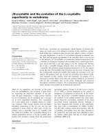
Fig. 2. Molecular model of human serine racemase, as described
by Baumgart et al. [12]. The calcium ions are depicted as yellow
spheres, the PLP moiety is shown in blue and the nucleotide analo-
gue phosphomethylphosphonic acid adenylate ester (AMP-PCP) is
shown in magenta. The molecular modelling was performed using
the crystal structures of the S. pombe serine racemase and the
E. coli Thr deaminase.
F. Baumgart and I. Rodrı
´
guez-Crespo Biochemistry of brain serine racemase
FEBS Journal 275 (2008) 3538–3545 ª 2008 The Authors Journal compilation ª 2008 FEBS 3541
counteract serine racemase activity in vivo by degrada-
tion of d-serine, is upregulated by
•
NO [20,21]. Subse-
quent biochemical proof for this model was provided
[22], pinning down the residue that becomes modified
and proposing a structural model for the action of
•
NO.
Apparently, cysteine 113 (out of seven cysteine residues
in the mouse and human serine racemase sequence) can
become nitrosylated, both in the recombinant enzyme
and in transfected cells, using the
•
NO donor, S-nitroso-
glutathione (GSNO). A molecular model of mouse ser-
ine racemase, based on a yeast homolog, reveals that
residue 113 lies in proximity to the putative ATP-bind-
ing region of the enzyme. Nitrosylation would therefore
lead to impaired nucleotide binding and inactivation of
the enzyme. It is noteworthy that GSNO is known to
modify cysteines not only with
•
NO but also with gluta-
thione, leading to protein glutathionylation, another
post-translational modification occurring under condi-
tions of oxidative ⁄ nitrosative stress. In fact, GSNO is
very frequently used in glutathionylating studies [23]. It
is thus conceivable that purified serine racemase
becomes modified by glutathione together with
•
NO.
Experiments with milder nitrosylating reagents that lack
a glutathione moiety would unambiguously demon-
strate if serine racemase is, in fact, modified by
•
NO.
Serine racemase-interacting proteins:
GRIP, PICK1 and Golga3
The carboxy-terminal end of both mouse and human
serine racemase display a -Val-serine-Val-COOH
sequence, a motif reminiscent of the type II consensus
sequence for binding to PSD95 ⁄ disc large ⁄ ZO-1
(PDZ) domains [24]. PDZ domains are among the
most ubiquitous protein–protein interaction motifs in
metazoan genomes and are especially important in the
nervous system for the assembly of synaptic complexes
and scaffolding [25,26]. After performing a yeast two-
hybrid screen of serine racemase against a rat hippo-
campus and cortex cDNA library, the hepta-PDZ
protein GRIP was identified as a binding partner of
serine racemase [18]. Out of the seven consecutive
PDZ modules found in GRIP, serine racemase was
found to bind specifically to the PDZ6 domain by
means of its C-terminal PDZ-binding motif. Previ-
ously, GRIP had been described to interact with gluta-
mate receptors of the AMPA ⁄ kainate type [27], where
it is responsible for proper trafficking and assembly of
the receptor and accessory proteins. GRIP can bind to
the glutamate receptor subunit 2 (GluR2) subunit of
AMPA receptors via PDZ4 ⁄ PDZ5, both PDZ domains
working in concert to establish binding [27,28]. The
finding of serine racemase interacting with the PDZ6
domain of GRIP and being activated was the first
report on cellular interaction partners of serine race-
mase and it raised several intriguing questions. It was
not clear whether GRIP directly activated serine race-
mase or if binding led to a translocation to the prox-
imity of AMPARs in vivo (Fig. 3). Furthermore, the
influence of the other PDZ domains of GRIP was not
investigated. Therefore, other proteins that become
associated with GRIP, using some of the other six
PDZ domains, might modulate the activity of serine
racemase. Conversely, d-serine might also change the
activity of some GRIP-associated proteins. When ser-
ine racemase ⁄ GRIP interactions were first studied, it
was proposed that GRIP was released from AMPARs
when they became stimulated and phosphorylated [18],
which would lead to GRIP interacting with serine
racemase in the cytosol where it would bind to and
activate serine racemase. With our own results we were
able to confirm the interaction of GRIP with serine
racemase via PDZ6 [12]. However, we observed that
binding to PDZ6 alone was not sufficient for activa-
tion. Rather, the presence of the rest of the C-terminal
region of GRIP, that is the PDZ7 module and a link-
ing segment between PDZ6 and PDZ7, was required
for full activation of serine racemase, both in vitro and
in vivo. Although these results do not necessarily pre-
clude a translocation process to AMPARs mediated by
GRIP, they do show the direct activation of serine
racemase by GRIP as a result of the concerted inter-
action of several PDZ modules, independent of the
subcellular localization. This PDZ crosstalk, where an
isolated PDZ domain is insufficient to carry out a
specific function, has also been observed in other
examples, for instance in the requirement of both
PDZ4 and PDZ5 for GRIP binding to GluR2 [27,28].
Interestingly, the activating effect of GRIP on serine
racemase results mainly in a change in V
max
. More-
over, the response curve to calcium remains unchanged
upon binding to GRIP under the experimental condi-
tions applied, which indicates that GRIP binding and
regulation by calcium must be regarded as independent
regulation pathways. It has been proposed that serine
racemase activation by GRIP takes place in the cytosol
after AMPAR phosphorylation and concomitant dis-
sociation of GRIP [18]. However, because nothing is
known about the kinetics of this process, the forma-
tion of a ternary complex among the GluR2 subunit
of the AMPA receptor, GRIP and serine racemase
cannot be discounted. It would be plausible that GRIP
brings serine racemase in close proximity to the gluta-
mate-activated channel, where serine racemase might
be close to other calcium channels. Although the
AMPAR is not a calcium channel, it is conceivable
Biochemistry of brain serine racemase F. Baumgart and I. Rodrı
´
guez-Crespo
3542 FEBS Journal 275 (2008) 3538–3545 ª 2008 The Authors Journal compilation ª 2008 FEBS
that in certain calcium microdomains serine racemase
could become exposed to temporarily high calcium
concentrations. To shut the system off, GluR2 could
become phosphorylated, in order to release GRIP and
serine racemase, abolishing the transient activation of
serine racemase by calcium (Fig. 3). Because GRIP
can bind to serine racemase, both in the presence and
absence of calcium, it is possible that some other
GRIP-interacting protein that also binds to PDZ6
might disrupt the serine racemase–GRIP interaction,
hence diminishing the activity of the former.
In a similar yeast two-hybrid screen using a human
hippocampal cDNA library, a different PDZ domain-
containing protein was found to interact with serine
racemase, also requiring the C-terminal binding motif
[30]. Protein interacting with C kinase 1 (PICK1) con-
tains one PDZ domain that is required for interacting
with protein kinase C (PKC) [30,31] or serine racemase.
It also contains a Bin ⁄ amphiphysin ⁄ Rys domain,
important for the interaction with lipids, and a coiled-
coil domain. Furthermore, it has been shown recently
that the PDZ domain of PICK1 is also capable of inter-
acting with lipid membranes, a property crucial for the
clustering of AMPAR and synaptic plasticity [32].
There are no data available regarding the effect of the
binding of PICK1 on serine racemase activity. There-
fore, biochemical characterization of the role of the
interaction of serine racemase and PICK1 is needed to
judge the importance of these observations. Surely the
interaction of PICK1 with PKC leads to the temptation
to speculate on a possible phosphorylation of serine
racemase by PKC [33]. As yet, however, there are no
data available, either on the details of the interaction of
PICK1 with serine racemase, or on the phosphorylation
of serine racemase.
Considering that the phosphorylation of Ser880 of
the GluR2 subunit of the AMPA receptor, positioned
at the carboxy-terminal end of the polypeptide chain,
disrupts its interaction with PDZ4 ⁄ PDZ5 of GRIP, it
is tempting to speculate that phosphorylation of
Ser336 of human serine racemase or of Thr336
of mouse serine racemase might also break their inter-
action with PDZ6 of GRIP. This putative phosphory-
latable residue is located at position -3 of the human
(Ser–Val–Ser–Val-COOH) and mouse (Thr–Val–Ser–
Val-COOH) sequences, respectively, and both are
inserted within amino acid sequences of type II con-
sensus PDZ domain-interacting partners [24,26]. It has
been proposed that PKCa phosphorylates serine race-
mase, probably brought into its proximity by PICK1
binding [33]. This hypothesis would rationalize a novel
mode of regulation of d-serine synthesis through the
activation of nonphosphorylated serine racemase by
the multi-PDZ domain GRIP. We have been unable to
identify PKCa as a kinase that modifies purified
recombinant serine racemase (unpublished data)
although perhaps this might be the case in vivo.In
addition, both the rat and cow serine racemases are
GluR2
AMPAR
GluR2
AMPAR
GluR2
AMPAR
GluR2
AMPAR
L-Ser
D-Ser
C
C
C
C
C
L-Ser
D-Ser
C
C
P
C
PP
P
AB
C
D
L-Ser
D-Ser
L-Ser
D-Ser
Fig. 3. Proposed modes of interaction
among serine racemase, GRIP and the
AMPAR. (A) A trimeric complex is assumed.
(B) Phosphorylation of the GluR2 subunit of
the AMPAR at Ser880 dissociates GRIP
binding, which remains bound to serine
racemase. (C) Serine racemase is active in
the cytoplasm and does not interact with
GRIP, whereas the latter associates with
the GluR2 subunit. (D) The simultaneous
phosphorylation of the GluR2 subunit of the
AMPAR together with the phosphorylation
of serine racemase releases GRIP to the
cytoplasm in the absence of any dual inter-
action.
F. Baumgart and I. Rodrı
´
guez-Crespo Biochemistry of brain serine racemase
FEBS Journal 275 (2008) 3538–3545 ª 2008 The Authors Journal compilation ª 2008 FEBS 3543
truncated in the carboxy-terminal end, hence lacking
GRIP-interacting sequences. Further experiments will
demonstrate if the activation of serine racemase by
GRIP and its binding to PICK1 is exclusively present
in certain mammals or if there is sequence splicing at
this region and both rat and cow do have longer
(as-yet unidentified) versions of serine racemase.
Consequently, at least four different modes of inter-
action can be envisaged among AMPAR, serine race-
mase and GRIP (Fig. 3). Although phosphorylation of
the GluR2 subunit of the AMPAR and the disruption
of its association with GRIP have been unambiguously
demonstrated, the putative carboxy-terminal phosphor-
ylation of serine racemase remains to be established. If
GRIP brings serine racemase towards the proximity of
the calcium channel, a theoretical modulation of the
synthesis of d-serine by calcium concentration can be
postulated, in accordance with recent data [18]. Using
a mouse brain lysate we observed a trimeric GluR2–
GRIP–serine racemase (data not shown) although we
do not know which mechanisms lead to the dissocia-
tion of serine racemase from GRIP. Nevertheless, in
the absence of the association with the AMPAR,
recombinant purified GRIP alone is able to increase
the activity of recombinant purified serine racemase
[12]. We have shown that certain GRIP amino acids,
present further down in the sequence than PDZ6, are
responsible for the majority of the observed activation
of serine racemase by GRIP [12].
In another study to identify binding partners of ser-
ine racemase using the yeast two-hybrid technology,
the Golgi-localized protein, Golgin subfamily A mem-
ber 3 (Golga3), was found to interact with serine race-
mase [34]. In this case, however, no PDZ interactions
with the C-terminal amino acid triplet of serine were
crucial for binding, but instead, the interaction was
established with its N-terminal 66 residues. Binding of
Golga3 increases d-serine synthesis. Intriguingly, this is
achieved through a decrease in ubiquitin ⁄ proteasomal
degradation of serine racemase, rather than by modula-
tion of the catalytic rate. Serine racemase was shown to
have an average half-life of about 4.5 h. When Golga3
and serine racemase were cotransfected, both serine
racemase stability and d-serine synthesis increased con-
siderably. Thus, it is important to note that in addition
to the modulators mentioned beforehand which directly
influence the catalysis of serine racemase, indirect
effects such as protein stability or subcellular localiza-
tion should be taken into account when investigating
the precise regulation of serine racemase-dependent
d-serine levels at glutamatergic synapses.
In conclusion, brain serine racemase, a member of
the type II-fold PLP-dependent racemases ⁄ dehydrata-
ses, shares several mechanistic properties with other
members of the same family, such as activation by
nucleotides or divalent cations, although its functional-
ity in the brain is also modulated through interaction
with specific glial or neuronal proteins.
References
1 Wolosker H, Sheth KN, Takahashi M, Mothet JP,
Brady RO Jr, Ferris CD & Snyder SH (1999) Purifica-
tion of serine racemase: biosynthesis of the neuromodu-
lator D-serine. Proc Natl Acad Sci U S A 96, 721–725.
2 Wolosker H, Blackshaw S & Snyder SH (1999) Serine
racemase: a glial enzyme synthesizing d-serine to regu-
late glutamate- N-methyl- d-aspartate neurotransmission.
Proc Natl Acad Sci U S A 96, 13409–13414.
3 Panizzutti R, De Miranda J, Ribeiro CS, Engelender S &
Wolosker H (2001) A new strategy to decrease N-methyl-
d-aspartate (NMDA) receptor coactivation: inhibition of
d-serine synthesis by converting serine racemase into an
eliminase. Proc Natl Acad Sci U S A 98, 5294–5299.
4 De Miranda J, Panizzutti R, Foltyn VN & Wolosker H
(2002) Cofactors of serine racemase that physiologically
stimulate the synthesis of the N-methyl-d-aspartate
(NMDA) receptor coagonist d-serine. Proc Natl Acad
Sci U S A 99, 14542–14547.
5 Cook SP, Galve-Roperh I, Martinez del Pozo A &
Rodriguez-Crespo I (2002) Direct calcium binding
results in activation of brain serine racemase. J Biol
Chem 277, 27782–27792.
6 Neidle A & Dunlop DS (2002) Allosteric regulation of
mouse brain serine racemase. Neurochem Res 27, 1719–
1724.
7 Fujitani Y, Nakajima N, Ishihara K, Oikawa T, Ito K
& Sugimoto M (2006) Molecular and biochemical char-
acterization of a serine racemase from Arabidopsis thali-
ana. Phytochemistry 67, 668–674.
8 Fujitani Y, Horiuchi T, Ito K & Sugimoto M (2007)
Serine racemases from barley, Hordeum vulgare L., and
other plant species represent a distinct eukaryotic
group: gene cloning and recombinant protein character-
ization. Phytochemistry 68, 1530–1536.
9 Abe K, Takahashi S, Muroki Y, Kera Y & Yamada
RH (2006) Cloning and expression of the pyridoxal
5¢-phosphate-dependent aspartate racemase gene from
the bivalve mollusk Scapharca broughtonii and charac-
terization of the recombinant enzyme. J Biochem
(Tokyo) 139, 235–244.
10 Wada M, Nakamori S & Takagi H (2003) Serine race-
mase homologue of Saccharomyces cerevisiae has
l-threo-3-hydroxyaspartate dehydratase activity. FEMS
Microbiol Lett 225, 189–193.
11 Ohnishi M, Saito M, Wakabayashi S, Ishizuka M,
Nishimura K, Nagata Y & Kasai S (2008) Purification
Biochemistry of brain serine racemase F. Baumgart and I. Rodrı
´
guez-Crespo
3544 FEBS Journal 275 (2008) 3538–3545 ª 2008 The Authors Journal compilation ª 2008 FEBS
and characterization of serine racemase from a hyper-
thermophylum Pyrobaculum islandicum. J Bacteriol 190,
1359–1365.
12 Baumgart F, Mancheno JM & Rodriguez-Crespo I
(2007) Insights into the activation of brain serine race-
mase by the multi-PDZ domain glutamate receptor
interacting protein, divalent cations and ATP. FEBS J
274, 4561–4571.
13 Wood WA & Gunsalus IC (1949) Serine and threonine
desaminaes of Escherichia coli; activators for a cell-free
enzyme. J Biol Chem 181, 171–182.
14 Whanger PD, Phillips AT, Rabinowitz KW, Piperno
JR, Shada JD & Wood WA (1968) The mechanism of
action of 5¢-adenylic acid-activated threonine dehydrase.
II. Protomer-oligomer interconversions and related
properties. J Biol Chem 243, 167–173.
15 Dunne CP, Gerlt JA, Rabinowitz KW & Wood WA
(1973) The mechanism of action of 5¢-adenylic acid-acti-
vated threonine dehydrase. IV. Characterization of
kinetic effect of adenosine monophosphate. J Biol Chem
248, 8189–8199.
16 Brocard JB, Rajdev S & Reynolds IJ (1993) Glutamate-
induced increases in intracellular free Mg
2+
in cultured
cortical neurons. Neuron 11, 751–757.
17 Schell MJ, Molliver ME & Snyder SH (1995) D-serine,
an endogenous synaptic modulator: localization to
astrocytes and glutamate-stimulated release. Proc Natl
Acad Sci U S A 92, 3948–3952.
18 Kim PM, Aizawa H, Kim PS, Huang AS, Wickrama-
singhe SR, Kashani AH, Barrow RK, Huganir RL,
Ghosh A & Snyder SH (2005) Serine racemase: activa-
tion by glutamate neurotransmission via glutamate
receptor interacting protein and mediation of neuronal
migration. Proc Natl Acad Sci U S A 102, 2105–2110.
19 Mothet JP, Pollegioni L, Ouanounou G, Martineau M,
Fossier P & Baux G (2005) Glutamate receptor activa-
tion triggers a calcium-dependent and SNARE protein-
dependent release of the gliotransmitter d-serine. Proc
Natl Acad Sci U S A 102, 5606–5611.
20 Shoji K, Mariotto S, Ciampa AR & Suzuki H (2006)
Regulation of serine racemase activity by d-serine and
nitric oxide in human glioblastoma cells. Neurosci Lett
392, 75–78.
21 Shoji K, Mariotto S, Ciampa AR & Suzuki H (2006)
Mutual regulation between serine and nitric oxide
metabolism in human glioblastoma cells. Neurosci Lett
394, 163–167.
22 Mustafa AK, Kumar M, Selvakumar B, Ho GP, Ehm-
sen JT, Barrow RK, Amzel LM & Snyder SH (2007)
Nitric oxide S-nitrosylates serine racemase, mediating
feedback inhibition of d-serine formation. Proc Natl
Acad Sci U S A 104, 2950–2955.
23 Klatt P & Lamas S (2000) Regulation of protein func-
tion by S-glutathiolation in response to oxidative and
nitrosative stress. Eur J Biochem 267, 4928–4944.
24 Hung AY & Sheng M (2002) PDZ domains: structural
modules for protein complex assembly. J Biol Chem
277, 5699–5702.
25 Sakarya O, Armstrong KA, Adamska M, Adamski M,
Wang IF, Tidor B, Degnan BM, Oakley TH & Kosik
KS (2007) A post-synaptic scaffold at the origin of the
animal kingdom. PLoS ONE 2, e506.
26 Kim E & Sheng M (2004) PDZ domain proteins of
synapses. Nat Rev Neurosci 5, 771–781.
27 Dong H, O’Brien RJ, Fung ET, Lanahan AA, Worley
PF & Huganir RL (1997) GRIP: a synaptic PDZ
domain-containing protein that interacts with AMPA
receptors. Nature 386, 279–284.
28 Feng W, Shi Y, Li M & Zhang M (2003) Tandem PDZ
repeats in glutamate receptor-interacting proteins have
a novel mode of PDZ domain-mediated target binding.
Nat Struct Biol 10, 972–978.
29 Matsuda S, Mikawa S & Hirai H (1999) Phosphoryla-
tion of serine-880 in GluR2 by protein kinase C pre-
vents its C terminus from binding with glutamate
receptor-interacting protein. J Neurochem 73, 1765–
1768.
30 Fujii K, Maeda K, Hikida T, Mustafa AK, Balkissoon
R, Xia J, Yamada T, Ozeki Y, Kawahara R, Okawa
M et al. (2006) Serine racemase binds to PICK1:
potential relevance to schizophrenia. Mol Psychiatry
11, 150–157.
31 Staudinger J, Lu J & Olson EN (1997) Specific interac-
tion of the PDZ domain protein PICK1 with the
COOH terminus of protein kinase C-alpha. J Biol Chem
272, 32019–32024.
32 Pan L, Wu H, Shen C, Shi Y, Jin W, Xia J & Zhang M
(2007) Clustering and synaptic targeting of PICK1
requires direct interaction between the PDZ domain
and lipid membranes. EMBO J 26, 4576–4587.
33 Mustafa AK, Kim PM & Snyder SH (2004) D-Serine as
a putative glial neurotransmitter. Neuron Glia Biol 1,
275–281.
34 Dumin E, Bendikov I, Foltyn VN, Misumi Y, Ikehara
Y, Kartvelishvily E & Wolosker H (2006) Modulation
of d-serine levels via ubiquitin-dependent proteasomal
degradation of serine racemase. J Biol Chem 281,
20291–20302.
F. Baumgart and I. Rodrı
´
guez-Crespo Biochemistry of brain serine racemase
FEBS Journal 275 (2008) 3538–3545 ª 2008 The Authors Journal compilation ª 2008 FEBS 3545