Báo cáo khoa học: The role of Ureaplasma nucleoside monophosphate kinases in the synthesis of nucleoside triphosphates potx
Bạn đang xem bản rút gọn của tài liệu. Xem và tải ngay bản đầy đủ của tài liệu tại đây (386.56 KB, 8 trang )
The role of Ureaplasma nucleoside monophosphate
kinases in the synthesis of nucleoside triphosphates
Liya Wang
Department of Molecular Biosciences, Swedish University of Agricultural Sciences, The Biomedical Centre, Uppsala, Sweden
Mycoplasmas (Mollicutes) are wall-less bacteria and
phylogenetically belong to the gram-positive bacteria.
Mollicutes are pathogens affecting humans, animals
and plants [1]. Ureaplasma is a human pathogen col-
onizing the urogenital tract and is the most common
cause of nonchlamydial nongonococcal urethritis. It
has also been implicated in infertility, spontaneous
abortion, stillbirth, and premature and perinatal mor-
bidity and mortality [2–4].
Mollicutes, in general, have low G + C content of
their genomes, and lack those genes necessary for
the synthesis of precursors for DNA, RNA and pro-
teins. For example, no de novo purine and pyrimi-
dine biosynthesis pathway exists [1]. Of all the
Mollicutes genomes sequenced to date, there is no
annotated ndk gene, coding for nucleoside diphos-
phate kinase, indicating that no homologue of any
known ndk gene or catalytic domain is present in
Mollicutes [5,6].
Nucleoside diphosphate kinase (NDK) catalyzes the
final step in ribonucleoside triphosphate (NTP) and
deoxynucleoside triphosphate (dNTP) biosynthesis.
NDK is involved in multiple cellular processes
including the control of cell growth and signalling by
Keywords
Mollicutes; nucleotide biosynthesis;
nucleoside diphosphate kinase; nucleoside
monophosphate kinase; Ureaplasma
Correspondence
L. Wang, Department of Molecular
Biosciences, Section of Veterinary Medical
Biochemistry, Swedish University of
Agricultural Sciences, The Biomedical
Centre, PO Box 575, SE-751 23 Uppsala,
Sweden
Fax: +46 18550762
Tel: +46 184714119
E-mail:
(Received 11 January 2007, revised 5
February 2007, accepted 14 February 2007)
doi:10.1111/j.1742-4658.2007.05742.x
Mollicutes are wall-less bacteria and cause various diseases in humans, ani-
mals and plants. They have the smallest genomes with low G + C content
and lack many genes of DNA, RNA and protein precursor biosynthesis.
Nucleoside diphosphate kinase (NDK), a house-keeping enzyme that plays
a critical role in the synthesis of nucleic acids precursors, i.e. NTPs and
dNTPs, is absent in all the Mollicutes genomes sequenced to date. There-
fore, it would be of interest to know how Mollicutes synthesize
dNTPs ⁄ NTPs without NDK. To answer this question, nucleoside mono-
phosphate kinases (NMPKs) from Ureaplasma were studied regarding their
role in the synthesis of NTPs ⁄ dNTPs. In this work, Ureaplasma adenylate
kinase, cytidylate kinase, uridylate kinase and thymidylate kinase were
cloned and expressed in Escherichia coli. The recombinant enzymes were
purified and characterized. These NMPKs are base specific, as indicated by
their names, and capable of converting (d)NMPs directly to (d)NTPs. The
catalytic rates of (d)NTPs and (d)NDP synthesis by these NMPKs were
determined using tritium-labelled (d)NMPs, and the rates for (d)NDP syn-
thesis, in general, were much higher (up to 100-fold) than that of (d)NTP.
Equilibrium studies with adenylate kinase suggested that the rates of
NTPs ⁄ dNTPs synthesis by NMPKs in vivo are probably regulated by the
levels of (d)NMPs. These results strongly indicate that NMPKs could sub-
stitute the NDK function in vivo.
Abbreviations
AdK, adenylate kinase; CMPK, cytidylate kinase; GMPK, guanylate kinase; NDK, nucleoside diphosphate kinase; NMPK, nucleoside
monophosphate kinase; TMPK, thymidylate kinase; UMPK, uridylate kinase.
FEBS Journal 274 (2007) 1983–1990 ª 2007 The Author Journal compilation ª 2007 FEBS 1983
providing either all the NTPs or a specific NTP such
as GTP, CTP or UTP [7–9]. NDK is a highly con-
served enzyme and is widely expressed. In humans,
eight ndk genes have been reported and the corres-
ponding enzymes have different tissue distribution
and subcellular localization. NDKs are also involved
in cell growth, differentiation and tumour metastasis,
etc. [8,9]. Null mutations in the ndk gene of Droso-
phila caused abnormalities in the development of the
larvae, leading to tissue necrosis and death at the pre-
pupal stage [10]. In prokaryotes such as Escherichia
coli, deletion of the ndk gene led to a mutator pheno-
type due to abnormal dCTP and dGTP pools [11].
However, the growth rate was not affected, suggesting
that other enzymes may be able to substitute the
NDK activity [12,13].
So far, no NDK activity was detected in total cell ex-
tracts or the chromatographic fraction of Mycoplasma
pneumoniae in an attempt to isolate the NDK
enzyme [14]. Thus, the question is how do Mollicutes
synthesize their dNTPs and NTPs without the NDK
enzyme?
Glycolytic enzymes such as pyruvate kinase and
phosphoglycerate kinase have been suggested to
replace the NDK activity in Mollicutes [14], and, in
that study, purine nucleoside diphosphates, e.g. ADP
and GDP, were converted to triphosphates with relat-
ively good efficiency; however, pyrimidine nucleoside
diphosphates had very low relative activity. Therefore,
there must be other alternative pathways that may
contribute to the synthesis of NTPs ⁄ dNTPs, especially
with regard to pyrimidine nucleotides.
Nucleoside monophosphate kinases (NMPK) cata-
lyze the reversible phosphorylation of a nucleoside
monophosphate (NMP) using a nucleoside triphos-
phate as phosphate donor, i.e. N
1
MP + N
2
TP ‹fi
N
1
DP + N
2
DP. Adenylate kinase (AdK) has been
suggested play a role in the synthesis of dNTPs and
NTPs using the reverse reaction and it was proposed
that AdK was the alternative enzyme in providing
nucleoside triphosphates in NDK-deficient E. coli
[12,13,15].
All Mollicutes species sequenced to date possess five
nucleoside monophosphate kinases, which have been
assigned as AdK, thymidylate kinase (TMPK), cyti-
dylate kinase (CMPK), uridylate kinase (UMPK), and
guanylate kinase (GMPK). In this work, four of the
Ureaplasma parvum nucleoside monophosphate
kinases, i.e. adenylate kinase, thymidylate kinase, cyti-
dylate kinase, and uridylate kinase, were cloned and
expressed in E. coli. The recombinant enzymes were
affinity purified and their role in dNTP and NTP syn-
thesis was investigated.
Results
Ureaplasma nucleoside monophosphate kinases:
cloning, expression and purification
Five NMPKs have been annotated in the genome
U. parvum, i.e. AdK (adk, UU251), TMPK (tmk,
UU020), CMPK (cmk, UU342), UMPK (pyrH,
UU513) and GMPK (gmk, UU213), based on the
sequence homology with NMPKs from other organ-
isms (GenBank accession number AF222894). There is
no experimental data regarding the functions of these
genes reported [5].
Open reading frames coding for AdK (UU251),
CMPK (UU342), UMPK (UU513), and TMPK
(UU020) were PCR amplified using the U. parvum
genomic DNA as template and cloned into the pET-
14b vector using the Nde I and BamH I restriction
sites. A 6-His tag and a thrombin cleavage site were
introduced to the N-terminus of the recombinant pro-
teins. Tryptophans, coded by UGA codons in UMPK
and AMPK, were mutated to UGG using the site-
directed mutagenesis method in order to express these
proteins in E. coli. Recombinant AdK, CMPK,
UMPK and TMPK were expressed in E. coli and puri-
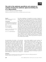
fied by metal affinity chromatography. SDS ⁄ PAGE
analysis showed that the dominant band corresponded
to the subunit molecular mass of each enzyme (Fig. 1).
The purified enzymes were used directly in the assays
without the removal of the His tag.
Substrate specificity of Ureaplasma nucleoside
monophosphate kinases
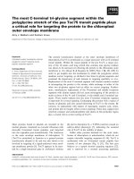
The substrate specificities of AdK, CMPK, UMPK
and TMPK were explored using a phosphoryl transfer
Fig. 1. SDS ⁄ PAGE analysis of purified recombinant Ureaplasma
nucleoside monophosphate kinases. Lanes 1–4, TMPK, UMPK,
AdK and CMPK. MW, molecular mass markers.
Ureaplasma nucleoside monophosphate kinases L. Wang
1984 FEBS Journal 274 (2007) 1983–1990 ª 2007 The Author Journal compilation ª 2007 FEBS
assay with
32
P-labelled ATP as phosphate donor and
natural (deoxy)ribonucleoside monophosphates as
acceptors. AdK used AMP and dAMP as the preferred
substrates (Fig. 2); both CMP and dCMP were effi-
ciently phosphorylated by CMPK (Fig. 2). UMPK
phosphorylated UMP and dUMP (Fig. 2) while
TMPK phosphorylated dTMP and dUMP (Fig. 2).
Products from the CMPK (with dCMP and CMP as
substrates) and the TMPK (with TMP as substrate)
reactions were re-analysed by TLC using a higher salt
concentration to separate the other nucleotides from
ATP. As shown in Fig. 3, substantial amounts of
dCTP, CTP and TTP were formed in these reactions.
Nucleoside di- and triphosphate synthesis
catalyzed by AdK
When tritium-labelled AMP was used as substrate in
AdK catalyzed reactions, the reaction products, as ana-
lysed by the TLC, were found to be [
3
H]ADP and
[
3
H]ATP. [
3
H]AMP was rapidly converted to [
3
H]ADP,
which reached a maximum after 3 min and then
declined, while the formation of [
3
H]ATP was increased
linearly with time within the first 15 min. After 30 min,
the reaction reached equilibrium (Fig. 4A). Further
experiments were designed to study the extent of
[
3
H]ATP and [
3
H]ADP synthesis using different phos-
phate donors. The [
3
H]ATP synthesis rates were slightly
influenced by the phosphate donors used, approxi-
mately two-fold; the highest rate was found with ATP
as donor and the lowest with GTP as donor (Table 1).
In order to measure the rate of [
3
H]ADP synthesis,
the reaction conditions was optimized, in terms of
AdK concentration, so that a first order reaction was
observed (Fig. 4B). As shown in Table 1, the rates of
[
3
H]ADP synthesis were approximately 40-fold higher
than that of [
3
H]ATP. As with [
3
H]ATP synthesis,
phosphate donors did not have major impact on the
Fig. 2. Substrate specificities of Ureaplasma
NMPKs. The reactions were performed as
described in Experimental procedures and
the products were separated by TLC and
visualized by autoradiography. The concen-
trations of nucleoside monophosphates and
[c-
32
P]ATP were as follows: 0.1 mM 1.
CMP; 2. dCMP; 3. AMP; 4. dAMP; 5. GMP;
6. dGMP; 7. UMP; 8. dUMP; 9. dTMP. The
reaction products were marked as close to
the product spot as possible.
Fig. 3. Direct formation of nucleoside triphosphates in NMPK cata-
lyzed reactions. Reaction mixtures 1 and 2 by CMPK and 9 by
TMPK, described in Fig. 2, were reanalysed by TLC (developed
using 0.32
M NaH
2
PO
4
) and the radiolabelled nucleoside triphos-
phate products were separated from ATP.
L. Wang Ureaplasma nucleoside monophosphate kinases
FEBS Journal 274 (2007) 1983–1990 ª 2007 The Author Journal compilation ª 2007 FEBS 1985
rates of [
3
H]ADP synthesis, although the highest
[
3
H]ADP synthesis rate was obtained when ATP was
the phosphate donor (Table 1).
Reaction equilibrium for AdK
In the reactions described above, the concentration of
phosphate donors was 1 mm, which was 10-fold higher
than that of [
3
H]AMP; however, at equilibrium
[
3
H]ATP was the dominant product. Therefore, a ser-
ies of reactions with varied ATP concentrations were
carried out to study the equilibrium of labelled adenine
nucleotides. As shown in Table 2, at equilibrium the
concentration of [
3
H]ATP was dependent on the con-
centration of ATP used in the reaction; a higher ATP
concentration yielded a higher concentration of
[
3
H]ATP. The equilibrium constants for labelled aden-
ine nucleotides were lowest with highest ATP concen-
tration tested (Table 2).
The equilibrium constants of labelled adenine nucleo-
tides with different phosphate donors at 1 mm concen-
tration was two-fold lower with ATP as compared
with CTP, UTP or GTP as phosphate donor
(Table 3).
0
20
40
60
80
100
0 20 40 60 80 100 120 140
Nucleotide concentration (µ
M
)
Min
ATP
ADP
AMP
A
0
20
40
60
80
100
0 5 10 15 20
Nucleotide concentration (µ
M
)
Min
ATP
AMP
ADP
B
Fig. 4. The synthesis of [
3
H]ADP and [
3
H]ATP from [
3
H]AMP cata-
lyzed by AdK. (A) The conditions for linear rate of [
3
H]ATP synthesis
were 0.2 l
M AdK, 100 lM [
3
H]AMP and 1 mM ATP ⁄ MgCl
2
in the
reaction buffer, as described in experimental procedures. (B) The
conditions for linear rate [
3
H]ADP synthesis were 1.0 nM AdK,
100 l
M [
3
H]AMP and 1 mM ATP ⁄ MgCl
2
in the reaction buffer, as
described in Experimental procedures.
Table 1. Rate of nucleoside di- and triphosphate synthesis by Urea-
plasma NMPKs (s
)1
). The concentrations of [
3
H]AMP, [
3
H]dCMP or
[
3
H]UMP were 100 lM, the concentrations of phosphate donors
were 1 m
M and the concentration of MgCl
2
was 10 mM. The values
were the means of two to four measurements with < 10% varia-
tion.
AdK CMPK UMPK
[
3
H]ADP [
3
H]ATP [
3
H]dCDP [
3
H]dCTP [
3
H]UDP [
3
H]UTP
ATP 181.6 4.6 32.3 0.31 6.6 0.03
UTP 134.2 3.4 27.3 0.33 0.16 0.007
GTP 94.7 1.9 26.4 0.24 0.003 < 0.001
CTP 128.1 3.5 1.4 0.16 0.004 < 0.001
Table 2. The effect of ATP concentration on the equilibrium con-
centration of labelled adenine nucleotides. The initial concentration
of [
3
H]AMP was 100 lM and the concentration of MgCl
2
was
10 m
M. The values are the means of 2–4 measurements with
< 10% variations. Equilibrium constants K
eq
of labelled adenine
nucleotides were calculated according to the following equations:
[
3
H]ATP + [
3
H]AMP fi 2[
3
H]ADP; K
eq
¼ ([
3
H]ADP)
2
⁄ ([
3
H]ATP)([
3
H]
AMP). The theoretical K
eq
for AdK is close to unity, as determined
by the equation. However, K
eq
values presented here refer to only
the labelled adenine nucleotide at equilibrium but not the reaction
equilibrium constants.
ATP
(m
M)
[
3
H]ATP
(l
M)
[
3
H]ADP
(l
M)
[
3
H]AMP
(l
M) K
eq
0.1 13 58 29 8.9
0.5 43 49 8 7.0
1.0 62 32 6 2.8
2.0 73 22 5 1.3
4.0 81 13 6 0.3
Table 3. Equilibrium concentrations of labelled adenine nucleotides
in AdK-catalyzed reactions. Equilibrium constants K
eq
of labelled
adenine nucleotides were calculated as described for Table 2. The
initial concentrations of phosphate donors were 1 m
M and the initial
concentration of phosphate acceptor [
3
H]AMP was 100 lM and the
concentration of MgCl
2
was 10 mM. The values were the means of
2–4 measurements with < 10% variations.
[
3
H]ATP [
3
H]ADP [
3
H]AMP K
eq
ATP 62 32 6 2.8
UTP 46 47 7 6.9
GTP 47 45 8 5.4
CTP 45 47 8 6.1
Ureaplasma nucleoside monophosphate kinases L. Wang
1986 FEBS Journal 274 (2007) 1983–1990 ª 2007 The Author Journal compilation ª 2007 FEBS
Nucleoside di- and triphosphate synthesis
catalyzed by CMPK
CMPK phosphorylated CMP and dCMP directly to
their triphosphate forms in assays with radiolabelled
ATP as phosphate donor (Fig. 3). The rate of
[
3
H]dCDP and [
3
H]dCTP synthesis from [
3
H]dCMP
was further studied using ATP, UTP, GTP or CTP as
phosphate donors. The rate for [
3
H]dCDP synthesis, in
general, is high, being 100-fold higher than that of
[
3
H]dCTP, except when CTP is used as a phosphate
donor (Table 1). ATP, UTP and GTP were good phos-
phate donors for both [
3
H]dCDP and [
3
H]dCTP syn-
thesis, while CTP was a poor phosphate donor, i.e.
20-fold lower for [
3
H]dCDP synthesis and two-fold
lower for [
3
H]dCTP synthesis (Table 1).
Nucleoside triphosphate synthesis catalyzed
by UMPK
Using [
3
H]UMP as substrate, the rates of [
3
H]UDP
and [
3
H]UTP synthesis were determined. Similar to
AdK and CMPK, the rate of [
3
H]UDP synthesis was
much higher than that of [
3
H]UTP (Table 1). ATP was
the most efficient phosphate donor for [
3
H]UDP syn-
thesis, while UTP, GTP and CTP were poor phosphate
donors. There was detectable formation of [
3
H]UTP in
the reaction with GTP and CTP as phosphate donor,
but the rates of [
3
H]UTP synthesis were very low
(Table 1).
Mechanism of phosphoryl transfer
Using [c-
32
P]ATP as phosphate donor, the mechanism
of phosphoryl transfer during CTP synthesis by CMPK
was studied. Initially, using either dCDP or CDP as the
substrate and [c-
32
P]ATP as the phosphate donor, no
radiolabelled dCTP formation was detected in the reac-
tion with dCDP as the substrate, but it was observed
when using CDP as the substrate. CTP was also
formed in the control reaction using dCMP as sub-
strate. However, as commercial CDP contains 4%
CMP, and the level of radiolabelled product (CTP) was
similar for both substrates (data not shown) and cor-
responded to the CMP content in the CDP solution
(4%), this accounted for the CTP product observed.
When the reaction was repeated with purer CDP and
run at shorter time intervals (90 s), no CTP was formed
in the CDP reaction, while CTP was formed in the con-
trol reactions with CMP as substrate. Thus, the CTP or
dCTP synthesis carried out by CMPK was not a direct
phosphorylation of CDP or dCDP by ATP, but rather
CTP or dCTP was formed in the reverse reaction in
two steps, i.e. (d)CMP + ATP* fi (d)CDP* + ADP;
and (d)CDP* + (d)CDP fi (d)CTP* + (d)CMP.
Discussion
The aim of the present study was to define the role
of Ureaplasma NMPKs in the synthesis of nucleo-
side triphosphates. Four Ureaplasma NMPKs were
cloned, expressed and the recombinant enzymes were
affinity purified. These NMPKs were shown to have
narrow substrate specificity regarding the phosphate
acceptors, i.e. they are base specific and each enzyme
has its own substrates sets (using the same nucleo-
tides as their names indicated), with little overlapping
activity. AdK, CMPK and UMPK phosphorylated
both ribos- respective deoxyribos- forms of nucleo-
tides efficiently. TMPK, however, was specific for
dTMP and only dUMP had some activity. At the
phosphate donor site, however, the specificities were
broader, e.g. all natural nucleoside triphosphates were
accepted as phosphate donors but with clear-cut pref-
erences, especially in case of UMPK. The less strin-
gent requirement for phosphate donors may be an
advantage, since the enzymes can use any phosphate
donors available.
It is known that reactions catalyzed by NMPKs are
reversible, which means that these enzymes can also
synthesize nucleoside triphosphates via the reverse
reaction. Using the tritium-labelled nucleoside mono-
phosphates as substrates, the rates of (d)NDP and
(d)NTP synthesis by these enzymes were investigated.
The results clearly showed that Ureaplasma NMPKs
are able to synthesize NTP ⁄ dNTPs via the reverse
reaction, but not sequential phosphorylation of NMP
by NTP as demonstrated here. This is also in agree-
ment with a recent study using AdK, where it was
clearly shown that the NDK-like activity of this
enzyme is the result of the reverse reaction [11]. The
capacity of Ureaplasma NMPKs to synthesize nucleo-
side triphosphates in general is lower than that of
diphosphates, which may implicate that the conversion
of nucleoside diphosphates to triphosphates is the rate-
limiting step.
Equilibrium studies with AdK showed that the
enzyme favours the reverse reaction, i.e. the highest
level of [
3
H]ATP formed from [
3
H]AMP was achieved
with the highest ATP concentration used in the assay.
ATP was a better phosphate donor to bring about
[
3
H]ATP formation from [
3
H]AMP as compared with
other nucleoside triphosphates. At physiologically rele-
vant ATP concentrations (2–4 mm), the level of
labelled [
3
H]AMP at equilibrium was the lowest among
all labelled adenine nucleotides, suggesting that the
L. Wang Ureaplasma nucleoside monophosphate kinases
FEBS Journal 274 (2007) 1983–1990 ª 2007 The Author Journal compilation ª 2007 FEBS 1987
supply of nucleoside monophosphates may regulate the
synthesis of nucleoside di- and triphosphates.
In Mollicutes, other enzymes exist that are capable
of synthesizing nucleoside triphosphates, e.g. pyvuvate
kinase, phosphoglycerate kinase, etc. [14]. Together
with NMPKs, they can provide cells with precursors
for both DNA and RNA synthesis. The relatively low
rates of (d)NTP synthesis may result in limited
(d)NTPs supply.
In the literature there are only few earlier studies
regarding the level of ribonucleotides and deoxyribo-
nucleotides in Mycoplasmas [16–18]. In Mycoplasma
mycoides subsp. mycoides the levels of dNTPs were
100-fold lower as compared with NTPs [16–18].
Interestingly, the sum of ATP and UTP was two-fold
higher than that of CTP and GTP, and the ratio of
dATP and dTTP to dCTP and dGTP was also 2 : 1
[16,17]. This fact is in accordance with the genome
composition of Mollicutes, which has a high A + T
content at > 70%. Studies carried out in our labora-
tory showed that the dTTP and dCTP levels were
below the detection limit when Ureaplasma was cul-
tured in the presence of tritium-labelled deoxycytidine
or thymidine, even though > 20% of the radioacti-
vity was recovered in the DNA fraction, indicating a
very low dNTP pool in Ureaplasma [19]. Similarly,
M. mycoides subsp. mycoides incubated with radiola-
belled thymidine monophosphate resulted also in a
high level of incorporation into DNA, but the level of
labelled dTTP was very low [18].
In organisms where NDK is present, the rate-limit-
ing step in the synthesis of dNTPs ⁄ NTPs is not the
conversion of (d)NDPs to (d)NTPs, as the catalytic
efficiency of NDK is high [7–9]. The regulatory mech-
anism in dNTPs production relies on allosteric
enzymes, e.g. ribonucleotide reductase, which to a
large extent regulates dNDP production and thereby
dNTP pools [20,21]. Disruption of the ndk gene in
E. coli, Saccharomyces cerevisiae, and Saccharomyces
pombe did not affect growth or morphology [11,22,23],
suggesting that NDK is not essential. Although Molli-
cutes lack the ndk gene, this work provided evidence
that NMPKs, probably together other enzymes such as
pyvuvate kinase [14], can replace NDK in providing
the cells with NTPs and dNTPs. A recent study sug-
gests that cells have a mechanism for arresting DNA
synthesis when the dNTP pool size is limiting [24]. The
doubling times for Mollicutes are usually much longer
when compared with other bacteria such as E. coli,
and the limitation of dNTP levels for DNA synthesis
could be the reason.
Mollicutes lack the de novo synthesis of purine and
pyrimidine bases and have to rely on salvage path-
ways for the biosynthesis of nucleotides required for
cellular processes. The work presented here clearly
showed that NMPKs are base specific and highly
efficient in the synthesis of NTPs and dNTPs.
NMPKs are essential enzymes for the survival of the
organism, as demonstrated recently in Mycoplasma
genitalium using transposon mutagenesis technique
[25]. Therefore, inhibition of these enzymes, especially
TMPK or UMPK, will most probably impair the
synthesis of both DNA and RNA precursors, which
eventually leads to cell death. Thus, these enzymes,
especially TMPK and UMPK, are potential targets
for future design of antibiotics against pathogenic
Mycoplasmas.
Experimental procedures
Materials
Radioactive substances [c-
32
P]ATP (3000 CiÆmmol
)1
) were
purchased from PerkinElmer LAS Inc. (Boston, MA, USA).
[2-
3
H]AMP (adenosine 5¢-monophosphate, 24.0 CiÆmmol
)1
)
was from Amersham Biosciences (Uppsala, Sweden).
[5-
3
H]dCMP (2¢-deoxycytidine 5¢-monophosphate, 21.9
CiÆmmol
)1
) and [5,6-
3
H]UMP (uridine 5¢-monophosphate,
32 CiÆmmol
)1
) were obtained from Moravek Biochemical,
Inc (Brea, CA, USA). Non-radioactive nucleotides were from
Sigma-Aldrich Sweden AB (Stockholm, Sweden).
Cloning, expression and purification of
Ureaplasma nucleoside monophosphate kinases
Primers used in PCR amplification of Ureaplasma nucleo-
side monophosphate kinases were designed according to the
DNA sequence of the respective enzyme in the database
(GenBank accession number of AE002122) and restriction
sites (Nde IorBamH I) were introduced to the 5¢-sequence
of the primers to facilitate subsequent cloning. PCR reac-
tions were carried out by a standard procedure using the
U. parvum genomic DNA (ATCC # 700970D) as template.
The amplified PCR fragments were digested with Nde I and
BamH I, purified on 1% agarose gel and cloned into the
pET-14b vector (Novagen, Madison, WI, USA) that had
been linearized with the same restriction enzymes. The
recombinant plasmids carrying the Ureaplasma nucleoside
monophosphate kinase genes were sequence verified using
the Bigdye terminator kit and ABI Prisma 310 genetic Ana-
lyzer (Applied Biosystems, Foster City, CA, USA). In order
to express the recombinant protein in E. coli UGA codons,
coding for Trp in AdK and UMPK, were mutated to UGG
using site-directed mutagenesis as described previously [26]
and sequence verified. Finally the plasmids carrying AdK,
CMPK, UMPK and TMPK were transformed into the
E. coli BL21 (DE3) bacteria for expression.
Ureaplasma nucleoside monophosphate kinases L. Wang
1988 FEBS Journal 274 (2007) 1983–1990 ª 2007 The Author Journal compilation ª 2007 FEBS
For production of recombinant protein, E. coli BL21
(DE3) harbouring the AdK, CMPK, UMPK or TMPK
plasmids was cultured in LB media with 50 lgÆmL
)1
carbe-
nicilin at 37 °C until the optical density at 600 nm reached
0.6. The cultures were then changed to the induction
temperature as indicated below and 0.1 mm isopropyl
b-d-1-thiogalactopyranoside was added and the cultures
were further incubated for 4 h. The temperature for induc-
tion was 30 °C for AdK and CMPK; 37 °C for UMPK;
28 °C for TMPK. Bacteria were harvested by centrifugation
and the pellets were resuspended in buffer containing 50 mm
Tris ⁄ HCl, pH 7.5, 0.2 m NaCl, 0.2 mm phenylmethylsulfo-
nyl fluoride and total proteins were extracted by sonication
at 18 W for a total of 3 min, with a pulse every 5 s. The
lysates were centrifuged at 30 000 g at 4 °C for 30 min
(XL-70 ultracentrifuge, Beckman Coulter, rotor type
RT50Ti) and the supernatant was loaded onto a metal affin-
ity column (TALON resin, BD Biosciences Clontech, Palo
Alto, CA, USA), which was equilibrated with 50 mm
Tris ⁄ HCl, pH 7.5, 0.2 m NaCl and 5 mm imidazole. The
column was subsequently washed with the same buffer con-
taining 30 mm imidazole and the recombinant proteins were
eluted with 300 mm imidazole and 50 mm Tris ⁄ HCl,
pH 7.5. Purified protein was analysed by SDS ⁄ PAGE and
protein concentrations were determined by Bio-Rad protein
assay with BSA as standard. Glycerol and dithiothreitol
were added to the purified enzymes to 10% and 2 mm,
respectively, and the enzymes were stored in aliquots at
)70 °C.
Enzyme assays
Phosphoryl transfer assays were performed essentially as
described previously [26]. Briefly, each reaction was per-
formed in a total volume of 20 lL containing 50 mm
Tris ⁄ HCl, pH 7.5, 0.5 mg.mL
)1
BSA, 5 mm dithiothreitol,
2mm MgCl
2
,15mm NaF, 0.1 mm nucleoside monophos-
phate, 0.1 mm [g-
32
P]ATP and 100 ng purified enzyme at
37 °C for 20 min and was stopped by heating at 70 °C for
2 min. After brief centrifugation (13 000 g, Biofuge 13, Her-
aeus Instruments, rotor type HFA 17.1, max 14 926 g),
1 lL of the supernatant was spotted onto a TLC plate (PEI-
cellulose; MERCK, VWR International AB, Stockholm,
Sweden) and dried. Authentic markers were also applied
onto the same TLC plates. The TLC plates were then devel-
oped in 0.2 m NaH
2
PO
4
for reactions with AdK and CMPK
and 0.1 m NaH
2
PO
4
for UMPK and TMPK. To separate
other nucleoside triphosphates from [g-
32
P]ATP, 0.32 m
NaH
2
PO
4
was used. The reaction products were visualized
by phosphoimagine analysis (Fuji ImageGause V3.1, Fuji
Photo Film Co., Ltd., Tilburg, the Netherlands). Authentic
markers were visualized under UV light.
NMPK assay with [
3
H]-labelled substrates were carried
out on the reaction mixture containing 50 mm Tris ⁄ HCl,
pH 7.5, 0.5 mgÆmL
)1
BSA, 5 mm dithiothreitol, 5 mm
MgCl
2
,15mm NaF, 0.1 mm
3
H-labelled substrate in a total
volume of 20 lL. The reaction was initiated by the addition
the enzyme (100 ngÆreaction
)1
for the determination of
NTP synthesis rates and 0.5–1 ngÆreaction
)1
for the deter-
mination of NDP synthesis rates) and incubated at 37 °C.
At each time point, 1 lL aliquot was withdrawn and spot-
ted directly onto a TLC plate (PEI-cellulose) and dried.
The TLC plate was then developed in 0.2 m NaH
2
PO
4
for
assays with AdK and CMPK and 0.1 m NaH
2
PO
4
for
assays with UMPK. Non-radioactive markers were spotted
onto the TCL plate and identified under UV light. The
reaction products were cut out and eluted with 0.5 mL of
0.1 m HCl and 0.2 m KCl, and then 2.5 mL of scintillation
fluid was added and the radioactivity counted.
The R
f
values for ATP, ADP, AMP, dCTP, dCDP and
dCMP in 0.2 NaH
2
PO
4
were 0.01, 0.28, 0.61, 0.03, 0.39
and 0.67, respectively. The R
f
values for UMP, UDP, UTP,
dTMP, dTDP and dTTP in 0.1 m NaH
2
PO
4
were 0.37,
0.21, 0.05, 0.36, 0.22, and 0.05, respectively.
Acknowledgements
This work was supported by grants from the Swedish
research council for Environment, Agricultural Scien-
ces, and Spatial Planning (FORMAS) and the Swedish
Research Council (VR).
References
1 Razin S, Yogev D & Naot Y (1998) Molecular biology
and pathogenicity of mycoplasmas. Microbiol Mol Biol
Rev 62, 1094–1156.
2 Cassell GH, Waites KB, Watson HL, Crouse DT &
Harasawa R (1993) Ureaplasma urealyticum intrauter-
ine infection: role in prematurity and disease in new-
borns. Clin Microbiol Rev 6 , 69–87.
3 Goncalves L, Chaiworapongsa T & Romero R (2002)
Intrauterine infection and prematurity. Ment Retard
Dev Disabil Res Rev 8, 3–13.
4 Waites KB, Katz B & Schelonka RL (2005) Mycoplas-
mas and ureaplasmas as neonatal pathogens. Clin
Microbiol Rev 18, 757–789.
5 Chakrabarty AM (1998) Nucleoside diphosphate kinase:
role in bacterial growth, virulence, cell signalling and
polysaccharide synthesis. Mol Microbiol 28, 875–882.
6 Lacombe ML, Milon L, Munier A, Mehus JG &
Lambeth DO (2000) The human Nm23 ⁄ nucleoside
diphosphate kinases. J Bioenerg Biomembr 32, 247–258.
7 Kimura N, Shimada N, Fukuda M, Ishijima Y,
Miyazaki H, Ishii A & Isikawa N (2000) Regulation of
cellular functions by nucleoside diphosphate kinases in
mammals. J Bioenerg Biomembr 32, 309–315.
8 Biggs J, Hersperger E, Steeg PS, Liotta LA & Shearn A
(1990) A Drosophila gene that is homologous to a
L. Wang Ureaplasma nucleoside monophosphate kinases
FEBS Journal 274 (2007) 1983–1990 ª 2007 The Author Journal compilation ª 2007 FEBS 1989
mammalian gene associated with tumor metastasis codes
for a nucleoside diphosphate kinase. Cell 63, 933–940.
9 Lu Q, Zhang X, Almaula N, Mathews CK & Inouye M
(1995) The gene for nucleoside diphosphate kinase func-
tions as a mutator gene in Escherichia coli. J Mol Biol
254, 337–331.
10 Lu Q & Inouye M (1996) Adenylate kinase comple-
ments nucleoside diphosphate kinase deficiency in
nucleotide metabolism. Proc Natl Acad Sci USA 93,
5720–5725.
11 Bernard MA, Ray NB, Olcott MC, Hendricks SP &
Mathews CK (2000) Metabolic functions of microbial
nucleoside diphosphate kinases. J Bioenergetics Biomem-
branes 32, 259–267.
12 Glass JI, Lefkowitz EJ, Glass JS, Heiner CR, Chen EY
& Cassell GH (2000) The complete sequence of the
mucosal pathogen Ureaplasma urealyticum. Nature 407,
757–762.
13 Pollack JD (2001) Ureaplasma urealyticum: an oppor-
tunity for combinatorial genomics. Trends Microbiol 9,
169–175.
14 Pollack JD, Myers MA, Dandekar T & Herrmann R
(2002) Suspected utility of enzymes with multiple
activities in the small genome Mycoplasma species: the
replacement of the missing ‘household’ nucleoside
diphosphate kinase gene and activity by glycolytic
kinases. OMICS 6, 247–258.
15 Willemoes M & Kilstrup M (2005) Nucleoside triphos-
phate synthesis catalysed by adenylate kinase is ADP
dependent. Arch Biochem Biophys 444, 195–199.
16 Mitchell A & Finch LR (1979) Enzymes of pyrimidine
metabolism in Mycoplasma mycoides subsp. mycoides.
J Bacteriol 137, 1073–1080.
17 Neale GA, Mitchell A & Finch LR (1983) Enzymes of
pyrimidine deoxyribonucleotide metabolism in Myco-
plasma mycoides subsp. mycoides. J Bacteriol 156, 1001–
1005.
18 Neale GA, Mitchell A & Finch LR (1984) Uptake and
utilization of deoxynucleoside 5¢-monophosphates by
Mycoplasma mycoides subsp. mycoides. J Bacteriol 158,
943–947.
19 Carnrot C, Wehelie R, Eriksson S, Bo
¨
lske G & Wang L
(2003) Molecular characterization of thymidine kinase
from Ureaplasma urealyticum: nucleoside analogues as
potent inhibitors of mycoplasma growth. Mol Microbiol
50, 771–780.
20 Reichard P (1988) Intractions between deoxyribonucleo-
tide and DNA synthesis. Annu Rev Biochem 57, 349–374.
21 Mathews CK (2006) DNA precursor metabolism and
genomic stability. FASEB J 20
, 1300–1314.
22 Fukuchi T, Nikawa J, Kimura N & Watanabe K (1993)
Isolation, overexpression and disruption of a Saccharo-
myces cerevisiae YNK gene encoding nucleoside diphos-
phate kinase. Gene 129, 141–146.
23 Izumiya H & Yamamoto M (1995) Cloning and func-
tional analysis of the ndk1 gene encoding nucleoside-
diphosphate kinase in Schizosaccharomyces pombe.
J Biol Chem 270, 27859–27864.
24 Koc A, Wheeler LJ, Mathews CK & Merrill GF (2004)
Hydroxyurea arrests DNA replication by a mechanism
that preserves basal dNTP pools. J Biol Chem 279,
223–230.
25 Glass JI, Assad-Garcia N, Alperovich N, Yooseph S,
Lewis MR, Maruf M, Hutchison CA 3rd, Smith HO &
Venter JC (2006) Essential genes of a minimal bacter-
ium. Proc Natl Acad Sci USA 103, 425–430.
26 Wang L, Westberg J, Bo
¨
lske G & Eriksson S (2001)
Novel deoxynucleoside-phosphorylating enzymes in
mycoplasmas: evidence for efficient utilization of deoxy-
nucleosides. Mol Microbiol 42, 1065–1073.
Ureaplasma nucleoside monophosphate kinases L. Wang
1990 FEBS Journal 274 (2007) 1983–1990 ª 2007 The Author Journal compilation ª 2007 FEBS