Báo cáo khoa học: The role of residue Thr249 in modulating the catalytic efficiency and substrate specificity of catechol-2,3-dioxygenase from Pseudomonas stutzeri OX1 ppt
Bạn đang xem bản rút gọn của tài liệu. Xem và tải ngay bản đầy đủ của tài liệu tại đây (260.03 KB, 14 trang )
The role of residue Thr249 in modulating the
catalytic efficiency and substrate specificity of
catechol-2,3-dioxygenase from Pseudomonas stutzeri OX1
Loredana Siani
1,
*, Ambra Viggiani
1,
*, Eugenio Notomista
1
, Alessandro Pezzella
2
and Alberto Di Donato
1
1 Dipartimento di Biologia Strutturale e Funzionale, Universita
`
di Napoli Federico II, Napoli and CEINGE-Biotecnologie Avanzate S.c.ar.l., Italy
2 Dipartimento di Chimica Organica e Biochimica, Universita
`
di Napoli Federico II, Italy
Several bacteria are capable of using aromatic hydro-
carbons as growth substrates [1–4]. The remarkable
range of substrates that can be metabolized endows
these microorganisms with the potential for bioremedi-
ating environmentally dangerous substances such as
benzene, toluene, xylene isomers, and polycyclic aro-
matic hydrocarbons and their derivatives [5–8].
Because of their toxicity, several of these compounds
are in the US Environmental Protection Agency prior-
ity pollutant list (). For example
Keywords
bioremediation; dioxygenase; enzyme
kinetics; protein expression; Pseudomonas
stutzeri
Correspondence
A. Di Donato, Dipartimento di Biologia
Strutturale e Funzionale, Universita
`
di Napoli
Federico II, Via Cinthia, I-80126 Napoli, Italy
Fax: +39 081 676710
Tel: +39 081 679143
E-mail:
*These authors contributed equally to this
work
(Received 27 January 2006, revised
28 March 2006, accepted 4 May 2006)
doi:10.1111/j.1742-4658.2006.05307.x
Bioremediation strategies use microorganisms to remove hazardous sub-
stances, such as aromatic molecules, from polluted sites. The applicability
of these techniques would greatly benefit from the expansion of the cata-
bolic ability of these bacteria in transforming a variety of aromatic com-
pounds. Catechol-2,3-dioxygenase (C2,3O) from Pseudomonas stutzeri OX1
is a key enzyme in the catabolic pathway for aromatic molecules. Its specif-
icity and regioselectivity control the range of molecules degraded through
the catabolic pathway of the microorganism that is able to use aromatic
hydrocarbons as growth substrates. We have used in silico substrate dock-
ing procedures to investigate the molecular determinants that direct the
enzyme substrate specificity. In particular, we looked for a possible
molecular explanation of the inability of catechol-2,3-dioxygenase to cleave
3,5-dimethylcatechol and 3,6-dimethylcatechol and of the efficient clea-
vage of 3,4-dimethylcatechol. The docking study suggested that reduction
in the volume of the side chain of residue 249 could allow the binding of
3,5-dimethylcatechol and 3,6-dimethylcatechol. This information was used
to prepare and characterize mutants at position 249. The kinetic and regio-
specificity parameters of the mutants confirm the docking predictions, and
indicate that this position controls the substrate specificity of catechol-2,3-
dioxygenase. Moreover, our results suggest that Thr249 also plays a previ-
ously unsuspected role in the catalytic mechanism of substrate cleavage.
The hypothesis is advanced that a water molecule bound between one of
the hydroxyl groups of the substrate and the side chain of Thr249 favors
the deprotonation ⁄ protonation of this hydroxyl group, thus assisting the
final steps of the cleavage reaction.
Abbreviations
C2,3O, catechol-2,3-dioxygenase; DHBD, 2,3-dihydroxybiphenyl-1,2-dioxygenase; DHND, 1,2-dihydroxynaphthalene dioxygenase; DHpCD,
2,3-dihydroxy-p-cumate dioxygenase; DMC, dimethylcatechol; ECD, extradiol ring cleavage dioxygenase; HPCD, 3,4-dihydroxyphenylacetate
(homoprotocatechuate)-2,3-dioxygenase; IBX, o-iodoxybenzoic acid; 3-MC, 3-methylcatechol; 4-MC, 4-methylcatechol; PH, phenol
hydroxylase; THTD, 2,4,5-trihydroxytoluene-5,6-dioxygenase; ToMo, toluene ⁄ o-xylene monooxygenase.
FEBS Journal 273 (2006) 2963–2976 ª 2006 The Authors Journal compilation ª 2006 FEBS 2963
long-term exposure of humans to benzene, toluene and
xylene could cause damage to the central nervous sys-
tem, liver and kidneys, chromosomal aberrations and
cancer [9–12].
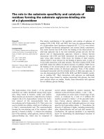
Extradiol ring cleavage dioxygenases (ECDs) are
Fe(II)-dependent enzymes that catalyze a crucial ring-
opening step in the catabolic pathways of microorgan-
isms capable of growing on aromatic compounds
[13–15]. ECDs cleave ortho-dihydroxylated aromatic
rings by catalyzing the addition of two atoms from
molecular oxygen at one of the C–C bonds adjacent to
the diol (metacleavage; Fig. 1) to produce nonaromatic
molecules that eventually enter central metabolic path-
ways [13,14]. ECDs comprise five evolutionarily related
subfamilies [16] that include catechol-2,3-dioxygenase
(C2,3O; EC 1.13.11.2) (subfamily 1), 2,3-dihydroxybi-
phenyl-1,2-dioxygenase (DHBD; EC 1.13.11.39) and
1,2-dihydroxynaphthalene dioxygenase (DHND, sub-
family 2), 3,4-dihydroxyphenylacetate-2,3-dioxygenase
(HPCD; EC 1.13.11.15) (subfamily 3), 2,3-dihydroxy-
p-cumate-3,4-dioxygenase (DHpCD, subfamily 5) and
2,4,5-trihydroxytoluene-5,6-dioxygenase (THTD, sub-
family 6). Even though the range of substrates that
can be oxidized by ECDs is broad, each enzyme in the
family displays restricted substrate specificity and reg-
ioselectivity. ECDs belonging to subfamilies 1, 2 and 3
cleave catechols substituted at positions 3 and ⁄ or 4 at
the bond adjacent to the diol and proximal to the sub-
stituent, as shown in Fig. 1A [17–23]. ECDs belonging
to subfamilies 5 and 6, such as DHpCD and THTD,
catalyze the transformation of 3,6-disubstituted and
4,5-disubstituted catechols, respectively [24–26], and
exhibit high regioselectivity by cleaving the bond prox-
imal to the alkylic group of the substrates as shown in
Fig. 1B,C [24–26]. The size of the substitutent that can
be accommodated by a subfamily varies. For instance,
C2,3Os can cleave catechols with small substituents at
positions 3 and 4, such as 3-methylcatechol (3-MC)
and 3,4-dimethylcatechol (3,4-DMC) [17,18], whereas
enzymes belonging to subfamily 2 act on catechols
with large substituents at the same positions [19–21,27]
(Fig. 1A).
The complete degradation of aromatic molecules is
initiated by monooxygenases and dioxygenases, which
produce dihydroxylated compounds in the upper
metabolic pathways [28,29]. These diols are cleaved
subsequently by ECDs. Since monooxygenases and
dioxygenases usually exhibit a wide range of substrate
specificity, they produce several dihydroxylated prod-
ucts, some of which are not always substrates for
ECDs and cannot be degraded further. As a conse-
quence, ECDs represent the gate that controls the flow
of molecules entering the lower metabolic pathways
[14,28,29], by reducing the range of aromatic com-
pounds that can be used by microorganisms as growth
substrates. Thus, enhancement of the catabolic poten-
tial of ECDs would represent a valuable tool for bio-
remediation strategies by widening the number of
substrates that can be consumed by bacteria that
depend on these enzymes for the utilization of specific
aromatic substrates as their primary source of carbon
and energy.
Pseudomonas stutzeri OX1 is an ideal model organ-
ism for these studies, since it can utilize benzene, tolu-
ene, and o-xylene, but not m-xylene and p-xylene, as
sole sources of carbon and energy [30]. Two NADH-
dependent monooxygenases—toluene ⁄ o-xylene mono-
oxygenase (ToMO) and phenol hydroxylase (PH)—act
sequentially in the microorganism [31] to convert aro-
matic hydrocarbons to the corresponding catechols.
These are cleaved by a C2,3O that is nearly identical
to the well-characterized enzyme from Pseudomonas
putida MT2 [18,32]. ToMO and PH are able to convert
o-xylene as well as m-xylene and p-xylene to 3,4-DMC,
3,5-DMC and 3,6-DMC, respectively (unpublished
results). However, P. stutzeri C2,3O can cleave only
3,4-DMC effectively [32], allowing this product to be
Fig. 1. Scheme of the reactions catalyzed by extradiol ring cleavage
dioxygenases (ECDs). Reactions catalyzed by (A) catechol-2,3-dioxy-
genases (C2,3Os), 2,3-dihydroxybiphenyl-1,2-dioxygenases (DHBDs)
and 3,4-dihydroxyphenylactetate-2,3-dioxygenase (HPCD), (B) by
2,3-dihydroxy-p-cumate dioxygenases (DHpCDs), and (C) by 2,4,5-
trihydroxytoluene-5,6,dioxygenases (THTDs).
Thr249 in catechol-2,3-dioxygenase function L. Siani et al.
2964 FEBS Journal 273 (2006) 2963–2976 ª 2006 The Authors Journal compilation ª 2006 FEBS
further metabolized through the lower pathway. This
is not possible in the case of 3,5-DMC and 3,6-DMC,
because of the very low activity of C2,3O towards
these compounds [32]. Thus, the restricted specificity
of C2,3O is the primary metabolic determinant that
limits the ability of P. stutzeri OX1 to efficiently grow
on xylene mixtures. Moreover, the inability of P. stut-
zeri C2,3O to cleave 3,5-DMC and 3,6-DMC also has
an adverse effect on the metabolism of the micro-
organism, since the NADH consumed by the mono-
oxygenase-catalyzed hydroxylations of m-xylene and
p-xylene cannot be restored by the lower pathway reac-
tions. This inefficiency results in a loss of metabolic
reducing power when P. stutzeri OX1 grows on xylene
mixtures. An understanding of the molecular determi-
nants that control the substrate specificity of P. stutzeri
C2,3O offers an opportunity to develop molecular
strategies aimed at adjusting the active site pocket of
C2,3O to control the products of the enzyme-catalyzed
reaction. Such adjustment could enhance the ability of
the microorganism to grow on substituted aromatic
compounds. Here, we report a study of the molecular
determinants of C2,3O substrate specificity carried out
by in silico substrate docking procedures followed by
the preparation and characterization of mutants at
position 249. Our findings indicate that Thr249 partici-
pates in the control of substrate specificity and plays a
previously unsuspected role in catalysis.
Results
Modeling of (di)methylcatechols in the active site
of C2,3O
C2,3Os from P. putida MT2 and P. stutzeri OX1 have
nearly identical C-terminal catalytic domains, except
for a single conservative substitution of leucine for
valine at position 225 in the P. stutzeri enzyme. Since
this substitution is 14 A
˚
from the catalytic iron atom,
it is likely that the active sites of the two C2,3Os are
structurally identical and that the crystal structure of
P. putida MT2 C2,3O (PDB accession code, 1mpy
[33]) would serve as an accurate model for investi-
gating the interactions of docked methylcatechols and
dimethylcatechols with the C2,3O substrate-binding
pocket.
The structures of two ECDs, DHBD from Pseudo-
monas KKS102 (1eim [34]), and HPCD from Brevibac-
terium fuscum (1q0c [35]), crystallized in their active
Fe(II) form with the substrate bound to the catalytic
metal, were used as templates for initial positioning of
catechols in the active site of C2,3O. The available
data suggest that the two structures (Fig. 2A,B) repre-
sent the catalytically competent enzyme–substrate com-
plex [34,35]. First, the catalytic C2,3O iron atom and
three ligands (His154, His214, Glu265) were superim-
posed on the corresponding atoms of DHBD (His145,
His209, Glu260) and HPCD (His155, His214, Glu267).
After superimposition of the active site atoms of
C2,3O on the corresponding atoms of DHBD and
HPCD, r.m.s.d. values were 0.35 A
˚
and 0.24 A
˚
, res-
pectively. Then, a (substituted) catechol molecule was
superimposed on the corresponding atoms of dihydroxy-
biphenylacetate or dihydroxyphenylacetate to obtain
two models of a catechol–C2,3O complex, named 1
and 2, respectively, in which the geometric parameters
of the metal center atoms are very similar to those
found in the DHBD and HPCD structures. The two
models were inspected to find close molecular contacts
between the catechol ring and the residues surrounding
the binding pocket. The two complexes were very sim-
ilar. In both structures, the largest contacts were found
between the plane of the substrate ring and the plane
of the imidazole ring of residue His246, which make p
contacts. However, it should be noted that in complex
2, based on the HPCD structure, the average distance
between the two interacting rings (3.0 A
˚
) is lower than
that measured in complex 1 (3.6 A
˚
). The same distance
is 3.6 A
˚
in the DHBD complex and 3.5 A
˚
in the
HPCD complex (Fig. 2A,B). No other close molecular
contacts were found in the two models. Given the high
similarity between the two complex models, complex 1,
based on the DHBD structure, was selected for further
analyses.
Owing to changes in the conformation of the back-
bone structures in C2,3O, the side chain of His246 is
shifted towards the substrate, resulting in a larger
overlap between the stacked rings. Moreover, the side
of the substrate ring opposite to His246 faces the edge
of the Phe191 side chain (Phe186 in DHBD, Trp192 in
HPCD) (not shown). The contacts between the edge of
the dihydroxylated substrate ring and the active site
pocket are probably involved in the determination of
substrate specificity. Inspection of the substrate CH
atoms at positions 3 and 4 reveals that they point
towards small cavities, indicated as subsites 1¢ and 2¢
in Fig. 2C, which are defined by residues Ile204,
Phe302, Ile291 and Leu248. Although the volume of
subsite 2¢ is smaller than that of subsite 1¢, these cavit-
ies are large enough to accommodate methyl substitu-
ents at positions 3 and 4, as verified by the docking of
3-MC, 4-MC and 3,4-DMC. A model of the complex
between C2,3O and 3,4-DMC is depicted in Fig. 2C.
In HPCD, in contrast to what is observed in the model
of the C2,3O complex, the cavity corresponding to
subsite 2¢ is larger and contains two arginine residues
L. Siani et al. Thr249 in catechol-2,3-dioxygenase function
FEBS Journal 273 (2006) 2963–2976 ª 2006 The Authors Journal compilation ª 2006 FEBS 2965
that interact with the carboxylate group of homoproto-
catechuate (Figs 1A and 2B). In DHBD, this region is
open to the solvent, thus allowing for the binding of
larger substituents (Fig. 2A).
The CH atoms of the substrate ring at positions 5
and 6 point towards the backbone of Leu248 and the
side chain of Thr249, respectively (Fig. 2C). Appar-
ently, the close contacts between these two residues
and the edge of the substrate ring could prevent bind-
ing of 3,6-DMC and 3,5-DMC, as shown in Fig. 2-
D,E,F. Thus, the binding of 3,5-DMC and 3,6-DMC
to the active site of C2,3O could be possible if the con-
formation of the active site changes with respect to the
one observed in the crystal structure of C2,3O upon
binding of the dimethylcatechols.
Since the CH atoms at position 5 of the substrate
ring point towards the backbone carbonyl group of
Leu248, replacement of the side chain at this position
would not be able to create space for accommodating
a methyl group at position 5 (Fig. 2D,E,F). The CH
atoms at position 6, however, contact the side chain of
residue Thr249. The tightest substrate–enzyme contacts
were located between the CH at position 6 and the
methyl group of the Thr249 side chain. In the four
protomers of C2,3O, the Thr249 side chain shows the
same orientation, probably due to a hydrogen bond
between the OH group of Thr249 and the oxygen
atom of the Leu248 carbonyl group (the two oxygen
atoms are at 2.7 A
˚
distance). A 180° rotation along
the Ca–Cb bond would minimize the interaction
between the side chain and the substrate bound in the
putative productive conformation. However, it would
also prevent formation of the hydrogen bond between
the Thr249 side chain and the backbone. A reduction
in the volume of this side chain might provide room
for housing a methyl substituent at this position and
allow for the binding of 3,6-DMC or 3,5-DMC, as
depicted in Fig. 2D,E.
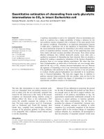
Fig. 2. Scheme of the active sites of 2,3-
dihydroxybiphenyl-1,2-dioxygenase (DHBD),
3,4-dihydroxyphenylactetate-2,3-dioxygenase
(HPCD) and catechol-2,3-dioxygenase
(C2,3O). (A) DHBD from Pseudomonas sp.
KKS102 with 2,3-dihydroxybiphenyl bound
(PDB code 1eim). (B) Brevibacterium fuscum
HPCD (PDB code 1q0c) with homoproto-
catechuate bound. (C,D) Pseudomonas
putida C2,3O (PDB code 1mpy) with 3,4-
dimethylcatechol (3,4-DMC) or 3,6-DMC,
respectively, docked in the active site.
Schemes in (E) and (F) illustrate the active
site of P. putida C2,3O with 3,5-DMC
docked in the active site in two different ori-
entations. Arrows indicate groups at distan-
ces between 3 A
˚
and 4.2 A
˚
. Arrows in bold
indicate groups at distances less than the
sum of the van der Waals’ radii. Hydrogen
bonds are shown as dotted lines. The
schemes of the side chains are shown only
when the side chain makes the closest con-
tact between the residue and the substrate.
Lines with round ends indicate stacking
between the ring of the substrate and
His240 in DHBD (A), His248 in HPCD (B),
and His246 in C2,3O (C–F).
Thr249 in catechol-2,3-dioxygenase function L. Siani et al.
2966 FEBS Journal 273 (2006) 2963–2976 ª 2006 The Authors Journal compilation ª 2006 FEBS
Based on the above observations, residue Thr249
was substituted in silico with valine, serine, alanine
and glycine, and the molecular contacts of docked
3,6-DMC and 3,5-DMC were reinspected. Mutation
T249V does not allow for a reduction of the steric hin-
drance with dimethylated substrates. Indeed, the valine
rotamer, which better fits the C2,3O active site, has
one of the methyl groups at approximately the same
position as the Thr249 methyl group that approaches
the substrate. On the contrary, the progressive reduc-
tion of the side chain of residue 249 caused by muta-
tion of threonine to serine, alanine and glycine creates
a new cavity (subsite 3¢) adjacent to CH atoms at posi-
tion 6, resulting in the reduction of steric hindrance
between a methyl group at this position and the pro-
tein. The reduction of steric hindrance caused by
mutations was estimated by measuring the radius of
the largest sphere that can be fitted to the active site,
using as center of the sphere the coordinates of the
carbon atom of the methyl group at position 6 of 3,6-
DMC bound as shown in Fig. 2D. The radius increa-
ses from 0.76 A
˚
—measured for wild-type C2,3O—to
0.98, 1.25 and 1.91 A
˚
for T249S C2,3O, T249A
C2,3O, and T249G C2,3O, respectively. As the radius
of a methyl group is 1.9–2 A
˚
, it can be predicted
that the ability of the mutants to bind dimethylcatech-
ols in a productive conformation should increase pro-
gressively, reaching its maximum in mutant T249G
C2,3O.
Kinetic parameters and regioselectivity of
wild-type and mutated C2,3O
To investigate the influence of the side chain of residue
Thr249 of C2,3O on the cleavage of 3,5-DMC and 3,6-
DMC, the catalytic properties of mutants were studied.
Based on the results of docking studies, three mutants
were produced by site-directed mutagenesis: T249S
C2,3O, T249A C2,3O, and T249G C2,3O. All of the
mutated proteins were active on catechol (Table 1),
and had an iron content similar to that of wild-type
C2,3O.
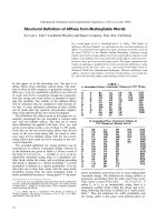
The regioselectivity of the wild-type and mutant
C2,3Os were determined by incubating them with
3-MC or 3,5-DMC, and analyzing the cleavage prod-
uct by NMR after extraction with ethyl acetate. For
all of the C2,3O variants, no aldehydic hydrogen was
detected in the product when 3-MC was used as a sub-
strate, indicating that other possible products of ring
cleavage distal to the methyl group, if present, were
below the detection limit (less than about 0.6–0.5% of
the cleavage product). On the other hand, when 3,5-
DMC was used as a substrate, the
1
H spectrum of the
product showed a signal at d 9.44, a value consistent
with that of an aldehydic hydrogen for a conjugate
aldehyde. Moreover, no signal that could be assigned
to hydrogen atoms of the product of ring cleavage
proximal to the methyl group at position 3 was ever
found at the expected field. This indicates that the
cleavage of 3,5-DMC is distal (‡ 99.0%) to the methyl
group at position 3 (Fig. 3).
Thus, the analysis above leads to the conclusion that
2-hydroxy-6-oxohepta-2,4-dienoic acid and 2-hydroxy-
3,5-dimethyl-6-oxohexa-2,4-dienoic acid (Fig. 3) are
the sole or main products of 3-MC and 3,5-DMC ring
cleavage, respectively (the NMR spectra of the clea-
vage products are shown in Supplementary Fig. 1).
The kinetic parameters of wild-type C2,3O were
determined on purified 3,5-DMC and 3,6-DMC
(Table 1). The K
m
values were found to be 74 lm and
21 lm, respectively, which are approximately 50 and
14 times higher than that measured on catechol. More-
over, the k
cat
values were found to be very low,
0.36 s
)1
for 3,5-DMC and 0.66 s
)1
for 3,6-DMC.
These values are about 0.2–0.5% of that measured
on catechol (180 s
)1
). Therefore, the low reactivity of
Fig. 3. Possible extradiol cleavage reactions for (A) 3,6-dimethyl-
catechol (3,6-DMC), (B), 3-methylcatechol (3-MC) and (C) 3,5-
dimethylcatechol (3,5-DMC) (C).
L. Siani et al. Thr249 in catechol-2,3-dioxygenase function
FEBS Journal 273 (2006) 2963–2976 ª 2006 The Authors Journal compilation ª 2006 FEBS 2967
C2,3O from P. stutzeri towards 3,5-DMC and 3,6-
DMC seems to depend on both weak binding and slow
catalysis.
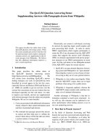
Figure 4 shows the kinetic parameters determined at
pH 7.5 using catechol, 3-MC, 3,5-DMC and 3,6-DMC
as a function of the radius of the largest sphere that
can be fitted to the active site of wild-type and
mutated C2,3O as described in the previous section.
This is a direct measurement of the volume of subsite
3¢ (Fig. 5), and hence of the ability of the enzyme to
bind 3,5-DMC and 3,6-DMC in an orientation similar
to those of catechol and 3-MC.
The K
m
values of catechol and 3-MC (Table 1) show
a regular, progressive increase as the volume of subsite
3¢ increases (Fig. 4A). In contrast, the K
m
values on
3,5-DMC and 3,6-DMC decrease with the increase of
the volume of subsite 3¢. Assuming that the kinetics of
C2,3O follow the Michaelis–Menten relationship, these
results indicate that a reduction of the volume of resi-
due 249 increases the affinity of the enzyme for 3,5-
DMC and 3,6-DMC and decreases the affinity for
smaller substrates.
Mutations at position 249 result in large and partly
unexpected variations in the k
cat
values (Table 1). For
the smaller substrates, catechol and 3-MC, the T249S
mutation has little or no effect on the catalytic con-
stants, whereas replacement of the threonine residue
with an alanine or a glycine residue causes a significant
reduction of the k
cat
values with respect to those meas-
ured for the wild-type enzyme; approximately four-fold
and 20-fold for catechol and 3-MC, respectively. On
the other hand, the behavior of the mutants is very
different in the case of dimethylcatechols. The T249S
mutation causes an increase in the k
cat
values on dime-
thylcatechols with respect to the wild-type enzyme. In
the case of 3,5-DMC, the k
cat
value is about eight
times higher than that of the wild-type enzyme. On the
contrary, mutations T249A and T249G have no signifi-
cant effect on the catalytic constants measured for
Fig. 4. Catalytic parameters of wild-type and mutant catechol-2,3-
dioxygenases (C2,3Os) measured at pH 7.5 are shown as functions
of the radii of subsite 3¢ shown in Fig. 5 (radii are: 0.76, 0.98, 1.25
and 1.91 A
˚
for wild-type, T249S, T249A and T249G C2,3O, respect-
ively). Filled circles, catechol; open circles, 3-methylcatechol
(3-MC); filled triangles, 3,6-dimethylcatechol (3,6-DMC); open trian-
gles, 3,5-DMC. For clarity in (B), the k
cat
⁄ K
m
values on catechol and
3-MC and the values on 3,5-DMC and 3,6-DMC are reported on dif-
ferent scales—on the left and on the right, respectively.
Table 1. Kinetic parameters of wild-type and mutated catechol-2,3-dioxygenase.
Substrate
Residue at position 249
Thr Ser Ala Gly
K
m
(lM) Catechol 1 ± 0.09 22.5 ± 2 37 ± 3 63.6 ± 5
3-MC 3.8 ± 0.4 11.5 ± 1 14.3 ± 1 26.6 ± 3
3,5-DMC 73.8 ± 6 57.5 ± 4 38.1 ± 3 23.7 ± 2
3,6-DMC 21.5 ± 2 9.7 ± 1 5.5 ± 0.6 7.4 ± 0.6
Catechol 180 ± 11 170 ± 10 48 ± 3 47.7 ± 3
k
cat
(s
)1
) 3-MC 118 ± 10 60.5 ± 6 6.3 ± 0.6 2.6 ± 0.3
3,5-DMC 0.36 ± 0.04 2.65 ± 0.18 1 ± 0.08 0.4 ± 0.03
3,6-DMC 0.66 ± 0.07 1.2 ± 0.1 0.4 ± 0.03 0.23 ± 0.02
Catechol 180 ± 27 7.5 ± 1 1.3 ± 0.18 0.8 ± 0.11
k
cat
⁄ K
m
(lM
)1
Æs
)1
) 3-MC 31.0 ± 5.8 5.3 ± 0.9 0.45 ± 0.07 0.1 ± 0.02
3,5-DMC 0.005 ± 0.0009 0.05 ± 0.007 0.026 ± 0.004 0.016 ± 0.002
3,6-DMC 0.03 ± 0.006 0.13 ± 0.02 0.074 ± 0.013 0.021 ± 0.003
Thr249 in catechol-2,3-dioxygenase function L. Siani et al.
2968 FEBS Journal 273 (2006) 2963–2976 ª 2006 The Authors Journal compilation ª 2006 FEBS
3,5-DMC and cause a small decrease in the catalytic
constants on 3,6-DMC.
Discussion
Bioremediation techniques are based on the use of
microorganisms to remove hazardous substances, such
as aromatic molecules, from polluted areas [5,6]. The
expansion of the catabolic potential of these bacteria
would greatly improve the applicability of these tech-
niques, by increasing the number of molecules that can
be metabolized by the microorganisms. ECD specificity
and regioselectivity control the range of molecules that
can be degraded through the catabolic pathways of
bacteria capable of using aromatic hydrocarbons as
growth substrates [14,28,29]. Knowledge of the
molecular determinants that direct their substrate
specificity is essential to tailor their active site to
transform a wider range of substrates, hence widening
the ability of the microorganism to grow on aromatic
compounds.
Members of the different subfamilies of ECD cata-
lyze the oxidative cleavage of a very wide range of
dihydroxylated aromatic substrates, ranging from the
simple ring of catechol to multiple substituted catech-
ols and polycyclic molecules [17–23]. Despite differ-
ences in their specificity, the catalytic residues seem to
be very well conserved. Six residues of the active site
are completely conserved [36,37]: the three ligands to
the catalytic metal (His154, His214, Glu265 in P. stut-
zeri C2,3O), two histidines that have been suggested to
act as acid–base catalysts (His199 and His246), and
Tyr255, which is responsible for the correct positioning
of the substrate [18,34,38].
The structures of two DHBDs, an Fe
2+
-dependent
HPCD and an Mn
2+
-dependent HPCD are available
in their reduced, active forms with the substrate bound
to the active site [34,35]. In each of these structures,
the substrate is bound similarly to both the catalytic
metal and the conserved residues in the active site
pocket. One of the substrate hydroxyl groups is posi-
tioned near the conserved tyrosine residue and is closer
to the metal atom than the other hydroxyl group
[34,35]. Available data suggest that the hydroxyl group
facing the conserved tyrosine is in the anionic form
[34,35].
To shed light on the specificity of the enzyme for
dimethylcatechols, the information above was used to
construct models of the complexes between P. stutzeri
C2,3O, a member of subfamily 1 ECDs, and different
substrates.
The models of the complexes indicate that the orien-
tation of the substrate in the active site pocket of the
C2,3O is very similar to that observed in the structure
of the DHBD and HPCD complexes. A closer compar-
ison of the X-ray structures and of our models of
C2,3O with bound catechols reveals that the residues
interacting with the first hydroxyl group that is
strongly coordinated to the metal atom, and those
interacting with the two faces of the substrate ring, are
conserved. The polypeptide regions that contact the
edge of the ring, however, are variable in the different
proteins. Thus, it is likely that the determinants of sub-
strate specificity reside in these regions.
The model of C2,3O with catechol bound in the act-
ive site pocket reveals the presence of two small sub-
sites, 1¢ and 2¢ (Fig. 2C), facing positions 3 and 4 of
the substrate ring. The volume of these cavities is large
enough to accommode methyl substituents at positions
3 and 4, thus providing a molecular scaffold to sup-
port C2,3O binding and cleavage of 3,4-DMC. Subsite
2¢, which is adjacent to position 4, is slightly smaller
Fig. 5. Scheme of possible binding of 3-methylcatechol (3-MC),
3,5-dimethylcatechol (3,5-DMC) and 3,6-DMC to catechol-2,3-
dioxygenase (C2,3O) active site. (A,B) Binding of 3-MC and 4-MC,
respectively, to the active site of wild-type Pseudomonas stutzeri
C2,3O. (C,E) Two possible orientations for the binding of 3-MC to
the active site of T246G C2,3O. (D,F) Binding of 3,5-DMC and
3,6-DMC, respectively, to the active site of T246G C2,3O.
L. Siani et al. Thr249 in catechol-2,3-dioxygenase function
FEBS Journal 273 (2006) 2963–2976 ª 2006 The Authors Journal compilation ª 2006 FEBS 2969
than subsite 1¢ facing position 3 of the catechol ring
(Fig. 2C). This difference could explain why 4-substi-
tuted catechols are more inactivating substrates than
3-substituted catechols [17,18]. The region of C2,3O
adjacent to positions 5 and 6 of the catechol ring pro-
vides no space for the binding of substituents at these
positions. This may suggest a structural basis for the
fact that C2,3O is not able to cleave catechols with
substituents at positions 3,5, or 3,6. (Fig. 2D,E,F). In
fact, a strong 20–70-fold decrease in the affinity of
P. stuzeri C2,3O for 3,5-DMC and 3,6-DMC with
respect to unsubstituted catechol and to 3-DMC is
found (Table 1).
The region facing positions 5 and 6 of the catechol
ring is mainly formed by a loop containing residues
246–249 in C2,3O (240–243 in DHBD and 248–251 in
HPCD) (Fig. 2). Multiple alignments of ECDs show
that this loop is well conserved within each subfamily
(Supplementary Fig. 2). The consensus sequence of the
loop is H-G-(L ⁄ I ⁄ V ⁄ F)-T in C2,3Os, H-(T ⁄ A ⁄ S ⁄ P)-
N-D in DHBDs and H-G-(V ⁄ I ⁄ L)-S in HPCDs.
Despite the differences in their primary structures, the
three different types of loop tightly contact positions 5
and 6 of the substrate ring in a very similar fashion
(Fig. 2). It should be noted that none of the members
of the C2,3O, DHBD or HPCD subfamilies have been
reported to cleave catechols with substituents at both
positions 3 and 5 or 3 and 6. Members of the
DHpCD subfamily, on the other hand, have been
reported to cleave 3,6-substituted catechols. This sub-
family has the loop consensus sequence H-P-(P ⁄ T)-S.
Unfortunately, there is no available structure for any
member of the DHpCD subfamily that could provide
insight into the contacts between the loop residues
and the substrate. Thus, the structure of 2,3-dihydrox-
y-p-cumate-3,4-dioxygenase from P. putida F1, a
member of the DHpCD subfamily, was modeled with
the substrate bound in the active site using the struc-
ture of DHBD from Burkholderia cepacia LB400
(1kmy [38]), as a template. We found (data not
shown) that the loop containing residues 235–238 of
DHpCD, with the sequence H-P-P-S, can assume a
conformation that easily accommodates the carboxy-
late group of the aromatic substrate dihydroxy-p-cu-
mate, whereas the isopropyl group of the substrate
can be housed in a cavity corresponding to subsite 1¢
of C2,3O (Fig. 2C). Moreover, the model indicates
that the carboxylate group can hydrogen bond to
Ser238 of the loop (data not shown). The model also
suggests that the active sites of other ECDs could be
enlarged to accommodate 3,6-disubstituted catechols
by inducing small changes to the loop 246–249 (C2,3O
numbering).
The modeling studies of the C2,3O complexes indi-
cate that the active site of this enzyme can accommo-
date one methyl group from 3,5-DMC or 3,6-DMC in
subsites 1¢ or 2¢, but not a second, because of the dif-
ferent structure of loop 246–249 of C2,3O (subsite 3¢)
with respect to that of the homologous loop 235–238
of DHpCD. Thus, the steric hindrance between the
second methyl group and loop 246–249 could force the
dimethylated substrate to bind in an orientation that is
not suitable for efficient catalysis. This hypothesis is
supported by the low affinity and low catalytic effi-
ciency of wild-type C2,3O on 3,5-DMC and 3,6-DMC
and by the results we have obtained from the study of
C2,3O Thr249 mutants.
The K
m
values in Fig. 4A indicate, as expected, that
the apparent affinity of dimethylcatechols for C23O
increases as the steric hindrance at position 249 decrea-
ses. Moreover, the 3,6-DMC K
m
values for wild-type
and mutant C2,3O are lower than those measured for
3,5-DMC and are in agreement with the models shown
in Fig. 5D,F. Figure 5F shows that the two methyl
groups of 3,6-DMC are housed in subsites 1¢ and 3¢,
whereas in the model of Fig. 5D, the methyl groups of
3,5-DMC are housed in subsites 2¢ and 3 ¢. The smaller
volume of subsite 2¢ compared to subsite 1¢ may
explain the lower affinity of wild-type and mutant
C2,3O for 3,5-DMC with respect to 3,6-DMC.
Interestingly, the progressive decrease of the dimen-
sion of the residue 249 side chain also causes an
increase in the K
m
values for catechol and 3-MC
(Fig. 4A). In the case of the smaller side chain, in
mutant T249G C2,3O, the K
m
values are 63 and seven
times higher, respectively, than those measured for the
wild-type enzyme, suggesting that residue Thr249
might make an energetic contribution to substrate
binding. Thr249 could contribute to substrate binding
either through van der Waals’ contacts as described in
Results, or a through a hydrogen bond network, dis-
cussed later in this section.
Thr249 mutants also give information on factors
that control the regioselectivity of C2,3O. 3-MC might
be cleaved at two different bonds (Fig. 3), yielding two
different extradiol cleavage products. All known ECDs
belonging to subfamilies 1, 2 and 3 catalyze only the
proximal cleavage [17,19–23] (Fig. 3). It has been
reported that this regioselectivity could depend either
on the reactivity of the substrate or on the asymmetry
of the active site that forces the binding of the sub-
strate in the monoanionic form [38–41]. The decrease
in K
m
values of mutant T249G C2,3O on 3,5-DMC
and 3,6-DMC could indicate that the T249G mutation
is successful in opening a new subsite (subsite 3¢) for
methyl binding. Thus, the presence of a new cavity in
Thr249 in catechol-2,3-dioxygenase function L. Siani et al.
2970 FEBS Journal 273 (2006) 2963–2976 ª 2006 The Authors Journal compilation ª 2006 FEBS
the active site pocket of mutant T249G C2,3O should
allow for the binding of 3-MC in two different orienta-
tions, i.e. with the methyl group housed in subsite 1¢
or in subsite 3¢ (Fig. 5C,E). As reported in Results, the
formation of the distal cleavage product (Fig. 3) has
never been observed, either with T249G C2,3O or with
the other two mutants. These data suggest that the
regioselectivity of the cleavage of 3-MC is proximal,
independently of the orientation of the substrate in the
binding site. Thus, the regioselectivity of cleavage
would be mainly controlled by the reactivity of the
substrate. This could explain the finding that wild-type
C2,3O and its mutants cleave 3,5-DMC only at the
bond proximal to the methyl group at position 5. This
regioselectivity could indicate that the methyl group at
this position is more activating than the methyl group
at position 3. This latter hypothesis is reinforced by
the observation that the k
cat
value of P. stutzeri C2,3O
for 4-MC is two times higher than the k
cat
value for
3-MC [18]. Figure 5B,D show that 4-MC and 3,5-
DMC could bind in the active sites of wild-type and
T249G C2,3O, respectively, with a similar orientation.
Thus, the methyl group at position 4 of 4-MC is geo-
metrically and chemically equivalent to the methyl
group at position 5 of 3,5-MC. Consequently, it could
be the reactivity of a substrate that bears a methyl sub-
stituent at an equivalent position—i.e. position 4 in
4-MC and position 5 in 3,5-DMC—that controls the
regioselectivity of the extradiol cleavage we have
observed in the case of 3,5-DMC.
Finally, the data reported in Table 1 show that resi-
due 249 also strongly influences the k
cat
values. More-
over, the variations observed in the k
cat
values are
significantly larger than those in the K
m
values; as a
consequence, the k
cat
and k
cat
⁄ K
m
values show similar
trends as a function of steric hindrance at the 3¢ sub-
site (Table 1 and Fig. 4B).
Mutation T249S increases the k
cat
and k
cat
⁄ K
m
val-
ues on 3,5-DMC and 3,6-DMC, with respect to those
measured using the wild-type enzyme (Table 1 and
Fig. 4B). This effect could depend on the relief of the
steric hindrance in the binding of dimethylcatechols at
the active site, which, in turn, might favor a more suit-
able orientation of the substrate for catalysis. How-
ever, mutations T249A and T249G cause instead small
variations in k
cat
and k
cat
⁄ K
m
values (Table 1) despite
the fact that their K
M
values on 3,5-DMC and 3,6-
DMC would suggest improved binding with respect to
the wild-type enzyme. Moreover, mutation T249S has
little or no effect on the k
cat
values on catechol and
3-MC (Table 1), whereas mutations T249A and T249G
reduce by four times the k
cat
values on catechol and 20
times those measured on 3-MC (Table 1).
These latter data are quite intriguing, and they sug-
gest that the hydroxyl group of Thr249 could play an
unsuspected role in catalysis. Its direct involvement in
the catalytic mechanism is unlikely, given the distance
(4 A
˚
or greater) between the oxygen atom of the
Thr249 side chain and the groups of the substrate
directly involved in the reaction. An analysis of the
active sites of DHBD structures in the presence and in
the absence of substrates and of the structure of
P. putida C2,3O suggests a possible hypothesis. In the
DHBD–substrate complex, a water molecule is bound
between the carboxylate group of Asp243 and the
hydroxyl group of the substrate (Supplementary
Fig. 3) [34]. A solvent molecule is also present in the
active site of each protomer of the P. putida C2,3O
structure [33], bound to the hydroxyl group of Thr249
at a position equivalent to that of the water molecule
bound to DHBD residue Asp243. Our modeling stud-
ies show that binding of the substrate to the active site
of P. putida C2,3O does not displace the water mole-
cule, which can bridge the hydroxyl group of residue
Thr249 and one of the hydroxyl groups of the sub-
strate, as in the DHBD–substrate complex (Supple-
mentary Fig. 3). Moreover, in the model of C2,3O
with 3,6-DMC and 3,5-DMC bound to the active site,
the water molecule contacts the methyl group located
in the 3¢ site (about 3 A
˚
between the oxygen atom and
the carbon atom of the methyl group). As a conse-
quence, the possible removal of the water molecule
due to mutations T249A and T249G should not make
a significant contribution to the decrease in steric hin-
drance between the substrate and the active site. This
observation and the reduced catalytic efficiency of
T249A C2,3O and T249G C2,3O with respect to the
wild-type enzyme and to T249S C2,3O would strongly
suggest that the bridging solvent molecule plays an
important role in catalysis.
Experimental procedures
Materials and general procedures
All chemicals were of the highest grade available and were
from Amersham Pharmacia Biotech (Amersham, UK),
Promega (Madison, WI, USA), New England Biolabs (Bev-
erly, MA, USA), Sigma (St Louis, MO, USA), or Appli-
Chem GmbH (Darmstadt, Germany).
SDS ⁄ PAGE was carried out according to the method of
Laemmli [43]. Protein concentration was determined colori-
metrically with the Bradford reagent [44], using bovine
serum albumin as a standard. Total iron content and Fe(II)
content were determined colorimetrically by complexation
with Ferene S [45].
L. Siani et al. Thr249 in catechol-2,3-dioxygenase function
FEBS Journal 273 (2006) 2963–2976 ª 2006 The Authors Journal compilation ª 2006 FEBS 2971
Bacterial strains and plasmids
Escherichia coli strain BL21(DE3) and plasmid pET22b(+)
were purchased from Novagen (Madison, WI, USA). Plas-
mid DNA purifications were performed by using the
Qiagen purification kit (Quiagen, Valencia, CA, USA). Bac-
terial transformation was carried out according to the
method of Sambrook et al. [46].
The construction of recombinant plasmid pET22b(+)
DXN ⁄ C2,3O used for the expression of wild-type P. stutzeri
C2,3O and the preparation of C2,3O mutants is described
elsewhere [18].
Construction of the expression vectors coding
for mutant C2,3Os
Mutant C2,3Os were produced by the Kunkel method [47],
starting from plasmid pET22b(+)DXN ⁄ C2,3O. The
sequences of the mutagenic oligonucleotides for T249S,
T249A and T249G were 5¢-GTCTTGCCGTGACTGAG
GCCGTGG-3¢,5¢-TCTTGCCGTGAGCGAGGCCGTGG
C-3¢ and 5¢-GGTCTTGCCGTGGCCGAGGCCGTGG-3¢,
respectively. The clones harboring the desired mutations
were identified by DNA sequencing and named
pET22b(+)DXN ⁄ (T249S)-C2,3O, pET22b(+)DXN ⁄ (T249A)-
C2,3O, and pET22b(+)DXN ⁄ (T249G)-C2,3O. The DNA
sequences of the three clones were verified by sequencing.
Expression and purification of C2,3Os
Wild-type and mutant C2,3O were expressed in E. coli
strain BL21(DE3), transformed with the appropriate
expression vector, purified and analyzed for quality as des-
cribed previously [18]. C2,3Os were stored at ) 80 °C under
a nitrogen atmosphere.
Synthesis and characterization of 3,5-DMC and
3,6-DMC
Synthesis of 3,5-DMC and 3,6-DMC was achieved by a
modification of the procedure described by Pezzella et al.
[48].
o-Iodoxybenzoic acid (IBX) was freshly prepared from
2-iodobenzoic acid as already described [49]. Solid IBX (2.5
equivalents) was added to a solution of 2,4-dimethylphenol
or 2,5-dimethylphenol (200 mg) in CHCl
3
⁄ MeOH 3 : 2 v ⁄ v
(40 mL) at ) 25 °C. A yellow–orange color developed and
the mixture was stirred for 24 h. Methanolic NaBH
4
(15 mg in 1 mL) was then added at ) 25 °C with vigorous
stirring until the color disappeared (usually within 5 min).
Excess NaBH
4
was removed by mild acidification with
acetic acid (200–500 lL). The mixture was then washed five
times with equal volumes of a saturated NaCl solution con-
taining 10% sodium dithionite buffered at pH 7.0 with
sodium phosphate. Evaporation of the organic layer even-
tually yielded 3,5-DMC or 3,6-DMC, which could be separ-
ated by preparative TLC (benzene ⁄ ethyl acetate ⁄ acetic acid
1 : 1 : 0.01) on silica.
1
H (13C) NMR spectra of products were recorded at
400.1 (100.6) MHz using a Bruker DRX ) 400 MHz instru-
ment fitted with a 5 mm
1
H ⁄ broadband gradient probe
with inverse geometry. Impurities were below
1
H-NMR
detection limits.
Spectral data of 3,5-DMC
Pale brown powder. UV(MeOH): k
max
281 nm. ESI(–) ⁄ MS
m ⁄ z: calculated for C
8
H
9
O
2
[M–H
+
] 137.061, determined
137.060.
1
H-NMR (CDCl
3
), d (p.p.m.) of selected signals:
2.19 (s, 3H, CH
3
), 2.21 (s, 3H, CH
3
), 6.51 (s, 2H), 6.53
(s, 2H).
Spectral data of 3,6-DMC
Pale brown powder. UV(MeOH): k
max
280 nm. ESI(–) ⁄ MS
m ⁄ z: calculated for C
8
H
9
O
2
[M–H
+
] 137.061, determined
137.060.
1
H-NMR (CDCl
3
), d (p.p.m.) of selected signals:
2.22 (s, 6H, CH
3
), 6.61 (s, 2H). On the basis of
1
H-NMR
and ESI ⁄ MS data, it was possible to confirm the structures
of 3,5-DMC and 3,6-DMC. Indeed, in the case of 3,5-
DMC, the presence of two aromatic signals at slightly dif-
ferent shifts, given the shielding effect of the OH group at
position 1, which is positioned para and ortho to hydrogen
3 and hydrogen 5, respectively, is consistent with the struc-
ture of catechol. In the case of 3,6-DMC, the
1
H-NMR
spectrum features only one aromatic and one methyl group
signal, as expected based on the symmetry of the molecule.
Also in this case, observed shifts are in agreement with
those predicted on the basis of the structure.
Determination of regioselectivity on 3-MC and
3,5-DMC
3-MC or 3,5-DMC were added to a solution containing
0.1 mgÆmL
)1
of wild-type or mutated C2,3O in 50 mm
Tris ⁄ HCl, pH 7.0, at 200 lm final concentration. After
5 min at 25 °C, the reaction was stopped by acidification to
pH 4.0 with H
3
PO
4
at 4 ° C, saturated with NaCl, and
extracted with ethyl acetate (3 · 100 mL). Evaporation of
the organic layer eventually furnished a pale yellow oil that
was directly characterized by
1
H-NMR (solvent CDCl
3
).
About 95–100% of 3-MC and 70–80% of 3,5-DMC were
converted, yielding 60–80 lmol of products. The determin-
ation of the structures of the cleavage products was done
only on the basis of the hydrogen atoms bound to sp
2
carbon atoms (see Supplementary Fig. 1 for details). The
signals of hydrogen atoms of methyl groups were not con-
sidered, as they do not allow us to discriminate distal and
Thr249 in catechol-2,3-dioxygenase function L. Siani et al.
2972 FEBS Journal 273 (2006) 2963–2976 ª 2006 The Authors Journal compilation ª 2006 FEBS
proximal cleavage products. Each spectrum was recorded at
least on two independent preparations in order to distin-
guish between background noise and reproducible weak sig-
nals. Under the conditions that we used, minor products,
with a relative abundance greater than about 0.5% and
1%, would have been detected using 3-MC and 3,5-DMC,
respectively, as substrates.
Spectral data of 3-MC cleavage product
1
H-NMR (CDCl
3
), d (p.p.m.) of selected signals: 6.27 (d,
1H, J ¼ 15.6 Hz), 6.37 (d, 1H, J ¼ 11.2 Hz), 7.55 (dd, 1H,
J ¼ 15.6, 11.2 Hz).
Spectral data of 3,5-DMC cleavage product
1
H-NMR (CDCl
3
), d (p.p.m.) of selected signals: 7.22 (s,
1H), 9.44 (s, 1H).
Enzyme assays
All assays were performed at 25 ° Cin50mm Tris ⁄ HCl
pH 7.5 or in 50 mm sodium phosphate pH 7.5 in a final
volume of 500 lL, by spectrophotometric determination of
the product of the reaction. Wild-type and mutant C2,3Os
were used to start the reaction. The amount of the fission
products was measured spectrophometrically using their
absorption extinction coefficients at the respective e
max
val-
ues. All ring cleavage products showed relative absorption
maxima in the range 375–395 nm. Substrates showed
absorption maxima in the range 270–280 nm and no
absorption at wavelengths corresponding to the e
max
values
of products. The absorption extinction coefficients are:
e
375
¼ 33 000 m
)1
Æcm
)1
for 2-hydroxymuconic semialde-
hyde, the product of catechol; e
388
¼ 13 400 m
)1
Æcm
)1
for
2-hydroxy-6-oxohepta-2,4-dienoic acid, the product of
3-MC; e
393
¼ 8230 m
)1
Æcm
)1
for 2-hydroxy-3,5-dimethyl-6-
oxohexa-2,4-dienoic acid, the product of 3,5-DMC; and
e
393
¼ 15 200 m
)1
Æcm
)1
for 2-hydroxy-3-methyl-6-oxohepta-
2,4-dienoic acid, the product of 3,6-DMC. Kinetic parame-
ters were determined by the program graphpad prism
(GraphPad Software ).
Analysis of ECD structures and modeling of
(di)methylcathecols into the active site of C2,3O
The structures of ECDs (PDB codes: 1mpy, 1eim, 1q0c)
were analyzed by swiss pdb viewer (asy.
org/spdbv/) and pymol (DeLano Scientific LLC, San Fran-
cisco, CA, USA). Catechol, 3-MC, 3,4-DMC, 3,5-DMC
and 3,6-DMC were modeled into the active site of C2,3O
from P. putida MT2 with a two-step procedure. First, swiss
pdb viewer was used to superimpose the structure of
C2,3O on the crystallographic complexes of DHBD and
HPCD with their respective substrates. Then, pymol was
used to manipulate the complexes in order to manually
minimize the number of nonbonded atoms at distances less
than the sum of their van der Waals’ radii. Catechol and
(di)methylcatechols were assumed to bind in the monoani-
onic, catecholate form. The phenolate oxygen was placed
on the same side as the conserved catalytic residue Tyr255
[18,33]. The aromatic ring was rotated using the iron atom
as rotation center in order to search for conformations that
fit the van der Waals’ volume of the substrate in the active
site. Coordinates for the (di)methylcatechols were generated
by the programs cs chemdraw pro and chem3d pro
(Cambridge Soft Corporation, Cambridge, MA, USA) and
energy minimized before docking the compounds into the
active site. The largest spheres that can be fitted to the act-
ive site of wild-type and mutant C2,3O were created using
the program caver ( />index.php).
Sequence alignments of ECDs and homology
modeling of DHpCD
Multiple sequence alignments of ECDs were prepared and
analyzed as described previously [16]. A model of the
DHpCD from P. putida F1 (accession code Q51976) was
generated using swiss pdb viewer and the Swiss Model ser-
ver [50]. The crystal structure of DHBD from B. cepacia
LB400 (1kmy [38]) served as the template. The alignment
between the sequences of the two proteins was extracted
from the multiple alignment of ECDs.
Acknowledgements
The authors are indebted to Dr Matthew H. Sazinsky,
Northwestern University Evanston, IL for critically
reading the manuscript. This work was supported by a
grant from the Ministry of University and Research
(PRIN ⁄ 2002).
References
1 Fetzner S & van der Meer JR (2000) Enzymes involved
in the aerobic bacterial degradation of N-heteroaromatic
compounds: molybdenum hydroxylases and ring-opening
2,4-dioxygenases. Naturwissenschaften 87, 59–69.
2 van der Meer JR (1997) Evolution of novel metabolic
pathways for the degradation of chloroaromatic com-
pounds. Antonie Van Leeuwenhoek 71, 159–178.
3 Whited GM & Gibson DT (1991) Toluene-4-mono-
oxygenase, a three component enzyme system that
catalyzes the oxidation of toluene to p-cresol in Pseu-
domonas mendocina KR1. J Bacteriolol 173, 3010–
3016.
L. Siani et al. Thr249 in catechol-2,3-dioxygenase function
FEBS Journal 273 (2006) 2963–2976 ª 2006 The Authors Journal compilation ª 2006 FEBS 2973
4 Sanseverino J, Applegate BM, King JM & Sayler GS
(1993) Plasmid-mediated mineralization of naphthalene,
phenanthrene, and anthracene. Appl Environ Microbiol
59, 1931–1937.
5 Timmis KN & Pieper DH (1999) Bacteria designed for
bioremediation. Trends Biotechnol 17, 200–204.
6 Diaz E (2004) Bacterial degradation of aromatic pollut-
ants: a paradigm of metabolic versatility. Int Microbiol
7, 173–180.
7 Samanta SK, Singh OV & Jain RK (2002) Polycyclic
aromatic hydrocarbons: environmental pollution and
bioremediation. Trends Biotechnol 20, 243–248.
8 Tan HM (1999) Bacterial catabolic transposons. Appl
Microbiol Biotechnol 51, 1–12.
9 Rana SV & Verma Y (2005) Biochemical toxicity of
benzene. J Environ Biol 26, 157–168.
10 Low LK, Meeks JR & Mackerer CR (1988) Health
effects of the alkylbenzenes. I. Toluene. Toxicol Ind
Health 4, 49–75.
11 Low LK, Meeks JR & Mackerer CR (1989) Health
effects of the alkylbenzenes. II. Xylenes. Toxicol Ind
Health 5, 85–105.
12 Schreiner CA (2003) Genetic toxicity of naphthalene: a
review. J Toxicol Environ Health B Crit Rev 6, 161–183.
13 Broderick JB (1999) Catechol dioxygenases. Essays
Biochem 34, 173–189.
14 Harayama S & Rekik M (1989) Bacterial aromatic ring-
cleavage enzymes are classified into two different gene
families. J Biol Chem 264, 15328–15333.
15 Harayama S & Rekik M (1993) Comparison of the
nucleotide sequences of the meta-cleavage pathway genes
of TOL plasmid pWW0 from Pseudomonas putida with
other meta-cleavage genes suggests that both single and
multiple nucleotide substitutions contribute to enzyme
evolution. Mol Gen Genet 239, 81–89.
16 Izzo V, Notomista E, Picardi A, Pennacchio F & Di
Donato A (2005) The thermophilic archaeon Sulfolobus
solfataricus is able to grow on phenol. Res Microbiol
156, 677–689.
17 Cerdan P, Wasserfallen A, Rekik M, Timmis KN &
Harayama S (1994) Substrate specificity of catechol 2,3-
dioxygenase encoded by TOL plasmid pWWO of Pseu-
domonas putida and its relationship to cell growth.
J Bacteriol 176, 6074–6081.
18 Viggiani A, Siani L, Notomista E, Birolo L, Pucci P &
Di Donato A (2004) The role of conserved residues
H246, H199 and Y255 in the catalysis of catechol 2,3-
dioxygenase from Pseudomonas stutzeri OX1. J Biol
Chem 279, 48630–48639.
19 Furukawa K & Arimura N (1987) Purification and
properties of 2,3-dihydroxybiphenyl dioxygenase
from polychlorinated biphenyl-degrading Pseudomonas
pseudoalcaligenes and Pseudomonas aeruginosa
carrying the cloned bphC gene. J Bacteriol 169,
924–927.
20 Kuhm AE, Stolz A, Ngai KL & Knackmuss HJ (1991)
Purification and characterization of a 1,2-dihydroxy-
naphthalene dioxygenase from a bacterium that
degrades naphthalenesulfonic acids. J Bacteriol 173,
3795–3802.
21 Eltis LD, Hofmann B, Hecht HJ, Lunsdorf H & Tim-
mis KN (1993) Purification and crystallization of 2,3-
dihydroxybiphenyl 1,2- dioxygenase. J Biol Chem 268,
2727–2732.
22 Wang YZ & Lipscomb JD (1997) Cloning, overexpres-
sion, and mutagenesis of the gene for homoprotocatech-
uate 2,3-dioxygenase from Brevibacterium fuscum.
Protein Expr Purif 10, 1–9.
23 Whiting AK, Boldt YR, Hendrich MP, Wackett LP &
Que L Jr (1996) Manganese (II)-dependent extradiol-
cleaving catechol dioxygenase from Arthrobacter globi-
formis CM-2. Biochemistry 35, 160–170.
24 Eaton RW (1996) p-Cumate catabolic pathway in Pseu-
domonas putida Fl: cloning and characterization of
DNA carrying the cmt operon. J Bacteriol 178, 1351–
1362.
25 Ninnekar HZ (1992) Purification and properties of 2,3-
dihydroxy-p-cumate-3,4-dioxygenase from Bacillus spe-
cies. Biochem Int 28, 97–103.
26 Haigler BE, Johnson GR, Suen WC & Spain JC (1999)
Biochemical and genetic evidence for meta-ring cleavage
of 2,4,5-trihydroxytoluene in Burkholderia sp. strain
DNT. J Bacteriol 181, 965–972.
27 Hatta T, Mukerjee-Dhar G, Damborsky J, Kiyohara H
& Kimbara K (2003) Characterization of a novel ther-
mostable Mn (II)-dependent 2,3-dihydroxybiphenyl 1,2-
dioxygenase from a polychlorinated biphenyl- and
naphthalene-degrading Bacillus sp. JF8. J Biol Chem
278, 21483–21492.
28 Jindrova E, Chocova M, Demnerova K & Brenner V
(2002) Bacterial aerobic degradation of benzene,
toluene, ethylbenzene and xylene. Folia Microbiol
(Praha) 47, 83–93.
29 Williams PA & Sayers JR (1994) The evolution of path-
ways for aromatic hydrocarbon oxidation in Pseudomo-
nas. Biodegradation 5, 195–217.
30 Baggi G, Barbieri P, Galli E & Tollari S (1987) Isola-
tion of a Pseudomonas stutzeri strain that degrades
o-xylene. Appl Environ Microbiol 53, 2129–2131.
31 Bertoni G, Bolognesi F, Galli E & Barbieri P (1996)
Cloning of the genes for and characterization of the
early stages of toluene catabolism in Pseudomonas stut-
zeri OX1. Appl Environ Microbiol 62, 3704–3711.
32 Arenghi FL, Berlanda D, Galli E, Sello G & Barbieri P
(2001) Organization and regulation of meta cleavage
pathway gene for toluene and o-xylene derivative degra-
dation in Pseudomonas stutzeri OX1. Appl Environ
Microbiol 67, 3304–3308.
33 Kita A, Kita S, Fujisawa I, Inaka K, Ishida T, Horiike K,
Nozaki M & Miki K (1999) An archetypical extradiol-
Thr249 in catechol-2,3-dioxygenase function L. Siani et al.
2974 FEBS Journal 273 (2006) 2963–2976 ª 2006 The Authors Journal compilation ª 2006 FEBS
cleaving catecholic dioxygenase: the crystal structure of
catechol 2,3-dioxygenase (metapyrocatechase) from
Ppseudomonas putida mt-2. Structure Fold Des 7, 25–34.
34 Sato N, Uragami Y, Nishizaki T, Takahashi Y, Sazaki
G, Sugimoto K, Nonaka T, Masai E, Fukuda M &
Senda T (2002) Crystal structures of the reaction inter-
mediate and its homologue of an extradiol-cleaving
catecholic dioxygenase. J Mol Biol 321, 621–636.
35 Vetting MW, Wackett LP, Que L Jr, Lipscomb JD &
Ohlendorf DH (2004) Crystallographic comparison of
manganese- and iron-dependent homoprotocatechuate
2,3-dioxygenases. J Bacteriol 186, 1945–1958.
36 Spence EL, Kawamukai M, Sanvoisin J, Braven H &
Bugg TD (1996) Catechol dioxygenases from Escherichia
coli (MhpB) and Alcaligenes eutrophus (MpcI): sequence
analysis and biochemical properties of a third family of
extradiol dioxygenases. J Bacteriol 178, 5249–5256.
37 Eltis LD & Bolin JT (1996) Evolutionary relationships
among extradiol dioxygenases. J Bacteriol 178, 5930–
5937.
38 Vaillancourt FH, Barbosa CJ, Spiro TG, Bolin JT,
Blades MW, Turner RF & Eltis LD (2002) Definitive
evidence for monoanionic binding of 2,3-dihydroxybi-
phenyl to 2,3-dihydroxybiphenyl 1,2-dioxygenase from
UV resonance Raman spectroscopy, UV ⁄ Vis absorption
spectroscopy, and crystallography. J Am Chem Soc 124,
2485–2496.
39 Shu L, Chiou YM, Orville AM, Miller MA, Lipscomb
JD & Que L Jr (1995) X-ray absorption spectroscopic
studies of the Fe (II) active site of catechol 2,3-dioxy-
genase. Implications for the extradiol cleavage mechan-
ism. Biochemistry 34, 6649–6659.
40 Reynolds MF, Costas M, Ito M, Jo DH, Tipton AA,
Whiting AK & Que L Jr (2003) 4-Nitrocatechol as a
probe of a Mn(II)-dependent extradiol-cleaving catechol
dioxygenase (MndD): comparison with relevant Fe(II)
and Mn(II) model complexes. J Biol Inorg Chem 8,
263–272.
41 Lin G, Reid G & Bugg TD (2001) Extradiol oxidative
cleavage of catechols by ferrous and ferric complexes of
1,4,7-triazacyclononane: insight into the mechanism of
the extradiol catechol dioxygenases. J Am Chem Soc
123, 5030–5039.
42 Que L Jr (1983) The catechol dioxygenases. Adv Inorg
Biochem 5, 167–199.
43 Laemmli U (1970) Cleavage of structural proteins dur-
ing the assembly of the head of bacteriophage T4.
Nature 227, 680–685.
44 Bradford MM (1976) A rapid and sensitive method for
the quantitation of microgram quantities of protein util-
izing the principle of protein-dye binding. Anal Biochem
72, 248–254.
45 Newman LM & Wackett LP (1995) Purification and
characterization of toluene 2-monooxygenase from Bur-
kholderia cepacia G4. Biochemistry 34, 14066–14076.
46 Sambrook J, Fritsch EF & Maniatis T (1989) Molecular
Cloning. A Laboratory Manual, 2nd edn. Cold Spring
Harbor Laboratory Press, Cold Spring Harbor, New
York.
47 Kunkel TA (1987) Rapid and efficient site-specific muta-
genesis without phenotypic selection. Proc Natl Acad
Sci USA 82, 488–492.
48 Pezzella A, Lista L, Napolitano A & d’Ischia M
(2005) An expedient one-pot entry to catecholestro-
gens and other catechol compounds via IBX-medi-
ated phenolic oxygenation. Tetrahedron Lett 46,
3541–3544.
49 Frigerio M, Santagostino M & Sputore S (1999) A user-
friendly entry to 2-iodoxybenzoic acid (IBX). J Org
Chem 64, 4537–4538.
50 Schwede T, Kopp J, Guex N & Peitsch MC (2003)
SWISS-MODEL: an automated protein homology-
modeling server. Nucleic Acids Res 31, 3381–3385.
Supplementary material
The following supplementary material is available
online:
Fig. S1.
1
H-NMR spectrum of the cleavage product
from T249S catechol-2,3-dioxygenase (C2,3O)-cata-
lyzed oxidation of 3-methylcatechol (3-MC) (A) and
3,5-dimethylcatechol (3,5-DMC) (B). Only the spectral
region (aromatic region) used for structural assignment
of cleavage products is shown. In (A), a typical spin
system is present, which can be attributed to three
adjacent protons on an sp
2
carbon chain bearing an
electron-withdrawing group. No signal that can be
attributed to an aldehydic group is visible at the expec-
ted fields (10.0–9.0 p.p.m.). In (B), the signal of the H4
proton is shifted at lower fields with respect to the sig-
nal of the corresponding hydrogen atom in the clea-
vage product of 3-MC, due to the releasing effect of
the methyl groups. No signal that can be attributed to
H3 and H5 protons of the proximal cleavage product
is visible at the expected fields (6.4–5.5 p.p.m.). The
signals at 6.523 and 6.500 p.p.m. correspond to the
two aromatic hydrogen atoms of the substrate (H4
and H6 of 3,5-DMC).
Fig. S2. Multiple alignment of selected extradiol ring
cleavage dioxygenases (ECDs). The loop 246–249
(Pseudomonas putida MT2 numbering) is highlighted.
Accession numbers of the sequences included in the
alignment are given in supplementary Table S1.
Fig. S3. Close-up of the active sites of 2,3-dihydroxybi-
phenyl-1,2-dioxygenase (DHBD) and catechol-2,3-
dioxygenase (C2,3O). (A) DHBD from Pseudomonas
sp. KKS102 with 2,3-dihydroxybiphenyl bound (PDB
code: 1eim). (B) Pseudomonas putida C2,3O (PDB
L. Siani et al. Thr249 in catechol-2,3-dioxygenase function
FEBS Journal 273 (2006) 2963–2976 ª 2006 The Authors Journal compilation ª 2006 FEBS 2975
code: 1mpy) with catechol docked into the active site.
Hydrogen bonds are shown as green dotted lines.
Distances are expressed in A
˚
. W indicates the water
molecules bound in the active sites. The active site of
DHBD from Burkholderia cepacia LB400 (PDB code:
1kmy) is very similar to that of Pseudomonas sp.
KKS102 enzyme shown in (A).
Table S1. Accession numbers of the sequences included
in the alignment shown in Fig. S2.
This material is available as part of the online article
from
Thr249 in catechol-2,3-dioxygenase function L. Siani et al.
2976 FEBS Journal 273 (2006) 2963–2976 ª 2006 The Authors Journal compilation ª 2006 FEBS