Báo cáo khoa học: Fatty acid regulation of adenylyl cyclase Rv2212 from Mycobacterium tuberculosis H37Rv doc
Bạn đang xem bản rút gọn của tài liệu. Xem và tải ngay bản đầy đủ của tài liệu tại đây (834.29 KB, 10 trang )
Fatty acid regulation of adenylyl cyclase Rv2212 from
Mycobacterium tuberculosis H37Rv
Amira Abdel Motaal
1
, Ivo Tews
2
, Joachim E. Schultz
1
and Ju
¨
rgen U. Linder
1
1 Abteilung Pharmazeutische Biochemie, Fakulta
¨
tfu
¨
r Chemie und Pharmazie, Universita
¨
tTu
¨
bingen, Germany
2 Biochemiezentrum der Universita
¨
t Heidelberg, Germany
Adenylyl cyclases (ACs) (EC 4.6.1.1) convert ATP to
the second messenger cAMP, which regulates a variety
of cellular functions, including virulence in several
pathogens, such as Mycobacterium tuberculosis [1–8].
Therefore, it is no surprise that ACs are subject to regu-
lation by both extracellular stimuli such as hormones,
availability of nutrients or osmotic pressure, and by
intracellular stimuli such as changes in pH or even
cAMP levels [9–11]. Currently, the catalytic domains of
AC isozymes are grouped into six classes based on
sequence similarities [12–14]. Class III contains by far
the largest number of ACs, including all mammalian
and many bacterial cyclases. All class III ACs must di-
merize to be active, because the substrate-binding sites
are formed at the dimer interface. On the basis of con-
served sequence differences, class III ACs are further
divided into subclasses IIIa–IIId [15]. The catalytic
domains of the class III ACs are most often linked to
additional protein domains, which in many instances
appear to impart peculiar regulatory features [15].
In the M. tuberculosis genome, 15 putative class III
AC genes of subclasses IIIa–IIId have been identified
that possess quite different domain compositions [15–
17]. Therefore, one may assume that each of these cyc-
lases participates in a distinct signalling pathway. To
date, the recombinant proteins of nine mycobacterial
AC genes have been shown to be catalytically active
in vitro [18–25]. In all of them, the catalytic domains
are associated with additionally distinct domains such
as hexahelical membrane anchors (Rv1625c, Rv1318–
Rv1320c, Rv3645), a pH-sensing domain (Rv1264),
AAA-ATPase and helix-turn-helix DNA-binding
domains (Rv0386), an a ⁄ b-hydrolase-like domain
(Rv1900c) and HAMP domains (Rv1318c, Rv1319c,
Keywords
adenylyl cyclase; cAMP; fatty acid;
Mycobacterium tuberculosis
Correspondence
J. U. Linder, Abteilung Pharmazeutische
Biochemie, Fakulta
¨
tfu
¨
r Chemie und
Pharmazie, Universita
¨
tTu
¨
bingen,
Morgenstelle 8, 72076 Tu
¨
bingen, Germany
Fax: +49 7071 295952
Tel: +49 7071 2974676
E-mail:
(Received 26 May 2006, revised 14 July
2006, accepted 17 July 2006)
doi:10.1111/j.1742-4658.2006.05420.x
Adenylyl cyclase Rv2212 from Mycobacterium tuberculosis has a domain
composition identical to the pH-sensing isoform Rv1264, an N-terminal
regulatory domain and a C-terminal catalytic domain. The maximal velo-
city of Rv2212 was the highest of all 10 mycobacterial cyclases investigated
to date (3.9 lmol cAMPÆmg
)1
Æmin
)1
), whereas ATP substrate affinity was
low (SC
50
¼ 2.1 mm ATP). Guanylyl cyclase side activity was absent. The
activities and kinetics of the holoenzyme and of the catalytic domain alone
were similar, i.e. in distinct contrast to the Rv1264 adenylyl cyclase, in
which the N-terminal domain is autoinhibitory. Unsaturated fatty acids
strongly stimulated Rv2212 activity by increasing substrate affinity. In
addition, fatty acids greatly enhanced the pH sensitivity of the holoenzyme,
thus converting Rv2212 to a pH sensor adenylyl cyclase. Fatty acid binding
to Rv2212 was modelled by homology to a recent structure of the N-ter-
minal domain of Rv1264, in which a fatty acid-binding pocket is defined.
Rv2212 appears to integrate three cellular parameters: ATP concentration,
presence of unsaturated fatty acids, and pH. These regulatory properties
open the possibility that novel modes of cAMP-mediated signal transduc-
tion exist in the pathogen.
Abbreviations
AAA, ATPase associated with a variety of cellular activities; AC, adenylyl cyclase; CHD, cyclase homology domain; HAMP, domain first
identified in histidine kinases, adenyl cyclases, methyl accepting chemotaxis proteins and phosphatases.
FEBS Journal 273 (2006) 4219–4228 ª 2006 The Authors Journal compilation ª 2006 FEBS 4219
Rv1320c, and Rv3645). Although several studies
have been published in recent years, the role of these
regulatory domains is just beginning to be revealed. In
the isoform Rv1625c, the large membrane anchor has
a prominent role in protein dimerization [26]. In the
four mycobacterial class IIIb ACs, the HAMP
domains appear to directly act as modulators of AC
activity, possibly transmitting signals that may be
picked up by a receptor function of their hexahelical
membrane domains [21]. The best investigated AC iso-
form is Rv1264, which contains an N-terminal pH sen-
sor module [25]. The structures of Rv1264 in an active
and an inhibited state have been determined by X-ray
crystallography. Rv1264 may enable M. tuberculosis to
counteract acidification of phagolysosomes during host
invasion and aid in intracellular survival [25].
Here we investigated the AC isoform Rv2212, which
has the same domain composition as Rv1264. Both the
recombinant Rv2212 holoenzyme and the isolated
catalytic domain (also called the cyclase homology
domain, CHD) were active in vitro. Unsaturated fatty
acids stimulated Rv2212 AC activity. We demonstrate
that the fatty acids are connected with a pH-sensing
function of the holoenzyme. Furthermore, the rather
low substrate affinity of Rv2212 suggests a potential
role as a cellular ATP gauge, i.e. as a sensor for the
prevailing energy status of the cell.
Results
Sequence features
The predicted M. tuberculosis Rv2212 gene product
has a domain composition identical to that of the AC
isoform Rv1264 (Fig. 1A). The C-terminal class IIIc
CHD of 177 amino acids shares 29% identity with that
of Rv1264 (41% similarity; for alignments see [20]).
The N-terminus of Rv2212 has 211 amino acids and is
21% identical (31% similar) to that of Rv1264, which
mediates pH sensing [20,25]. Similar N-terminal
domains are exclusively found in related actinobacteri-
al ACs [20]. Irrespective of the identical domain organ-
ization, the limited similarity of the N-termini of
Rv2212 and Rv1264 suggested that Rv2212 may be
regulated in a different way from Rv1264.
AC activity of Rv2212
212)388
and Rv2212
1)388
The boundaries of the Rv2212 catalytic domain, S212
to the C-terminal D388, were defined by sequence
comparisons with other bacterial class IIIa ACs. The
catalytic domain Rv2212
212)388
and the holoenzyme
Rv2212
1)388
were expressed in Escherichia coli as sol-
uble proteins and purified to homogeneity by affinity
chromatography (Fig. 1B). Both displayed AC activity
with a pH optimum of 6.5. Activity was Mn
2+
-
dependent. With up to 10 mm Mg
2+
, AC activity was
below the detection limit of 0.5 nmol cAMPÆmg
)1
Æ
min
)1
. Guanylyl cyclase activity was absent. We consis-
tently observed that AC activity varied among different
protein preparations. At 0.5 mm ATP, the activity
of Rv2212
212)388
was 487 ± 365 nmol cAMPÆmg
)1
Æ
min
)1
(SD, n ¼ 31, range 104–2105), and that of
Rv2212
1)388
was 377 ± 357 nmol cAMPÆmg
)1
Æmin
)1
(SD, n ¼ 58, range 82–1735). This clearly demonstra-
ted that the N-terminal domain of Rv2212 was
not autoinhibitory as in Rv1264 [20]. The excessive
variability in enzyme activity did not correlate with
technical parameters such as the method of cell lysis,
the extent of protein purification and duration or con-
ditions of storage. Furthermore, individual assays were
linear with respect to time and protein concentration.
Protein aggregation was excluded because gel filtration
chromatography reproducibly yielded a single symmet-
rical peak corresponding to a dimer (not shown).
Obviously, the affinity of the AC monomers was very
high and only dimers existed in solution. Furthermore,
there was no indication of charge heterogeneity, as evi-
dent from a single symmetrical peak that was obtained
upon anion exchange chromatography (data not
shown). Finally, we have never observed such a fluctu-
ation of AC activities in any of the recombinant myco-
bacterial ACs that we have reported on previously,
such as Rv1625c, Rv1264, Rv3645, Rv1318c, Rv1319c,
Rv1320c, and Rv0386 [19–21,23–25]. Therefore, the
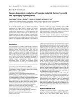
Fig. 1. Mycobacterium tuberculosis adenylyl cyclase (AC) Rv2212.
(A) Predicted domain composition of Rv2212. CHD, cyclase homol-
ogy domain of class III ACs. (B) Purity of the recombinant ACs
Rv2212
212)388
(CHD) and Rv2212
1)388
(holoenzyme) proteins.
SDS ⁄ PAGE, 15%, stained with Coomassie blue. Lane 1, 2.3 lgof
Rv2212
212)388
; lane 2, 2.8 lg of Rv2212
1)388
; molecular weight
markers on the left.
Mycobacterium tuberculosis AC Rv2212 A. Abdel Motaal et al.
4220 FEBS Journal 273 (2006) 4219–4228 ª 2006 The Authors Journal compilation ª 2006 FEBS
most likely explanation for these highly variable activ-
ities of Rv2212 in vitro is that the dimeric enzyme can
exist in several, interconvertible states with different
catalytic activities. Kinetic analysis of Rv2212
212)388
yielded a V
max
of 7.5 lmol cAMPÆmg
)1
Æmin
)1
(Table 1), an SC
50
of 3.3 mm ATP, and a Hill coeffi-
cient of 1.8, which indicated strong positive coopera-
tivity for the substrate and was consistent with the
dimeric nature of bacterial class III ACs with two sub-
strate-binding sites. The properties of the Rv2212
1)388
holoenzyme were similar (V
max
¼ 3.9 lmol cAMPÆ
mg
)1
Æmin
)1
,SC
50
¼ 2.1 mm ATP, Hill coefficient 1.8;
Table 1). Thus, Rv2212 has a higher V
max
than any
other mycobacterial AC studied to date (range 0.007–
2.1 lmol cAMPÆmg
)1
Æmin
)1
). Furthermore, ATP sub-
strate affinity was rather low compared to other myco-
bacterial ACs, which had SC
50
concentrations in the
range 0.06–1.2 mm ATP [19–25].
Stimulation of Rv2212 by fatty acids
The response of Rv2212 proteins to pH changes was
modest. Rv2212
1)388
had a five-fold higher activity
at pH 6.5 compared to activity at pH 9, and with
Rv2212
212)388
the maximal activity difference in activ-
ity was three-fold between pH 6.5 and 7.6. This was in
striking contrast to the pH-sensing AC Rv1264, which
is stimulated 110-fold upon a shift from pH 9.0 to
pH 6.0 [25]. The modest pH sensitivity of Rv2212 indi-
cated that its N-terminus probably connects to differ-
ent or additional regulatory inputs.
It has been reported that the AC from Brevibacteri-
um liquefaciens, which has an identical domain compo-
sition to Rv1264 and Rv2212, is strongly activated by
pyruvate, other a-ketocarbonic acids, glycine, alanine
and lactate [27]. Therefore, we examined the effects
of various metabolites on Rv2212 activity. At 1 mm
concentrations, d-galactose, d-mannose, l-arabinose,
l-rhamnose, d-glucose, d-fructose, fructose 1,6-bis-
phosphate, glucose 6-phosphate, dl-threonine, l-iso-
leucine, l-valine, l-asparagine, l-histidine, l-aspartic
acid, d-alanine, l-alanine, l -cysteine, l-leucine, glycine,
sodium chloride, potassium chloride, sodium citrate,
sodium acetate, sodium bicarbonate, NADH, glyoxylic
acid, a-ketoglutarate, pyruvate and phosphoenolpyru-
vate did not significantly affect Rv2212
1)388
activity.
Three-fold stimulation was obtained with 100 lm pal-
mitic acid (not shown). Oleic, linoleic, linolenic and
arachidonic acids at 100 lm produced strong activa-
tion (Table 2). To determine whether the activation
was specific for the Rv2212 AC, we established dose–
response curves for oleic and linoleic acids using the
class IIIc ACs Rv2212 and Rv1264, and the class IIIa
AC Rv1625c, a membrane-bound isoform of unrelated
Table 1. Kinetic analysis of Rv2212. Values are means ± SD. Numbers of experiments are in parentheses. *P < 0.001 compared to the
respective control value with Rv2212
1)388
.
Parameter
Rv2212
1)388
(n ¼ 8)
Rv2212
212)388
(n ¼ 6)
Rv2212
1)388
(n ¼ 8)
100 l
M
linoleic acid
Rv2212
1)388
(n ¼ 4)
100 l
M
oleic acid
Rv2212
1)388
(n ¼ 4)
100 l
M
arachidonic
acid
Rv2212
1)388
(n ¼ 2)
170 l
M
polidocanol
Rv2212
1)388
(n ¼ 2)
100 l
M
Brij 35
Rv2212
1)388
(n ¼ 2)
100 l
M
Triton X-100
V
max
(lmol cAMPÆmg
)1
Æmin
)1
)
3.9 ± 0.6 7.5 ± 0.1* 4.1 ± 0.7 3.2 ± 0.9 2.9 ± 0.7 3.8 ± 0.2 3.1 ± 0.1 2.9 ± 0.1
SC
50
(mM) 2.1 ± 0.5 3.3 ± 0.2* 0.9 ± 0.1* 1.0 ± 0.2* 0.7 ± 0.3* 0.3 ± 0.1* 0.3 ± 0.1* 0.8 ± 0.1*
Hill coefficient 1.8 ± 0.1 1.8 ± 0.1 1.3 ± 0.1* 1.2 ± 0.2* 1.0 ± 0.1* 1.1 ± 0.2* 1.1 ± 0.1* 1.7 ± 0.1
Table 2. Stimulation of Rv2212 by fatty acids and detergents. The numbers of experiments are in parentheses. NT, not tested. To be com-
parable, ‘fold stimulation’ was measured at 100 l
M of each fatty acid and at 119 lM polidocanol, 105 lM Triton X-100 and 119 lM Nonidet
P-40, respectively. Values are means ± SD. All stimulations were highly significant (P<0.005).
Rv2212
1)388
Rv2212
212)388
EC
50
(lM) Fold stimulation EC
50
(lM) Fold stimulation
Linoleic acid 56 ± 6 (2) 6.0 ± 3.3 (13) 78 ± 4 (4) 2.6 ± 1.1 (9)
Oleic acid 41 ± 4 (2) 7.5 ± 2.5 (3) 65 ± 1 (2) 2.2 ± 0.1 (2)
Arachidonic acid 10 ± 1 (4) 8.4 ± 0.8 (4) 10 ± 1 (2) 2.4 ± 0.4 (2)
Polidocanol 16 ± 1 (2) 11.0 ± 0.4 (2) 30 ± 3 (2) 7.0 ± 0.3 (2)
Triton X-100 16 ± 1 (2) 5.0 ± 0.7 (2) NT NT
Nonidet P-40 15 ± 1 (2) 4.3 ± 0.2 (2) NT NT
A. Abdel Motaal et al. Mycobacterium tuberculosis AC Rv2212
FEBS Journal 273 (2006) 4219–4228 ª 2006 The Authors Journal compilation ª 2006 FEBS 4221
domain composition (Fig. 2A,B). Only Rv2212 was sti-
mulated 9–13-fold by fatty acids with EC
50
concentra-
tions around 50 lm (Fig. 2; Table 2), whereas Rv1625c
was totally unresponsive and Rv1264 was stimulated a
mere two-fold. This established a specificity of the
effect of unsaturated fatty acids for the Rv2212 iso-
form. Furthermore, the activation was specific to the
Rv2212 holoenzyme, because the catalytic domain
Rv2212
212)388
was stimulated only 2–3-fold (Table 2;
Fig. 2A,B). Thus the stimulation was dependent on the
presence of the N-terminal domain of Rv2212. Because
the high variability in the AC activity of Rv2212 was
reflected in a large variability of the extent of stimula-
tion, we examined the effect in more detail.
Fatty acids are prototypical detergents. We there-
fore investigated whether the activation of Rv2212
was due to the detergent-like properties or whether it
was the result of specific molecular interactions
between the protein and the unsaturated fatty acids.
Indeed, nonionic detergents such as polidocanol sti-
mulated the Rv2212 holoenzyme 4–11-fold (Table 2;
Fig. 2C). However, the effect of the detergents was
identical for the catalytic domain alone and for the
Rv2212 holoenzyme, and hence was unspecific
(Table 2; Fig. 2C). We conclude that the stimulation
of Rv2212 by unsaturated fatty acids only partly
results from their amphipathic nature and distinct dif-
ferences exist when compared to the effects of non-
ionic detergents.
Biochemical properties of Rv2212 activation
by fatty acids and polidocanol
Because of the unusually large variability in basal and
linoleic acid-stimulated activities, we investigated whe-
ther the extent of activation was dependent on the pre-
set basal activity that each preparation of recombinant
protein displayed (Fig. 3). In 13 assays, the activities
of Rv2212
1)388
were 0.37 ± 0.34 and 1.43 ± 0.51
lmol cAMPÆmg
)1
Æmin
)1
in the absence and presence
Fig. 2. Effect of fatty acids and polidocanol on Rv2212. Basal activ-
ity is set to 100%. (A) Effect of oleic acid examined at respective
optimal pH conditions. Rv2212
1)388
, filled squares, pH 6.5;
Rv2212
212)388
, open squares, pH 6.5; Rv1264, circles, pH 5.7;
Rv1625c, triangles, pH 7.5. Starting at 70 l
M, stimulation by oleic
acid is highly significant (P<0001) for Rv2212
1)388
, Rv2212
212)388
and Rv1264. For each fatty acid dilution, controls were carried out
with the solvent alone. There were no significant solvent effects.
(B) Effect of linoleic acid [symbols as in (A)]. All stimulations at
70 l
M linoleic acid and higher were highly significant (P<0001) for
Rv2212
1)388
, Rv2212
212)388
and Rv1264. (C) Effect of polidocanol
on Rv2212
1)388
(squares) and Rv2212
212)388
(circles) at pH 6.5. Sti-
mulations at 50 l
M polidocanol and above were highly significant
(P<0001) for both proteins. SD values are indicated by vertical
bars (n ¼ 2).
Mycobacterium tuberculosis AC Rv2212 A. Abdel Motaal et al.
4222 FEBS Journal 273 (2006) 4219–4228 ª 2006 The Authors Journal compilation ª 2006 FEBS
of 100 lm linoleic acid, respectively (± SD). Thus, the
variabilities were 92% and 35% in basal and stimula-
ted AC activities, respectively. A plot of the stimula-
tion factors versus basal activities illustrated this
correlation (Fig. 3A). Rv2212
1)388
was stimulated most
when the basal AC activity was below 0.4 lmol
cAMPÆmg
)1
Æmin
)1
, whereas the potency of linoleic acid
was rather modest in those instances where basal AC
activity was already above 1 lmol cAMPÆmg
-1
Æmin
-1
(see insert in Fig. 3A). Under standard assay condi-
tions, AC activity stimulated by 100 lm linoleic acid
was in the range 0.9–2.3 lmol cAMPÆmg
)1
Æmin
)1
and
thus fairly independent of basal activity, which varied
by more than an order of magnitude (0.09–1.18). One
interpretation is that the mycobacterial Rv2212 holo-
enzyme, when heterologously expressed in E. coli, was
isolated as a mixture of low-activity and high-activity
states, and that the activation by linoleic acid was
actually a conversion of the protein to a uniformly
high-activity state. Thus, linoleic acid activation was
only modest in cases where the purification a priori
yielded an AC protein with a large fraction already in
an activated state. Obviously, this applied exclusively
to the Rv2212
1)388
holoenzyme, because the
Rv2212
212)388
catalytic domain displayed fundament-
ally different features of activation by linoleic acid. In
nine assays, the AC activities of Rv2212
212)388
were
0.373 ± 0.206 and 0.924 ± 0.488 lmol cAMPÆmg
)1
Æ
min
)1
(SD) in the absence and presence of 100 lm
linoleic acid, respectively; that is, the variability of
basal and stimulated activities in individual protein
preparations were identical (54%; Fig. 3B). Thus, the
lesser activation of the catalytic domain Rv2212
212)388
by linoleic acid was due to a totally different mode of
activation compared to stimulation of the holoenzyme
Rv2212
1)388
.
Fatty acid activation of Rv2212
1)388
was then exam-
ined kinetically (Fig. 4; Table 1). Stimulation was
caused by an increase in substrate affinity and a con-
comitant loss of cooperativity, whereas V
max
remained
unaffected. Consequently, fatty acid stimulation
decreased with increasing substrate concentration. At
5mm ATP, hardly any stimulation was observed
(Fig. 4; Table 1). The kinetic basis of stimulation by
polidocanol was identical (Table 1). We also tested the
kinetics of activation by Brij 35, a lauryl alcohol deriv-
ative, which has a 50% higher mean molecular mass.
The effect of Brij 35 was identical to that of polidocan-
ol, demonstrating that chain length did not matter
(Table 1). Triton X-100, which contains the bulkier
octylphenyl moiety, also increased substrate affinity
but did not reduce cooperativity (Table 1). Obviously,
diverse classes of detergents operated by different
mechanisms, possibly reflecting different binding sites.
Next, we determined the pH dependence of
Rv2212
1)388
activation. pH profiles of basal and stimu-
lated activities showed marked differences (Fig. 5). The
basal AC activity of Rv2212
1)388
decreased five-fold
from pH 6.5 to pH 9 (n ¼ 6). However, in the pres-
ence of 100 lm oleic, linoleic or arachidonic acid, it
decreased 20-fold, 19-fold and 26-fold, respectively.
Obviously, the unsaturated fatty acids considerably
bolstered the pH sensitivity of Rv2212
1)388
. Further-
more, in a kinetic analysis at pH 9, we did not obtain
substrate saturation even at 8 mm ATP, irrespective of
the absence or presence of fatty acids (not shown).
This demonstrated that the pH increase led to a
Fig. 3. Correlation analysis of adenylyl cyclase (AC) stimulation by
linoleic acid versus corresponding basal activity of the holoenzyme
Rv2212
1)388
(A) (n ¼ 13) and the catalytic domain Rv2212
212)388
(B) (n ¼ 9). Open circles represent the average of four data groups.
Group 1, basal activity < 0.3 lmolÆmg
)1
Æmin
)1
; group 2, 0.3–0.5;
group 3, 0.5–0.8; group 4, > 0.8). SD values for both dimensions
are shown whenever they exceed the symbol size.
A. Abdel Motaal et al. Mycobacterium tuberculosis AC Rv2212
FEBS Journal 273 (2006) 4219–4228 ª 2006 The Authors Journal compilation ª 2006 FEBS 4223
decrease in substrate affinity. The specificity of this
effect with regard to fatty acids was emphasized by
comparison with the detergent polidocanol. In the
presence of polidocanol, the activity of Rv2212
1)388
was almost unchanged between pH 5.5 and pH 9.
Although polidocanol activated Rv2212, this effect
was pH-independent, i.e. the detergent abrogated the
pH response of the AC, corroborating unequivocally
that the effects of fatty acids and nonionic detergents
on Rv2212 are mediated by distinctly different mecha-
nisms.
Discussion
The class IIIc AC Rv2212 is the 10th AC isoform with
proven enzyme activity out of 15 putative cyclases
from M. tuberculosis, and like the other nine isoforms
uses only Mn
2+
as a cofactor [18,21,24]. By domain
composition and sequence similarity it is closely related
to the mycobacterial Rv1264 isoform, yet the bio-
chemical properties and regulation are different. The
main differences between Rv2212 and Rv1264 are the
lack of autoinhibition by the N-terminal domain of
Rv2212, its remarkable response to fatty acids, and the
high V
max
of the holoenzyme at its pH optimum
[20,25]. The biochemical analysis of the Rv2212 iso-
form was severely complicated by the exceptional vari-
ability in basal enyzme activities. Although in the
holoenzyme this variability was greatly reduced by
addition of linoleic acid, the extent of stimulation was
diminished in protein preparations with high basal
activity. We excluded the possibility that the variability
in AC activity was due to different levels of copurified
fatty acids or detergents, because treatment of the
recombinant protein with Bio-Beads SM-2 to remove
lipids did not affect enzyme activity (data not shown).
Therefore, we assume that (a) heterologous expression
in E. coli resulted in a mixture of conformations of
different specific activities, and that (b) fatty acids
Fig. 4. Kinetic analyses of the stimulation of Rv2212
1)388
by
100 l
M linoleic acid (A) and by 170 lM polidocanol (B). Squares,
basal activity; circles, stimulated activity. SD values are shown
(n ¼ 2–8). Stimulation by both compounds is highly significant
(P<0.001) for 0.1–1.0 m
M ATP and significant (P<0.05) at 2 mM
ATP.
Fig. 5. pH dependence of Rv2212
212)388
basal activity (open tri-
angles), Rv2212
1)388
basal activity (open squares), and Rv2212
1)388
in the presence of 100 lM linoleic acid (circles), arachidonic acid
(closed rhombi), oleic acid (closed triangles), and 170 l
M polidocanol
(open rhombi). The buffer systems used were: acetic acid ⁄ NaOH
(pH 5 and 5.6), Bistris ⁄ HCl (pH 5.6–7.3) and Tris ⁄ HCl (pH 7.3–9).
Vertical lines show SD values (n ¼ 2–5). The stimulations at pH 6.5
were compared to the respective activities at pH 9. Significance
(P<0.05) was observed for basal and stimulated Rv2212
1)388
.
Mycobacterium tuberculosis AC Rv2212 A. Abdel Motaal et al.
4224 FEBS Journal 273 (2006) 4219–4228 ª 2006 The Authors Journal compilation ª 2006 FEBS
induced a more uniform high-activity state. The fatty
acids, however, exerted a second, important effect in
sharpening the pH profile of Rv2212
1)388
, i.e. in indu-
cing pH sensing. In the presence of unsaturated fatty
acids, Rv2212 showed a 20-fold increase in activity
from pH 9 to pH 6.5, a response that is comparable to
the 110-fold stimulation of the pH-sensing isoform
Rv1264 by a similar pH shift [25]. This stimulation
was not due to the detergent-like properties, because a
nonionic detergent such as polidocanol stimulated
Rv2212; however, it failed to promote pH sensing. We
acknowledge that two mechanisms are possible to
explain the pH dependence of the effect of fatty acids.
One possibility is that fatty acids bind at any pH but
activate only at lower pH values. The other possibility
is that fatty acids neither bind nor activate at high pH.
Currently, it is impossible to experimentally distinguish
these possibilities. Because in the related AC Rv1264
oleic acid appears to be a protein constituent at any
pH (see below), we favour pH-independent binding of
the fatty acids to Rv2212.
The kinetic bases of the fatty acid and pH-sensing
responses of Rv2212 were similar. In both instances,
activation was mainly due to an increase in ATP sub-
strate affinity. In contrast, the pH response of Rv1264
is predominantly mediated by an increase in V
max
[25].
Prima facie this would argue for different mechanisms
of pH sensing in the two isoforms. The structural
transition upon acidification of Rv1264 has been eluci-
dated (see scheme in Fig. 6A). In the active state
(pH 6), the catalytic domains align as a closed dimer
capable of binding ATP and of catalysis. In the in-
active state (pH 8), the catalytic domains are drawn
apart by extended a-helices such that they are neither
able to bind ATP nor to catalyse cAMP formation. In
Rv2212, the pronounced change in substrate affinity
may be explained by an ability of ATP itself to shift
the equilibrium towards an active state even at high
pH and in the absence of activators.
Structural analysis also suggests a binding site for
fatty acids in Rv2212. In a recent high-resolution crys-
tallographic study, we investigated the N-terminal reg-
ulatory domain of Rv1264 and identified a binding
site for fatty acids. The dimer of N-terminal domains
contains two hydrophobic tunnels like cul-de-sacs. A
number of different crystal forms (protein databank
codes 2EV1, 2EV2, 2EV3 and 2EV4), as well as MS
analysis, have allowed us to show that a fatty acid
may be an intrinsic constituent of the Rv1264 N-ter-
minus. We found that oleic acid is present in the
Rv1264 crystal at any pH (unpublished results). This
would explain the rather small stimulation of Rv1264
by added fatty acids (see above). In light of the data
presented here, the physiological ligand for the N-ter-
minal domain of Rv2212 is probably also an unsatur-
ated fatty acid. To test whether the N-terminal
domain of Rv2212 could also contain a tunnel for the
uptake and binding of fatty acid molecules, we
attempted modelling (Fig. 6B,C). The results demon-
strate that fatty acid binding is compatible with the
three-dimensional model of Rv2212, but they do not
explain the apparent differences in biochemical
responses between the two enzymes. Such an analysis
would clearly require an experimentally determined
structure of Rv2212.
The activity of Rv2212 is governed by unsaturated
fatty acids, pH and ATP concentration in vitro, but
Fig. 6. Models of stimulation of Rv2212 by fatty acids. (A) Sche-
matic representation of Rv2212 holoenzymes in the inhibited and
active state according to crystal structures of Rv1264 [25]. In the
inhibited state, the catalytic domains are recruited by the N-terminal
domains and unable to bind ATP. In Rv2212, fatty acids and a pH
shift synergize to cause activation. Alternatively, a high ATP con-
centration may substitute for this. (B) Homology model of the
dimeric Rv2212 N-terminal domain, viewed from the top. N and C
indicate the N-terminus and C-terminus, respectively. Oleic acid is
modelled with carbon atoms in green and oxygen in red. The
arrows indicate the access sites of the ligand-binding tunnels. (C)
Side view of (B).
A. Abdel Motaal et al. Mycobacterium tuberculosis AC Rv2212
FEBS Journal 273 (2006) 4219–4228 ª 2006 The Authors Journal compilation ª 2006 FEBS 4225
what is the physiological relevance of these parameters?
Viewing the results together, a significant response to
fatty acids and consequently pH can only be expected if
the ATP concentration is equal to or lower than the
SC
50
value (0.9–2.1 mm; Table 1). The ATP content of
M. tuberculosis is 130, 270 and 520 pg per 10
6
viable
cells during chronic infection, during acute infection
and in vitro, respectively [28]. Taking into account the
reported average volume of a mycobacterium of
0.96 femtoliters [29], this translates into intracellular
ATP concentrations of 0.27, 0.55 and 1.1 mm. Thus, it
is conceivable that Rv2212 operates as an ATP sensor
in vivo, integrating two other signals, the presence of
certain fatty acids and the intracellular pH. The cyto-
plasmic concentration of free fatty acids in M. tuber-
culosis is unknown. Free fatty acids are the substrate for
fatty acyl-AMP ligases, which are crucial for the synthe-
sis of certain complex lipids of the cell envelope [30].
Thus the presence of free fatty acids in the mycobacte-
rial cytoplasm appears certain. Furthermore, mycobac-
teria have been shown to accumulate triacylglycerols as
intracellular inclusions [31] that may contain some free
fatty acids. Analysis of these lipid inclusions showed
palmitic, stearic, oleic, palmitoleic and lignoceric acids
to be major constituents. Thus these fatty acids are at
least transiently present in the cytoplasm, and triacyl-
glyerol synthesis actually is thought to serve detoxifica-
tion of free fatty acids [31–33].
Taken together, the results suggest that Rv2212 is a
biochemical integrator of three different signals, with
cAMP as the output. Its properties are distinct from
those of the other nine AC isoforms investigated to
date. Some functional redundancy may exist concern-
ing the isoform Rv1264, because both enzymes are
able to respond to low pH. The results presented here
are one further step towards understanding signal
transduction through cAMP in M. tuberculosis.
Experimental procedures
Materials
Genomic DNA from M. tuberculosis was a gift of E Boett-
ger (University of Zu
¨
rich, Medical School). Radiochemicals
were from Hartmann Analytik (Braunschweig, Germany).
All enzymes were purchased from either Roche Diagnostics
(Mannheim, Germany) or New England Biolabs (Frank-
furt, Germany). pQE30 and Ni-nitrilotriacetic acid ⁄ agarose
were from Qiagen (Hilden, Germany). Fine chemicals were
purchased from Merck (Darmstadt, Germany), Roche
Diagnostics, Roth (Karlsruhe, Germany) or Sigma
(Taufkirchen, Germany). Bio-Beads SM-2, a removal agent
for organic compounds including fatty acids and detergents,
were from Bio-Rad (Munich, Germany) and were used
according to the manufacturer’s instructions.
Rv2212 constructs
The annotated ORF of gene Rv2212 (GenBank Accession
Number BX842579, NP_216728, 378 amino acids) starts
with a GTG codon, although an in-frame ATG start codon
is just 10 codons (30 bp) upstream. Therefore, we extended
the ORF and included the N-terminal decapeptide
MGVPAGTLRQ. The ORF (388 codons) was amplified by
PCR using specific primers and genomic DNA as a tem-
plate. BamHI and HindIII restriction sites were added at
the 5¢-end and 3¢-end, respectively, and the PCR product
was inserted into pQE30. This added an N-terminal
MRGSH
6
GS tag. Similarly, the catalytic domain
(Rv2212
212)388
) was fitted with a 5¢ BamHI and a 3¢
HindIII site and inserted into pQE30. The fidelity of all
constructs was verified by double-stranded DNA sequen-
cing. Primer sequences are available on request.
Expression and purification of proteins
Plasmids with either Rv2212
1)388
or Rv2212
212)388
were
transformed into E. coli BL21(DE3)[pREP4]. Protein
expression was induced with 60 lm isopropyl-thio-b-d-gal-
actoside for 4–5 h at 22 °C. Bacteria were collected by
centrifugation at 2600 g for 15 min with a Centricon H-401
centrifuge, A8.24 rotor (Kontron-Hermle, Gosheim, Ger-
many), washed once with buffer (50 mm Tris ⁄ HCl, 1 mm
EDTA, pH 8), frozen in liquid nitrogen and stored at
) 80 °C. For purification, cells from 200 to 400 mL of cul-
ture were suspended in 20 mL of lysis buffer (50 mm
Tris ⁄ HCl, 0.02% a-monothioglycerol, pH 8), lysed by soni-
cation, and treated with 0.2 mgÆml
)1
lysozyme (30 min,
0 °C). Subsequently, 5 mm MgCl
2
and 20 lgÆml
)1
DNaseI
were added (30 min). After centrifugation (31 000 g for
30 min at 0 °C, Centricon H-401, A8.24 rotor), 15 mm imi-
dazole (pH 8) and 250 mm NaCl (final concentrations) were
added to the supernatant. Protein was equilibrated for a
minimum of 60 min with 250 lLofNi
2+
-nitrilotriacetic
acid ⁄ agarose slurry on ice, and then transferred to a mini-
column and successively washed with 10 mL of buffer A
(lysis buffer containing 15 mm imidazole, 250 mm NaCl and
5mm MgCl
2
) and 5 mL of buffer B (lysis buffer with
15 mm imidazole and 5 mm MgCl
2
). The protein was eluted
with 0.3 mL of buffer C (lysis buffer with 150 mm imidazole
and 2 mm MgCl
2
). After addition of 20% glycerol (v ⁄ v),
proteins were stored at ) 20 °C.
AC assays
AC activity was measured at 37 °C for 10 min in a volume
of 100 lL [34]. Standard reactions contained 50 mm
Mycobacterium tuberculosis AC Rv2212 A. Abdel Motaal et al.
4226 FEBS Journal 273 (2006) 4219–4228 ª 2006 The Authors Journal compilation ª 2006 FEBS
Bistris ⁄ HCl (pH 6.5), 22% glycerol, 3 mm MnCl
2
, 500 l m
[a
32
P]ATP and 2 mm [2,8-
3
H]cAMP. Substrate kinetics
were analysed with 0.1–6 mm ATP and 10 mm MnCl
2
. Kin-
etic constants were derived from Hill plots. Throughout,
100 nm Rv2212
1)388
and 200 nm Rv2212
212)388
were used.
Solutions of fatty acids and detergents
Fatty acids were prepared as 5 mm solutions in 1 mm Tris.
Further dilutions were made with AC assay buffer. Deter-
gents were prepared as 10% solutions in the AC assay
buffer and further diluted with the same buffer.
Homology modelling
The homology model of Rv2212 was constructed from
amino acids 4–186 with the high-resolution structure of the
N-terminal domain of Rv1264 as a template (protein data-
bank accession number 2EV1), using the program what if
[35]. Briefly, a sequence alignment and the template struc-
ture are input, and the program first mutates all disparate
residues to glycine, and then places the side chains in
reverse order of degrees of freedom for the individual resi-
dues using the rotamer search procedure, usually resulting
in bulky residues being placed first. The model is then
manually corrected, geometrized and minimized using pro-
cedures implemented in what if.
Acknowledgements
This work was supported by the Deutsche Forschungs-
gemeinschaft. AM was supported by a scholarship
of the Deutscher Akademischer Austauschdienst
(DAAD).
References
1 Botsford JL & Harman JG (1992) Cyclic AMP in pro-
karyotes. Microbiol Rev 56, 100–122.
2 D’Souza CA & Heitman J (2001) Conserved cAMP sig-
naling cascades regulate fungal development and viru-
lence. FEMS Microbiol Rev 25, 349–364.
3 Gross A, Bouaboula M, Casellas P, Liautard JP &
Dornand J (2003) Subversion and utilization of the host
cell cyclic adenosine 5¢-monophosphate ⁄ protein kinase
A pathway by Brucella during macrophage infection.
J Immunol 170, 5607–5614.
4 Petersen S & Young GM (2002) Essential role for cyclic
AMP and its receptor protein in Yersinia enterocolitica
virulence. Infect Immun 70, 3665–3672.
5 Smith RS, Wolfgang MC & Lory S (2004) An adenylate
cyclase-controlled signaling network regulates Pseudo-
monas aeruginosa virulence in a mouse model of acute
pneumonia. Infect Immun 72, 1677–1684.
6 Wolfgang MC, Lee VT, Gilmore ME & Lory S (2003)
Coordinate regulation of bacterial virulence genes by a
novel adenylate cyclase-dependent signaling pathway.
Dev Cell 4, 253–263.
7 Rickman L, Scott C, Hunt DM, Hutchinson T, Menen-
dez MC, Whalan R, Hinds J, Colston MJ, Green J &
Buxton RS (2005) A member of the cAMP receptor
protein family of transcription regulators in Mycobac-
terium tuberculosis is required for virulence in mice and
controls transcription of the rpfA gene coding for a
resuscitation promoting factor. Mol Microbiol 56, 1274–
1286.
8 Spreadbury CL, Pallen MJ, Overton T, Behr MA,
Mostowy S, Spiro S, Busby SJ & Cole JA (2005)
Point mutations in the DNA- and cNMP-binding
domains of the homologue of the cAMP receptor
protein (CRP) in Mycobacterium bovis BCG: impli-
cations for the inactivation of a global regulator
and strain attenuation. Microbiology 151, 547–556.
9 Peterkofsky A, Reizer A, Reizer J, Gollop N, Zhu PP &
Amin N (1993) Bacterial adenylyl cyclases. Prog Nucleic
Acid Res Mol Biol 44, 31–65.
10 Tang WJ & Hurley JH (1998) Catalytic mechanism and
regulation of mammalian adenylyl cyclases. Mol Phar-
macol 54, 231–240.
11 Sunahara RK & Taussig R (2002) Isoforms of mamma-
lian adenylyl cyclase: multiplicities of signaling. Mol
Interv 2, 168–184.
12 Barzu O & Danchin A (1994) Adenylyl cyclases: a het-
erogeneous class of ATP-utilizing enzymes. Prog Nucleic
Acid Res Mol Biol 49, 241–283.
13 Sismeiro O, Trotot P, Biville F, Vivares C & Danchin A
(1998) Aeromonas hydrophila adenylyl cyclase 2: a new
class of adenylyl cyclases with thermophilic properties
and sequence similarities to proteins from hyperthermo-
philic archaebacteria. J Bacteriol 180, 3339–3344.
14 Cotta MA, Whitehead TR & Wheeler MB (1998) Iden-
tification of a novel adenylate cyclase in the ruminal
anaerobe, Prevotella ruminicola D31d. FEMS Microbiol
Lett
164, 257–260.
15 Linder JU & Schultz JE (2003) The class III adenylyl
cyclases: multi-purpose signalling modules. Cell Signal
15, 1081–1089.
16 McCue LA, McDonough KA & Lawrence CE (2000)
Functional classification of cNMP-binding proteins and
nucleotide cyclases with implications for novel regula-
tory pathways in Mycobacterium tuberculosis. Genome
Res 10, 204–219.
17 Shenoy AR & Visweswariah SS (2004) Class III nucleo-
tide cyclases in bacteria and archaebacteria: lineage-
specific expansion of adenylyl cyclases and a dearth of
guanylyl cyclases. FEBS Lett 561, 11–21.
18 Reddy SK, Kamireddi M, Dhanireddy K, Young L,
Davis A & Reddy PT (2001) Eukaryotic-like adenylyl
A. Abdel Motaal et al. Mycobacterium tuberculosis AC Rv2212
FEBS Journal 273 (2006) 4219–4228 ª 2006 The Authors Journal compilation ª 2006 FEBS 4227
cyclases in Mycobacterium tuberculosis H37Rv: cloning
and characterization. J Biol Chem 276, 35141–35149.
19 Guo YL, Seebacher T, Kurz U, Linder JU & Schultz
JE (2001) Adenylyl cyclase Rv1625c of Mycobacterium
tuberculosis: a progenitor of mammalian adenylyl
cyclases. EMBO J 20, 3667–3675.
20 Linder JU, Schultz A & Schultz JE (2002) Adenylyl
cyclase Rv1264 from Mycobacterium tuberculosis has an
autoinhibitory N-terminal domain. J Biol Chem 277,
15271–15276.
21 Linder JU, Hammer A & Schultz JE (2004) The effect
of HAMP domains on class IIIb adenylyl cyclases from
Mycobacterium tuberculosis. Eur J Biochem 271, 2446–
2451.
22 Shenoy AR, Sreenath NP, Mahalingam M & Viswes-
wariah SS (2005) Characterization of phylogenetically
distant members of the adenylate cyclase family from
mycobacteria: Rv1647 from Mycobacterium tuberculosis
and its orthologue ML1399 from M. leprae. Biochem J
387, 541–551.
23 Castro LI, Hermsen C, Schultz JE & Linder JU (2005)
Adenylyl cyclase Rv0386 from Mycobacterium tuber-
culosis H37Rv uses a novel mode for substrate selection.
FEBS J 272, 3085–3092.
24 Sinha SC, Wetterer M, Sprang SR, Schultz JE & Linder
JU (2005) Origin of asymmetry in adenylyl cyclases:
structures of Mycobacterium tuberculosis Rv1900c.
EMBO J 24, 663–673.
25 Tews I, Findeisen F, Sinning I, Schultz A, Schultz JE &
Linder JU (2005) The structure of a pH-sensing myco-
bacterial adenylyl cyclase holoenzyme. Science 308,
1020–1023.
26 Guo YL, Kurz U, Schultz A, Linder JU, Dittrich D,
Keller C, Ehlers S, Sander P & Schultz JE (2005) Inter-
action of Rv1625c, a mycobacterial class IIIa adenylyl
cyclase, with a mammalian congener. Mol Microbiol 57,
667–677.
27 Takai K, Kurashina Y, Suzuki-Hori C, Okamoto H &
Hayaishi O (1974) Adenylate cyclase from Brevibacter-
ium liquefaciens. I. Purification, crystallization, and
some properties. J Biol Chem 249, 1965–1972.
28 Dhople AM & Ryon DL (2000) ATP content of Myco-
bacterium tuberculosis grown in vivo and in vitro.
Microbios 101, 81–88.
29 Cox RA (2004) Quantitative relationships for specific
growth rates and macromolecular compositions of
Mycobacterium tuberculosis, Streptomyces coelicolor
A3(2) and Escherichia coli B ⁄ r: an integrative theoretical
approach. Microbiology
150, 1413–1426.
30 Trivedi OA, Arora P, Sridharan V, Tickoo R, Mohanty
D & Gokhale RS (2004) Enzymic activation and trans-
fer of fatty acids as acyl-adenylates in mycobacteria.
Nature 428, 441–445.
31 Garton NJ, Christensen H, Minnikin DE, Adegbola
RA & Barer MR (2002) Intracellular lipophilic inclu-
sions of mycobacteria in vitro and in sputum. Micro-
biology 148, 2951–2958.
32 McCarthy C (1971) Utilization of palmitic acid by
Mycobacterium avium. Infect Immun 4, 199–204.
33 Weir MP, Langridge WH 3rd & Walker RW (1972)
Relationships between oleic acid uptake and lipid meta-
bolism in Mycobacterium smegmatis . Am Rev Respir Dis
106, 450–457.
34 Salomon Y, Londos C & Rodbell M (1974) A highly
sensitive adenylate cyclase assay. Anal Biochem 58, 541–
548.
35 Vriend G (1990) WHAT IF: a molecular modeling and
drug design program. J Mol Graph 8, 52–56, 29.
Mycobacterium tuberculosis AC Rv2212 A. Abdel Motaal et al.
4228 FEBS Journal 273 (2006) 4219–4228 ª 2006 The Authors Journal compilation ª 2006 FEBS