Báo cáo khoa học: Structure–activity relationships of fowlicidin-1, a cathelicidin antimicrobial peptide in chicken docx
Bạn đang xem bản rút gọn của tài liệu. Xem và tải ngay bản đầy đủ của tài liệu tại đây (434.6 KB, 13 trang )
Structure–activity relationships of fowlicidin-1,
a cathelicidin antimicrobial peptide in chicken
Yanjing Xiao1,*, Huaien Dai2,*, Yugendar R. Bommineni1, Jose L. Soulages3, Yu-Xi Gong2,
Om Prakash2 and Guolong Zhang1
1 Department of Animal Science, Oklahoma State University, Stillwater, OK, USA
2 Department of Biochemistry, Kansas State University, Manhattan, KS, USA
3 Department of Biochemistry and Molecular Biology, Oklahoma State University, Stillwater, OK, USA
Keywords
antibiotic resistance; antimicrobial peptide;
cathelicidin; chicken; structure–activity
relationship
Correspondence
O. Prakash, Department of Biochemistry,
Kansas State University, Manhattan,
KS 66506, USA
Fax: +1 785 532 7278
Tel: +1 785 532 2345
E-mail:
G. Zhang, Department of Animal Science,
Oklahoma State University, Stillwater,
OK 74078, USA
Fax: +1 405 744 7390
Tel: +1 405 744 6619
E-mail:
*These authors contributed equally to this
paper
(Received 4 February 2006, revised 21
March 2006, accepted 5 April 2006)
doi:10.1111/j.1742-4658.2006.05261.x
Cationic antimicrobial peptides are naturally occurring antibiotics that are
actively being explored as a new class of anti-infective agents. We recently
identified three cathelicidin antimicrobial peptides from chicken, which
have potent and broad-spectrum antibacterial activities in vitro (Xiao Y,
Cai Y, Bommineni YR, Fernando SC, Prakash O, Gilliland SE & Zhang
G (2006) J Biol Chem 281, 2858–2867). Here we report that fowlicidin-1
mainly adopts an a-helical conformation with a slight kink induced by glycine close to the center, in addition to a short flexible unstructured region
near the N terminus. To gain further insight into the structural requirements for function, a series of truncation and substitution mutants of fowlicidin-1 were synthesized and tested separately for their antibacterial,
cytolytic and lipopolysaccharide (LPS)-binding activities. The short C-terminal helical segment after the kink, consisting of a stretch of eight amino
acids (residues 16–23), was shown to be critically involved in all three functions, suggesting that this region may be required for the peptide to interact with LPS and lipid membranes and to permeabilize both prokaryotic
and eukaryotic cells. We also identified a second segment, comprising three
amino acids (residues 5–7) in the N-terminal flexible region, that participates in LPS binding and cytotoxicity but is less important in bacterial
killing. The fowlicidin-1 analog, with deletion of the second N-terminal
segment (residues 5–7), was found to retain substantial antibacterial
potency with a significant reduction in cytotoxicity. Such a peptide analog
may have considerable potential for development as an anti-infective agent.
Cathelicidins are a major family of animal antimicrobial peptides with hallmarks of a highly conserved prosequence (cathelin domain) and an extremely variable,
antibacterially active sequence at the C terminus [1–3].
The exact microbicidal mechanism for this family of
antimicrobial peptides is not clearly understood. However, it is generally believed that the electrostatic interaction between the C-terminal cationic peptides with
anionic lipids followed by membrane permeabilization
is mainly responsible for killing prokaryotic cells.
Because of such a nonspecific membrane-lytic mechanism, many cathelicidins kill a variety of bacteria at low
micromolar concentrations with much less chance of
developing resistance [4–6]. More importantly, they are
equally active against antibiotic-resistant bacterial
strains, with some demonstrating synergism in killing
bacteria with conventional antibiotics or structurally
different antimicrobial peptides [7–9]. One side-effect
Abbreviations
EC50, 50% effective concentration; LPS, lipopolysaccharide; MDCK, Madin–Darby canine kidney cells; MIC, minimum inhibitory
concentration; SAR, structure–activity relationship; TFE, trifluoroethanol.
FEBS Journal 273 (2006) 2581–2593 ª 2006 The Authors Journal compilation ª 2006 FEBS
2581
Structure–activity relationships of fowlicidin-1
Y. Xiao et al.
that is commonly associated with cathelicidins as
potential therapeutic agents is their cytotoxicity
towards mammalian host cells [4–6]. However, the
concentrations that are required for cathelicidins to
exert an appreciable cytolytic effect are often higher
than the concentrations which exert bactericidal
effects.
Structure–activity relationship (SAR) studies of
cathelicidins revealed that cationicity, amphipathicity,
hydrophobicity and helicity (helical content) are
among the most important determinants of their
microbicidal and cytolytic activities [10,11]. However,
in general there is no simple correlation between any
of these physicochemical properties and peptide functions. A delicate balance of these parameters often dictates the antimicrobial potency and target selectivity
[10,11]. Moreover, the domain that is responsible for
cytotoxicity can sometimes be separated from that
responsible for antimicrobial activity [12,13]. Therefore, it is possible that strategic manipulation of structural and physicochemical parameters of cathelicidins
may maximize their antimicrobial activity while reducing their cytotoxicity.
We and others have recently identified three novel
chicken cathelicidins [14–16], which are called fowlicidins 1–3 in this report. All three fowlicidins share little
similarity with mammalian cathelicidins in the C-terminal sequence [16]. Putatively mature fowlicidin-1, a
linear peptide of 26 amino acid residues, was found to
be broadly active against a range of Gram-negative
and Gram-positive bacteria with a potency similar to
that of SMAP-29 [16]. However, fowlicidin-1 also displayed considerable cytotoxicity towards human erythrocytes and mammalian epithelial cells, with 50% lysis
in the range of 6–40 lm [16].
To understand the mechanism of action of fowlicidin-1 in greater detail, we determined its tertiary
structure by NMR spectroscopy in this study. Fowlicidin-1 was shown to be composed of an a-helical
segment with a slight kink near the center and a
flexible unstructured region at the N-terminal end. A
series of deletion and substitution mutants of fowlicidin-1 were further synthesized and tested separately
for their antibacterial, lipopolysaccharide (LPS) binding and cytolytic activities. The regions responsible
for each of these functions have been revealed. In
addition, we identified a fowlicidin-1 analog with
deletion of the N-terminal flexible region that retains
the antibacterial potency but which has substantially
reduced cytotoxicity. Such a peptide analog may represent an excellent candidate as a novel antimicrobial
agent against bacteria that are resistant to conventional antibiotics.
2582
Results
Solution structure of fowlicidin-1
To determine the secondary structure of fowlicidin-1,
CD spectroscopy was performed in increasing
concentrations of the structure-promoting agents
trifluoroethanol (TFE) and SDS. As shown in Fig. 1A,
fowlicidin-1 was largely unstructured in the aqueous
solution, but underwent a significant transition to a
typical a-helical conformation following the addition
of TFE. The a-helical content of fowlicidin-1 increased
dose-dependently from 10% in 50 mm phosphate buffer to 81% in 60% TFE, with a concomitant reduction
of the random coiled structure. Significant a-helical
content (81%) was similarly observed in the presence
of 0.25% or 0.5% SDS (Fig. 1B).
Fig. 1. CD spectra of fowlicidin-1 in different concentrations of trifluoroethanol (TFE) (A) and SDS micelles (B). The CD spectra of the
peptides were acquired at 10 lM in 50 mM potassium phosphate
buffer, pH 7.4, with or without different concentrations of TFE or
SDS micelles.
FEBS Journal 273 (2006) 2581–2593 ª 2006 The Authors Journal compilation ª 2006 FEBS
Y. Xiao et al.
Structure–activity relationships of fowlicidin-1
Because of adoption of a well-defined structure in
the presence of TFE or SDS, subsequent NMR experiments were carried out in 50% deuterated TFE. The
spectra acquired at 35 °C gave good chemical shift
dispersion with limited spectral overlap, enabling the
assignment of most spin systems for fowlicidin-1 (Supplementary material Table S1, Figs S1 and S2). The
complete proton resonance assignments were obtained
for the peptide using spin system identification and
sequential assignments [17] from 2D NMR spectra
recorded at 35 °C. Some ambiguities, caused by overlapping signals, were also solved by the comparative
use of spectra recorded at 10 °C and 35 °C. In these
assignments, Ha(i)-Hd(i+1:Pro) (dad) or Ha(i)-Ha(i +
1:Pro) (daa) instead of daN were used for Pro7, which
showed strong dad NOEs, indicating that Pro7 in
fowlicidin-1 has the trans configuration.
Stereo-specific assignments of b-methylene protons
were obtained by using information on 3JHaHb coupling constants estimated qualitatively from short-mixing time TOCSY spectra combined with intraresidue
NH-Hb and Ha-Hb NOEs. Qualitative analysis of
short- and medium-range NOEs, 3JHNHa coupling constants, and slowly exchanging amide proton patterns
was used to characterize the secondary structure of
fowlicidin-1. The sequential and medium distance
NOE connectivities, as well as the Ca-proton chemical
shift index (DCaH) [18] are illustrated in Fig. 2. A
number of nonsequential daN(i, i +3) and dab(i, i +3)
NOEs, which are clearly characteristics of a-helical
conformation, were observed for fowlicidin-1 from
Leu8 to Lys25. A continuous stretch of dNN(i, i +1)
also extended from Leu8 to Lys25, except for Gly16.
The helicity of fowlicidin-1 was further supported by
the chemical shift index (Fig. 2).
To determine the tertiary structure of fowlicidin-1,
a total of 247 NOE distance constraints, involving
5
10
15
20
25
Fig. 2. Schematic diagram of sequential and medium distance NOE
connectivities and CaH chemical shift index for fowlicidin 1. The thickness of the bar reflects the strength of the NOE connectivities.
90 inter-residue, 81 sequential and 76 medium range
constraints, were used in the structural calculations
(Table 1). Of 100 conformers calculated, 20 structures
with the lowest energy were retained for further analysis.
All 20 structures were in good agreement with the
experimental data, with no distance violations of
˚
> 0.3 A and no angle violations of > 5°. A Ramachandran plot was also produced by procheck-nmr [19],
showing that 76.1% of the residues are in the most favored region, and 21.8 and 1.1% are in additional and
generously allowed regions, respectively (Table 1).
The minimized average structure is shown in Fig. 3A,
indicating that fowlicidin-1 is primarily an a-helical peptide consisting of a helical segment from Leu8 to Lys25
and a disordered region near the N terminus from Arg1
to Pro7. No unambiguous long range NOEs for the first
four N-terminal residues were observed (Fig. 2), indicative of their extremely flexible nature. A closer examination revealed that the long helix of fowlicidin-1 is
further composed of two short, but perfect, a-helical
segments (Leu8–Ala15 and Arg21–Lys25) with a slight
bend between Gly16 and Tyr20, as a result of the presence of Gly16 (Fig. 3A). A superimposition of the backbones of the 20 lowest energy structures best fitted to
residues 8–16 or residues 17–25 indicated that the two
short helices are highly rigid, but with some degree of
Table 1. Structural statistics of the 20 lowest energy structures of
fowlicidin-1.
NOE constraints
Total
247
Intraresidue (|i-j| ¼ 0)
90
Sequential (|i-j| ¼ 1)
81
Medium range (|i-j| £ 4)
76
Constraints ⁄ residue
9.5
Energies (kcalỈmol)1)
Overall
31.76 ± 1.24
Bonds
1.46 ± 0.12
Angles
18.61 ± 0.39
Improper
1.09 ± 0.13
van der Waals
5.30 ± 0.96
NOE
5.30 ± 0.63
˚
Pairwise RMSDs for residues 1–26 (A)
Backbone
2.98 ± 0.98
Heavy atoms
4.48 ± 0.96
˚
RMSDs to mean structure (backbone ⁄ heavy atoms) (A)
Residues 1–26
1.76 ⁄ 2.50
Residues 8–16
0.28 ⁄ 0.98
Residues 17–25
0.48 ⁄ 1.96
Percentage of residues in regions of /–w space
Core
76.1%
Allowed
21.8%
Generously allowed
1.1%
Disallowed
0.9%
FEBS Journal 273 (2006) 2581–2593 ª 2006 The Authors Journal compilation ª 2006 FEBS
2583
Structure–activity relationships of fowlicidin-1
Y. Xiao et al.
segment of the helix was superimposed (Table 1). It is
noteworthy that the angle between the two helical axes
could not be measured because of a lack of NOEs in the
Gly16 region and fluidity between the two segments.
However, flexibility of the ‘hinge’ is somewhat restricted
by the side chains of nearby residues, such as Tyr17
(Fig. 3A).
A
N
C
N
Design and physicochemical properties
of fowlicidin-1 analogs
C
In contrast with most cathelicidins containing a highly
cationic, amphipathic a-helix [10], the central helical
region (residues 6–23) of fowlicidin-1 is highly hydrophobic, containing only two cationic residues (Arg11
and Arg21) and two uncharged polar residues (Thr12
and Gln18) (Fig. 4A). Positively charged residues are
B
N
N
A
A22
R11
A15
L8
L19
N18
T12
P7
I14
I23
Hyd
ro
phob ic
Hydr o
philic-
C
C
G16
C
V9
R21
N
N
Y20
I10
Y17
W6
V13
Fowlicidin-1(6-23)
B
A22
R11
A15 L8
L19
K18
L12
C
K14
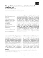
Fig. 3. Solution structure of fowlicidin-1. (A) Ribbon stereo-diagram
of the restrained minimized average structure of fowlicidin-1. (B)
Stereo-diagrams of the backbone trace of the 20 lowest energy
structures of fowlicidin-1, with residues 8–16 overlaid. (C) Stereodiagrams of the backbone trace of the 20 lowest energy structures
of fowlicidin-1, with residues 17–25 overlaid. This figure was generated using MOLMOL.
flexibility in between (Fig. 3B,C). The superimposition
of the two short helical segments of the 20 final structures against an averaged structure resulted in a rmsd
˚
value of backbone of < 0.5 A (Table 1). Greater flexibility between the helices was revealed when only one
2584
I23
Hyd
ro
phob ic
Hydr o
philic-
K7
C
L16
V9
R21
I10
Y20
Y17
W6
V13
Fowlicidin-1(6-23)-KLKLK
Fig. 4. Helical wheel projections of the central helical regions
(residues 6–23) of fowlicidin-1 (A) and its substitution mutant,
fowlicidin-1-K7L12K14L16K18 (B). The representation shows the
amphipathic structure of the helical region. Charged residues are
indicated on a black background, and polar uncharged residues are
on a gray background. The mutated residues are circled. Note a
significant enhancement in amphipathicity of the mutant peptide
relative to the native peptide.
FEBS Journal 273 (2006) 2581–2593 ª 2006 The Authors Journal compilation ª 2006 FEBS
Y. Xiao et al.
Structure–activity relationships of fowlicidin-1
Table 2. Fowlicidin-1 and its analogs.
Mass
Peptide
Sequence
Charge
Length
Calculated
Observed
Fowlicidin-1 (1–26)
Fowl-1 (1–15)
Fowl-1 (1–23)
Fowl-1 (8–26)
Fowl-1 (5–26)
Fowl-1-L16
Fowl1-K7L12K14L16K18
RVKRVWPLVIRTVIAGYNLYRAIKKK
RVKRVWPLVIRTVIA
RVKRVWPLVIRTVIAGYNLYRAI
LVIRTVIAGYNLYRAIKKK
VWPLVIRTVIAGYNLYRAIKKK
RVKRVWPLVIRTVIALYNLYRAIKKK
RVKRVWKLVIRLVKALYKLYRAIKKK
+8
+4
+5
+5
+5
+8
+11
26
15
23
19
22
26
26
3141.9
1807.3
2758.4
2220.8
2603.2
3199.0
3271.2
3141.6
1807.6
2757.2
2220.9
2600.3
3197.3
3271.1
instead highly concentrated at both ends. To probe the
impact of N- and C-terminal cationic regions and two
short helical segments on antibacterial, LPS-binding,
and cytolytic activities of fowlicidin-1, several N- and
C-terminal deletion mutants were designed (Table 2).
All mutants have fewer net positive charges than the
parent peptide, in addition to missing one or two
structural components.
To investigate further the influence of helicity on the
functional properties, Gly16 of fowlicidin-1 was
replaced with a helix-stabilizing residue, leucine, to
give rise to fowlicidin-1-L16. Such a variant minimized
the bend and flexibility between two short helices, as
modeled by modeller [20] (data not shown), without
significantly altering any other structural or physicochemical characteristics. Another substitution variant,
fowlicidin-1-K7L12K14L16K18, was designed mainly
for significant augmentation of its amphipathicity. This
mutant has cationic residues clearly aligned along one
side and hydrophobic residues aligned along the
opposite side of the helix (compare Fig. 4A with 4B).
The net charge of this mutant increased from +8 to
+11, as compared with the parent peptide. Replacement of two helix-breaking residues, Pro7 and Gly16,
with helix-stabilizing residues, lysine and leucine,
respectively, also enhanced the helical content of fowlicidin-1-K7L12K14L16K18 by concomitant reduction
of the kink in the center and extension of the helix at
the N terminus. Along with simultaneous enhancement
of amphipathicity, cationicity and helicity, it is understandable that such a peptide variant also has reduced
hydrophobicity in the helical region as a result of
incorporation of several positively charged residues.
Consistent with the modeling results, two substitution
mutants showed increased a-helical contents in the
presence of 50% TFE by CD spectroscopy, relative to
the parent peptide (data not shown).
All peptides were synthesized commercially by the
standard solid-phase method and ordered at > 95%
purity. The molecular mass and purity of each synthetic
peptide were further confirmed by MS (Table 2).
Antibacterial activities of fowlicidin-1
and its analogs
Two representative Gram-negative bacteria (Escherichia coli ATCC 25922 and Salmonella enterica serovar
Typhimurium ATCC 14028) and two Gram-positive
bacteria (Listeria monocytogenes ATCC 19115 and Staphylococcus aureus ATCC 25923) were used to test the
antibacterial potency of fowlicidin-1 and its analogs in a
modified broth microdilution assay, as described previously [16,21]. Compared with the parent peptide, the
analog with deletion of three C-terminal lysines [fowlicidin-1(1–23)], or of four [fowlicidin-1(5–26)] or seven
[fowlicidin-1(8–26)] N-terminal residues, retained much
of the bactericidal activity (Table 3), suggesting that the
cationic residues at both ends are dispensable for its
antibacterial activity, but all or part of the central
hydrophobic a-helical region between residues 8 and 23
plays a major role in killing bacteria. However, the
peptide analog that is composed entirely of the central
hydrophobic a-helix (residues 8–23), with a net charge
of +2, became insoluble in 0.01% acetic acid and
therefore was excluded from antibacterial assays.
To examine further the differential role of the
N- and C-terminal short helical segments in antibacterial potency, fowlicidin-1(1–15), with omission of the
C-terminal helical region after the kink at Gly16, was
tested against the four bacterial strains and was found
to have a less than twofold reduction in minimum inhibitory concentration (MIC) towards Gram-negative bacteria, but a seven- to 18-fold reduction in MIC towards
Gram-positive bacteria (Table 3), suggesting that the
C-terminal short helix (residues 16–23) is critical in
maintaining antibacterial potency against Gram-positive but not Gram-negative bacteria. This is consistent
with earlier observations that activity of cationic antimicrobial peptides against Gram-negative bacteria is generally more tolerant to structural changes [10].
In contrast to our expectations, two substitution mutants (fowlicidin-1-L16 and fowlicidin-1K7L12K14L16K18) with significant improvement in
FEBS Journal 273 (2006) 2581–2593 ª 2006 The Authors Journal compilation ª 2006 FEBS
2585
Structure–activity relationships of fowlicidin-1
Y. Xiao et al.
Table 3. Functional properties of fowlicidin-1 and its analogs. EC50, 50% effective concentration; MIC, minimum inhibitory concentration;
LPS, lipopolysaccharide.
Antibacterial activity (MIC, lM)
Cytolytic activity (EC50, lM)
LPS-binding activity
Peptide
S. aureus
Listeria
Salmonella
E. coli
Hemolytic
Cytotoxic
(EC50, lM)
Fowlicidin-1 (1–26)
Fowl-1 (1–15)
Fowl-1 (1–23)
Fowl-1 (8–26)
Fowl-1 (5–26)
Fowl-1-L16
Fowl-1-KLKLK
0.5
13.8
1.1
2.8
0.6
2.0
1.9
2.0
13.8
2.3
5.6
2.4
3.9
> 7.6
2.0
3.5
2.3
2.8
2.4
2.0
1.9
4.0
6.9
4.5
5.6
4.8
7.8
> 7.6
6
> 443
38
> 360
11
3
1
15
> 443
40
159
9
15
11
11
> 443
39
> 260
10
9
6
helicity, amphipathicity and ⁄ or cationicity, were found
to have reduced antibacterial activity relative to the
wild-type peptide (Table 3), reinforcing the notion that
an intricate balance, rather than a simple enhancement
in those structural parameters, dictates the antibacterial potency of the a-helical antimicrobial peptides
[10,11]. It is noteworthy that all peptide analogs
showed similar kinetics in killing bacteria as the fulllength peptide, with maximal activities being reached
30 min after incubation with bacteria in the presence
or absence of 100 mm NaCl (data not shown). It is
not clear why fowlicidin-1-K7L12K14L16K18 largely
maintained its potency against S. aureus and Sal. enterica serovar Typhimurium, but failed to completely
inhibit the growth of E. coli and L. monocytogenes,
even at the highest concentration (7.6 lm ẳ
25 lgặmL)1) tested.
Cytotoxicity of fowlicidin-1 and its analogs
To map the region that is responsible for the lysis of
eukaryotic cells and to identify a peptide analog with
reduced cytolytic activity, all deletion and substitution
mutants of fowlicidin-1 were tested individually against
human erythrocyte and Madin-Darby canine kidney
cells (MDCK) for their toxicity, as previously described [13,16,22]. As summarized in Table 3, Fowlicidin-1 exhibited considerable toxicity towards
erythrocytes and epithelial cells with 50% effective
concentrations (EC50) in the range of 6–15 lm. Deletion of the last three lysines [fowlicidin-1(1–23)] resulted in a modest (less than fourfold) reduction in
toxicity, while truncation of the entire C-terminal short
helix [fowlicidin-1(1–15)] caused the almost complete
loss of lytic activity towards both erythrocytes and
epithelial cells, indicating that the C-terminal helix
(residues 16–23), but not the last three lysines, is a critical determinant of cytotoxcity.
Relative to the full-length peptide, fowlicidin-1(5–26)
maintained a similar lytic activity, whereas fowlicidin2586
1(8–26) only caused minimal 20% lysis of human red
blood cells at 360 lm, the highest concentration tested
(data not shown), suggesting the possible presence of
another cytotoxicity determinant in the N-terminal
unstructured segment between residues 5 and 7. Consistent with these results, a significant, > 10-fold
reduction, in the killing of MDCK cells was also
observed with fowlicidin-1(8–26) (Table 3). Because of
the fact that two peptide analogs, fowlicidin-1(1–15)
and fowlicidin-1(8–26), each containing one cytolytic
determinant, had substantially reduced toxicity, it is
likely that the two lytic sites (residues 5–7 and 16–23)
act in a synergistic manner in the lysis of eukaryotic
cells (i.e. the presence of one determinant facilitates
the action of the other).
The single substitution of Gly16 for leucine (fowlicidin-1-L16) did not lead to any obvious alterations in
the killing of eukaryotic cells (Table 3). In contrast,
fowlicidin-1-K7L12K14L16K18, with a nearly perfect
amphipathic helix in the center, showed a sixfold
increase in the lysis of red blood cells, but only slightly
higher lytic activity against mammalian epithelial cells
(Table 3). This suggested that the amphipathic helix
has a stronger binding affinity and permeability
towards erythrocyte membranes than to epithelial
membranes, perhaps as a result of the difference in the
lipid composition of the two host cell types.
LPS-binding activity of fowlicidin-1
and its analogs
Binding and disrupting anionic LPS, the major outer
membrane component of Gram-negative bacteria, is
often the first step for antimicrobial peptides to interact with bacteria and permeabilize membranes [10].
Several cathelicidins, including human LL-37 ⁄ hCAP18 [21,23], rabbit CAP-18 [24] and sheep SMAP-29
[25], have been shown to bind and neutralize LPS with
an EC50 at low micromolar concentrations. We have
also demonstrated that fowlicidin-1 has at least two
FEBS Journal 273 (2006) 2581–2593 ª 2006 The Authors Journal compilation ª 2006 FEBS
Y. Xiao et al.
Structure–activity relationships of fowlicidin-1
LPS-binding sites [16]. To map the regions involved in
the binding of fowlicidin-1 to LPS, the N- and C-terminal deletion mutants were mixed with LPS, and
their ability to bind LPS and to inhibit LPS-mediated
procoagulant activation was measured by a chromogenic Limulus amoebocyte assay [21,25]. As shown in
Fig. 5A, fowlicidin-1(1–23) and fowlicidin-1(5–26) had
similar affinities for LPS to the full-length peptide,
with an EC50 in the range of 10–39 lm (Table 3),
suggesting that LPS-binding sites are likely to be
located in the central helical region between residues 5
and 23.
Residues 5–7 are clearly involved in LPS binding
and may constitute the core region of one LPS-binding
site, because fowlicidin-1(8–26) showed a > 15-fold
LPS Binding (%)
A
100
75
50
25
0
0.5
5
50
500
Peptide (µM)
LP S B i ndi ng ( % )
B
100
80
reduction in binding to LPS relative to fowlicidin-1(5–
26), which had a similar affinity for LPS to the fulllength peptide. The other LPS-binding site is probably
located in the C-terminal short helix between residues
16 and 23, because deletion of that region [fowlicidin1(1–15)] resulted in a > 25-fold reduction in LPS binding, as compared with fowlicidin-1(1–23) (Fig. 5A,
Table 3). It is important to note that two LPS-binding
sites of fowlicidin-1 are located in the same regions
where the two cytotoxicity determinants reside. This is
perhaps not surprising, given that sequences which
interact with anionic LPS or phospholipids on bacterial membranes are probably involved in interactions
with eukaryotic cell membranes, which is a prerequisite
for cytotoxicity. In fact, the hemolytic domain of
SMAP-29 was also shown to overlap with an LPSbinding site at the C-terminal end [25].
To determine whether the two LPS-binding sites act
in a synergistic manner, an equimolar mixture of fowlicidin-1(1–15) and fowlicidin-1(8–26), each containing
one LPS-binding site, was incubated with LPS and
measured for the ability to bind to LPS. As shown in
Fig. 5A, the mixture displayed an enhanced affinity for
LPS, approaching that of the full-length peptide, indicative of the synergistic nature of the two LPS-binding
sites. Both substitution mutants, fowlicidin-1-L16 and
fowlicidin-1-K7L12K14L16K18,
showed
minimal
changes in LPS-binding affinity, relative to the native
peptide (Fig. 5B), suggesting that a simultaneous
enhancement in helicity, cationicity and amphipathicity
has little impact on the interactions of peptides with
LPS and possibly also with bacterial membranes,
which may explain why the antibacterial activities of
both mutants remained largely unchanged (Table 3).
60
Discussion
40
20
0
0.1
1
10
100
Peptide (µM)
Fig. 5. Lipopolysaccharide (LPS)-binding isotherms of the deletion
(A) and substitution (B) mutants of fowlicidin-1. The 50% effective
concentration (EC50 value), indicated by a dotted line in each panel,
was defined as the peptide concentration that inhibited LPS-mediated procoagulant activation by 50%. Panel A: n, fowlicidin-1(1–26);
s, fowlicidin-1(8–26); n, fowlicidin-1(1–15); m, fowlicidin-1(5–26); r,
fowlicidin-1(1–23); d, fowlicidin-1(8–26) + fowlicidin-1(1–15). Panel
B: n, fowlicidin-1(1–26); m, fowlicidin-1-L16; and ., fowlicidin-1KLKLK. Data shown represent the means ± SEM of three independent experiments.
Cathelicidins are highly conserved from birds to mammals in the prosequence, but are extremely divergent
in the C-terminal mature sequence [1–3]. Cathelicidinlike molecules have also been found in the hagfish, the
most ancient extant jawless fish with no adaptive
immune system [26]. With the finding that fowlicidin-1
adopts an a-helix (Fig. 3), it is now evident that at
least one cathelicidin in the a-helical conformation is
present in each of the fish, bird and mammalian species examined. This suggests that, in addition to the
prosequence, cathelicidins appear to be conserved in
the mature region structurally and presumably also
functionally. It is plausible that the presence of additional structurally different cathelicidins in certain animal species may help the hosts to cope better with
unique microbial insults in the ecological niche where
FEBS Journal 273 (2006) 2581–2593 ª 2006 The Authors Journal compilation ª 2006 FEBS
2587
Structure–activity relationships of fowlicidin-1
Y. Xiao et al.
each species inhabits, given the fact that different cathelicidins appear to possess a nonoverlapping antimicrobial spectrum [6] and that some act synergistically in
combinations in killing microbes [7]. On the other
hand, the innate host defense of animal species (such
as primates and rodents) which contain a single cathelicidin, may be compensated for by the presence of a
large number of other antimicrobial peptides such as
a- and b-defensins [27,28]. Conversely, pig and cattle
have multiple cathelicidins, but no a-defensins have
been reported.
Our NMR studies revealed that, in addition to a
short flexible unstructured region at the N terminus,
fowlicidin-1 is primarily composed of two short a-helical segments connected by a slight kink caused by
Gly16 near the center (Fig. 3). Interestingly, such a
helix–hinge–helix structural motif is not uncommon for
cathelicidins. Mouse cathelicidin CRAMP [22], bovine
BMAP-34 [29] and porcine PAMP-37 [30] all adopt a
helix–hinge–helix structure, with the hinge occurring at
the central glycine (Fig. 6). In fact, none of the linear,
naturally occurring cathelicidins are strictly a-helical.
Besides peptides with helix–hinge–helix structures, a
few other linear cathelicidins consist of an N-terminal
helix followed by nonhelical and mostly hydrophobic
tails, such as rabbit CAP-18 [31], sheep SMAP-29 [25],
and bovine BMAP-27 and -28 [12] (Fig. 6).
In addition to cathelicidins, a scan of over 150
helical antimicrobial peptides revealed that glycine is
frequently found near the center and acts as a hinge to
increase flexibility in many other protein families [10]
(Fig. 6). The presence or insertion of such a hinge in
the helix has been shown, in many cases, to be desirable, attenuating the toxicity of peptides to host cells
while maintaining comparable antimicrobial potency
with the peptides that have no hinge sequences [10,11].
Mutation of the hinge sequence with a helix-stabilizing
residue, such as leucine, will generally result in an
increase in cytotoxicity and, in several cases, anti
microbial potency. However, substituting Gly16 of
fowlicidin-1-L16 for leucine did not enhance the antibacterial or cytolytic activity (Table 3), probably as a
result of the relatively low flexibility of the wild-type
peptide.
A careful comparison of fowlicidin-1 with other
a-helical cathelicidins indicated that the a-helix (residues
8–23) of fowlicidin-1 is much more hydrophobic and
much less amphipathic than most of the mammalian
cathelicidins (Fig. 6). The positive charges of fowlicidin-1 are more concentrated in the nonhelical regions
at both ends. Because high hydrophobicity is often
associated with strong cytotoxicity [10,11], it is perhaps
not surprising to see that fowlicidin-1 is relatively more
toxic than many other cathelicidins. Interestingly, fowlicidin-1 is structurally more similar to melittin, a helical peptide found in honey bee venom that has a
curved hydrophobic helix with positively charged residues located primarily at the C-terminal end [32]
(Fig. 6). Like fowlicidin-1, melittin displays considerable antibacterial and hemolytic activities. An attempt
to reduce the hydrophobicity and enhance the amphipathicity of the helical region of fowlicidin-1 to make
fowlicidin-1-K7L12K14L16K18 led to a dramatically
increased toxicity, particularly towards erythrocytes,
with a minimum change in the antibacterial activity
against certain bacteria (Table 3). This is consistent with
an earlier conclusion that an amphipathic helix is more
essential for interactions with zwitteronic lipid membranes on eukaryotic cells than for anionic lipids on
prokaryotic cells [33].
Fig. 6. Alignment of representative linear a-helical antimicrobial peptides demonstrating the conservation of a kink induced by glycine near
the center. Putatively mature fowlicidin-1 sequence is aligned with representative cathelicidins (mouse CRAMP, rabbit CAP18, bovine
BMAP34 and BMAP28, sheep SMAP34 and SMAP29, and porcine PMAP37) as well as three insect peptides (fruit fly cecropin A1, a putative porcine cecropin P1, and honey bee melittin). Dashes are inserted to optimize the alignment, and conserved residues are shaded. Note
that each peptide aligned has an a-helix N-terminal to the conserved glycine (boxed) near the center, followed by either a helical or an
unstructured tail. The only exception is CRAMP, which has a kink at Gly11 instead of Gly18 [22].
2588
FEBS Journal 273 (2006) 2581–2593 ª 2006 The Authors Journal compilation ª 2006 FEBS
Y. Xiao et al.
Fig. 7. Schematic drawing of the distribution of functional determinants of fowlicidin-1. Note that the C-terminal helix from Gly16 to
Ile23 is indispensable for antibacterial, cytolytic and lipopolysaccharide (LPS)-binding activities, whereas the three residues (Val5–Pro7)
in the N-terminal unstructured region constitute the core of the second determinant that is critically involved in cytotoxicity and LPS
binding, but less significant in the bactericidal activity. The N-terminal helix (Leu8–Ala15) also presumably facilitates the interactions
of the C-terminal helix (Gly16–Ile23) with lipid membranes.
Our SAR data revealed the regions that are
responsible for each of the antibacterial, LPS-binding
and cytolytic activities of fowlicidin-1 (Fig. 7). The
C-terminal a-helix after the kink (residues 16–23), consisting of a stretch of eight amino acids, is required for
all three functions, suggesting that this region is probably a major site for the peptide to interact with LPS
and lipid membranes and to permeabilize both bacterial and eukaryotic cells. It is not surprising to see the
presence of two lipophilic tyrosines (Tyr17 and Tyr20)
that might be critical in mediating membrane interactions for fowlicidin-1. However, the a-helix before the
kink at Gly16 is also likely to be involved in membrane penetration, because the minimum length
required for a helical peptide to traverse membranes
and exert antimicrobial and lytic activities is % 11–14
residues [34].
Another region, comprising three amino acids in the
N-terminal flexible region (residues 5–7), is also
involved in both LPS binding and cytotoxicity, but is
not so important in bacterial killing (Fig. 7). It is interesting to note that among the three residues in this
region, it is Trp6 which is known to have a preference
for insertion into lipid bilayers at the membrane–water
interface [35,36]. Because of such membrane-seeking
ability, inclusion of tryptophan often renders peptides
with a higher affinity for membranes and more
potency against bacteria [37,38]. It is not known why
tryptophan is not significantly involved in the antibacterial activity of fowlicidin-1.
It is noteworthy that the N-terminal helix of many
cathelicidins plays a major role in LPS binding and
bacterial killing, while the C-terminal segment is either
dispensable for antimicrobial activity or more involved
in cytotoxicity [12,25,39,40]. However, the C-terminal
helix after the kink of fowlicidin-1 is more important
Structure–activity relationships of fowlicidin-1
in killing bacteria than the N-terminal helix. Such a
marked difference in the distribution of functional
domains along the peptide chain between fowlicidin-1
and other cathelicidins is probably because of a more
pronounced hydrophobic nature of the helix and the
presence of an additional highly flexible segment at the
N terminus of fowlicidin-1.
One aim of our study was to identify peptide analog(s) with better therapeutic potential. Fowlicidin-1(1–
23) and fowlicidin-1(5–26) had only a marginal effect
on either antibacterial potency or cytotoxicity, whereas
fowlicidin-1(1–15) exhibited minimal toxicity up to
443 lm, but with an obvious decrease in antibacterial
activity particularly against Gram-positive bacteria,
implying less desirable therapeutic relevance of these
peptide analogs as a broad-spectrum antibiotic. Fowlicidin-1-L16 and fowlicidin-1-K7L12K14L16K18 also
had a more pronounced reduction in antibacterial
activity than in toxicity, therefore with reduced clinical
potential. In contrast, fowlicidin-1(8–26) with the
N-terminal toxicity determinant (residues 5–7) deleted
and the C-terminal antibacterial domain (residues
16–23) left unaltered, had a slight reduction in MIC
against bacteria, but with > 10-fold reduction in toxicity towards mammalian epithelial cells and negligible
toxicity towards erythrocytes (Table 3). Coupled with
its smaller size, this peptide analog may represent a
safer and more attractive therapeutic candidate than
the parent peptide. Given the fact that fowlicidin-1 is
broadly effective against several common bacterial
strains implicated in cystic fibrosis, including S. aureus,
Klebiella pneumoniae and Pseudomonas aeruginosa, in a
salt-independent manner [16], its analog, fowlicidin1(8–26), might prove useful in controlling chronic respiratory infections of cystic fibrosis patients. These
results also suggested the usefulness of systematic SAR
studies in improving the safety and target specificity of
antimicrobial peptides.
Experimental procedures
Peptide synthesis
Fowlicidin-1 was synthesized using the standard solid-phase
method of SynPep (Dublin, CA, USA) and its analogs were
synthesized by either Sigma Genosys (Woodlands, TX,
USA) or Bio-Synthesis (Lewisville, TX, USA) (Table 1).
The peptides were purified through RP-HPLC and purchased at > 95% purity. The mass and purity of each peptide were further confirmed by 15% Tris-Tricine PAGE
(data not shown) and by MALDI-TOF MS (Table 1) using
the Voyager DE-PRO instrument (Applied Biosystems,
Foster City, CA, USA) housed in the Recombinant
FEBS Journal 273 (2006) 2581–2593 ª 2006 The Authors Journal compilation ª 2006 FEBS
2589
Structure–activity relationships of fowlicidin-1
Y. Xiao et al.
DNA ⁄ Protein Resource Facility of Oklahoma State University.
CD spectroscopy
To determine the secondary structure of fowlicidin-1, CD
spectroscopy was performed with a Jasco-715 spectropolarimeter (JASCO, Tokyo, Japan), using a 0.1-cm path length
cell over the 180–260 nm range, as previously described
[41]. The spectra were acquired at 25 °C every 1 nm with a
2 s averaging time per point and a 1 nm band pass. Fowlicidin-1 (10 lm) was measured in 50 mm potassium phosphate buffer, pH 7.4, with or without different
concentrations of TFE (0, 10, 20, 40 and 60%) or SDS micelles (0.25 and 0.5%). Mean residue ellipticity (MRE) was
expressed as [h]MRE (degỈcm)2Ỉdmol)1). The contents of six
types of the secondary structural elements, including regular
and distorted a-helix, regular and distorted b-sheet, turns,
and unordered structures, were analyzed with the program
selcon3 using a 29-protein data set of basic spectra [42].
NMR spectroscopy
2D[1H-1H] NMR experiments for fowlicidin-1 were performed as previously described [43,44]. Briefly, NMR data
were acquired on an 11.75T Varian UNITYplus spectrometer (Varian, Palo Alto, CA, USA), operating at
500 MHz for 1H, with a 3-mm triple-resonance inverse
detection probe. The NMR sample of fowlicidin-1, consisting of 4 mm in water containing 50% deuterated TFE
(TFE-d3; Cambridge Isotope Laboratories, Andover, MA,
USA) and 10% D2O, was used to record spectra at 10, 20,
30 and 35 °C. The spectra acquired at 35 °C were determined to provide the optimal resolution of overlapping
NMR resonances. These spectra were processed and analyzed using Varian software, vnmr Version 6.1C, on a
Silicon Graphics (Mountain View, CA, USA) Octane workstation. The invariant nature of the NMR chemical shifts
and line widths upon 10-fold dilution indicated that fowlicidin-1 was monomeric in solution at the concentration used
for 2D NMR analysis. A total of 512 increments of 4K
data points were collected for these 2D NMR experiments.
The high digital resolution DQF-COSY spectra were recorded using 512 increments and 8K data points in t1 and t2
dimensions. Sequential assignments were carried out by
comparison of cross-peaks in a NOESY spectrum with
those in a TOCSY spectrum acquired under similar experimental conditions. NOESY experiments were performed
with 200, 300, 400 and 500 ms mixing times. A mixing time
of 200 ms was used for distance constraints measurements.
The NOE cross-peaks were classified as strong, medium,
weak and very weak based on an observed relative number
of contour lines. TOCSY spectra were recorded by using
MLEV-17 for isotropic mixing for 35 and 100 ms at a B1
field strength of 7 KHz.
2590
Water peak suppression was obtained by low-power irradiation of the water peak during relaxation delay. The residual
TFE methylene peak was considered as a reference for the
chemical shift values. The temperature dependences of amide
proton chemical shifts were measured by collecting data from
10 °C to 35 °C in steps of 5 °C by using a variable temperature probe. All experiments were zero-filled to 4K data points
in the t1 dimension and, when necessary, the spectral resolution was enhanced by Lorenzian-Gaussian apodization.
Structure calculations
For structure calculations, NOE-derived distance restraints
were classified into four ranges (1.8–2.7, 1.8–3.5, 1.8–4.0
˚
and 1.8–5.0 A) according to the strong, medium, weak and
very weak NOE intensities. Upper distance limits for
NOEs, involving methyl protons and nonstereospecifically
assigned methylene protons, were corrected appropriately
˚
for center averaging [45]. In addition, a distance of 0.5 A
was added to the upper distance limits only for NOEs
involving the methyl proton after correction for center
averaging [46]. The distance restraints were then used to
create initial peptide structures starting from extended
structures using the program cns (version 1.1) [47]. cns
uses both a simulated annealing protocol and molecular
dynamics to produce low energy structures with the minimum distance and geometry violations. In general, default
parameters supplied with the program were used with 100
structures for each cns run. The final round of calculations
began with 100 initial structures, and 20 best structures
with the lowest energy were selected and analyzed with
molmol [48] and procheck-nmr [19]. Structure figures
were generated by using molmol. The structures of fowlicidin-1 analogs were further modeled by using modeller
[20], based on the parent peptide.
Antibacterial assay
Two representative species of Gram-negative bacteria
(E. coli ATCC 25922 and S. enterica serovar Typhimurium
ATCC 14028) and two representative species of Gram-positive bacteria (L. monocytogenes ATCC 19115 and S. aureus
ATCC 25923) were purchased from the ATCC (Manassas,
VA, USA) and tested separately against fowlicidin-1 and its
analogs by using a modified broth microdilution assay, as
described previously [16,21]. Briefly, overnight cultures of
bacteria were subcultured for an additional 3–5 h at 37 °C
in trypticase soy broth to the mid-log phase, washed with
10 mm sodium phosphate buffer, pH 7.4, and suspended to
5 · 105 colony-forming units per mL in 1% cation-adjusted
Mueller Hinton broth (BBL, Cockeysville, MD, USA),
which was prepared by a 1 : 100 dilution of conventional
strength Mueller Hinton broth in 10 mm phosphate buffer.
If necessary, 100 mm NaCl was added to test the influence
of salinity on antibacterial activity. Bacteria (90 lL) were
FEBS Journal 273 (2006) 2581–2593 ª 2006 The Authors Journal compilation ª 2006 FEBS
Y. Xiao et al.
Structure–activity relationships of fowlicidin-1
then dispensed into 96-well plates, followed by the addition,
in duplicate, of 10 lL of serially diluted peptides in 0.01%
acetic acid. After overnight incubation at 37 °C, the MIC
value of each peptide was determined as the lowest concentration that gave no visible bacterial growth. The antibacterial assays were repeated at least three or four times for
each bacterial strain, with less than a twofold difference in
MIC values observed in all cases, and representative MIC
values are tabulated in Table 3.
Hemolysis assay
The hemolytic activity of fowlicidin-1 and its mutants were
determined essentially as described previously [13,22].
Briefly, fresh anticoagulated human blood was collected,
washed twice with NaCl ⁄ Pi, diluted to 0.5% in NaCl ⁄ Pi,
and 90 lL was dispensed into 96-well plates. Serial twofold
dilutions of peptides were added in duplicate to erythrocytes and incubated at 37 °C for 2 h. Following centrifugation at 800 g for 10 min, the supernatants were transferred
to new 96-well plates and monitored by measuring the
absorbance (A) at 405 nm for released hemoglobin. Controls for 0 and 100% hemolysis consisted of cells suspended
in NaCl ⁄ Pi only and in 1% Triton X-100, respectively. Percentage hemolysis (%) was calculated as follows:
Percentage hemolysis %ị ẳ ẵA405nm;peptide A405nm;NaCl=Pi Þ=
ðA405nm;1%TritonXÀ100 À
A405nm;NaCl=Pi Þ Â 100:
The EC50 of the hemolytic activity was defined as the peptide concentration that caused 50% lysis of erythrocytes.
Cytotoxicity assay
The toxic effect of fowlicidin-1 and its analogs on mammalian epithelial cells was evaluated with MDCK cells by using
alamarBlue dye (Biosource, Carlsbad, CA, USA) as previously described [16]. Briefly, cells were seeded into 96-well
plates at 1.5 · 105 cells per well and allowed to grow overnight in Dulbecco’s modified Eagle’s medium (DMEM) containing 10% fetal bovine serum. Cells were then washed once
with DMEM, followed by the addition of 90 lL of fresh
DMEM, together with 10 lL of serially diluted peptides in
0.01% acetic acid in triplicate. After incubation for 18 h,
10 lL of alamarBlue dye was added to cells for 6 h at 37 °C
in a humidified 5% CO2 incubator before the fluorescence
was read with excitation at 545 nm and emission at 590 nm.
Percentage cell death was calculated as follows:
Percentage cell death ẳ ẵ1 Fpeptide Fbackground ị=
Faceticacid À Fbackground Þ Â 100;
where Fpeptide is the fluorescence of cells exposed to different concentrations of peptides, Facetic acid is the fluorescence
of cells exposed to 0.01% acetic acid only, and Fbackground
is the background fluorescence of 10% alamarBlue dye in
cell culture medium without cells. Cytotoxicity (EC50) of
individual peptides was defined as the peptide concentration
that caused 50% cell death.
LPS-binding assay
The binding of LPS to fowlicidin-1 and its analogs was
measured by a kinetic chromogenic Limulus amebocyte lysate assay (Kinetic-QCL 1000 kit; BioWhittaker, Walkersville, MD, USA), as previously described [21,25]. Briefly,
25 lL of serially diluted peptide was added in duplicate to
25 lL of E. coli O111:B4 LPS containing 0.5 endotoxin
unitsỈmL)1 and incubated for 30 min at 37 °C, followed by
incubation with 50 lL of the amoebocyte lysate reagent for
10 min. The absorbance at 405 nm was measured at 10 and
16 min after the addition of 100 lL of chromogenic substrate, Ac-Ile-Glu-Ala-Arg-p-nitroanilide. Percentage LPS
binding was calculated as follows:
Percentage LPS binding ẳ ẵDD1 DD2 ỵ DD3Þ=DD1 Â 100;
where DD1 represents the difference in absorbance between
10 and 16 min for the sample containing LPS only, DD2
represents the difference in absorbance between 10 and
16 min for the samples containing LPS and different concentrations of peptides, and DD3 represents the difference
in absorbance between 10 and 16 min for the samples containing different concentrations of peptides with no LPS.
The EC50 of the LPS-binding activity was defined as the
peptide concentration that inhibited LPS-mediated procoagulant activation by 50%.
Protein Data Bank accession code
The atomic co-ordinates and structural factors of putatively
mature fowlicidin-1 have been deposited under accession
code 2AMN in the Protein Data Bank, Research Collaboratory for Structural Bioinformatics, Rutgers University,
New Brunswick, NJ ( />
Acknowledgements
This work was supported by grants from the National
Science Foundation (Grants MCB0236039 and
EPS0236913), Oklahoma Center for the Advancement
of Science and Technology (Grant HR03-146), and
Oklahoma Agricultural Experiment Station (Project
H-2507).
We are grateful to Ulrich Melcher, Chang-An Yu,
Michael Massiah, Rodney Geisert, and anonymous
reviewers for critical reading of the manuscript and
constructive comments. We also thank Steve Hartson
for helping with mass spectrometry and Amar Patil for
Tris-Tricine polyacrylamide gel electrophoresis.
FEBS Journal 273 (2006) 2581–2593 ª 2006 The Authors Journal compilation ª 2006 FEBS
2591
Structure–activity relationships of fowlicidin-1
Y. Xiao et al.
References
1 Zanetti M (2004) Cathelicidins, multifunctional peptides
of the innate immunity. J Leukoc Biol 75, 39–48.
2 Zaiou M & Gallo RL (2002) Cathelicidins, essential
gene-encoded mammalian antibiotics. J Mol Med 80,
549–561.
3 Lehrer RI & Ganz T (2002) Cathelicidins: a family of
endogenous antimicrobial peptides. Curr Opin Hematol
9, 18–22.
4 Zasloff M (2002) Antimicrobial peptides of multicellular
organisms. Nature 415, 389–395.
5 Hancock RE & Patrzykat A (2002) Clinical development of cationic antimicrobial peptides: from natural to
novel antibiotics. Curr Drug Targets Infect Disord 2,
79–83.
6 Zanetti M, Gennaro R, Skerlavaj B, Tomasinsig L &
Circo R (2002) Cathelicidin peptides as candidates for a
novel class of antimicrobials. Curr Pharm Des 8, 779–793.
7 Yan H & Hancock RE (2001) Synergistic interactions
between mammalian antimicrobial defense peptides.
Antimicrob Agents Chemother 45, 1558–1560.
8 Levy O, Ooi CE, Weiss J, Lehrer RI & Elsbach P
(1994) Individual and synergistic effects of rabbit granulocyte proteins on Escherichia coli. J Clin Invest 94,
672–682.
9 Nagaoka I, Hirota S, Yomogida S, Ohwada A & Hirata
M (2000) Synergistic actions of antibacterial neutrophil
defensins and cathelicidins. Inflamm Res 49, 73–79.
10 Tossi A, Sandri L & Giangaspero A (2000) Amphipathic, alpha-helical antimicrobial peptides. Biopolymers
55, 4–30.
11 Dathe M & Wieprecht T (1999) Structural features of
helical antimicrobial peptides: their potential to
modulate activity on model membranes and biological
cells. Biochim Biophys Acta 1462, 71–87.
12 Skerlavaj B, Gennaro R, Bagella L, Merluzzi L, Risso
A & Zanetti M (1996) Biological characterization of
two novel cathelicidin-derived peptides and identification of structural requirements for their antimicrobial
and cell lytic activities. J Biol Chem 271, 28375–
28381.
13 Shin SY, Park EJ, Yang ST, Jung HJ, Eom SH, Song
WK, Kim Y, Hahm KS & Kim JI (2001) Structureactivity analysis of SMAP-29, a sheep leukocyte-derived
antimicrobial peptide. Biochem Biophys Res Commun
285, 1046–1051.
14 van Dijk A, Veldhuizen EJ, van Asten AJ & Haagsman
HP (2005) CMAP27, a novel chicken cathelicidin-like
antimicrobial protein. Vet Immunol Immunopathol 106,
321–327.
15 Lynn DJ, Higgs R, Gaines S, Tierney J, James T, Lloyd
AT, Fares MA, Mulcahy G & O’Farrelly C (2004) Bioinformatic discovery and initial characterisation of nine
2592
16
17
18
19
20
21
22
23
24
25
26
27
28
novel antimicrobial peptide genes in the chicken. Immunogenetics 56, 170–177.
Xiao Y, Cai Y, Bommineni YR, Fernando SC, Prakash
O, Gilliland SE & Zhang G (2006) Identification and
functional characterization of three chicken cathelicidins
with potent antimicrobial activity. J Biol Chem 281,
2858–2867.
Wuthrich K (1986) NMR of Proteins and Nucleic Acids.
John Wiley and Sons Inc., New York.
Wishart DS, Sykes BD & Richards FM (1992) The chemical shift index: a fast and simple method for the
assignment of protein secondary structure through
NMR spectroscopy. Biochemistry 31, 1647–1651.
Laskowski RA, Rullmannn JA, MacArthur MW,
Kaptein R & Thornton JM (1996) AQUA and PROCHECK-NMR: programs for checking the quality of
protein structures solved by NMR. J Biomol NMR 8,
477–486.
Fiser A & Sali A (2003) Modeller: generation and
refinement of homology-based protein structure models.
Methods Enzymol 374, 461–491.
Turner J, Cho Y, Dinh NN, Waring AJ & Lehrer RI
(1998) Activities of LL-37, a cathelin-associated antimicrobial peptide of human neutrophils. Antimicrob
Agents Chemother 42, 2206–2214.
Yu K, Park K, Kang SW, Shin SY, Hahm KS & Kim
Y (2002) Solution structure of a cathelicidin-derived
antimicrobial peptide, CRAMP as determined by NMR
spectroscopy. J Pept Res 60, 1–9.
Larrick JW, Hirata M, Balint RF, Lee J, Zhong J &
Wright SC (1995) Human CAP18: a novel antimicrobial
lipopolysaccharide-binding protein. Infect Immun 63,
1291–1297.
Larrick JW, Hirata M, Zheng H, Zhong J, Bolin D,
Cavaillon JM, Warren HS & Wright SC (1994) A novel
granulocyte-derived peptide with lipopolysaccharideneutralizing activity. J Immunol 152, 231–240.
Tack BF, Sawai MV, Kearney WR, Robertson AD,
Sherman MA, Wang W, Hong T, Boo LM, Wu H,
Waring AJ et al. (2002) SMAP-29 has two LPS-binding
sites and a central hinge. Eur J Biochem 269, 1181–
1189.
Uzzell T, Stolzenberg ED, Shinnar AE & Zasloff M
(2003) Hagfish intestinal antimicrobial peptides are
ancient cathelicidins. Peptides 24, 1655–1667.
Patil A, Hughes AL & Zhang G (2004) Rapid evolution
and diversification of mammalian alpha-defensins as
revealed by comparative analysis of rodent and primate
genes. Physiol Genomics 20, 1–11.
Patil AA, Cai Y, Sang Y, Blecha F & Zhang G (2005)
Cross-species analysis of the mammalian beta-defensin
gene family: presence of syntenic gene clusters and preferential expression in the male reproductive tract.
Physiol Genomics 23, 5–17.
FEBS Journal 273 (2006) 2581–2593 ª 2006 The Authors Journal compilation ª 2006 FEBS
Y. Xiao et al.
29 Gennaro R, Scocchi M, Merluzzi L & Zanetti M (1998)
Biological characterization of a novel mammalian antimicrobial peptide. Biochim Biophys Acta 1425, 361–368.
30 Tossi A, Scocchi M, Zanetti M, Storici P & Gennaro R
(1995) PMAP-37, a novel antibacterial peptide from pig
myeloid cells. cDNA cloning, chemical synthesis and
activity. Eur J Biochem 228, 941–946.
31 Chen C, Brock R, Luh F, Chou PJ, Larrick JW, Huang
RF & Huang TH (1995) The solution structure of the
active domain of CAP18- a lipopolysaccharide binding
protein from rabbit leukocytes. FEBS Lett 370, 46–52.
32 Terwilliger TC, Weissman L & Eisenberg D (1982) The
structure of melittin in the form I crystals and its implication for melittin’s lytic and surface activities. Biophys
J 37, 353–361.
33 Dathe M, Schumann M, Wieprecht T, Winkler A,
Beyermann M, Krause E, Matsuzaki K, Murase O &
Bienert M (1996) Peptide helicity and membrane surface
charge modulate the balance of electrostatic and hydrophobic interactions with lipid bilayers and biological
membranes. Biochemistry 35, 12612–12622.
34 Blondelle SE & Houghten RA (1992) Design of model
amphipathic peptides having potent antimicrobial activities. Biochemistry 31, 12688–12694.
35 Yau WM, Wimley WC, Gawrisch K & White SH
(1998) The preference of tryptophan for membrane
interfaces. Biochemistry 37, 14713–14718.
36 Cook GA, Prakash O, Zhang K, Shank LP, Takeguchi
WA, Robbins A, Gong YX, Iwamoto T, Schultz BD &
Tomich JM (2004) Activity and structural comparisons
of solution associating and monomeric channel-forming
peptides derived from the glycine receptor m2 segment.
Biophys J 86, 1424–1435.
37 Deslouches B, Phadke SM, Lazarevic V, Cascio M, Islam
K, Montelaro RC & Mietzner TA (2005) De novo generation of cationic antimicrobial peptides: influence of
length and tryptophan substitution on antimicrobial
activity. Antimicrob Agents Chemother 49, 316–322.
38 Subbalakshmi C, Bikshapathy E, Sitaram N & Nagaraj
R (2000) Antibacterial and hemolytic activities of single
tryptophan analogs of indolicidin. Biochem Biophys Res
Commun 274, 714–716.
39 Larrick JW, Hirata M, Shimomoura Y, Yoshida M,
Zheng H, Zhong J & Wright SC (1993) Antimicrobial
activity of rabbit CAP18-derived peptides. Antimicrob
Agents Chemother 37, 2534–2539.
40 Tossi A, Scocchi M, Skerlavaj B & Gennaro R (1994)
Identification and characterization of a primary antibacterial domain in CAP18, a lipopolysaccharide binding
protein from rabbit leukocytes. FEBS Lett 339, 108–112.
41 Soulages JL, Arrese EL, Chetty PS & Rodriguez V
(2001) Essential role of the conformational flexibility of
Structure–activity relationships of fowlicidin-1
42
43
44
45
46
47
48
helices 1 and 5 on the lipid binding activity of apolipophorin-III. J Biol Chem 276, 34162–34166.
Sreerama N, Venyaminov SY & Woody RW (2000)
Estimation of protein secondary structure from circular
dichroism spectra: inclusion of denatured proteins with
native proteins in the analysis. Anal Biochem 287,
243–251.
Dhanasekaran M, Baures PW, VanCompernolle S,
Todd S & Prakash O (2003) Structural characterization
of peptide fragments from hCD81-LEL. J Pept Res 61,
80–89.
Yu XQ, Prakash O & Kanost MR (1999) Structure of a
paralytic peptide from an insect, Manduca sexta. J Pept
Res 54, 256–261.
Wuthrich K, Billeter M & Braun W (1983) Pseudostructures for the 20 common amino acids for use in
studies of protein conformations by measurements of
intramolecular proton-proton distance constraints with
nuclear magnetic resonance. J Mol Biol 169, 949–961.
Clore GM, Gronenborn AM, Nilges M & Ryan CA
(1987) Three-dimensional structure of potato carboxypeptidase inhibitor in solution. A study using nuclear
magnetic resonance, distance geometry, and restrained
molecular dynamics. Biochemistry 26, 8012–8023.
Brunger AT, Adams PD, Clore GM, DeLano WL, Gros
P, Grosse-Kunstleve RW, Jiang JS, Kuszewski J, Nilges
M, Pannu NS et al. (1998) Crystallography & NMR
system: a new software suite for macromolecular structure determination. Acta Crystallogr D Biol Crystallogr
54, 905–921.
Koradi R, Billeter M & Wuthrich K (1996) molmol: a
program for display and analysis of macromolecular
structures. J Mol Graph 14, 51–55.
Supplementary material
The following supplementary material is available
online:
Fig. S1. Fingerprint region of a 500-MHz 2D [1H, 1H]TOCSY NMR spectrum of fowlicidin-1 in deuterated
trifluoroethanol (TFE): H2O (1 : 1) and at 35 °C.
Fig. S2. Fingerprint (NH-NH) region of a 500-MHz
2D [1H, 1H]-NOESY NMR spectrum of fowlicidin-1
in deuterated trifluoroethanol (TFE): H2O (1 : 1) and
at 35 °C.
Table S1. Proton chemical shift assignments of fowlicidin 1 in deuterated trifluoroethanol (TFE): H2O (1 : 1)
and at 35 °C.
This material is available as part of the online article
from
FEBS Journal 273 (2006) 2581–2593 ª 2006 The Authors Journal compilation ª 2006 FEBS
2593