Báo cáo khoa học: Constitutive expression of the human peroxiredoxin V gene contributes to protection of the genome from oxidative DNA lesions and to suppression of transcription of noncoding DNA pdf
Bạn đang xem bản rút gọn của tài liệu. Xem và tải ngay bản đầy đủ của tài liệu tại đây (510.99 KB, 11 trang )
Constitutive expression of the human peroxiredoxin V
gene contributes to protection of the genome from
oxidative DNA lesions and to suppression of transcription
of noncoding DNA
Andrey Kropotov
1,2,3
, Vladimir Serikov
1
, Jung Suh
1
, Alexandra Smirnova
2
, Vladimir Bashkirov
4
,
Boris Zhivotovsky
3
and Nikolai Tomilin
1,2
1 Children’s Hospital Oakland Research Institute, CA, USA
2 Institute of Cytology, Russian Academy of Sciences, St Petersburg, Russia
3 Institute of Environmental Medicine, Division of Toxicology, Karolinska Institutet, Stockholm, Sweden
4 Applied Biosystems, Foster City, CA, USA
Peroxiredoxins belong to the family of ubiquitously
expressed proteins involved in antioxidant defense and
redox signaling [1]. In mammals, six different peroxire-
doxins have been identified (PRDX1–PRDX6), which
show little sequence homology. Homozygous inactiva-
tion of the PRDX1 gene in mice leads to accumulation
of 8-oxoguanine (8-oxoG) in cell DNA, a decrease in
lifespan, and an increase in tumor incidence [2]. PRDX2
knockout in mice affects the lifespan of erythrocytes [3],
and this protein also regulates platelet-derived growth
factor signaling [4]. Mitochondrial PRDX3 regulates
apoptotic signaling [5] and has an important antioxi-
dant function in the cardiovascular system [6]. PRDX4
plays a regulatory role in the activation of the trans-
cription factor NF-kappaB [7], and PRDX6 knockout
mice have a significantly lower survival rate [8].
Keywords
heterochromatin; oxidative stress;
8-oxoguanine; peroxiredoxin V; transcription
Correspondence
N. V. Tomilin, Institute of Cytology, Russian
Academy of Sciences, 194064 St
Petersburg, Russia
Fax: +7 812 2470341
Tel: +7 812 2475512
E-mail:
(Received 20 March 2006, accepted 10 April
2006)
doi:10.1111/j.1742-4658.2006.05265.x
Peroxiredoxins belong to a family of antioxidant proteins that neutralize
reactive oxygen species. One member of this family, peroxiredoxin I
(PRDX1), suppresses DNA oxidation. Peroxiredoxin V (PRDX5) has been
cloned as a transcriptional corepressor, as a peroxisomal ⁄ mitochondrial
antioxidant protein, and as an inhibitor of p53-dependent apoptosis. Pro-
moters of mammalian PRDX5 genes contain clusters of antioxidant
response elements, which can bind the transcription factor NRF2. How-
ever, we found that expression of the human PRDX5 gene in situ was not
stimulated by the oxidative agent menadione. Silencing of the NRF2 gene
in the absence of oxidative stress by specific siRNA did not decrease
PRDX5 protein concentration. We also constructed clones of human lung
epithelial cells A549 with siRNA-mediated knockdown of the PRDX5 gene.
This led to a significant increase in 8-oxoguanine formation in cell DNA.
In the PRDX5 knockdown clone, an increase in transcripts containing
sequences of alpha-satellite and satellite III DNAs was also detected, sug-
gesting that this protein may be required for silencing of heterochromatin.
Together, these results suggest that constitutively expressed PRDX5 gene
plays an important role in protecting the genome against oxidation and
may also be involved in the control of transcription of noncoding DNA.
Abbreviations
ARE, antioxidant response element; CSE, cigarette smoke extract; EpRE, electrophile response element; FITC, fluorescein isothiocyanate;
GFP, green fluorescent protein; NRF2, nuclear factor erythroid-derived 2-like 2; 8-oxoG, 7,8-dihydro-8-oxoguanine; PRDX5, peroxiredoxin V;
siRNA, small interfering RNA.
FEBS Journal 273 (2006) 2607–2617 ª 2006 The Authors Journal compilation ª 2006 FEBS 2607
Mammalian PRDX5 was initially identified as a tran-
scriptional corepressor [9–11], a peroxisomal antioxidant
protein [12], a mitochondrial antioxidant protein, per-
oxynitrite reductase [13,14] and an inhibitor of p53-
dependent apoptosis [15]. Antiapoptotic activity of the
human recombinant PRDX5 gene has also been detec-
ted in transfected muscle cells from dystrophin-deficient
mice [16] and in transfected human tendon cells [17].
Recent data indicate that this protein suppresses accu-
mulation of etoposide-induced DNA double-strand
breaks and may be involved in the global regulation of
transcription through its association with Cajal bodies
[18] where transcription complexes are assembled [19]. It
has been also shown that overexpression of human
PRDX5 in mitochondria of Chinese hamster cells
decreases formation of 8-oxoG in mitochondrial DNA
[20], but the role of this protein in protecting nuclear
DNA from oxidation in human cells and the mecha-
nisms of regulation of the PRDX5 gene are not estab-
lished.
In mammalian cells, the transcriptional response to
antioxidants and electrophiles is governed by promo-
ter-associated antioxidant response elements (AREs),
which are identical with electrophile response elements
(EpREs), which bind a common set of transcription
factors and thus induce Phase II enzymes and stress
proteins [21]. Of the ARE ⁄ EpRE-binding proteins, an
important role belongs to the nuclear factor erythroid-
derived 2-like 2 (NFE2L2; GenBank accession number
NM_006164) also termed NRF2 [21,22]. This protein
is kept inactive by KEAP1, phosphorylated during
stress, and binds to ARE ⁄ EpRE motifs replacing
BACH1, a negative regulator of these elements [21,23].
It has recently been suggested that a balance between
active NRF2 and BACH1 inside the nucleus influences
up-regulation or down-regulation of ARE-mediated
gene expression [24]. PRDX1 gene expression is known
to be activated in mouse macrophages in a NRF2-
dependent manner by oxidative stress and electrophilic
agents [25] and in mouse lungs by cigarette smoke [22].
Induction of the PRDX1 gene depends on conserved
ARE ⁄ EpREs in this gene which apparently positively
respond to binding of NRF2.
We investigated transcriptional regulation of the
PRDX5 gene the promoter of which contains ARE ⁄
EpRE and can bind transcription factor NRF2. How-
ever, we found that expression of PRDX5 protein is
not induced in several human cell lines by oxidative
stress. Using previously made plasmids for small inter-
fering RNA (siRNA)-mediated silencing of the human
PRDX5 gene, we also constructed stable clones of the
cultured airway epithelial cell line A549, which has
significant down-regulation of this gene clone, and
analyzed the concentration of 8-oxoG and transcrip-
tion of some families of DNA repeats in these clones.
Results
PRDX5 expression is not induced by oxidative
stress in human cell lines
Treatment of cells with menadione (2-methyl-1,4-naph-
thoquinone) leads to enhanced formation of active
oxygen species and depletion of cellular glutathione,
GSH [29]. Menadione induced oxidative stress in A549
cells, which was evident from an increase in 2¢,7¢-
dichlorofluorescein diacetate fluorescence (Fig. 1A)
and from depletion of glutathione (GSH; not shown),
induced by 3 h incubation in 50 lm menadione. We
further examined whether PRDX5 protein is induced
by menadione in A549 cells and found that treatment
with 50 lm menadione did not significantly affect the
PRDX5 protein concentration (Fig. 1B,C). To ensure
that this effect was not specific to A549 cells, the
PRDX5 concentration after treatment of other human
cell lines (U1810, HeLa, A431 and HEF4) was ana-
lyzed. None showed an increase in PRDX5 concentra-
tion after treatment with menadione (Fig. 1D). Similar
A
B
DC
Fig. 1. No induction of PRDX5 by the oxidative agent menadione.
(A) 2¢,7¢-dichlorofluorescein diacetate fluorescence (indicative of oxi-
dative stress) in A549 cells treated for 3 h with 50 l
M menadione
or 0.5 m
M H
2
O
2
. For methods see ref [18]. (B) Representative im-
munoblot of A549 cells. (C) Quantitation of relative PRDX5 amounts
3 h after menadione obtained in three independent experiments
(mean ± SD). (D) PRDX5 immunoblotting results obtained in experi-
ments with four other human cell lines (U1810, HEF4, A431 and
HeLa).
Transcription of noncoding DNA A. Kropotov et al.
2608 FEBS Journal 273 (2006) 2607–2617 ª 2006 The Authors Journal compilation ª 2006 FEBS
results were also reproducibly obtained at different
times after treatment of A549 and U1810 cells with
0.1–1.0 mm H
2
O
2
(not shown), indicating that expres-
sion of the human PRDX5 gene did not apparently
respond to oxidative stress under these conditions.
It is known that, in mouse macrophages, expression
of the PRDX1 gene is induced by oxidative stress agents
including menadione, and this induction depends on the
transcription factor NRF2 [25]. As the consensus
sequence of ARE ⁄ EpRE, which binds NRF2, is known
(RTGAYNNNGCR) [22], the location of the AREs in
mammalian PRDX5 genes was examined using the pro-
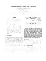
gram patser. Data presented in Fig. 2 show that clus-
ters of potential AREs are present near CpG islands in
human, mouse and rat PRDX5 genes, suggesting that
ARE-binding transcription factors may be involved in
the regulation of these genes. It should be noted that,
although the exact positions of the AREs are not con-
served between different mammalian species, at least
50% of all AREs in all of these genes are located within
promoter CpG islands (Fig. 2), despite significant diver-
gence of their noncoding sequences (not shown).
To study whether NRF2 is actually required for
basal expression of the human PRDX5 gene in the
absence of oxidative stress, we used specific siRNA
introduced into A549 cells by transient transfection.
NRF2 is required not only for inducible but also
for constitutive expression of some genes [23]. About
20-fold silencing of NRF2 expression was reproducibly
detected 72 h after NRF2 siRNA transfection of A549
cells (Fig. 3A), but this did not significantly decrease
the amount of PRDX5 protein (Fig. 3B). This indi-
Fig. 2. Distribution of potential AREs in mammalian PRDX5 genes predicted by the computer program PATSER ( />PATSER was run with the following command line options: bin ⁄ patser -A a:t 0.3 c:g 0.2 –m mp ⁄ patser.2005-08-11.202429. matrix -b 1 -c -d1
-ls 6 -f tmp ⁄ patser. CpG islands (CpGi) were predicted using a program available at default settings (55% GC,
0.65 ratio ObsCpG ⁄ ExpCpG, 500-bp length, 100-bp gap between adjacent islands). Genomic sequences and intron–exon structures were
extracted from the Ensemble Genome Browser ().
A
B
EDC
Fig. 3. Modulation of NRF2 and PRDX5 expression. (A,B) Results
of introduction into A549 cells of NRF2 siRNA. RNA transfection
agent and antibodies to NRF2 were obtained from Santa Cruz Bio-
tech. Tranfections were performed according to the manufacturer’s
instructions in 12-well plates, and immunoblots were performed
72 h after transfection. (C,D) Inhibition of NRF2 and PRDX5 by CSE
in A549 cells. CSE was prepared as described in ref [30]. (D)
Mean ± SD values relative to control (taken as 1) obtained in three
experiments. (E) Immunoblotting results were obtained using two
stable clones of A549 cells (Control and KD-1, lanes 1 and 2; their
isolation is described in the legend to Fig. 5), and A549 cells transi-
ently transfected with plasmid (lane 3, 48 h after transfection)
expressing under the control of the CMV promoter the short
( 17 kDa) form of PRDX5 (S-PRDX5) started from its second initi-
ation codon [11]. This is the main form of the PRDX5 protein
detectable by immunoblotting in human cells. It can be seen that
down-regulation (lane 2) or up-regulation (lane 3) of PRDX5 slightly
affects the amount of NRF2.
A. Kropotov et al. Transcription of noncoding DNA
FEBS Journal 273 (2006) 2607–2617 ª 2006 The Authors Journal compilation ª 2006 FEBS 2609
cates that NRF2 is not required for basal expression
of the PRDX5 gene, or that the effect of NRF2 is
masked by other transcription factors.
NFR2 may be still be required for maintenance of
high PRDX5 gene expression under conditions of oxi-
dative stress. We previously found that expression of
the PRDX5 gene is strongly inhibited by treatment of
human or rat cells with cigarette smoke extract (CSE)
[30], which can be potentially explained by CSE-medi-
ated down-regulation of NRF2. Here we examined
how CSE affects expression of NRF2, and found
(Fig. 3C,D) that 3 h treatment with 5% CSE induces
an approximately fivefold decrease in NRF2 protein,
which is consistent with the view that NRF2 may be
required for maintenance of high PRDX5 expression
during CSE-mediated stress. To exclude a possible
inverse effect of PRDX5 down-regulation on NRF2
expression, we examined NRF2 in A549 cells overex-
pressing PRDX5 and in A549 KD-1 cells with siRNA-
mediated knockdown of the PRDX5 gene (described
below). NRF2 concentration was not significantly
changed in cells with down-regulated PRDX5 (Fig. 3E,
lane 2) or in overexpressing cells (Fig. 3E, lane 3),
indicating that NRF2 expression does not depend on
PRDX5. It appears therefore that inhibition of
PRDX5 by CSE can be explained by a CSE-induced
decrease in NRF2 (Fig. 3C,D).
Role of PRDX5 in suppression of DNA oxidation
(formation of 8-oxoG)
To study the potential significance of PRDX5 in pro-
tection of the human genome (nuclear DNA) from oxi-
dation, we constructed a stable clone of A549 cells
(KD-1) with greatly decreased expression of this gene
mediated by specific siRNA (Fig. 4). KD-1 cells prolif-
erated normally, but were more sensitive to menadi-
one-induced cell death, which was measured by
quantitation of the population of SubG1 cells 24 h
after exposure to 50 lm menadione (Fig. 4C).
8-OxoG was analysed in the KD-1 clone using a
flow cytometric method based on the ability of 8-oxoG
to bind avidin [27]. As a control, we used a stable
clone of A549 cells obtained after transfection of
the plasmid encoding green fluorescent protein (GFP)-
siRNA which showed a normal level of expression of
the PRDX5 gene (see above). Figure 5A (left bars)
shows the results of analysis of 8-oxoG in the control
and KD-1 cells. In the KD-1 cells, the concentration
of endogenous 8-oxoG was significantly increased,
indicating that PRDX5 contributed to the genome def-
ense against spontaneous oxidative lesions in DNA
induced by endogenous factors. However, we were
unable to detect a significant decrease in GSH in this
clone compared with control cells under standard con-
ditions (not shown). This indicates that the increased
spontaneous DNA oxidation (Fig. 5A) may depend on
a GSH-independent mechanism. KD-1 cells were also
characterized by a minor increase in 8-oxoG as a result
of a 3-h treatment with 50 lm menadione (Fig. 5A).
This increase may be underestimated, if menadione
suppresses the knockdown effect of PRDX5 siRNA,
but we found that the decrease in PRDX5 observed in
KD-1 cells was not affected by menadione (Fig. 5B).
A small effect of PRDX5 on menadione-induced
DNA oxidation was also found when human recombin-
ant full L-PRDX5 (Long-PRDX5) was added to the
medium [Dulbecco’s modified Eagle’s medium
(DMEM) without serum and phenol red] in which cells
were exposed to 50 lm menadione (data not shown).
This protein, expressed in Escherichia coli (Fig. 5C)
showed catalase-like activity (Fig. 5D). However, in the
presence of another oxidative agent, CSE, significant
suppression of DNA oxidation by L-PRDX5 was detec-
ted (Fig. 5E).
This suppression may be caused by neutralization
of oxidative compounds of CSE in the medium by
L-PRDX5. Also, L-PRDX5 may act inside cells, as it
is capable of penetrating membranes because of the
A
BC
Fig. 4. Construction of a plasmid expressing PRDX5 siRNA and
isolation of the PRDX5 stable knockdown clone. The control clone
was obtained after transfection of A549 cells with a plasmid encod-
ing GFP–siRNA, and the KD-1 clone was isolated after transfection
of A549 cells with a plasmid encoding PRDX5 siRNA (A). The relat-
ive amount of PRDX5 protein but not PRDX1 protein is greatly
decreased in KD-1 cells as seen in (B), lane 2. The KD-1 clone also
shows higher sensitivity to cell death induced by 24 h of treatment
with 50 l
M menadione (C), which was detected by analysis of
SubG1 cells using flow cytometry as described in ref [18]. Other
details are described in Experimental procedures.
Transcription of noncoding DNA A. Kropotov et al.
2610 FEBS Journal 273 (2006) 2607–2617 ª 2006 The Authors Journal compilation ª 2006 FEBS
presence of a full N-terminus-covering mitochondrial
targeting signal [13]. Taken together, our results indicate
that PRDX5 may be involved in defense against sponta-
neous DNA oxidation and DNA oxidation induced by
CSE, and can suppress menadione-induced cell death,
making a small contribution to protection from oxida-
tion of nuclear DNA induced by menadione.
Transcription of satellite DNA in the PRDX5
knockdown clone
The human PRDX5 gene was originally cloned as an
in vitro corepressor of transcription of Alu repeats [11].
Later we also detected colocalization of this protein
with Cajal bodies [18] where transcription complexes
are assembled [19]. Here using real-time PCR, we stud-
ied whether changes in the PRDX5 KD-1 clone influ-
ence the level of transcripts complementary to human
DNA repeats. A similar approach has been recently
used by others to show increased transcription of some
mouse DNA repeats in cells deficient in H3-K9-specific
histone methyltransferase Suv39 h [33] and mouse
cells deficient in the ribonuclease Dicer [34], which is
involved in processing and formation of siRNA.
We compared cDNAs synthesized using random pri-
mer on total RNAs from the PRDX5 KD-1 clone and
from the control clone. The sequences of primers used in
real-time PCR are shown in Table 1. Control and
KD-1 cDNAs showed in real-time PCR almost identical
amplification curves with primers to the human b-actin
gene (Fig. 6A), indicating uniform reverse transcription
of both RNA samples. Similar results were obtained
with primers amplifying internal Alu sequences, but
primers against a-satellite and satellite III DNA (ampli-
fication products are seen in Fig. 6B) showed consistent
differences between threshold cycles for KD-1 and
A
B
E
DC
Fig. 5. Analysis of 7,8-dihydro-8-oxoguanine (8-oxoG) in the PRDX5 knockdown clone KD-1 (A,B) and in A549 cells treated with recombinant
human L-PRDX5 (C–E). (A) Results of flow cytometry: the ordinate shows the shift (M1 gate) in the green fluorescence histogram obtained
after incubation of cells with FITC–avidin as described in Experimental procedures. The M1 gate was adjusted to 1 with the control clone C
(expressing GFP–siRNA) in the absence of menadione. Values are mean ± SD obtained from four experiments. Student’s test for the left pair
of bars (no treatment) showed P ¼ 0.0032, and for the right pair of bars (menadione) the P ¼ 0.032. In (B) KD-1 cells were treated with 50 l
M
menadione (MEN) for 3 h, and immunoblotting was carried out as described in the legend to the Fig. 2. (C) Purity of recombinant L-PRDX5
( 23 kDa) determined by electrophoresis in a 10% acrylamide gel stained with Coomassie Brilliant Blue. (D) Catalase activity of L-PRDX5.
Reaction mixtures (0.5 mL) contained 100 l
M Hepes buffer, pH 7.5, 2 mM dithiothreitol, the indicated amount of PRDX5 per ml, and 100–
130 l
M H
2
O
2
. The reaction was stopped by mixing 100 mL aliquots with 0.75 mL 12.5% trichloroacetic acid. Then 200 lL10mM
Fe(NH
4
)
2
(SO
4
)
2
and 100 lL2.5M KSCN were added, and A
450
was measured. Cells grown on plastic in DMEM containing 10% fetal calf
serum and phenol red were washed with NaCl ⁄ P
i
, and the DMEM without fetal calf serum and phenol red was added (2 mL per 60-mm dish)
followed by the addition of CSE (2% final concentration) and L-PRDX5 (50 lgÆmL
)1
final concentration). After incubation at 37 °CinaCO
2
incu-
bator for 3 h, cells were washed with NaCl ⁄ P
i
, detached from the plastic using trypsin ⁄ EDTA, and 8-oxoG was analysed as described above.
A. Kropotov et al. Transcription of noncoding DNA
FEBS Journal 273 (2006) 2607–2617 ª 2006 The Authors Journal compilation ª 2006 FEBS 2611
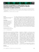
control cDNA probes (Fig. 6B, right section, Fig. 6C)
with KD-1 samples reaching the amplification threshold
about two cycles earlier than the controls at different
dilutions. This corresponds to an approximately four-
fold higher abundance of a-satellite and satellite III
RNA in KD-1 cells than in control cells (Fig. 6C).
Therefore, knockdown of the PRDX5 gene stimulated
transcription of some families of human satellite DNA,
which may be caused by direct involvement of PRDX5
in silencing of the transcription of heterochromatin.
Discussion
In this study we found that PRDX5 gene expression in
cultured human A549 cells as well as in several other
human cell lines is not induced by oxidative stress.
This happens despite the presence of the conserved
cluster of potential AREs, which can serve as binding
sites for the transcription factor NRF2 in the 5¢ pro-
moter region of mammalian PRDX5 genes. NRF2 is
known to be involved in oxidative stress-induced acti-
vation of the PRDX1 gene in mouse macrophages [25]
and mouse lungs [22]. NRF2 is also involved in activa-
tion of many other antioxidant response genes [21]. As
NRF2 sites in the PRDX5 promoter may be involved
in the maintenance of constitutive expression of
PRDX5 in the absence of oxidative stress, we exam-
ined whether it can be inhibited by specific NRF2
siRNA, but found that PRDX5 gene expression was
not silenced by NRF2 siRNA. This indicates that
other transcription factors may control constitutive
(basal) expression of the PRDX5 gene. It should be
noted that the potential binding site for the ubiquitous
transcription factor NF-1 is located 1 kb upstream of
the PRDX5 transcription start.
Higher expression of PRDX5 than other peroxire-
doxins in different types of normal human tissues was
found using Affimetrix gene expression arrays; the
relevant information is available free at http://
www.GeneCards.org. Apparently, human PRDX5 gene
promoter has efficient transcription cis-regulatory ele-
ments for its high constitutive expression, which may
be required for neutralization of reactive oxygen species
continuously produced by mitochondria [31]. A signifi-
cant fraction of PRDX5 is known to be located in
mitochondria [13] and associated with the mitochond-
rial matrix (A. V. Kropotov et al., unpublished results).
However, NRF2 may be required for the maintenance
of high PRDX5 gene expression under oxidative stress,
as we found that NRF2 was strongly down-regulated
by CSE, as well as PRDX5 in our earlier study [30].
Here we also constructed a clone of human cells
with siRNA-mediated down-regulation of PRDX5
(KD-1) and found a significantly increased concentra-
tion of endogenous 8-oxoG in this clone. This suggests
that constitutive basal expression of the PRDX5 gene
contributes to the antioxidant defense of the human
genome. The 8-oxoG content of nuclear DNA from
mouse tissues estimated by quantitative analysis
(HPLC and electrospray detection in isolated DNA) is
0.1–0.4 per 10
6
bp [32]. Assuming that the basal
steady-state concentration of 8-oxoG in control A549
cells is the same as in mouse tissues and that the dip-
loid genome size is 7000 Mbp, it can be calculated
that the observed relative 2.5-fold increase in 8-oxoG
in the PRDX5 KD-1 clone (Fig. 5A) corresponds to
> 1000 additional potentially mutagenic DNA lesions
per nucleus in PRDX5 knockdown cells.
PRDX5 may also be involved in defense against
DNA oxidation induced in A549 cells by external stress
Table 1. Sequences of primers used in real-time PCR. B-ACT, beta
actin; ASAT, alpha-satellite; ALU, Alu repeat; S3T, satellite III. D,
direct; R, reverse.
Name
Primer
length (nt) Sequence (5¢ to 3¢)
PCR product
length (nt)
B-ACT-D 21 CATGTACGTTGCTATCCAGGC
B-ACT-R 21 CTCCTTAATGTCACGCACGAT 250
ASAT-D 22 TCTTTGTGATGTGTGCATTCAA
ASAT-R 20 TATTCCCGTTTCCAACGAAG 132
ALU-D 20 ACGAGGTCAGGAGATCGAGA
ALU-R 19 GATCTCGGCTCACTGCAAG 174
S3T-D 19 AATCAACCCGAGTGCAATC
S3T-R 22 TCCATTCCATTCCTGTACTCGG 160
Fig. 6. Real-time PCR analysis of transcription in PRDX5 knockdown clone. (A, left section) Photograph of a 3% acrylamide gel with amplifi-
cation products obtained with b-actin primers (Table 1) using two different dilutions (1 : 9 and 1 : 729) of template cDNA from the control
clone (lanes 1 and 3) and the same dilutions of cDNA from the KD-1 clone (lanes 2 and 4). Control reactions with b-actin primers but without
template are shown in lanes 5 and 6. Size markers (25-bp ladder from Invitrogen) are shown in lane 7. (A, right section) Kinetics of real-time
DNA amplification with numbers corresponding to the lanes in the left section. (B, left section) Photograph of a 3% agarose gel with amplifi-
cation products obtained with alpha-satellite (ASAT, lane 1) and satellite III (S3T, lane 3) primers (Table 1) of cDNA from the control clone
(dilution 1 : 200). Lane 2 shows size markers. The expected size of the product with ASAT primers is 132 bp and with S3T primers
)160 bp. (B, right section) Kinetics of amplification with ASAT primers of identical amounts of cDNA (1 : 25 dilution) from the control clone
(curve P) and from the KD-1 clone (curve D). (C) Fold difference between cDNA isolated from the control and KD-1 clones calculated from
standard curves. Mean ± SD values of four to six PCRs are shown.
Transcription of noncoding DNA A. Kropotov et al.
2612 FEBS Journal 273 (2006) 2607–2617 ª 2006 The Authors Journal compilation ª 2006 FEBS
A
B
C
A. Kropotov et al. Transcription of noncoding DNA
FEBS Journal 273 (2006) 2607–2617 ª 2006 The Authors Journal compilation ª 2006 FEBS 2613
agents such as CSE (Fig. 5E) and in suppression of cell
death induced by menadione (Fig. 4C). However, the
contribution of PRDX5 to menadione-induced DNA
oxidation was small (Fig. 5A), indicating that other fac-
tors may be more important in this menadione effect
than PRDX5.
As PRDX5 was originally isolated as a transcript-
ional corepressor [11], we studied whether transcript-
ion is affected in KD-1 cells. No change in the
abundance of b-actin gene transcripts or of transcripts
containing Alu or LINE1 sequences was found in the
KD-1 clone, but a significant increase in the abundance
of transcripts of a-satellite and satellite III DNA was
reproducibly detected. Strong induction of transcrip-
tion of satellite III DNA (which is located in hetero-
chromatin of chromosome 9) by heat shock has been
documented by other groups [33,34]. In real-time PCR
analysis of Alu transcripts, primers matching very con-
served internal Alu subsequences were used, which are
present in 3¢ untranslated segments of mRNA of many
different genes and can mask possible contributions of
RNA polymerase III-dependent Alu transcripts. How-
ever, inserts of conserved a-satellite and satellite III
sequences are present in a very small number of entries
of the human dbEST database (< 10 per 7 · 10
6
sequences), indicating that the detected increase in
abundance of satellite DNA transcripts is probably
associated with a true increase in heterochromatin tran-
scription in cells with down-regulated PRDX5. The
mechanism of this increase remains unclear.
Transcription of heterochromatin is known to be
controlled by a negative regulatory loop involving
siRNA produced by the nuclease Dicer and histone
H3-K9 trimethylation [35–37]. If PRDX5 is involved
in maturation of these RNA-induced transcription
silencing complexes, its knockdown may lead to activa-
tion of heterochromatin transcription. Alternatively,
an increased transcription of some satellite DNAs in
the PRDX5 knockdown clone may be an indirect con-
sequence of changes in cellular redox state, affecting a
redox-sensitive transcription factor, e.g. NF-kappaB.
This factor regulates gene expression of a large number
of cytokine and other immune response genes [38], but
its role in transcription of heterochromatin is not known.
Experimental procedures
Cell culture
Human lung carcinoma line A549 and epidermoid carci-
noma line A431 were obtained from ATCC and cultured in
DMEM containing d-glucose (4.5 g Æ L
)1
) supplemented with
10% fetal calf serum, 100 UÆmL
)1
penicillin, and
100 lgÆmL
)1
streptomycin (complete DMEM) in a CO
2
incubator at 37 °C. Human embryonic fibroblasts HEF4
and HeLa cells were obtained from the Cell Culture Collec-
tion of the Institute of Cytology RAS (St Petersburg), and
the nonsmall cell lung carcinoma line U1810 was obtained
from the Cell Line Collection of the Karolinska Institutet
(Stockholm).
Construction of siRNA plasmids
Vector mU6pro with U6 RNA gene promoter was obtained
from D. L. Turner [26] ( />dLturner.vectors/rna_interference_vectors). NEO gene was
amplified using PCR from the Rc-CMV plasmid (Strata-
gene, La Jolla, CA, USA) and subcloned into mU6pro
between ApaI and HindIII sites upstream of the U6 promo-
ter. Hairpin oligonucleotides for RNAi were subcloned
between BbsI and XbaI restriction sites immediately down-
stream from the U6 promoter. Sequences (5¢ to 3¢)of
complementary hairpin oligonucleotides targeting PRDX5
cDNA (exons 5 and 6 underlined) were: TTT
GAGA
ACCTCTTGAGACGTCGATGACGTCTCAAGAGGTTC
TCTTTTT (top strand) and CTAGAAAAAGAGAA
CCTCTTGAGACG TCATC GACGTCTCAAGAGGTTCT
(bottom strand). Targeted segments are present in all spli-
cing variants of PRDX5 mRNA and therefore should
silence all these variants. For the control plasmid, we used
hairpin oligonucleotides targeting GFP (sequences under-
lined) gene: TTT
GAAGAAGTCGTGCTGCTTCATGGA
AGCAGCACGACTTCTTCTTTTT (top strand) and CTA
GAAAAA
GAAGAAGTCGTGCTGCTTCCATAAGCAG
CACGACTTCTT (bottom strand). Bacterial clones with
insertions in these oligonucleotides were identified using
PCR and sequenced with vector primers GCTACATTTTA
CATGATAGGCTTGG (U6-forward) and CACAGGAAA
CAGCTATGACCAT (M13-rev).
Isolation of stable PRDX5 knockdown (KD) and
control clones of A549 cells
The cells, grown on 24-well plates, were transfected with
mU6neo plasmids, which expressed siRNA against PRDX5
or GFP using Lipofectamine 2000 (Invitrogen, Carlsbad,
CA, USA). Plasmid DNA (0.8 lg) or Lipofectamine (1.6 lL)
was diluted in 50 lL Opti-MEM, incubated for 5 min, then
mixed and incubated for 20 min before addition to cells.
After overnight incubation at 37 °CinaCO
2
incubator,
DMEM with 10% fetal calf serum was added, and incuba-
tion was continued for 10 h. Then cells were replated at
1 : 10–1 : 40 dilution on to the six-well plates and selection
with G418 (1 mgÆmL
)1
) was started the next day. Individual
clones growing on G418 were analyzed using western blot-
ting with antibodies to PRDX5 as well as the control clone
obtained after transfection of A549 cells with GFP siRNA
expressing mU6neo plasmid. One of the clones (KD-1) which
Transcription of noncoding DNA A. Kropotov et al.
2614 FEBS Journal 273 (2006) 2607–2617 ª 2006 The Authors Journal compilation ª 2006 FEBS
showed > 90% inhibition of PRDX5 protein on blots (com-
pared with the control during several passages after isolation)
was used for further experiments. PRDX5 KD-1 cells derived
from the A549 line showed normal morphology, normal
GSH ⁄ GSSG ratio, and growth characteristics in the medium
with G418 similar to cells of the control clone. Therefore,
down-regulation of the PRDX5 gene did not significantly
affect proliferation of A549 and cell cycle progression.
Expression of PRDX5 in E. coli
To express full-length PRDX5 (L-PRDX5) starting from the
first AUG codon in its coding sequence, the corresponding
DNA fragment was PCR-amplified from a plasmid with full
PRDX5 cDNA using primers BE-1 (GGCGGATCCATGG
GACTAGCTGGCGTGTGCG) and BE-2 (GGCGA
ATTCTTATCAGAGCTGTGAGATGATATTGGG) and
DNA polymerase PfuI. The resulting fragment was purified,
treated with Bam H1 ⁄ EcoRI, subcloned into the BamH1 ⁄
EcoRI-cleaved GST-fusion vector pGEX-6P-1, and trans-
formed into E. coli BL21. Bacteria were grown at 30 °Cin
Luria–Bertani medium until A
600
¼ 1.0. GST-fusion protein
synthesis was induced with 1.0 mm isopropyl b-d-thiogal-
actoside (final concentration), and bacteria were further
grown for 3–4 h. Cells were harvested by centrifugation, and
the pellet was resuspended and washed with TB buffer
(9.1 mm Hepes, 55 mm MgCl
2
,15mm CaCl
2
, 250 mm KCl,
adjusted to pH 6.7). The cell pellet containing GST-fusion
protein was resuspended in NaCl ⁄ P
i
lysis buffer (140 mm
NaCl, 2.7 mm KCl, 10 mm Na
2
HPO
4
, 1.8 mm KH
2
PO
4
adjusted to pH 7.4) supplemented with lysozyme
(1 mgÆmL
)1
; Sigma, St Louis, MO, USA), Complete
TM
Protease Inhibitor Cocktail (Roche, Alameda, CA, USA),
10 mm MgCl
2
, and DNAse I (10 UÆmL
)1
; Roche). Cells
were effectively lysed by repeated freezing ⁄ thawing. The
lysate was cleared by centrifugation at 70 000 g and 4 °C.
The supernatant was loaded on to a GSTrap FF column
(Amersham Biosciences, Piscataway, NJ, USA; Glutathione
Sepharose
TM
4 Fast Flow). After GST-fusion protein bind-
ing to the column, bound material was washed with NaCl ⁄ P
i
,
pH 7.4. Then the buffer was replaced with PreScission Prote-
ase buffer (50 mm Tris ⁄ HCl, 100 mm NaCl, 1 mm EDTA,
1mm dithiothreitol, pH 8.0), and rapid on-column GST-
fusion protein cleavage was performed. The enzyme contains
a noncleavable GST-affinity tag for optimum on-column
cleavage, and the column remains online, connected to the
purification system, eliminating the loss of any material.
Proteolytic digestion used 2 U enzyme per 100 lg bound
GST-fusion protein. PreScission Protease was diluted in
4.5 mL PreScission buffer and manually injected on to the
column at an increased flow rate of 5–7 mLÆmin
)1
, and the
system was incubated for 12–16 h at 4 °C. Before elution, a
1-mL GSTrap FF column (pre-equilibrated with PreScission
buffer) was connected downstream to the GSTrap FF pro-
teolytic cleavage column. The GSTrap FF affinity column
acts as a filter to capture any released cleaved GST protein,
uncleaved GST-fusion protein, and unbound PreScission
Protease. Cleaved protein is eluted immediately upon flow
startup with PreScission buffer. The elution peak containing
the cleaved target protein (L-PRDX5) was eluted first
through the 1-mL buffering GSTrap column.
Analysis of 8-oxoG in DNA
This analysis is based on previous observations that avidin
binds with high affinity to 8-oxoG in DNA [27,28]. This
approach was efficiently used previously for detection of
8-oxoG in mouse knockout cells with targeted disruption of
the PRDX1 gene [2]. Here we used fluorescein isothiocya-
nate (FITC)–avidin (Sigma) and flow cytometry (Beckton–
Dickinson, Franklin Lakes, NJ, USA; FACS Calibur) for
detection of 8-oxoG. Cells were treated with the oxidative
agent menadione (50 lm; Sigma) for the indicated times in
serum-free and phenol red-free DMEM (Invitrogen),
detached from the plastic with trypsin ⁄ EDTA, washed in
NaCl ⁄ P
i
, and fixed in 2% formaldehyde at 4 °C and then
in 80% ethanol at )20 °C. Other steps before FACS analy-
sis were performed as described in the instructions to the
OxyDNA fluorimetric kit (catalogue no. 500095) produced
by Calbiochem (San Diego, CA, USA). FITC–avidin bind-
ing was quantified by relative peak shift (M1 gate) in the
FACS histograms obtained.
Immunoblotting
Cells were scraped off into cold lysis buffer composed of
1% Triton X-100, 2 mm EDTA, 2 mm dithiothreitol,
0.25 mgÆmL
)1
leupeptin, 0.25 mgÆmL
)1
pepstatin A,
0.4 mgÆmL
)1
aprotinin and 0.1 mm phenylmethanesulfonyl
fluoride. Samples were separated by SDS ⁄ PAGE (10% gels),
and then separated proteins were transferred to membranes,
where they were blocked overnight at 4 °C with 5% nonfat
dry milk in TBST (10 mm Tris ⁄ HCl, pH 8.0 ⁄ 150 mm
NaCl ⁄ 0.1% Tween 20). The blot was rinsed twice with TBST
and incubated for 2 h at room temperature with rabbit poly-
clonal PRDX5 antibody (1 : 2000 dilution) [11] or commer-
cial antibodies to actin and NRF2 (1 : 1000 dilution) in
TBST containing 5% BSA. The membrane was washed for
15 min with TBST and incubated with goat anti-mouse IgG
conjugated with horseradish peroxidase in 5% milk for 1 h,
and then washed three times with TBST and developed with
enhanced chemiluminescence reagent (Amersham). Bands on
radioautographs were quantified using the Image J program
(NIH).
Transfections of A549 cells with NRF2 siRNA
Here we used siRNA, antibodies, transfection reagents
and protocols produced and recommended by Santa Cruz
A. Kropotov et al. Transcription of noncoding DNA
FEBS Journal 273 (2006) 2607–2617 ª 2006 The Authors Journal compilation ª 2006 FEBS 2615
Biotechnology (Santa Cruz, CA, USA). In preliminary
experiments, the efficiency of RNA transfection of A549 cells
was examined using FITC-labeled short RNA and analysis
by flow cytometry. In these experiments, a significant shift in
mean fluorescence was detected 24 h after transfection, indi-
cating high efficiency of RNA uptake by A549 cells.
RNA extraction, cDNA synthesis, and real-time PCR
Total RNA was extracted from A549 cells using Trizol
Reagent (Invitrogen). cDNA was synthesized using the
Invitrogen SuperScript III First Strand Synthesis System
with random hexamer primers for 50 min at 50 °C. After
being heated for 5 min at 85 °C and hydrolysed with
RNase H for 20 min at 37 °C, the resulting cDNA was
used in real-time PCR (Universal PCR SYBR Mastermix;
Applied BioSystems) in a final volume of 25 l L in 96-well
plates in an automated fluorimeter (ABI PRISM 7000).
Standard amplification conditions were used: 2 min at
50 °C, 10 min at 95 °C, 40 cycles of 15 s at 95 °C and 60 s
at 60 °C. Sequences of oligonucleotide primers used in
RT-PCR are shown in Table 1.
Computational approaches
Genomic sequences were exported from the Ensemble Data-
base () along with 2 kb of flank-
ing sequences on both sides of the genes. To analyze the
distribution of binding sites for NRF2 in PRD5X genes, the
patser program ( was used with
the following command line options: bin ⁄ patser -A a:t 0.3
c:g 0.2 -m tmp ⁄ patser.2005-08-11.202429.matrix -b 1 -c -d1
-ls 6 -f tmp ⁄ patser with the lower threshold estimation
through the weight score 6. The position ⁄ weight matrix for
NRF2 was composed using known consensus binding
sequences for NRF2 [22]. The position of the CpG islands
in the PRDX genes was determined using a program avail-
able at default settings (55%
GC, 0.65 ratio ObsCpG ⁄ ExpCpG, 500-bp length, 100-bp
gap between adjacent islands).
Acknowledgements
This research was supported in part by Philip Morris
USA Inc. and by Philip Morris International (to V.S.),
by Swedish (3829-B04–09XAC) and Stockholm
(041502) Cancer Societies, Swedish Research Council
(K2006–31X-02471-39-3) and INTAS Genomics
(05-1000004-7755) grants (to B.Z.), and by the Russian
Fund of Basic Research grants 04-04-49292 and 04-04-
293 (to N.T.). A.K. was also supported by a grant
from the Institute of Environmental Medicine, Kar-
olinska Institutet, from the Wenner-Gren Foundation
(Sweden), and the Russian Science Foundation.
References
1 Wood ZA, Poole LB & Karplus PA (2003) Peroxire-
doxin evolution and the regulation of hydrogen perox-
ide signaling. Science 300, 650–653.
2 Neumann CA, Krause DS, Carman CV, Das S,
Dubey DP, Abraham JL, Bronson RT, Fujiwara Y,
Orkin SH & Van Etten RA (2003) Essential role for the
peroxiredoxin Prdx1 in erythrocyte antioxidant defence
and tumour suppression. Nature 424, 561–565.
3 Lee TH, Kim SU, Yu SL, Kim SH, Park DS, Moon HB,
Dho SH, Kwon KS, Kwon HJ, Han YH, et al. (2003)
Peroxiredoxin II is essential for sustaining life span of
erythrocytes in mice. Blood 101, 5033–5038.
4 Choi MH, Lee IK, Kim GW, Kim BU, Han YH, Yu
DY, Park HS, Kim KY, Lee JS, Choi C, et al. (2002)
Regulation of PDGF signalling and vascular remodel-
ling by peroxiredoxin II. Nature 435, 347–353.
5 Chang TS, Cho CS, Park S, YuS, Kang SW & Rhee
SG (2004) Peroxiredoxin III, a mitochondrion-specific
peroxidase, regulates apoptotic signaling by mitochon-
dria. J Biol Chem 279, 41975–41984.
6 Araki M, Nanri H, Ejima K, Murasato Y, Fujiwara T,
Nakashima Y & Ikeda M (1999) Antioxidant function
of the mitochondrial protein SP-22 in the cardiovascular
system. J Biol Chem 274, 2271–2278.
7 Zhang Y, Emmanuel N, Kamboj G, Chen J, Shurafa
M, Van Dyke DL, Wiktor A & Rowley JD (2004)
PRDX4, a member of the peroxiredoxin family, is fused
to AML1 (RUNX1) in an acute myeloid leukemia
patient with a t (X; 21) (p22;q22). Genes Chromosomes
Cancer 40, 365–370.
8 Wang X, Phelan SA, Forsman-Semb K, Taylor EF,
Petros C, Brown A, Lerner CP & Paigen B (2003) Mice
with targeted mutation of peroxiredoxin 6 develop nor-
mally but are susceptible to oxidative stress. J Biol
Chem 278, 25179–25190.
9 Kropotov AV & Tomilin NV (1996) A human B-box-
binding protein downregulated in adenovirus 5-trans-
formed human cells. FEBS Lett 386, 43–46.
10 Kropotov AV & Tomilin NV (1997) Evidence for a reg-
ulatory protein complex on RNA polymerase III pro-
moter of human retroposons of Alu family. Genetica
(Dortrecht) 98, 223–233.
11 Kropotov A, Sedova V, Ivanov V, Sazeeva N, Tomilin A,
Krutilina R, Oei SL, Griesenbeck J, Buchlow G & Tomi-
lin N (1999) A novel human DNA-binding protein with
sequence similarity to a subfamily of redox proteins
which is able to repress RNA-polymerase-III-driven tran-
scription of the Alu-family retroposons in vitro. Eur J
Biochem 260, 336–346.
12 Yamashita H, Avraham S, Jiang S, London R, Van
Veldhoven PP, Subramani S, Rogers RA & Avraham H
(1999) Characterization of human and murine PMP20
Transcription of noncoding DNA A. Kropotov et al.
2616 FEBS Journal 273 (2006) 2607–2617 ª 2006 The Authors Journal compilation ª 2006 FEBS
peroxisomal proteins that exhibit antioxidant activity
in vitro. J Biol Chem 274, 29897–29904.
13 Knoops B, Clippe A, Bogard C, Arsalane K, Wattiez R,
Hermans C, Duconseille E, Falmagne O & Bernard A
(1999) Cloning and characterization of AOEB166, a
novel mammalian antioxidant enzyme of the peroxire-
doxin family. J Biol Chem 274, 30451–30458.
14 Dubuisson M, Vander Stricht D, Clippe A, Etienne F,
Nauser T, Kissner R, Koppenol WH, Rees JF &
Knoops B (2004) Human peroxiredoxin 5 is a peroxyni-
trite reductase. FEBS Lett 571, 161–165.
15 Zhou Y, Kok KH, Chun AC, Wong CM, Wu HW, Lin
MC, Fung PC, Kung H & Jin DY (2000) Mouse
peroxiredoxin V is a thioredoxin peroxidase that inhibits
p53-induced apoptosis. Biochem Biophys Res Commun
268, 921–927.
16 Mikhailov VM, Kropotov AV, Zelenin AV, Krutilina RI,
Kolesnikov VA, Zelenina IA, Baranov AN, Shtein GI,
Ostapenko OV, Tomilin NV & Baranov VS (2002) The
BCL-xL and ACR-1 genes promote differentiation and
reduce apoptosis in muscle fibers of mdx mice. Genetika
(Moscow) 38, 1445–1450.
17 Yuan J, Murrell GA, Trickett A, Landtmeters M, Kno-
ops B & Wang MX (2004) Overexpression of antioxidant
enzyme peroxiredoxin 5 protects human tendon cells
against apoptosis and loss of cellular function during oxi-
dative stress. Biochim Biophys Acta 1693, 37–45.
18 Kropotov AV, Grudinkin PS, Pleskach NM,
Gavrilov BA, Tomilin NV & Zhivotovsky B (2004)
Downregulation of peroxiredoxin V stimulates forma-
tion of etoposide-induced double-strand DNA breaks.
FEBS Lett 572, 75–79.
19 Gall JG (2001) A role for Cajal bodies in assembly of the
nuclear transcription machinery. FEBS Lett 498, 164–167.
20 Banmeyer I, Marchand C, Clippe A & Knoops B (2005)
Human mitochondrial peroxiredoxin 5 protects from
mitochondrial DNA damages induced by hydrogen per-
oxide. FEBS Lett 579, 2327–2333.
21 Numazawa S & Yoshida T (2004) Nrf2-dependent gene
expressions: a molecular toxicological aspect. J Toxicol
Sci 29, 81–89.
22 Rangasamy T, Cho CY, Thimmulappa RK, Zhen I,
Srisuma SS, Kensler TW, Yamamoto M, Petrache I,
Tuder RM & Biswal S (2004) Genetic ablation of Nrf2
enhances susceptibility to cigarette smoke-induced
emphysema in mice. J Clin Invest 114, 1248–1259.
23 Nguyen T, Sherratt PJ, Nioi P, Yang CS & Pickett CB
(2005) Nrf2 controls constitutive and inducible expres-
sion of ARE-driven genes through a dynamic pathway
involving nucleocytoplasmic shuttling by Keap1. J Biol
Chem 280, 32485–32492.
24 Dhakshinamoorthy S, Jain AK, Bloom DA & Jaiswal
AK (2005) Bach1 competes with Nrf2 leading to
negative regulation of the antioxidant response element
(ARE)-mediated NAD(P)H:quinone oxidoreductase 1
gene expression and induction in response to antioxi-
dants. J Biol Chem 280, 16891–16900.
25 Ishii T, Itoh K, Takahashi S, Sato H, Yanagawa T,
Katoh Y, Bannai S & Yamamoto M (2000) Transcrip-
tion factor Nrf2 coordinately regulates a group of oxi-
dative stress-inducible genes in macrophages. J Biol
Chem 275, 16023–16029.
26 Yu JU, DeRuiter SL & Turner DL (2002) RNA inter-
ference by expression of short-interfering RNAs and
hairpin RNAs in mammalian cells. Proc Natl Acad Sci
USA 99, 6047–6052.
27 Struthers L, Patel R, Clark J & Thomas S (1998) Direct
detection of 8-oxodeoxyguanosine and 8-oxoguanine by
avidin and its analogues. Anal Biochem 255, 20–31.
28 Chen SK, Tsai MH, Hwang JJ & Chang WP (2001)
Determination of 8-oxoguanine in individual cell
nucleus of gamma-irradiated mammalian cells. Radiat
Res 155, 832–836.
29 Orrenius S (1985) Oxidative stress studied in intact mam-
malian cells. Philo s Trans R S oc Lond B B iol S ci 311, 673–677.
30 Serikov VB, Leutenegger C, Krutilina RI, Kropotov AV
& Tomilin NV (2006) Cigarette smoke extract inhibits
expression of antioxidant protein peroxiredoxin V. Inhal
Toxicol 18, 79–92.
31 Balaban RS, Nemoto S & Finkel T (2005) Mitochon-
dria, oxidants, and aging. Cell 120, 483–495.
32 Hamilton ML, Van Remmen H, Drake JA, Yang H,
Guo ZM, Kewitt K, Walter CA & Richardson A (2001)
Does oxidative damage to DNA increase with age? Proc
Natl Acad Sci USA 98, 10469–10474.
33 Jolly C, Metz A, Govin J, Vigneron M, Turner BM,
Khochbin S & Vourc’h C (2004) Stress-induced transcrip-
tion of satellite III repeats. J Cell Biol 164, 25–33.
34 Rizzi N, Denegri M, Chiodi I, Corioni M, Valgardsdot-
tir R, Cobianchi F, Riva S & Biamonti G (2004) Tran-
scriptional activation of a constitutive heterochromatic
domain of the human genome in response to heat
shock. Mol Biol Cell 15, 543–551.
35 Martens JH, O’Sullivan RJ, Braunschweig U, Opravil S,
Radolf M, Steinlein P & Jenuwein T (2005) The profile of
repeat-associated histone lysine methylation states in the
mouse epigenome. EMBO J 24, 800–812.
36 Kanellopoulou C, Muljo SA, Kung AL, Ganesan S,
Drapkin R, Jenuwein T, Livingston DM & Rajewsky K
(2005) Dicer-deficient mouse embryonic stem cells are
defective in differentiation and centromeric silencing.
Genes Dev 19, 489–501.
37 Murchison EP, Partridge JF, Tam OH, Cheloufi S &
Hannon GJ (2005) Characterization of Dicer-deficient
murine embryonic stem cells. Proc Natl Acad Sci USA
102, 12135–12140.
38 Kabe Y, Ando K, Hirao S, Yoshida M & Handa H
(2005) Redox regulation of NF-kappaB activation: dis-
tinct redox regulation between the cytoplasm and the
nucleus. Antioxid Redox Signal 7, 395–403.
A. Kropotov et al. Transcription of noncoding DNA
FEBS Journal 273 (2006) 2607–2617 ª 2006 The Authors Journal compilation ª 2006 FEBS 2617