Báo cáo khoa học: Replacement of helix 1¢ enhances the lipid binding activity of apoE3 N-terminal domain pot
Bạn đang xem bản rút gọn của tài liệu. Xem và tải ngay bản đầy đủ của tài liệu tại đây (420.56 KB, 10 trang )
Replacement of helix 1¢ enhances the lipid binding activity
of apoE3 N-terminal domain
Katherine A. Redmond
1
, Conrad Murphy
1
, Vasanthy Narayanaswami
1
, Robert S. Kiss
2
,
Paul Hauser
1
, Emmanuel Guigard
3
, Cyril M. Kay
3
and Robert O. Ryan
1
1 Lipid Biology in Health and Disease Research Group, Children’s Hospital Oakland Research Institute, CA, USA
2 Lipoprotein and Atherosclerosis Group, University of Ottawa Heart Institute, Ottawa, Ontario, Canada
3 Department of Biochemistry and Protein Engineering Network of Centres of Excellence, University of Alberta, Edmonton, Canada
Human apolipoprotein E (apoE) is comprised of two
structural domains, a 22 kDa N-terminal (NT) domain
and a 10 kDa C-terminal (CT) domain [1,2]. Studies
conducted with isolated domains reveal that the NT
domain contains amino acids responsible for binding
to members of the low-density lipoprotein receptor
(LDLR) family [3]. Several lines of evidence have led
to a consensus that localizes the receptor binding
region to residues 134–150 [4]. In the absence of lipid,
however, the isolated NT domain is not recognized by
Keywords
apolipoprotein E; fluorescence
spectroscopy; low-density lipoprotein; low-
density lipoprotein receptor; phospholipids
Correspondence
R. O. Ryan, Children’s Hospital Oakland
Research Institute, 5700 Martin Luther King
Jr. Way, Oakland, CA 94609, USA
Fax: +1 510 450 7910
Tel: +1 510 450 7645
E-mail:
(Received 3 November 2005, revised 1
December 2005, accepted 5 December
2005)
doi:10.1111/j.1742-4658.2005.05089.x
The N-terminal domain of human apolipoprotein E (apoE-NT) harbors
residues critical for interaction with members of the low-density lipoprotein
receptor (LDLR) family. Whereas lipid free apoE-NT adopts a stable four-
helix bundle conformation, a lipid binding induced conformational adapta-
tion is required for manifestation of LDLR binding ability. To investigate
the structural basis for this conformational change, the short helix connect-
ing helix 1 and 2 in the four-helix bundle was replaced by the sequence
NPNG, introducing a b-turn. Recombinant helix-to-turn (HT) variant
apoE3-NT was produced in Escherichia coli, isolated and characterized.
Stability studies revealed a denaturation transition midpoint of 1.9 m
guanidine hydrochloride for HT apoE3-NT vs. 2.5 m for wild-type apoE3-
NT. Wild-type and HT apoE3-NT form dimers in solution via an
intermolecular disulfide bond. Native PAGE showed that reconstituted
high-density lipoprotein prepared with HT apoE3-NT have a diameter in
the range of 9 nm and possess binding activity for the LDLR on cultured
human skin fibroblasts. In phospholipid vesicle solubilization assays, HT
apoE3-NT was more effective than wild-type apoE3-NT at inducing a time
dependent decrease in dimyristoylphosphatidylglycerol vesicle light scatter-
ing intensity. In lipoprotein binding assays, HT apoE3-NT protected
human low-density lipoprotein from phospholipase C induced aggregation
to a greater extent that wild-type apoE3-NT. The results indicate that a
mutation at one end of the apoE3-NT four-helix bundle markedly enhan-
ces the lipid binding activity of this protein. In the context of lipoprotein
associated full-length apoE, increased lipid binding affinity of the N-ter-
minal domain may alter the balance between receptor-active and -inactive
conformational states.
Abbreviations
ANS, 8-anilino-1-naphthalene sulfonate; apo, apolipoprotein; CT, carboxy (C) terminal; DMPC, dimyristoylphosphatidylcholine; DMPG,
dimyristoylphosphatidylglycerol; FAFA, fatty acid free albumin; HDL, high-density lipoprotein; HT, helix-to-turn; LDL, low-density lipoprotein;
LDLR, low-density lipoprotein receptor; NT, amino (N) terminal; PL-C, phospholipase C; rHDL, reconstituted high density lipoprotein;
WT, wild-type.
558 FEBS Journal 273 (2006) 558–567 ª 2006 The Authors Journal compilation ª 2006 FEBS
the LDLR. On the other hand, upon association with
lipid, apoE-NT binds efficiently to the LDLR [3].
X-ray crystallography of apoE3-NT has yielded a
high-resolution structure [5,6]. In the absence of lipid,
this domain exists as an elongated globular four-helix
bundle. In this conformation the amphipathic a-helices
orient their hydrophobic faces toward the center of the
bundle. At the same time, polar faces of individual
helical segments interact with the aqueous environ-
ment. The interior of the bundle contains several leu-
cine residues that align to form a leucine zipper-like
motif. In addition to the four helices that comprise the
bundle, apoE3-NT possesses a short helix, termed helix
1¢, that connects helix 1 and 2. Despite the fact that
the residues comprising this helix are highly conserved
across species, the structural or functional role of this
region of the protein remains unknown.
Weisgraber [4] proposed an open conformation model
in which the loop segment connecting helix 2 and 3 in
the bundle functions as a hinge about which the protein
opens to expose its hydrophobic interior. According to
this model, helix 1¢ resides in a location where it could
play a role in lipid surface recognition and ⁄ or initiation
of lipid binding. Moreover, it is conceivable that helix 1¢
undergoes a lipid dependent conformational change that
increases exposure of the hydrophobic interior of the
bundle, perhaps functioning in a manner similar to the
lid segment of lipases [7]. Studies of the lipid interaction
properties of apoE3-NT have been facilitated by its
ability to transform dimyristoylphosphatidylcholine
(DMPC) bilayer vesicles into discrete disc complexes [3].
Raussens et al. [8] investigated the structural organiza-
tion of apoE3-NT in DMPC complexes by infrared
spectroscopy. These authors presented a model wherein
the NT domain adopts an open conformation, circum-
scribing the perimeter of the disc bilayer with its major
helical axes aligned perpendicular to the fatty acyl
chains of DMPC. Support for this molecular orientation
has come from studies employing fluorescence reson-
ance energy transfer to evaluate distance relationships
between specific sites in the protein as a function of lipid
binding [9,10]. Also, Lu et al. [11] reported that apoE-
NT dependent transformation of DMPC bilayer vesicles
into disc complexes is abolished when helical segments
in the bundle are tethered by disulfide bond engineering.
An important consideration with regard to the lipid
interaction properties of apoE3-NT relates to the high
intrinsic stability of this domain. Denaturation studies
revealed that the NT domain is exceptionally stable
compared to either the C-terminal domain or other
apolipoproteins [1]. Likewise, lipoprotein binding stud-
ies showed that the isolated NT domain has a low
affinity for lipoprotein particles [12]. Several studies
have investigated the molecular basis of this property
with the general finding that various external factors
including solution pH, ionic strength or the presence
of chaotropic agents, modulates the lipid binding activ-
ity of apoE3-NT [13,14]. In the present study we
employed a protein engineering approach, removing
helix 1¢ and replacing it with a sequence predicted to
adopt a b-turn. The resulting variant protein displays
a marked enhancement in lipid binding activity.
Results
Design of helix-to-turn apoE3-NT
On the basis of studies with structurally related helix
bundle apolipoproteins [15,16], we hypothesized that
residues comprising a short connector helix in apoE3-
NT function in recognition or initiation of lipid bind-
ing, leading to helix bundle opening and formation
of a stable binding interaction. Using X-ray structure
data [5] as a guide, residues comprising helix 1¢ [SE-
QVQEELLS(44–53)] were deleted and replaced by a
sequence predicted to adopt a b-turn, NPNG [17]. We
hypothesized that the resulting helix-to-turn (HT) vari-
ant apoE3-NT would adopt a stable solution confor-
mation that retains the protein’s four-helix bundle
molecular architecture and LDLR binding capability,
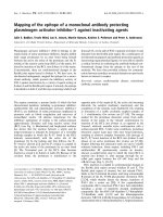
yet would display altered lipid binding activity. A rib-
bon diagram of apoE3-NT depicting the introduced
change is shown in Fig. 1.
Bacterial expression and characterization
of HT apoE3-NT
Wild-type (WT) and HT apoE3-NT were isolated
from the supernatant fraction of bacteria cultures as
described by Fisher et al. [18]. SDS ⁄ PAGE analysis
under reducing conditions revealed that the HT
apoE3-NT has a faster mobility than wild-type
apoE3-NT, consistent with the changes introduced
into the amino acid sequence (Fig. 2). Mass spectro-
metry of HT apoE3-NT gave rise to a monomer
mass ¼ 20 310 Da (calculated mass ¼ 20 318 Da) versus
21 191 Da for WT apoE3-NT. SDS ⁄ PAGE under
nonreducing conditions revealed the presence of
disulfide linked homo-dimers. ApoE3-NT contains a
single cysteine residue (at position 112) that is
known to form disulfide bonds [19]. The data pre-
sented indicate that WT and HT apoE3-NT exist in
solution as a mixture of monomers and disulfide
linked homo-dimers. This finding was corroborated
by sedimentation equilibrium experiments conducted
in the analytical ultracentrifuge, wherein evidence for
K. A. Redmond et al. apoE3-NT domain lipid binding
FEBS Journal 273 (2006) 558–567 ª 2006 The Authors Journal compilation ª 2006 FEBS 559
the presence of monomers and dimers was obtained
for both WT and HT apoE3-NT.
Stability properties of HT apoE3-NT
The effect of guanidine hydrochloride concentration
on the tryptophan fluorescence emission properties of
WT and HT apoE3-NT was monitored by fluorescence
spectroscopy. In the absence of guanidine hydrochlo-
ride, WT apoE3-NT gave rise to a tryptophan fluo-
rescence emission wavelength maximum of 346 nm
(excitation 280 nm) while the corresponding value for
HT apoE3-NT was 348 nm. Upon exposure to high
concentrations of guanidine hydrochloride, both pro-
teins displayed an 8 nm red shift in wavelength of
tryptophan fluorescence emission maximum. Plots of
guanidine hydrochloride concentration vs. the percent
maximal change in Trp fluorescence emission wave-
length maximum (Fig. 3) revealed a transition mid-
point at 2.5 m for WT apoE3-NT and a corresponding
transition at 1.9 m guanidine hydrochloride for HT
apoE3-NT.
Fluorescent dye binding
To evaluate the extent to which HT apoE3-NT mani-
fests altered exposure of hydrophobic sites in the pro-
tein, the effect of HT apoE3-NT and WT apoE3-NT
on the fluorescence emission intensity of 8-anilino-1-
naphthalene sulfonate (ANS) was examined (Fig. 4).
In the absence of protein, ANS has a low quantum
yield with an emission wavelength maximum of
515 nm (excitation 395 nm). Introduction of WT
apoE3-NT induced a 35 nm blue shift in ANS fluores-
cence emission wavelength maximum together with an
1234
Fig. 2. SDS ⁄ PAGE analysis of apolipoproteins. Proteins were elec-
trophoresed on a 4–20% (w ⁄ v) acrylamide gradient SDS slab gel
under reducing (lanes 1 and 2) or nonreducing (lanes 3 and 4) con-
ditions and was stained with Coomassie Blue. Lanes 1 and 3, WT
apoE3-NT; lanes 2 and 4, HT apoE3-NT.
Fig. 3. Effect of guanidine hydrochloride on apoE3-NT tryptophan
fluorescence emission. Indicated amounts of guanidine hydrochlo-
ride were added to WT apoE3-NT and HT apoE3-NT in buffer
(20 m
M sodium phosphate, pH 7.4) and, at each concentration, the
wavelength of maximum fluorescence emission (excitation 280 nm)
was determined. d, WT apoE3-NT; s, HT apoE3-NT.
Fig. 1. Ribbon diagram depicting WT apoE3-NT and HT apoE3-NT.
Arrows indicate the position of cysteine 112. The figure was pre-
pared using PDB coordinates (code 1lpe) and the
PYMOL program
().
apoE3-NT domain lipid binding K. A. Redmond et al.
560 FEBS Journal 273 (2006) 558–567 ª 2006 The Authors Journal compilation ª 2006 FEBS
enhancement in quantum yield. HT apoE3-NT induced
a similar blue shift in ANS fluorescence emission wave-
length maximum as well as a greater enhancement in
quantum yield. Given that these incubations contained
equivalent amounts of apolipoprotein, the data indi-
cate that HT apoE3-NT possesses more ANS access-
ible hydrophobic binding sites than WT apoE3-NT.
When examined under reducing conditions, the same
trend in ANS accessibility between WT and HT
apoE3-NT was observed.
LDLR binding activity of the HT apoE3-NT
To evaluate the ability of WT and HT apoE3-NT
to serve as ligands for the LDLR on cultured human
skin fibroblasts, lipid associated apolipoproteins were
employed. HT and WT apoE3-NT were complexed
with DMPC and the resulting particles characterized
by native gradient PAGE and tryptophan fluorescence
quenching. The reconstituted high-density lipoproteins
(rHDL) generated with WT or HT apoE3-NT migra-
ted as homogeneous populations of particles with
diameters in the range of 9 nm. Likewise, potassium
iodide quenching studies of the four tryptophan resi-
dues in apoE3-NT gave rise to Stern–Volmer quench-
ing constant (Ksv) values of 4.62 m
)1
± 0.1 and
4.77 m
)1
± 0.7 for WT and HT apoE3-NT rHDL,
respectively, indicating similar solvent exposure of Trp
residues in these lipid particles. Human skin fibroblasts
were grown to confluence in lipoprotein deficient
serum, transferred to 4 °C and incubated with
125
I-labeled LDL in the absence or presence of
competitor ligand.
125
I-labeled LDL binding in the
absence of competitor (labeled LDL alone) was taken
as 100% (Fig. 5). Inclusion of a 50-fold excess of
unlabeled LDL (50· unlabeled LDL) resulted in a
marked decrease in
125
I-labeled LDL binding. Like-
wise, WT apoE3-NT–DMPC was shown to be an
effective competitor of
125
I-labeled LDL binding.
Given that the level of reduction of
125
I-labeled LDL
binding observed with HT apoE3-NT–DMPC com-
plexes at 50 lgÆmL
)1
was similar to that observed with
WT apoE3-NT–DMPC, we conclude that the HT
mutation does not compromise the LDLR binding
activity of this protein.
Dimyristoylphosphatidylglycerol vesicle
solubilization studies
A hallmark feature of exchangeable apolipoproteins is
their ability to solubilize certain phospholipid bilayer
Fig. 4. Effect of apolipoproteins on ANS fluorescence emission.
ANS (1 m
M)in10mM sodium phosphate, pH 7.0, was excited at
395 nm and emission was monitored from 405 to 600 nm. Curve
(a) ANS in buffer at pH 7.0; curve (b) ANS plus 5 l
M WT apoE3-NT;
curve (c) ANS plus 5 l
M HT apoE3-NT.
0
20
40
60
80
100
Labeled LDL alone
50X unlabeled LDL
W
T apoE3-NT DMPC
HT apoE3-NT DMPC
125
I-LDL bound (%)
Fig. 5. LDLR binding activity of apoE3-NT. Human skin fibroblasts
were incubated with DMEM containing 1 mgÆmL
)1
FAFA and
2 lgÆmL
)1 125
I-labeled LDL in the absence or presence of competi-
tors at 4 °Cfor2h.
125
I-labeled LDL binding to fibroblasts treated
with serum free medium in the absence of competitor ligand (cor-
responding to 23 742 c.p.m. per mg cell protein) was taken as
100% (bar 1). Binding in the presence of competitors is expressed
as percentage of control. Incubations of cells with
125
I-labeled LDL
were conducted with the following competitors: a 50-fold excess
of unlabeled LDL; 50 lg WT apoE3-NT–DMPC complexes and
50 lg HT apoE3-NT–DMPC complexes. Values reported are the
average of three determinations ± SD.
K. A. Redmond et al. apoE3-NT domain lipid binding
FEBS Journal 273 (2006) 558–567 ª 2006 The Authors Journal compilation ª 2006 FEBS 561
vesicles, transforming them into discoidal complexes.
To determine the effect of the amino acid sequence
alteration introduced into HT apoE3-NT on the kinet-
ics of apoE3-NT lipid binding activity, apolipoprotein
dependent dimyristoylphosphatidylglycerol (DMPG)
vesicle solubilization was monitored as a function of
time (Fig. 6). Whereas DMPG vesicle light scattering
intensity did not change upon incubation at 23 °C
in buffer alone, inclusion of WT apoE3-NT induces a
time dependent reduction in light scattering intensity
(T
1 ⁄ 2
¼ 75 s). By comparison, HT apoE3-NT dis-
played a marked enhancement in lipid binding activity,
inducing clearance of the turbid vesicle substrate with
aT
1 ⁄ 2
< 10 s. The same differences in lipid binding
activity were observed when disulfide bonds in WT
and HT apoE3-NT were reduced with 1 mm dithio-
threitol, indicating that the presence of disulfide bon-
ded homodimeric apolipoprotein does not interfere
with the lipid interaction properties of these proteins.
Interaction with lipoproteins
To examine the ability of HT apoE3-NT to bind
spherical lipoproteins, human LDL was incubated with
phospholipase C (PL-C) in the presence or absence
of HT apoE3-NT or WT apoE3-NT. PL-C induces
hydrolysis of LDL phosphatidylcholine, generating
diacylglycerol moieties that destabilize LDL structural
integrity, resulting in particle aggregation and sample
turbidity development. In studies of this phenomenon
Liu et al. [20] showed that exchangeable apolipopro-
teins bind to PL-C modified LDL and prevent lipo-
protein aggregation. In control incubations lacking
exogenous apolipoprotein, PL-C induces a rapid
increase in LDL sample turbidity (Fig. 7). WT apoE3-
NT showed a limited ability to protect LDL from
PL-C induced turbidity development while HT apoE3-
NT conferred nearly full protection. These studies were
extended by evaluating the effect of apolipoprotein
concentration on their ability to protect LDL from
PL-C induced aggregation (Fig. 8A). Whereas WT
apoE3-NT was unable to fully protect LDL from lipo-
lysis-induced aggregation at any concentration exam-
ined, HT apoE3-NT was more effective, consistent
with formation of a stable binding interaction. In a
competition experiment, wherein equal amounts of
WT and HT apoE3-NT were incubated with LDL and
PL-C, HT apoE3-NT preferentially associated with
LDL (Fig. 8B). In the absence of PL-C, no apoE3-NT
was recovered in the LDL density range. These data
confirm the higher lipid affinity of HT apoE3-NT ver-
sus its WT counterpart.
Discussion
An important aspect of apoE function relates to the
fact that it manifests LDLR binding activity only
when lipid associated. Early studies showed that
apoE conformational status affects clearance of
Fig. 6. Effect of apolipoproteins on DMPG vesicle light scattering
intensity. DMPG vesicles (600 nmoles phospholipid) were incuba-
ted in buffer at 23 °C at pH 7.0. Sample right angle light scatter
intensity was monitored as a function of time. Curve (a) DMPG
vesicles in buffer; curve (b) DMPG vesicles plus 5 nmoles WT
apoE3-NT; curve (c) DMPG vesicles plus 5 nmoles HT apoE3-NT.
Fig. 7. Effect of apolipoproteins on PL-C induced aggregation of
human LDL. Human LDL (100 lg protein) was incubated at 37 °C
in the absence (s) or presence of PL-C (0.9 units) with no apolipo-
protein (d), 100 lg WT apoE3-NT (
n) or HT apoE3-NT (h). Sample
absorbance at 340 nm was determined after 90 min. Values repre-
sent mean ± SD (n ¼ 3).
apoE3-NT domain lipid binding K. A. Redmond et al.
562 FEBS Journal 273 (2006) 558–567 ª 2006 The Authors Journal compilation ª 2006 FEBS
triacylglycerol-rich lipoproteins [21]. Although apoE
may be present, some particles remain receptor inac-
tive. Using monoclonal antibodies Krul et al. [22]
showed that expression of specific apoE epitopes on
lipoprotein particles correlates with LDLR binding
ability. When considered in light of available structural
data and localization of the LDLR recognition
sequence to helix 4 in the NT domain, these observa-
tions are consistent with the concept that the conform-
ational status of the NT domain modulates the
receptor recognition properties of apoE. More specific-
ally, a conformational transition in the NT domain
from its receptor inactive globular four-helix bundle to
an ‘open’ lipid-bound conformation is considered to be
necessary and sufficient to confer receptor-recognition
properties to the protein. Whereas the precise structure
is not known, in the case of reconstituted HDL, evi-
dence suggests apoE adopts an extended conformation
around the periphery of these discoidal particles [23].
Structural and biophysical data on full-length apoE
have led to the concept that the CT domain mediates
initial contact with lipoprotein surfaces, effectively
anchoring the NT domain at the particle surface [24,25].
In this manner, depending on physiological conditions,
the NT domain may exist in one of two alternate con-
formational states. Given that the NT domain is an
independently folded structural element within apoE
that, when lipid associated, possesses full LDLR bind-
ing activity, studies of this domain in isolation may pro-
vide insight into the conformational transition that
occurs upon lipid interaction as well as factors that
modulate lipid surface recognition and⁄ or initiation of
lipid binding. Based on studies with an unrelated helix
bundle apolipoprotein [15] we hypothesized that helix 1¢
may play a role in lipid-induced NT domain conforma-
tional opening. Characterization studies revealed that
both proteins exist in solution as a population of mono-
mers and disulfide-linked homodimers. When exposed
to increasing concentrations of guanidine hydrochlo-
ride, WT apoE3-NT and HT apoE3-NT denature, indu-
cing in a red shift in tryptophan fluorescence emission
maximum. Whereas, the transition midpoint observed
for WT apoE3 is similar to that reported earlier [1], the
corresponding transition for HT apoE3-NT occurred at
a lower guanidine hydrochloride concentration (2.5 m
versus 1.9 m), indicating structural alteration of the pro-
tein reduces its ability to resist guanidine hydrochloride
induced denaturation. Despite this difference, HT
apoE3-NT adopts a solution conformation that remains
far more stable than several other members of the apo-
lipoprotein family [1]. When associated with DMPC,
HT apoE3-NT competed with
125
I-labeled LDL for
binding to the LDLR on cultured human skin fibro-
blasts. Taken together, these data indicate that the HT
mutation did not compromise the ability of this domain
to adopt a stable solution conformation or interfere
with its function as a ligand for the LDLR. The results
also showed that helix 1¢ is not essential for recognition
or initiation of lipid binding. Indeed, HT apoE3-NT
displayed enhanced lipid-binding activity compared to
the WT protein. This result may be a reflection of muta-
tion-induced structural alteration of the protein wherein
potential lipid binding sites may be exposed. Thus, it
appears that helix 1¢ plays a structural role, serving to
maintain the integrity of the helix bundle in the absence
of lipid, perhaps by contributing to sequestration of the
hydrophobic interior of the protein.
A
B
Fig. 8. ApoE3-NT interaction with PL-C treated LDL. (A) Human
LDL (100 lg) and PL-C (0.9 units) were incubated at 37 °C in the
presence of specified amounts of WT apoE3-NT (s) or HT apoE3-
NT (d). Sample absorbance at 340 nm was determined after
90 min. Values represent mean ± SD (n ¼ 3). (B) SDS ⁄ PAGE analy-
sis of apolipoprotein associated with PL-C treated LDL. Human LDL
(200 lg) was incubated with 400 lg each of WT apoE3-NT and HT
apoE3-NT in the absence and presence of PL-C (1.8 units) at 37 °C.
After 90 min the sample was subjected to density gradient ultra-
centrifugation and the LDL fraction recovered. The sample was dia-
lyzed against deionized water, lyophilized, resuspended in sample
treatment buffer (reducing) and separated by SDS ⁄ PAGE. Lane 1,
WT apoE3-NT standard; lane 2, HT apoE3-NT standard; lane 3, mix-
ture of WT and HT apoE3-NT; lane 4, LDL density fraction from
incubation with PL-C; lane 5, LDL density fraction from incubation
without PL-C.
K. A. Redmond et al. apoE3-NT domain lipid binding
FEBS Journal 273 (2006) 558–567 ª 2006 The Authors Journal compilation ª 2006 FEBS 563
It is conceivable that, in WT apoE3-NT, helix 1¢
repositions during lipid interaction to reveal hydropho-
bic sites in the protein, facilitating opening of the helix
bundle by helix 1 and 2 moving away from helix 3 and
4, as depicted by Weisgraber [4] and in Fig. 9A. Alter-
natively, the flexible segment connecting helix 2 and 3
(residues 79–90, termed the 80 s loop) could play a
role in apoE-NT interaction with lipid surfaces [6]. In
this scheme the helix bundle opens via helix 1 and 4
moving away from helix 2 and 3, with the segments
connecting helix 1 and 2 and helix 3 and 4 serving as
‘hinges’ (Fig. 9B). It has been suggested that negatively
charged side chains of glutamate residues may be
attracted to the quaternary amino group of phosphat-
idylcholine at the lipid surface, while the flexibility of
this region facilitates the required conformational
change [6]. Whereas long-range mutation induced
structural alterations could affect the 80 s loop and be
responsible for the results presented here, two observa-
tions implicate a mechanism whereby the helix bundle
opens via the loop connecting helix 2 and 3. First, the
enhanced phospholipid vesicle solubilization activity
and increased binding to modified lipoproteins of HT
apoE3-NT compared to WT apoE3-NT is likely to
have arisen from increased exposure of hydrophobic
sites in the protein normally protected by helix 1¢ and
second, the strong phospholipid vesicle solubilization
activity observed with the anionic phospholipid,
DMPG, would not be expected if the 80 s loop, which
contains a cluster of negatively charged amino acids,
initiated contact with the lipid surface. Further work,
including mutations within the 80 s loop will be
required to elucidate the precise mechanism whereby
the NT domain initiates contact with lipid surface to
undergo the conformational transition that culminates
in LDLR recognition. Another goal will be to evaluate
whether the increased lipid binding activity of HT
apoE3-NT is maintained in the context of full-length
apoE. It is conceivable that an NT domain with
increased lipid binding activity will result in a greater
proportion of lipoprotein associated full-length apoE
molecules that adopt a receptor-active conformation.
Experimental procedures
Lipoproteins, apoE and site directed mutagenesis
Human LDL was obtained from Intracel (Frederick, MD,
USA). A plasmid vector encoding HT apoE3-NT was cre-
ated by DNA amplification using mutagenic oligonucleotide
primers and WT apoE3-NT pET 22b plasmid vector, as
described elsewhere [26]. WT and HT apoE3-NT were pro-
duced and isolated from Escherichia coli under identical
conditions, as described by Fisher et al. [18].
Analytical procedures
Protein concentrations were determined by absorbance
spectroscopy (280 nm) or the bicinchoninic acid assay
(Pierce Chemical Co., Rockford, IL, USA) with bovine
serum albumin as the standard. SDS ⁄ PAGE was performed
on 4–20% (w ⁄ v) acrylamide slab gels run at a constant
30 mA for 1.5 h. Gels were stained with Gel Code (Pierce
Chemical Co.) stain according to the manufacturer’s
instructions. Mass spectrometry was performed on a Bruker
Autoflex MALDI-TOF (Bruker Daltonics, Billerica, MA,
USA) instrument equipped with a SCOUT MTP ion
source. Samples were spotted onto a Scout 384 plate using
a matrix of sinapinic acid saturated in 30% acetonitrile ⁄
70% water ⁄ 0.1% trifluoroacetic acid. Ions were accelerated
A
B
Fig. 9. Scheme of possible lipid binding-
induced conformational changes in apoE3-
NT. Models were adapted from X-ray crystal
structure of apoE3-NT using the program
PYMOL. Labels denote specific a-helices (H1–
H4) identified in the helix bundle structure.
apoE3-NT domain lipid binding K. A. Redmond et al.
564 FEBS Journal 273 (2006) 558–567 ª 2006 The Authors Journal compilation ª 2006 FEBS
at +20 kV and masses were detected in linear mode with
Protein A used as external calibrant.
Fluorescence spectroscopy
Fluorescence spectra were obtained using a PerkinElmer LS
50B luminescence spectrometer (Boston, MA, USA). For dye
binding experiments, incubations were carried out in 400 lL
20 mm sodium phosphate buffer (pH 7.0) containing 1 mm
ANS [27], in the absence and presence of 5 lm WT
apoE3-NT or HT apoE3-NT. Samples were excited at
395 nm (slit width 3 nm) and emission monitored between
405 and 600 nm (3 nm slit width). For guanidine hydro-
chloride unfolding experiments, samples were incubated
overnight at given denaturant concentrations in order to
attain equilibrium. Subsequently, the samples were excited at
280 nm and scanned from 300 to 375 nm (3.0 nm slit width).
For quenching studies, samples were excited at 295 nm and
emission was monitored from 300 to 350 nm. A stock
solution of potassium iodide contained 1 mm thiosulfate to
prevent formation of free iodine. Quenching data were analy-
zed by the Stern–Volmer equation: F
0
⁄ F ¼ 1 + Ksv [Q]
where F
0
and F represent the emission maximum in the
absence and presence of quencher, respectively. The collision-
al quenching constant, Ksv, was determined from the slope
of plots of F
0
⁄ F versus [Q] (quencher concentration).
Analytical ultracentrifugation
Sedimentation equilibrium experiments were conducted at
20 °C in a Beckman XL-I analytical ultracentrifuge (Fuller-
ton, CA, USA) using absorbance optics, as described by
Laue and Stafford [28]. Aliquots (110 lL) of the sample
solution were loaded into six sector charcoal filled epon
(CFE) sample cells, allowing three concentrations to be run
simultaneously. Runs were performed at a minimum of
three different speeds and each speed was maintained until
there was no significant difference in r
2
⁄ 2 versus absorbance
scans taken 2 h apart to ensure that equilibrium was
achieved. Sedimentation equilibrium data were evaluated
using the nonlin program (J.W. Lary, Rockville, CT,
USA), which employs a nonlinear least squares curve-fitting
algorithm described by Johnson et al. [29]. The data set
obtained at a protein concentration of 0.25 mgÆmL
)1
at
19 000 r.p.m. (rotor type, Beckman An50Ti) was omitted
due to unexplained signal noise. The protein’s partial
specific volume (0.73 mgÆg
)1
) and the solvent density
(1.0047 gÆmL
)1
) were estimated using the sednterp program
(University of New Hampshire, Durham, NH, USA) [30].
LDLR binding assay
Human skin fibroblasts were grown to approximately 60%
confluence in the presence of DMEM with 10% fetal
bovine serum. Fibroblasts were then grown to 100% con-
fluence in DMEM with 10% lipoprotein-deficient serum.
At confluence, cells were cooled on ice for 30 min, washed
twice with NaCl ⁄ P
i
containing 1 mgÆmL
)1
fatty acid-free
albumin (FAFA), then incubated with DMEM containing
1mgÆmL
)1
FAFA, 2 lgÆmL
)1 125
I-labeled LDL and differ-
ent amounts of receptor binding competitor for 2 h at
4 °C. The medium was removed, and the cells were washed
five times with chilled NaCl ⁄ P
i
-FAFA and two times with
chilled NaCl ⁄ P
i
. Cells were released from the surface of the
dishes by incubation with 0.1 m NaOH for 1 h at 24 °C
and cell-associated radioactivity was measured on a Cobra
II Auto-Gamma Counter (PerkinElmer, Woodbridge,
Ontario, Canada). Competitor ligands were prepared by
cosonication of DMPC bilayer vesicles and a specified
apoE3-NT, resulting in formation of disk complexes.
DMPG vesicle solubilization studies
DMPG bilayer vesicles were prepared by extrusion through
a 200 nm filter as described by Weers et al. [31]. Stock solu-
tions of protein and lipid vesicles were prepared in 20 mm
sodium phosphate, pH 7.0, in the presence or absence of
1mm dithiothreitol. Six hundred nanomoles DMPG was
incubated at 23 °C in a thermostated cell holder in the
absence or presence of 5 nmoles apolipoprotein (sample
volume ¼ 400 lL). Sample right angle light scattering
intensity was monitored on a PerkinElmer LS 50B lumines-
cence spectrometer, with the excitation and emission mono-
chromaters set at 600 nm (3 nm slit width).
Lipoprotein binding assay
Human LDL was incubated for 90 min at 37 °Cinthe
presence of Bacillus cereus phospholipase C (0.9 U per
100 lg LDL protein). Where indicated, apolipoprotein
(0–400 lg per 100 lg LDL protein) was included in the
reaction mixture. Incubations were conducted in 50 mm
Tris ⁄ HCl, pH 7.5, 150 mm NaCl and 2 mm CaCl
2
in a total
sample volume of 200 lL. Sample absorbance at 340 nm
was determined on a Spectramax 340 microtiter plate rea-
der (Sunnyvale, CA, USA). Note that the extent of turbid-
ity development induced by incubation of LDL with PL-C
varies with age of the LDL preparation such that LDL
samples stored at 4 °C for one week generate more turbid-
ity than a fresh preparation of LDL under identical condi-
tions. As a result, final turbidity values vary in different
experiments.
Acknowledgements
We thank Jennifer A. Beckstead for assistance with
mass spectrometry and Dr Carl A. Fisher for assist-
ance with Figs 1 and 9. Supported by grants from the
K. A. Redmond et al. apoE3-NT domain lipid binding
FEBS Journal 273 (2006) 558–567 ª 2006 The Authors Journal compilation ª 2006 FEBS 565
California Tobacco Related Disease Research Program
(12RT-0014) and the National Institutes of Health
(HL-64159).
References
1 Wetterau JR, Aggerbeck LP, Rall SC Jr & Weisgraber
KH (1988) Human apolipoprotein E3 in aqueous solu-
tion I. Evidence for two structural domains. J Biol
Chem 263, 6240–6248.
2 Aggerbeck LP, Wetterau JR, Weisgraber KH, Wu
C-SC & Lindgren FT (1988) Human apolipoprotein
E3 in aqueous solution II. Properties of the amino-
and carboxyl-terminal domains. J Biol Chem 263,
6249–6258.
3 Innerarity TL, Friedlander BJ, Rall SC Jr, Weisgraber
KH & Mahley RW (1983) The receptor-binding domain
of human apolipoprotein E. Binding of apolipoprotein
E fragments. J Biol Chem 258, 12341–12347.
4 Weisgraber KH (1994) Apolipoprotein E: structure–
function relationships. Adv Protein Chem 45, 249–302.
5 Wilson C, Wardell MR, Weisgraber KH, Mahley RW
& Agard DA (1991) Three dimensional structure of the
LDL receptor-binding domain of human apolipoprotein
E. Science 252, 1817–1822.
6 Segelke BW, Forstner M, Knapp M, Trakhanov SD,
Parkin S, Newhouse YM, Bellamy HD, Weisgraber KH
& Rupp B (2000) Conformational flexibility in the apo-
lipoprotein E amino-terminal domain structure deter-
mined from three new crystal forms: implications for
lipid binding. Protein Sci 9, 886–897.
7 Dugi KA, Dichek HL & Santamarino-Fojo S (1995)
Human hepatic and lipoprotein lipase: the loop covering
the catalytic site mediates lipase substrate specificity.
J Biol Chem 270, 25396–25401.
8 Raussens V, Fisher CA, Goormaghtigh E, Ryan RO &
Ruysschaert J-M (1998) The LDL receptor active con-
formation of apolipoprotein E. Helix organization in
N-terminal domain-phospholipid disc particles. J Biol
Chem 273, 25825–25830.
9 Fisher CA & Ryan RO (1999) Lipid binding-induced
conformational changes in the N-terminal domain of
apolipoprotein E. J Lipid Res 40, 93–99.
10 Fisher CA, Narayanaswami V & Ryan RO (2000) The
lipid associated conformation of the receptor binding
domain of human apolipoprotein E. J Biol Chem 275,
33601–33606.
11 Lu B, Morrow JA & Weisgraber KH (2000) Conforma-
tional reorganization of the four helix bundle of human
apolipoprotein E in binding to phospholipid. J Biol
Chem 275, 20775–20781.
12 Weisgraber KH (1990) Apolipoprotein E distribution
among human plasma lipoproteins: role of cysteine-
arginine interchange at position 112. J Lipid Res 31,
1503–1511.
13 Weers PMM, Narayanaswami V & Ryan RO (2001)
Modulation of the lipid binding properties of the
N-terminal domain of human apolipoprotein E3. Eur J
Biochem 268, 3728–3735.
14 Morrow JA, Hatters DM, Lu B, Ho
¨
chtl P, Oberg KA,
Rupp B & Weisgraber KH (2002) Apolipoprotein E4
forms a molten globule. A potential basis for its associa-
tion with disease. J Biol Chem 277, 50380–50385.
15 Narayanaswami V, Wang J, Schieve D, Kay CM &
Ryan RO (1999) A molecular trigger of lipid-binding
induced opening of a helix bundle exchangeable apoli-
poprotein. Proc Natl Acad Sci USA 96, 4366–4371.
16 Wang J, Sykes BD & Ryan RO (2002) Structural basis
for the conformational adaptability of apolipophorin
III, a helix-bundle exchangeable apolipoprotein. Proc
Natl Acad Sci USA 99, 1188–1193.
17 Wilmot CM & Thornton JM (1990) Beta-turns and
their distortions: a proposed new nomenclature. Protein
Eng 3, 479–493.
18 Fisher CA, Wang J, Sykes BD, Kay CM, Francis G &
Ryan RO (1997) Bacterial overexpression, isotope
enrichment and NMR analysis of the N-terminal
domain of human apolipoprotein E. Biochem Cell Biol
75, 45–53.
19 Weisgraber KH & Shinto LH (1991) Identification of
the disulfide-linked homodimer of apolipoprotein E3 in
plasma. Impact on receptor binding activity. J Biol
Chem 266, 12029–12034.
20 Liu H, Scraba DG & Ryan RO (1993) Prevention of
phospholipase-C induced aggregation of low-density
lipoprotein by amphipathic apolipoproteins. FEBS Lett
316, 27–33.
21 Gianturco SH, Gotto AM Jr, Hwang SC, Karlin JB,
Lin AHY, Prasad SC & Bradley WA (1983) Apolipo-
protein E mediates uptake of S
f
100–400 hypertryglycer-
idemic very low-density lipoproteins by the low-density
lipoprotein receptor pathway in normal human fibro-
blasts. J Biol Chem 258, 4526–4533.
22 Krul ES, Tikkanen M & Schonfeld G (1988) Hetero-
geneity of apolipoprotein E epitope expression on
human lipoproteins: importance for apolipoprotein E
function. J Lipid Res 29, 1309–1325.
23 Narayanaswami V, Maiorano JN, Dhanasekaran P,
Ryan RO, Phillips MC, Lund-Katz S & Davidson WS
(2004) Helix orientation of the functional domains in
apolipoprotein E in discoidal high density lipoprotein
particles. J Biol Chem 279, 14273–14279.
24 Narayanaswami V & Ryan RO (2000) The molecular
basis of exchangeable apolipoprotein function. Biochim
Biophys Acta 1483, 15–36.
25 Saito H, Dhanasekaran P, Baldwin F, Weisgraber KH,
Lund-Katz A & Phillips MC (2001) Lipid binding
induced conformational change in human apolipopro-
tein E. Evidence for two lipid bound states on spherical
particles. J Biol Chem 276, 40949–40954.
apoE3-NT domain lipid binding K. A. Redmond et al.
566 FEBS Journal 273 (2006) 558–567 ª 2006 The Authors Journal compilation ª 2006 FEBS
26 Narayanaswami V, Szeto SSW & Ryan RO (2001) Lipid
association-induced N- and C-terminal domain reorga-
nization in human apolipoprotein E3. J Biol Chem 276,
37853–37860.
27 Stryer L (1965) The interaction of a naphthalene dye
with apomyoglobin and apohemoglobin. A fluorescent
probe of non-polar binding sites. J Mol Biol 13, 482–
495.
28 Laue TM & Stafford WF III (1999) Modern applica-
tions of analytical ultracentrifugation. Annu Rev Biophys
Biomol Struct 28, 75–100.
29 Johnson ML, Correia JJ, Yphantis DA & Halvorson
HR (1981) Analysis of data from the analytical ultra-
centrifuge by nonlinear least-squares techniques. Biophys
J 36, 575–588.
30 Laue TM, Shah BD, Ridgeway TM & Pelletier SL
(1991) Computer-aided interpretation of analytical sedi-
mentation data for proteins. In Analytical Ultracentrifu-
gation in Biochemistry and Polymer Science (SE
Harding, AJ Rowe, JC Horton, eds), pp. 90–125. Royal
Society of Chemistry, Cambridge, UK.
31 Weers PMM, Narayanaswami V, Kay CM & Ryan RO
(1999) Interaction of an exchangeable apolipoprotein
with phospholipid vesicles and lipoprotein particles.
Role of leucines 32, 34, and 95 in Locusta migratoria
apolipophorin III. J Biol Chem 274, 21804–21810.
K. A. Redmond et al. apoE3-NT domain lipid binding
FEBS Journal 273 (2006) 558–567 ª 2006 The Authors Journal compilation ª 2006 FEBS 567