Báo cáo khóa học: Cloning and expression of murine enzymes involved in the salvage pathway of GDP-L-fucose ppt
Bạn đang xem bản rút gọn của tài liệu. Xem và tải ngay bản đầy đủ của tài liệu tại đây (940.22 KB, 9 trang )
Cloning and expression of murine enzymes involved
in the salvage pathway of GDP-
L
-fucose
L
-fucokinase and GDP-
L
-fucose pyrophosphorylase
Jaana Niittyma¨ki
1
, Pirkko Mattila
2
, Christophe Roos
2
, Laura Huopaniemi
1
, Solveig Sjo¨ blom
1
*
and Risto Renkonen
1,3
1
Department of Bacteriology and Immunology, Haartman Institute and Biomedicum, University of Helsinki;
2
MediCel, Helsinki;
3
HUCH Laboratory Diagnostics, Helsinki University Central Hospital, Finland
In the salvage pathway of GDP-
L
-fucose, free cytosolic
fucose is phosphorylated by
L
-fucokinase to form
L
-fu-
cose-1-phosphate, which is then further converted to
GDP-
L
-fucose in the reaction catalyzed by GDP-
L
-fucose
pyrophosphorylase. We report here the cloning and
expression of murine
L
-fucokinase and GDP-
L
-fucose
pyrophosphorylase. Murine
L
-fucokinase is expressed as
two transcripts of 3057 and 3270 base pairs, encoding
proteins of 1019 and 1090 amino acids with predicted
molecular masses of 111 kDa and 120 kDa respectively.
Only the longer splice variant of
L
-fucokinase was enzy-
matically active when expressed in COS-7 cells. Murine
GDP-
L
-fucose pyrophosphorylase has an open reading
frame of 1773 base pairs encoding a protein of 591 amino
acids with a predicted molecular mass of 65.5 kDa. GDP-
L
-fucose, the reaction product of GDP-
L
-pyrophosphory-
lase, was identified by HPLC and MALDI-TOF MS
analysis. The tissue distribution of murine
L
-fucokinase
and GDP-
L
-fucose pyrophosphorylase was investigated by
quantitative real time PCR, which revealed high expres-
sion of
L
-fucokinase and GDP-
L
-fucose pyrophosphory-
lase in various tissues. The wide expression of both
enzymes can also be observed from the large amount of
data collected from a number of expressed sequence tag
libraries, which indicate that not only the de novo pathway
alone, but also the salvage pathway, could have a signi-
ficant role in the synthesis of GDP-
L
-fucose in the cytosol.
Keywords: GDP-
L
-fucose;
L
-fucokinase; GDP-
L
-fucose
pyrophosphorylase; salvage pathway; molecular cloning.
L
-Fucose is an important monosaccharide in the complex
carbohydrates of mammals. It decorates N- and O-linked
glycoproteins and glycolipids [1] or is covalently linked to
some serine or threonine residues of proteins [2]. Various
functions have been established in biological processes for
fucose residues that are present in the terminal chains of
oligosaccharides of membrane bound or secreted molecules
[3]. Fucosylated glycans form ABO and Lewis blood group
antigens in humans [4,5]. Glycans that contain a(1,3)-
fucosylated modifications, e.g. sialyl Lewis x-type glycans,
have an important role in inflammation. They initiate
extravasation of leukocytes by mediating their tethering
and rolling on the endothelium by decorating the leukocyte
and endothelial cell counter receptors for selectin family of
cell adhesion molecules [6,7]. Fucosylation also seems to
play an important role in fertilization [8,9], development
[10–13], tumor metastasis [14] and programmed cell death
[15].
Fucosylation requires GDP-
L
-fucose as a donor of fucose
and as a substrate for fucosyltransferases. Two different
cytosolic pathways lead to formation of GDP-
L
-fucose. The
constitutively active de novo pathway involves conversion of
GDP-a-
D
-mannose to GDP-b-
L
-fucose by two enzymes,
GDP-
D
-mannose-4,6-dehydratase (GMD) and GDP-4-
keto-6-deoxy-
D
-mannose-3,5-epimerase-4-reductase (FX)
[16,17]. In the alternative biosynthetic pathway, i.e. the
ÔsalvageÕ metabolism,
L
-fucokinase synthesizes
L
-fucose-1-
phosphate from
L
-fucose and ATP. GDP-
L
-fucose
pyrophosphorylase further catalyzes the formation of
GDP-
L
-fucose from
L
-fucose-1-phosphate and GTP. The
salvage pathway utilizes fucose obtained from extracellular
sources or from intracellular degradation of glycoproteins
and glycolipids [Fig. 1].
Correspondence to: R. Renkonen, Department of Bacteriology and
Immunology, Haartman Institute and Biomedicum, PO Box 63,
FIN-00014 University of Helsinki, Helsinki, Finland.
Fax: + 358 9 1912 5155, Tel: + 358 9 1912 5111,
E-mail:
Abbreviations: CDS, coding sequence; EST, expressed sequence tag;
FX, GDP-4-keto-6-deoxy-
D
-mannose-3,5-epimerase-4-reductase;
GMD, GDP-
D
-mannose-4,6-dehydratase; LADII, leukocyte
adhesion deficiency type 2.
Enzymes: GDP-mannose 4,6-dehydratase (EC 4.2.1.47); GDP-4-
keto-6-deoxy-
D
-mannose 3,5-epimerase/4-reductase (EC 1.1.1.187);
L
-fucokinase (EC 2.7.1.52); GDP-
L
-fucose pyrophosphorylase
(EC 2.7.7.30).
Note: Nucleotide sequence data are available in the DDBJ/EMBL/
GenBank databases under the accession numbers AJ297482,
AJ534942 and AJ276067.
*Present address: Department of Biosciences, Division and Genetics,
University of Helsinki, Finland.
(Received 6 October 2003, accepted 30 October 2003)
Eur. J. Biochem. 271, 78–86 (2004) Ó FEBS 2003 doi:10.1046/j.1432-1033.2003.03904.x
L
-fucokinase and GDP-
L
-fucose pyrophosphorylase were
first discovered in pig liver [18,19]. To date
L
-fucokinase has
been partially purified and characterized from porcine liver
[19] and thyroid gland [20], and purified to apparent
homogeneity from pig kidney [21]. Furthermore, the gene
encoding human fucokinase has been identified [22]. GDP-
L
-fucose pyrophosphorylase has been purified from porcine
kidney and the corresponding gene has been cloned from
human [23].
In the present study we have cloned the murine genes
coding for the enzymes involved in the salvage pathway
of GDP-
L
-fucose.
L
-fucokinase and GDP-
L
-fucose pyro-
phosphorylase were expressed in COS-7 cells, and the
enzyme activities were determined.
Experimental procedures
Cloning of mouse
L
-fucokinase
Based on the sequence from three published pig fuco-
kinase peptides [21], a portion of the mouse fucokinase
sequence was resolved through tBlastn and FastA-
searches of the EMBL/GenBank/DDBJ sequence data-
base [24,25]. Using expressed sequence tags (ESTs),
primers corresponding to putative fucokinase sequence
were designed. A region of the mouse fucokinase
sequence was amplified by PCR from the cDNA of
mouse kidney (QuickClone cDNA, Clontech, Palo Alto,
CA, USA), cloned into blunt II-TOPO vector (Invitro-
gen, Carlsbad, CA, USA) and sequenced. This sequence
was used as a query tool for further sequence database
searches and sequence alignments. The IMAGE clone
4190449 (accession number BF538673) was obtained
from MRC geneservice (Cambridge, UK) and sequenced.
This clone, which was identified as containing a putative
fucokinase, contained the full coding sequence (CDS) of
L
-fucokinase. RT-PCR was performed to confirm the
relevance of the IMAGE clone sequence. Mouse kidney
total RNA (Ambion, Inc., Austin, TX, USA) was used
as a template in the first strand cDNA synthesis
(Superscript First Strand synthesis system for RT-PCR,
Invitrogen). Primers for RT-PCR were designed accord-
ing to the sequence data gained from IMAGE clone
4190449. The gene-specific primer for in vitro reverse
transcription, was 5¢-TAGCAGCAGACTTGAAGAGG
TA-3¢. PCR was performed by using the forward primer
5¢-GCCAGAATGGAGCAGTCAGAGGGAGTC-3¢ and
the reverse primer 5¢-GCAGCTCTAGGTGGTGCCCA
CTTCAGAG-3¢. The PCR products were cloned into
pCRÒ-XL-TOPOÒ vector (Invitrogen) and sequenced.
Two clones were identified displaying two putative splice
variants [Fig. 2].
Expression of fucokinase cDNAs
The two splice variants of fucokinase were subcloned into
the XbaI site of a pQM vector containing a C-terminal
E2-Tag/A (Quattromed Ltd, Tarto, Estonia). The forward
primer was 5¢-ATCTCTAGAATGGAGCAGTCAGA
G-3¢ and the reverse primer was 5¢-ATCTCTAGAGGT
GGTGCCCACTTC-3¢. All primers contained the XbaI
restriction enzyme recognition site (underlined in the
oligonucleotide sequences). Long and short splice variants
of fucokinase were transiently transfected into COS-7 cells
by lipofectamine 2000 (Invitrogen) according to the manu-
facturer’s instructions. After 48 h, the transfected cells were
lysed in 50 lLof50m
M
Tris/HCl (pH 7.8), 150 m
M
NaCl,
1% (v/v) Triton X-100, and incubated on ice for 1 h with a
protease inhibitor cocktail (BD, Erembodegem, Belgium).
Protein concentrations were determined using bicinchoninic
acid protein reagent (Pierce Chemical Co., Rockford, IL,
USA).
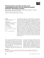
Fig. 1. Synthesis of GDP-
L
-fucose in mammals. The constitutively
active de novo pathway converts GDP-
D
-mannose into GDP-
L
-fucose
via oxidation, epimerization and reduction catalyzed by two enzymes,
GMD and FX. In the alternative salvage pathway, free fucose is
delivered to cytosol from extracellular sources (shown) or from lyso-
somal degradation of glycoconjugates (not shown).
L
-fucose is phos-
phorylated by
L
-fucokinase to form
L
-fucose-1-phosphate, which is
converted to GDP-
L
-fucose in the reaction catalyzed by GDP-
L
-fucose
pyrophosphorylase. GDP-
L
-fucose is then transported into the Golgi.
Fig. 2. Gene structures of the short and the long splice variants of mouse
fucokinase (A) and human fucokinase (B). The long splice variant of
mouse fucokinase contains exons 1–20, 21a, 22, 23a and 24. The short
splice variant contains exons 1–20, 21b, 23b and 24. The human
fucokinase has a similar gene structure to the long splice variant of
mouse fucokinase at the 3¢ end.
Ó FEBS 2003 The salvage pathway of GDP-
L
-fucose in mouse (Eur. J. Biochem. 271)79
COS-7 cell lysates (30 lg) were detected by Western
blotting using anti-(E2-Tag) primary mAb (Quattromed)
and anti-(mouse IgG) HRP-conjugated secondary anti-
body. Detection was performed using enhanced chemilu-
minescence (Amersham Biosciences, Bucks, UK) according
to standard methods.
Fucokinase activity assay
Cell lysate (100 lg) was assayed in a 100 lL reaction
mixture containing 50 m
M
Tris/HCl (pH 8.0), 5 m
M
MgSO
4
, 150 000 c.p.m.
L
-[
3
H]fucose (specific activity
63.0 CiÆmmol
)1
, Amersham), 0.1 m
M
fucose (Sigma,
St Louis, MO, USA), 5 m
M
ATP and 5 m
M
NaF (Sigma)
in final concentrations. The reaction mixture was incubated
at 37 °C for 30 min and terminated with 100 lL of ethanol.
The incubation mixture was applied to two 10 cm DEA-
Bond Elut column (Varian, Palo Alto, CA, USA), which
was then washed with four column volumes of 10 m
M
NH
4
HCO
3
to remove the unbound material. The [
3
H]fu-
cose-1-P was eluted with 2 mL of 250 m
M
NH
4
HCO
3
.The
eluate (400 lL) was counted with liquid scintillation and
luminescence counter (Wallac Trilux, Turku, Finland).
Cloning of GDP-
L
-fucose pyrophosphorylase
The 3¢ end of the pyrophosphorylase gene was cloned from
the mouse kidney UNI-ZAP XR lambda cDNA library
(Stratagene, La Jolla, CA, USA) by screening of approxi-
mately 1 · 10
6
recombinant plasmids. The published
human GDP-
L
-fucose pyrophosphorylase (accession num-
ber AF017445) [23] was used in a BLAST search to locate
mouse ESTs corresponding to the putative pyrophospho-
rylase. According to the EST sequence (AA422658), the
forward primer 5¢-GAGTATTCTAGATTGGGGCCT
GA-3¢ and reverse primer 5¢-TGTGGACTGCACGCA
TTTTCC-3¢ were designed. PCR was performed using
mouse liver cDNA (QuickClone cDNA, Clontech) as a
template. The 330 bp PCR product was labelled with
[
32
P]dCTP[aP] using the Multiprime DNA labelling kit
(Amersham Biosciences, Buckinghamshire, UK) according
to the manufacturer’s protocol. The labelled probe was used
in colony hybridization according to standard methods.
Theentire5¢ end was resolved by 5¢ RACE-PCR (Robust
RT-PCR kit, Finnzymes, Espoo, Finland) using mouse
kidney mRNA (Clontech) as a template. PCR was
performed using the 5¢ RACE synthesis primer AP1
(Clontech), 5¢-CCATCCTAATACGACTCACTATAGG
GC-3¢ and the gene-specific reverse primer, 5¢-GACTCC
AGGCCTCATGTTTGAGGGGAAATCCACGTAC-3¢.
The second round PCR was performed with a nested
adaptor primer AP2 (Clontech), 5¢-ACTCACTATAGG
GCTCGAGCGGC-3¢ together with a nested gene-specific
primer, 5¢-CAAACACTCAAGGGAACAAAG-3¢.All
PCR products were cloned into pCRÒ-Blunt II-TOPOÒ
vector (Invitrogen).
The enzymatic activity of GDP-fucose pyrophosphorylase
The coding sequence of pyrophosphorylase was amplified
by PCR using the forward primer, 5¢-AAT
GGTACC
ATGGCGTCTCTCCGCGA-3¢ and the reverse pri-
mer, 5¢-CAC
GGATCCTTAAGATTTCTCTAAATCAG-3¢
creating KpnI and BamHI restriction enzyme recognition
sites (underlined), respectively. Subcloning of the PCR
product into a pCDNA3.1(+) vector (Invitrogen) and
the transient transfection into COS-7 cells were per-
formed as above. The cells were lysed on ice with 50 m
M
Tris/HCl (pH 7.5) including protease inhibitor cocktail
(Pharmingen), with sonication for 3 · 15 s (Branson
Sonifier 450, Heinemann, Schwa
¨
bich Gmund, Austria).
Cell lysates (60 lg) were incubated in a 50 lL reaction
mixture containing 0.5
M
Tris/HCl (pH 7.8), 200 m
M
MgCl
2
,10m
M
b-
L
-Fuc-1P (Sigma), 100 m
M
GTP, 0.5 U
inorganic pyrophosphorylase (Sigma) at 37 °C for
30 min.
Nucleotide sugars were purified from the cell lysates as
described by Rabina et al. [26] and analyzed by ion-pair
reversed-phase HPLC on a Discovery HS C18 column
(0.46 · 25 cm; Supelco Inc., Pennsylvania, PE, USA) at a
flow rate of 1 mLÆmin
)1
. A linear gradient of 0–1.5% (v/v)
acetonitrile in 20 m
M
triethyammonium acetate buffer
(pH 7.0) over 35 min was used and the effluent was
monitored at 254 nm. The amount of synthesized GDP-
L
-fucose was calculated using the peak areas of external
nucleotide sugar standards GDP-
D
-mannose, GDP-
D
-rhamnose [27] and GDP-
L
-fucose (Calbiochem, San
Diego, CA, USA). The fraction containing the putative
GDP-
L
-fucose was collected from the HPLC assay for
further analysis with MALDI-TOF MS.
MALDI-TOF-MS
MALDI-TOF MS was performed with a Biflex III mass
spectrometer (Bruker Daltonics, Bremen, Germany). Nuc-
leotide sugars were investigated in a 2,4,6-trihydroxy-
acetonephenone–acetonitrile–aqueous ammonium citrate
matrix as described previously [26], utilizing the reflector
negative-ion mode with delayed extraction. External calib-
ration was performed with TDP-
D
-rhamnose (a generous
gift from P. Messner, Universita
¨
tfu
¨
r Bodenkultur Wien,
Wien, Austria) and UDP-GlcNAc (Sigma). GDP-
L
-fucose
(Calbiochem) was used as a positive control.
Reverse transcription and quantitative real time PCR
Fucokinase and pyrophosphorylase mRNA expression in
different tissues was detected by quantitative real time PCR.
Ambion’s Mouse Total RNA (kidney, liver, brain, ovary,
testicle, heart, lung, spleen) was used for the first strand
cDNA synthesis. For each tissue, 1 lg of total RNA was
reverse transcribed with random hexamers using the Invi-
trogen SuperScriptÒ cDNA synthesis kit according to the
manufacturer’s instructions. Parallel reactions in the absence
of SuperScript IIÒ (–RT controls) were performed to assess
the degree of contaminating genomic DNA. The resulting
cDNA samples were subjected to real time quantitative PCR
assay [28] to detect the expression levels of pyrophosphory-
lase and long and short splice variants of fucokinase. Primers
and probes were designed using the
PRIMER EXPRESS
program
(Version 1, PE Applied Biosystems, Foster City, CA, USA),
a software tool provided with the ABI 7000 Sequence
Detection System (PE Applied Biosystems). Forward and
reverse primers were positioned as close as possible to each
80 J. Niittyma
¨
ki et al. (Eur. J. Biochem. 271) Ó FEBS 2003
other without overlapping the probe. Probes were synthes-
ized incorporating the fluorescent reporter FAM (6-carboxy-
fluorescein) at the 5¢ end and the quencher TAMRA
(6-carboxy-tetramethyl-rhodamine) at the 3¢ end.
One microlitre of freshly synthesized cDNA was ampli-
fied in a total volume of 25 lL containing 1· Universal
Master Mix (PE Applied Biosystems) on an ABI Prism 7000
Sequence Detection System. Assays for each transcript were
carried out as duplicates on the same plate and real time
PCR amplification was repeated twice. Any inefficiencies in
RNA input or reverse transcription were corrected by
normalization to a housekeeping gene (18S rRNA Control
Reagents, PE Applied Biosystems). Primer concentrations
used were 900 n
M
/300 n
M
(forward/reverse) for the long
splice variant of fucokinase, 900 n
M
/900 n
M
(forward/
reverse) for the short splice variant of fucokinase, and
300 n
M
/900 n
M
(forward/reverse) for pyrophosphorylase.
The concentration of the double labelled probe was 200 n
M
for the long variant of fucokinase and pyrophosphorylase,
and 300 n
M
for the short fucokinase splice variant. Relative
amounts of the three mRNAs analyzed were based on
standard curves (Applied Biosystems User Bulletin 2)
prepared by a serial dilution of control cDNA.
Results
Cloning of putative mouse
L
-fucokinase and sequence
analysis
Using the three known pig fucokinase peptides [21] as
probes, part of the putative murine fucokinase sequence was
identified from mouse genomic sequence from the EMBL/
GenBank/DDBJ database. This sequence was cloned from
mouse kidney cDNA and used as a query in order to find
thecompletesequencefromthedatabase.TheIMAGE
clone 4190449 contained the full CDS of a putative mouse
L
-fucokinase, which was utilized in the design of primers for
RT-PCR. Two putative cDNAs of different sizes were
cloned representing two splice variants of
L
-fucokinase. The
long splice variant of
L
-fucokinase consisted of 3270 bp,
encoding a protein of 1090 amino acids. The sequence of the
shorter cDNA was similar to the sequence of the IMAGE
clone 4190449, consisting of 3057 bp. The short splice
variant did not code for amino acids 921–992 present in the
long splice variant, thus the short version consisted of 1019
amino acids [Fig. 3].
The long splice variant of
L
-fucokinase contains exons
1–20, 21a, 22, 23a and 24 whereas the short one contains
exons 1–20, 21b, 23b and 24. As can be seen in Fig. 2, exons
21b and 23b are wholly included in the longer variants of
these exons (21a and 23a respectively). The splice junction
from exon 20 to exon 21a or 21b is not affected, neither is
the splice junction between exon 23a or 23b, and exon 24. In
conclusion, the alternative splicing maintains the reading
frame along the entire protein, therefore the protein variants
are identical in the amino-terminal end up to the alternative
splice area, in addition to the carboxy-terminal end after the
alternative splice area.
There are three methionine codons (ATG) within a
300 bp region at the upstream end of the longest open
reading frame in the mouse fucokinase mRNA sequence
(accession number AJ534942). The first ATG is estimated
to be the most probable CDS initiation site based on a
probabilistic model using multiple parameters, including the
Kozak translation initiation signal, as implemented in the
GENSCAN
analysis tool [28].
Expression of fucokinase in mammalian cells
The two splice variants of the murine fucokinase genes were
expressed in COS-7 cells in frame with a 10-amino acid E2-
Tag present in the pQM vector. The molecular masses
of fucokinase proteins were determined by Western blot
analysis; the tagged long splice variant had a mass of
125 kDa and the tagged short splice variant a mass of
115 kDa. Both E2-Tagged splice variants had slightly
greater molecular masses than the predicted 120 and
111 kDa proteins, respectively [Fig. 4].
The production of
L
-fucose-1-phosphate from
L
-[
3
H]fu-
cose and ATP was measured in order to determine whether
the expressed splice variants of fucokinase were functionally
active. The long splice variant showed significant enzyme
activity; the specific enzyme activity was determined to be
598.5 pmolÆmg
)1
Æh
)1
in transfected COS-7 cells. The activity
of the short splice variant was only marginally higher
(13.7 pmolÆmg
)1
Æh
)1
) than the background in the COS-7
cells (11.4 pmolÆmg
)1
Æh
)1
). Human
L
-fucokinase, IMAGE
clone 4179554 (AJ441184) [22], was also transfected into
COS-7 cells and assayed in regard to fucokinase activity. The
specific enzyme activity of the human
L
-fucokinase was the
same level, 12.3 pmolÆmg
)1
Æh
)1
, as the activity of the shorter
mouse splice variant and the vector control (Fig. 5A).
L
-fucokinase activity is present in many different tissues,
and exhibits high activity in kidney [21]. The COS-7 cell line
is derived from monkey kidney cells and thus has some
intrinsic fucokinase activity. In order to discriminate the
possible fucokinase activity of a short splice variant from the
kidney cell backround, the short splice variant was also
transfected into epithelial HeLa S3-cells. The relatively weak
enzymatic activity of the short splice variant could be
detected in HeLa cells; the specific enzyme activity was
30.6 pmolÆmg
)1
Æh
)1
whereas the specific activity of the mock
control was 8.4 pmolÆmg
)1
Æh
)1
(Fig. 5b).
Cloning of murine GDP-
L
-fucose pyrophosphorylase
The cloned human pyrophosphorylase (accession number
AF017445) [23] was used as a query in BLAST searches to
find a mouse EST corresponding to the putative pyro-
phosphorylase. Using this mouse EST as a probe, the 3¢ end
of the GDP-
L
-fucose pyrophosphorylase was cloned from a
mouse kidney cDNA library by screening 1 · 10
6
recom-
binant plasmids. The 5¢ end of the sequence was resolved by
the RACE-PCR method, using mouse kidney mRNA as the
template as described in Experimental procedures. The
isolated cDNA consisted of 3480 bp, and the predicted CDS
encoded a protein of 591 amino acids [Fig. 6].
Pyrophosphorylase activity assay and the identification
of GDP-
L
-fucose
Because we could detect only a faint protein band in SDS/
PAGE from cell lysate with the estimated molecular mass of
65.5 kDa that relates to GDP-
L
-fucose pyrophosphorylase
Ó FEBS 2003 The salvage pathway of GDP-
L
-fucose in mouse (Eur. J. Biochem. 271)81
(data not shown), we decided to identify accurately the
product of a GDP-
L
-pyrophosphorylase assay. The cell
lysate expressing the pyrophosphorylase gene was incubated
with
L
-fucose-1-phosphate and GTP, and the resulting
product of the reaction was analyzed by ion-pair reversed-
phase HPLC. The analysis revealed a peak with the same
retention time as the GDP-
L
-fucose standard (F) at
29.6 min in a sample containing the pyrophosphorylase,
whereas the vector control gave only a faint peak at
29.7 min [Fig. 7]. The peak was purified and subjected to
further analysis by MALDI-TOF MS, which gave a single
peak at 588.08 m/z, thus being identical to the GDP-
L
-
fucose control.
Quantitative PCR and tissue distribution levels
of
L
-fucokinase and pyrophosphorylase
The primer and probe sequences and their positions in the
mRNA sequence, for GDP-
L
-fucose pyrophosphorylase
and the short and long splice variants of
L
-fucokinase, are
listed in Table 1.
Various mouse tissues were analyzed for the expression of
the three enzymes (GDP-
L
-fucose pyrophosphorylase, and
short and long splice variants of
L
-fucokinase) involved in
the salvage pathway of GDP-
L
-fucose, to elucidate the
possible differences between the various tissues. Moreover,
the ratio of long to short splice variants of
L
-fucokinase in
Fig. 3. Nucleotide sequence and deduced amino acid sequence of mouse
L
-fucokinase. The predicted amino acid sequence for the coding area of the
long splice variant of fucokinase consists of 1090 amino acids. Due to alternative splicing, the amino acids 921–992 (bold letters) are not coded in
the short splice variant of fucokinase. The amino acids corresponding to the published peptide sequences of pig fucokinase [21] are underlined. The
sequence data of the short splice variant is available in the EMBL/GenBank/DDBJ Nucleotide Sequence Databases under Accession No. AJ297482
and the long splice variant under the Accession No. AJ534942.
82 J. Niittyma
¨
ki et al. (Eur. J. Biochem. 271) Ó FEBS 2003
different tissues was also determined (Table 2). Relative
expression levels, shown in Fig. 8, were calculated following
normalization to 18S RNA. In the subsequent calculations,
expression levels of those enzymes found in mouse liver were
assigned a relative expression value of one. The expression
of both splice variants of
L
-fucokinase was found to be
relatively high in brain, ovary, testis and kidney. In spleen,
heart and lung the expression was lower. When calculating
the ratio between the long and short splice variants of
fucokinase it could be seen that the long splice variant was
more abundantly expressed in liver, kidney, ovary, testis,
spleen and heart. In the lung the expression levels were
equal, whereas in brain the expression of the short splice
Fig. 5. Fucokinase activities of the cell lysates of COS-7 cells (A) and
HeLa cells (B) transfected with the fucokinase cDNAs. Enzyme activity
is expressed as pmol of
L
-[
3
H]fucose incorporated onto ATP per hour
devided by the total protein content. (A) Enzyme activities of COS-7
cells, transfected with the short and long splice variants of mouse
fucokinase (mFK) and human fucokinase (hFK, AJ441184). (B)
Fucokinase activities of HeLa cells transfected with vector or the short
splice variant of mouse fucokinase.
Fig. 4. Western blot analysis of the expressed murine
L
-fucokinase in
COS-7 cells detected with E2-Tag antibodies. Lane 1, negative COS-7
cell control; lane 2, short splice variant of mouse fucokinase and lane 3,
long splice variant of mouse fucokinase.
Fig. 6. Nucleotide sequence and deduced amino acid sequences of murine
GDP-
L
-fucose pyrophosphorylase. The 3.5 kb nucleotide sequence
predicts an amino acid sequence of 590 residues for the coding region
of GDP-
L
-fucose pyrophosphorylase. The sequence data is available in
the EMBL/GenBank/DDBJ Nucleotide Sequence Databases under
Accession No. AJ276067.
Ó FEBS 2003 The salvage pathway of GDP-
L
-fucose in mouse (Eur. J. Biochem. 271)83
variant was higher than that of the long
L
-fucokinase splice
variant. The expression pattern of GDP-
L
-fucose pyro-
phosphorylase resembles the pattern of
L
-fucokinase, i.e.
expression was high in brain, ovary, testis and kidney.
Again, the expression levels were lower in liver, spleen, heart
and lung (Fig. 8C).
Discussion
The de novo synthesis of GDP-
L
-fucose, that converts GDP-
D
-mannose to GDP-
L
-fucose, is evolutionary conserved and
the enzymes involved in this pathway have been cloned
from several bacteria [17], plants [29] and mammals [30]. In
addition, the de novo synthesis of GDP-
L
-fucose has been
characterized in silico from the fruit fly [31]. The alternative
pathway of GDP-
L
-fucose synthesis, the salvage pathway,
allows cells to activate monosaccharides that come from
nutrition or from lysosomal degradation of glycoproteins
and glycolipids. The sugars are phosphorylated by kinases
and activated by pyrophosphorylases. To date the salvage
pathway of GDP-
L
-fucose has been identified only in
mammals [21,23]. The specific salvage pathway is also found
for UDP-galactose, UDP-glucuronic acid and UDP-N-
acetylgalactosamine [32].
The salvage pathway of GDP-
L
-fucose involves
L
-fuco-
kinase which catalyzes the transfer of phosphate from ATP
to free
L
-fucose, forming
L
-fucose-1-phosphate. GDP-
L
-
fucose pyrophophorylase then condensates
L
-fucose-
1-phosphate with GTP to form GDP-
L
-fucose. In the
present study we have cloned the murine enzymes involved
in the salvage pathway of GDP-
L
-fucose and expressed
them as functionally active enzymes. Two splice variants of
L
-fucokinase were cloned, but only the long splice variant
was enzymatically active when expressed in mammalian
cells. The short splice variant did not show significant
Table 1. Probe and primer sequences in quantitative PCR. FK short, short splice variant of
L
-fucokinase; FK long, long splice variant of
L
-fucokinase; PP, GDP-l-fucose pyrophosphorylase; F, forward primer; R, reverse primer; P, probe.
Target gene Primer/Probe sequence Starting position in mRNA Length of amplicon
FK short F 5¢-CTGAGGGTTTGTCCCAGAA-3¢ 2786 103 bp
R5¢-GGCTTTGGCCATACGCATAC-3¢ 3081
P
a
5¢-ACGGCCAGCGGCTCGCA-3¢ 3037
FK long F 5¢-GCAGGACGTGCTGAGGAACT-3¢ 2865 64 bp
R5¢-CAGTCTGCGGGCATTCTGT-3¢ 2911
P
a
5¢-CCACAACGGGCAACCGAGCG-3¢ 2889
PP F 5¢-AGCTGGGCTTACAATCCATAGCT-3¢ 1118 79 bp
R5¢-TGAATGACACAGGCTGTTCCA-3¢ 1176
P5¢-AGTGTCTCTCCAAGTGTTCCTGAGCGCT-3¢ 1144
a
Probe is antisense strand.
Table 2. Ratio of long splice variant to short splice variant of
L
-fucokinase.
Tissue Ratio
Liver 1.52
Kidney 1.34
Ovary 1.40
Testis 1.28
Spleen 3.21
Brain 0.77
Heart 1.15
Lung 0.99
Fig. 7. Ion-pair reversed-phase HPLC analysis of the product of the
enzymatic reaction catalyzed by GDP-
L
-fucose pyrophosphorylase. (A)
Vector control in COS-7 cell lysate; (B) putative mouse pyrophos-
phorylase in COS-7 cell lysate; (C) GDP-sugar standards, 500 pmol of
each. M, GDP-
D
-mannose, 18.6 min; R, GDP-
D
-rhamnose, 24.4 min;
F, GDP-
L
-fucose, 29.6 min.
84 J. Niittyma
¨
ki et al. (Eur. J. Biochem. 271) Ó FEBS 2003
enzymatic activity, but was expressed abundantly in many
tissues, especially in brain, which may indicate an uniden-
tified role for this variant.
When comparing both splice variants of mouse
L
-fuco-
kinase cDNA sequence with the previously published human
L
-fucokinase cDNA sequence (accession number AJ441184)
[22], it can be observed that the human fucokinase cDNA is
similar to the long splice variant of mouse fucokinase cDNA
at the 3¢ splice region. The first and third methionines in the
murine sequence, in the upstream end of the CDS, are also
found in the human sequence (e.g. BC032542) while the
second one has evolved into a leucine. Although the
beginning of the human CDS has been proposed to start
from the position that corresponds to the third ATG in the
murine sequence [22], we suggest that the first ATG would be
a better starting codon than the third one; indeed, it is
predicted to be the first triplet in the CDS by several gene
prediction tools, e.g.
GENSCAN
analysis tool [28]. Further-
more, a high degree of sequence similarity exists between the
mouse and the human cDNA sequences upstream of the
third ATG, suggesting that this segment is part of the CDS.
In conclusion, we propose that the CDS starts not at the third
but at the first ATG in the murine sequence, and that the
human CDS starts at the corresponding position. Thus, we
suggest that the human CDS of
L
-fucokinase becomes 94
amino acids longer than the corresponding CDS in the
previous study [22].
L
-fucose is a fundamental component of many mamma-
lian glycoproteins and glycolipids. Fucosylation requires
GDP-
L
-fucose as a donor of
L
-fucose, and a specific
fucosyltransferase to catalyze the transfer of
L
-fucose to
the acceptor molecules. The synthesis of GDP-
L
-fucose and
its import into the Golgi lumen for a specific fucosyltrans-
ferase is essential for selectin-dependent leukocyte traffick-
ing and for normal human development. Leukocyte
adhesion deficiency type 2 (LADII), also known as a
congenital disorder of glycosylation IIc, is a rare human
disorder of fucose metabolism in which the patient suffers
from recurrent infection, persistent leukocytosis and severe
mental and growth retardation [33,34]. Missense mutations
in a Golgi-localized GDP-fucose transporter lead to parti-
ally defective function and are responsible for the defective
fucosylation in LADII patients [35,36]. Studies with LADII
patients show that oral supplementation of fucose can
restore selectin ligands and correct the immunodeficiency
[37,38]. In this scenario, GDP-
L
-fucose is synthesized from
oral fucose through the salvage pathway, which elevates the
amountofGDP-
L
-fucose in the cytosol, leading to
enhanced GDP-fucose uptake into the Golgi [35]. In a
study by Smith et al. [39], the targeted disruption of the FX
locus in the mouse ablates the de novo pathway for GDP-
fucose synthesis from GDP-mannose causing adult animals
to lack almost completely the fucosylated glycans in
multiple tissues, leading to symptoms similar to those of
LADII. The FX-deficient mice are completely dependent on
dietary fucose, which restores the synthesis of GDP-fucose
through the salvage pathway.
The salvage metabolism accounts for approximately only
10% of the intracellular pool of GDP-
L
-fucose [40].
However, the enzymes of the salvage pathway are expressed
with relatively high intensities in various animal tissues, e.g.
brain, ovary, testis, kidney and liver, as shown by the
quantitative real time PCR analysis in the present study and
also in previous studies [21,23]. The wide expression of the
enzymes involved in the salvage pathway of GDP-
L
-fucose
can also be deduced from the large amount of data available
from different EST libraries (e.g. .
gov/UniGene). Our analysis of the expression of the enzymes
involved in the salvage pathway of GDP-
L
-fucose indicates
that not only the de novo pathway alone, but also the salvage
pathway could have an essential role in the synthesis of
GDP-
L
-fucose in the cytosol. The importance and the
regulatory mechanisms of the enzymes in the salvage
pathway of GDP-
L
-fucose have not been elucidated, thus
futher studies are needed.
Acknowledgements
We thank Tuula Kallioinen and Sirkka-Liisa Kauranen for skilled
technical assistance in molecular biology, and Kati Vena
¨
la
¨
inen and
Leena Penttila
¨
for assistance in HPLC and MALDI-TOF MS analysis.
The work was supported in part by Research Grants of the Academy of
Finland, the Technology Development Center (TEKES), Helsinki, the
Sigrid Juselius Foundation, and the Helsinki University Central
Hospital Fund (EVO).
Fig. 8. Tissue expression patterns of murine
L
-fucokinase short and long
splice variants and GDP-
L
-fucose pyrophosphorylase. The expression
levels of the long splice variant of
L
-fucokinase (A), the short splice
variant of
L
-fucokinase (B) and GDP-
L
-fucose pyrophosphorylase (C)
were detected by quantitative real time PCR. The mRNA expression
levels in each tissue were expressed relative to expression in the liver.
Ó FEBS 2003 The salvage pathway of GDP-
L
-fucose in mouse (Eur. J. Biochem. 271)85
References
1. Walz,G.,Aruffo,A.,Kolanus,W.,Bevilacqua,M.&Seed,B.
(1990) Recognition by ELAM-1 of the sialyl-Lex determinant on
myeloid and tumor cells. Science 250, 1132–1135.
2. Harris, R.J. & Spellman, M.W. (1993) O-linked fucose and other
post-translational modifications unique to EGF modules. Glyco-
biology 3, 219–224.
3. Staudacher, E., Altmann, F., Wilson, I.B. & Marz, L. (1999)
Fucose in N-glycans: from plant to man. Biochim. Biophys. Acta
1473, 216–236.
4. Greenwell, P. (1997) Blood group antigens: molecules seeking a
function? Glycoconjugate J. 14, 159–173.
5. Lloyd, K.O. (2000) The chemistry and immunochemistry of blood
group A, B, H, and Lewis antigens: past, present and future.
Glycoconjugate J. 17, 531–541.
6. Lasky, L.A. (1992) Selectins: interpreters of cell-specific carbo-
hydrate information during inflammation. Science 258, 964–969.
7. Lowe, J.B. (1997) Selectin ligands, leukocyte trafficking, and fuc-
osyltransferase genes. Kidney Int. 51, 1418–1426.
8. Mori, E., Hedrick, J.L., Wardrip, N.J., Mori, T. & Takasaki, S.
(1998) Occurrence of reducing terminal N-acetylglucosamine
3-ulfate and fucosylated outer chains in acidic N-glycans of porcine
zona pellucida glycoproteins. Glycoconjugate J. 15, 447–456.
9. Johnston, D.S., Wright, W.W., Shaper, J.H., Hokke, C.H., Vanden
Eijnden, D.H. & Joziasse, D.H. (1998) Murine sperm-zona bind-
ing, a fucosyl residue is required for a high affinity sperm-binding
ligand. A second site on sperm binds a nonfucosylated, beta-ga-
lactosyl-capped oligosaccharide. J. Biol. Chem. 273, 1888–1895.
10. Xiang, J. & Bernstein, I.A. (1992) Differentiative changes in
fucosyltransferase activity in newborn rat epidermal cells.
Biochem. Biophys. Res. Commun. 189, 27–32.
11. Ruggiero-Lopez, D., Biol, M.C., Louisot, P. & Martin, A. (1991)
Participation of an endogenous inhibitor of fucosyltransferase
activities in the developmental regulation of intestinal fucosylation
processes. Biochem. J. 279, 801–806.
12. Moloney, D.J., Panin, V.M., Johnston, S.H., Chen, J., Shao, L.,
Wilson,R.,Wang,Y.,Stanley,P.,Irvine,K.D.,Haltiwanger,R.S.
& Vogt, T.F. (2000) Fringe is a glycosyltransferase that modifies
Notch. Nature 406, 369–375.
13. Bruckner, K., Perez, L., Clausen, H. & Cohen, S. (2000) Glyco-
syltransferase activity of Fringe modulates Notch–Delta inter-
actions. Nature 406, 411–415.
14. Fukuda, M. (1996) Possible roles of tumor-associated carbo-
hydrate antigens. Cancer Res. 56, 2237–2244.
15. Hiraishi, K., Suzuki, K., Hakomori, S. & Adachi, M. (1993) Le(y)
antigen expression is correlated with apoptosis (programmed cell
death). Glycobiology 3, 381–390.
16.Tonetti,M.,Sturla,L.,Bisso,A.,Benatti,U.&DeFlora,A.
(1996) Synthesis of GDP-
L
-fucose by the human FX protein.
J. Biol. Chem. 271, 27274–27279.
17.Tonetti,M.,Sturla,L.,Bisso,A.,Zanardi,D.,Benatti,U.&
De Flora, A. (1998) The metabolism of 6-deoxyhexoses in
bacterial and animal cells. Biochimie 80, 923–931.
18. Ishihara, H., Massaro, D.J. & Heath, E.C. (1968) The metabolism
of
L
-fucose. III. The enzymatic synthesis of beta-
L
-fucose
1-phosphate. J. Biol. Chem. 243, 1103–1109.
19. Ishihara, H. & Heath, E.C. (1968) The metabolism of
L
-fucose. IV.
The biosynthesis of guanosine diphosphate
L
-fucose in porcine
liver. J. Biol. Chem. 243, 1110–1115.
20. Kilker, R.D., Shuey, D.K. & Serif, G.S. (1979) Isolation and
properties of porcine thyroid fucokinase. Biochim. Biophys. Acta
570, 271–283.
21. Park, S.H., Pastuszak, I., Drake, R. & Elbein, A.D. (1998) Puri-
fication to apparent homogeneity and properties of pig kidney
L
-fucose kinase. J. Biol. Chem. 273, 5685–5691.
22. Hinderlich,S.,Berger,M.,Blume,A.,Chen,H.,Ghaderi,D.&
Bauer, C. (2002) Identification of human
L
-fucosekinaseami-
noacid sequence. Biochem. Biophys. Res. Commun. 294, 650–654.
23. Pastuszak, I., Ketchum, C., Hermanson, G., Sjoberg, E.J., Drake,
R. & Elbein, A.D. (1998) GDP-
L
-fucose pyrophosphorylase.
Purification, cDNA cloning, and properties of the enzyme. J. Biol.
Chem. 273, 30165–30174.
24. Altschul, S.F., Madden, T.L., Schaffer, A.A., Zhang, J., Zhang,
Z., Miller, W. & Lipman, D.J. (1997) Gapped BLAST and PSI-
BLAST: a new generation of protein database search programs.
Nucleic Acids Res. 25, 3389–3402.
25. Pearson, W.R. (1990) Rapid and sensitive sequence comparison
with FASTP and FASTA. Methods Enzymol. 183, 63–98.
26. Rabina, J., Maki, M., Savilahti, E.M., Jarvinen, N., Penttila, L. &
Renkonen, R. (2001) Analysis of nucleotide sugars from cell
lysates by ion-pair solid-phase extraction and reversed-phase
high-performance liquid chromatography. Glycoconjugate J. 18,
799–805.
27. Maki,M.,Jarvinen,N.,Rabina,J.,Roos,C.,Maaheimo,H.,
Mattila, P. & Renkonen, R. (2002) Functional expression of
Pseudomonas aeruginosa GDP-4-keto-6-deoxy-
D
-mannose reduct-
ase which synthesizes GDP-rhamnose. Eur. J. Biochem. 269,
593–601.
28. Heid, C.A., Stevens, J., Livak, K.J. & Williams, P.M. (1996) Real
time quantitative PCR. Genome Res. 6, 986–994.
29. Bonin, C.P., Potter, I., Vanzin, G.F. & Reiter, W.D. (1997) The
MUR1 gene of Arabidopsis thaliana encodes an isoform of GDP-
D
-mannose-4,6-dehydratase, catalyzing the first step in the de novo
synthesis of GDP-
L
-fucose. Proc. Natl Acad. Sci. USA 94,
2085–2090.
30. Reitman, M.L., Trowbridge, I.S. & Kornfeld, S. (1980) Mouse
lymphoma cell lines resistant to pea lectin are defective in fucose
metabolism. J. Biol. Chem. 255, 9900–9906.
31. Roos,C.,Kolmer,M.,Mattila,P.&Renkonen,R.(2002)Com-
position of Drosophila melanogaster proteome involved in fuco-
sylated glycan metabolism. J. Biol. Chem. 277, 3168–3175.
32. Bulter, T. & Elling, L. (1999) Enzymatic synthesis of nucleotide
sugars. Glycoconjugate J. 16, 147–159.
33. Etzioni, A., Frydman, M., Pollack, S., Avidor, I., Phillips, M.L.,
Paulson, J.C. & Gershoni-Baruch, R. (1992) Brief report:
recurrent severe infections caused by a novel leukocyte adhesion
deficiency. N. Engl. J. Med. 327, 1789–1792.
34. Becker, D.J. & Lowe, J.B. (1999) Leukocyte adhesion deficiency
type II. Biochim. Biophys. Acta 1455, 193–204.
35.Luhn,K.,Wild,M.K.,Eckhardt,M.,Gerardy-Schahn,R.&
Vestweber, D. (2001) The gene defective in leukocyte adhesion
deficiency II encodes a putative GDP-fucose transporter. Nat.
Genet. 28, 69–72.
36. Lubke, T., Marquardt, T., Etzioni, A., Hartmann, E., von Figura,
K. & Korner, C. (2001) Complementation cloning identifies CDG-
IIc, a new type of congenital disorders of glycosylation, as a GDP-
fucose transporter deficiency. Nat. Genet. 28, 73–76.
37. Marquardt, T., Luhn, K., Srikrishna, G., Freeze, H.H., Harms, E.
& Vestweber, D. (1999) Correction of leukocyte adhesion defi-
ciency type II with oral fucose. Blood 94, 3976–3985.
38. Luhn, K., Marquardt, T., Harms, E. & Vestweber, D. (2001)
Discontinuation of fucose therapy in LADII causes rapid loss
of selectin ligands and rise of leukocyte counts. Blood 97, 330–332.
39. Smith, P.L., Myers, J.T., Rogers, C.E., Zhou, L., Petryniak, B.,
Becker, D.J., Homeister, J.W. & Lowe, J.B. (2002) Conditional
controlofselectinligandexpressionandglobalfucosylationevents
in mice with a targeted mutation at the FX locus. J. Cell Biol. 158,
801–815.
40. Yurchenco, P.D. & Atkinson, P.H. (1977) Equilibration of
fucosyl glycoprotein pools in HeLa cells. Biochemistry 16,
944–953.
86 J. Niittyma
¨
ki et al. (Eur. J. Biochem. 271) Ó FEBS 2003