Báo cáo khóa học: Protein assembly of photosystem II and accumulation of subcomplexes in the absence of low molecular mass subunits PsbL and PsbJ pdf
Bạn đang xem bản rút gọn của tài liệu. Xem và tải ngay bản đầy đủ của tài liệu tại đây (399.38 KB, 12 trang )
Protein assembly of photosystem II and accumulation of subcomplexes
in the absence of low molecular mass subunits PsbL and PsbJ
Marjaana Suorsa
1
, Ralph E. Regel
2
, Virpi Paakkarinen
1
, Natalia Battchikova
1
, Reinhold G. Herrmann
2
and Eva-Mari Aro
1
1
Department of Biology, Plant Physiology and Molecular Biology, University of Turku, Finland;
2
Botanisches Institute der
Ludwig-Maximilians Universita
¨
t, Mu
¨
nchen, Germany
The protein assembly and stability of photosystem II (PSII)
(sub)complexes were studied in mature leaves of four plastid
mutants of tobacco (Nicotiana tabacum L), each having one
of the psbEFLJ operon genes inactivated. In the absence of
psbL, no PSII core dimers or PSII–light harvesting complex
(LHCII) supercomplexes were formed, and the assembly of
CP43 into PSII core monomers was extremely labile. The
assembly of CP43 into PSII core monomers was found to be
necessary for the assembly of PsbO on the lumenal side of
PSII. The two other oxygen-evolving complex (OEC) pro-
teins, PsbP and PsbQ, were completely lacking in DpsbL.In
the absence of psbJ, both intact PSII core monomers and
PSII core dimers harboring the PsbO protein were formed,
whereas the LHCII antenna remained detached from the
PSII dimers, as demonstrated by 77 K fluorescence meas-
urements and by the lack of PSII–LHCII supercomplexes.
The DpsbJ mutant was characterized by a deficiency of PsbQ
and a complete lack of PsbP. Thus, both the PsbL and PsbJ
subunits of PSII are essential for proper assembly of the
OEC. The absence of psbE and psbF resulted in a complete
absence of all central PSII core and OEC proteins. In con-
trast, very young, vigorously expanding leaves of all psb-
EFLJ operon mutants accumulated at least traces of D2,
CP43 and the OEC proteins PsbO and PsbQ, implying
developmental control of the expression of the PSII core and
OEC proteins. Despite severe problems in PSII assembly, the
thylakoid membrane complexes other than PSII were pre-
sent and correctly assembled in all psbEFLJ operon mutants.
Keywords: oxygen-evolving complex; photosystem II
assembly; photosystem II small subunits; psbEFLJ operon;
tobacco.
Photosystem II (PSII) is a multisubunit pigment–protein
complex that catalyses electron transfer from water to the
plastoquinone pool with concomitant evolution of oxygen.
The PSII reaction center core consists of the D1 and
D2 proteins, cytochrome b
559
(Cyt b
559
), the chloro-
phyll a-binding antenna proteins CP43 and CP47, and a
number of low molecular mass (LMM) proteins, the
functions and locations of which in PSII are still largely
unknown. They include both chloroplast-encoded (PsbH, I,
J, K, L, M, N, T and Z) and nucleus-encoded (PsbR, W and
X) proteins with generally only one membrane-spanning
helix [1]. During the past few years, enormous progress has
been made in determining the structure of PSII [2–4]. The
functional form of PSII is apparently a dimer [5]. The
oxygen-evolving complex (OEC) situated on the lumenal
side of PSII is composed of the PsbO (33 kDa), PsbP
(23 kDa) and PsbQ (17 kDa) proteins in higher plants. PSII
dimers further associate with the light-harvesting complex II
(LHCII) to form PSII–LHCII supercomplexes, the minor
antenna proteins CP24, CP26 and CP29 probably serving as
linker proteins [2,5,6]. It has been suggested that several
LMM proteins participate in PSII dimerization [7,8].
However, despite the available structure of PSII at 3.8
and 3.7 A
˚
resolution [3,4], the exact locations and roles of
most of the LMM proteins in the assembly and stability of
PSII remain to be determined.
Today it is a challenge to resolve the assembly steps of
PSII. Various approaches have been fruitful in analysing the
primary assembly steps of PSII [9]. The best-characterized
LMM proteins of PSII, the a and b subunits of Cyt b
559
,
probably function as an assembly core, which is required for
the synthesis of the D2 protein [10]. Indeed, it has been
shown that Cyt b
559
and the D2 protein exist as a complex
in etiolated barley leaves [11]. The full-length D1 protein,
however, is synthesized only in the light and is cotransla-
tionally associated with the D2–Cyt b
559
complex [12].
Radiolabeling experiments have demonstrated that the
subsequent assembly steps include association of CP47
followed by that of CP43 [13]. Labeling experiments,
however, are unable to reveal all the different steps in the
sequential and hierarchical assembly of multisubunit PSII.
In particular, the assembly of the LMM subunits, except the
Cyt b
559
subunits, has been difficult to address. This is
because separation of the various subcomplexes after
assembly of each of the LMM subunits is not possible
Correspondence to E M. Aro, Department of Biology, Plant Physio-
logy and Molecular Biology, FIN-20014 University of Turku,
Finland. Fax: + 358 2333 5549, Tel.: + 358 2333 5931,
E-mail: evaaro@utu.fi
Abbreviations: BN, Blue-native; Cyt, cytochrome; LHCII, light-
harvesting complex II; LMM, low molecular mass; OEC, oxygen-
evolving complex; PSI, photosystem I; PSII, photosystem II.
(Received 5 September 2003, revised 28 October 2003,
accepted 4 November 2003)
Eur. J. Biochem. 271, 96–107 (2004) Ó FEBS 2003 doi:10.1046/j.1432-1033.2003.03906.x
because of resolution problems and, furthermore, only some
of the LMM subunits of PSII incorporate [
35
S]methionine.
Another approach to understanding the assembly of
LMM subunits into PSII is to use specific PSII protein
deletion mutants and to analysethe ability of such mutants to
form various PSII subassemblies. This approach has only
seldom been taken because of technical problems, and, when
applied, the fractionation of PSII subcomplexes has been
based in sucrose-density centrifugation with limited resolu-
tion capacity [14]. Moreover, none of the numerous studies
with Synechocystis 6803 mutants of the LMM subunits of
PSII has addressed the PSII assembly process as such, but
instead the focus has been on functional properties of PSII
and the overall synthesis or composition of thylakoid
polypeptides. Furthermore, despite remarkable similarities
between cyanobacterial and chloroplast PSIIs [15], many of
the PSII LMM subunits, which are completely dispensable
for the assembly of PSII in Synechocystis, are necessary for
the formation of functional PSII in the respective LMM
mutants of Chlamydomonas reinhardtii.Representative
examples of differential effectson the formation of functional
PSII in Synechocystis and Chlamydomonas are the deletion
mutants of psbH [16,17], psbI [18,19] and psbK [20,21].
However, it is not known at which assembly step these
proteins are crucial for the formation of functional PSII in
Chlamydomonas. So far only a few studies have seriously
searched for PSII assembly intermediates in the absence of
any particular LMM subunit, in either Synechocystis or
chloroplasts of Chlamydomonas and higher plants.
The psbEFLJ operon of plant chloroplasts encodes four
distinct LMM subunits of PSII, the a and b subunits of
Cyt b
559
(encoded by the psbE and psbF genes) and two
other small subunits, PsbL and PsbJ. Deletion of the psbE
gene in Chlamydomonas [22] or the psbF gene in Synecho-
cystis [23] resulted in loss of PSII activity. Similarly, the psbL
deletion mutant of Synechocystis was not capable of PSII
oxygen evolution [24]. The crucial role of PsbL has been
suggested to be related to the function of the acceptor side of
PSII at the level of Q
A
[25,26]. On the other hand, the psbJ
deletion mutants of cyanobacteria were capable of slow
photoautotrophic growth [27,28], whereas the growth of
DpsbJ tobacco mutants was completely dependent on an
external energy source [28,29], possibly because of an
incorrectly assembled OEC [29].
Recently, very young leaves of tobacco psbEFLJ operon
mutants were characterized in terms of functional, structural
and biogenetic aspects [14]. To differentiate the mechanisms
related to the rapid growth and division of chloroplasts in
young leaves from mechanisms of the PSII assembly process
as such, we mainly focused on mature, but not old, leaves of
tobacco psbEFLJ operon mutants, where partial disassem-
bly and assembly of PSII is constantly occurring because of
turnover of the reaction center D1 protein [30]. In
particular, the role of PsbL and PsbJ in the assembly and
stability of PSII was addressed. To maximize separation of
PSII subcomplexes, we applied 2D Blue-native (BN) gel
electrophoresis followed by protein identification with
immunoblotting and MS. In addition, comparative analysis
of both very young and mature leaves was performed to
examine the developmental aspects of PSII core and OEC
protein accumulation in the psbEFLJ operon mutants with
impaired PSII assembly.
Materials and methods
Transformation of tobacco chloroplasts
Tobacco (Nicotiana tabacum cv. Petit Havanna) psbEFLJ
operon mutants were constructed by replacing portions of
the four individual genes of the operon with a terminator-
less aadA gene cassette. A similar cassette with a terminator
wasalsoinsertedintoanEcoRV site, located in the 3¢ UTR
of the operon, to generate the RV control plants. The
plasmid construct and the transformation, selection and
culture of the transformants is described in detail elsewhere
[14,28,31]. Mutants and controls (wild-type and RV plants)
were aseptically grown in MS medium [32] supplemented
with 3% (w/v) sucrose under low light conditions
( 10 lmol photonsÆm
)2
Æs
)1
)at25°C. Mature, fully
expanded green leaves, but not senescing ones (hereafter
referred to as mature leaves), were used for all experiments
except the one in Fig. 1B where, for comparison, rapidly
expanding small and very young leaves (hereafter referred to
as young leaves) were used.
Isolation of thylakoid membranes
Leaves were briefly homogenized in 50 m
M
Hepes/KOH,
pH 7.5, containing 330 m
M
sorbitol, 2 m
M
EDTA, 1 m
M
MgCl
2
,5m
M
ascorbate, 0.05% BSA and 10 m
M
NaF,
filtered through Miracloth and centrifuged at 2500 g for
4minat4°C. The pellet was resuspended in 50 m
M
Hepes/
KOH, pH 7.5, containing 5 m
M
sorbitol and 10 m
M
NaF
and centrifuged at 2500 g for 4 min at 4 °C. The thylakoid
pellet was resuspended in 50 m
M
Hepes/KOH, pH 7.5,
containing 100 m
M
sorbitol, 10 m
M
MgCl
2
and 10 m
M
NaF, centrifuged at 2500 g for 3 min at 4 °C, and finally
resuspended in the same buffer. Chlorophyll was extracted
in 80% (v/v) buffered acetone (2.5 m
M
Hepes/NaOH,
pH 7.5) and quantitated as described [33].
BN-PAGE, SDS/PAGE and protein identification
Blue-native PAGE (BN-PAGE) was performed as des-
cribed previously [34] with slight modifications. Thylakoid
membrane suspensions containing 20 lg chlorophyll were
used as starting material. Thylakoids were washed with
50 m
M
BisTris/HCl, pH 7.0, containing 330 m
M
sorbitol
and 0.25 lgÆlL
)1
Pefabloc (Roche), sedimented at 3500 g
for 2 min at 4 °C, and resuspended in 25 m
M
BisTris/HCl,
pH 7.0, containing 20% (w/v) glycerol and 0.25 lgÆlL
)1
Pefabloc. Thylakoids were then solubilized with 1% (w/v)
n-dodecyl b-
D
-maltoside (0.5 mg chlorophyllÆmL
)1
)and
incubated on ice for 2 min. After centrifugation at 18000 g
for 15 min at 4 °C, the supernatant was supplemented with
0.1 vol sample buffer (100 m
M
BisTris/HCl, pH 7.0, 0.5
M
e-amino-n-caproic acid, 30% (w/v) sucrose, 50 mgÆmL
)1
Serva blue G) and subjected to BN-PAGE with a gradient
of 5–12% acrylamide in the separation gel. The electro-
phoresis was performed at 2 °C, 95 V overnight, followed
by a progressive increase in voltage to 200 V for 4–5 h.
After the run, a lane of BN-PAGE was cut out, solubilized
with 5% (v/v) 2-mercaptoethanol in the sample buffer [35]
for 40 min and run in the second dimension in SDS/PAGE
with 15% acrylamide and 6
M
urea. After electrophoresis,
Ó FEBS 2003 Low molecular mass proteins in the assembly of PSII (Eur. J. Biochem. 271)97
gels were silver-stained or electroblotted on to a poly(viny-
lidene difluoride) membrane. Western blotting with chemi-
luminescence detection was performed with standard
techniques using protein-specific antibodies (D1, D2, PsbE,
CP43, CP47, PsbO, PsbP, PsbQ, Cyt f, Lhcb1,2, CP26,
CP29) or an antibody raised against the PSI complex. The
AIS Analytical Imaging Station (version 3.0 rev 1.7;
Imaging Research Inc., Brock University, St Catharines,
Ontario, Canada) was used for quantitation of the Western
blots. For each quantitation, a minimum of three inde-
pendent Western blots was used.
Several protein components of PSII, OEC and LHCII
complexesaswellasCytb
6
f and PSI were also identified by
MS MALDI-TOF analysis. Protein in-gel digestion with
modified trypsin (Promega) and sample preparation for MS
analysis were performed manually [36]. Samples were
loaded on to the target plate by the dried droplet method
using a-cyano-4-hydroxycinnamic acid as a matrix.
MALDI-TOF analysis was performed in reflector mode
on a Voyager-DE PRO mass spectrometer (Applied Bio-
systems, Foster City, CA, USA). Internal mass calibration
of spectra was based on trypsin autodigestion products
(842.5094 and 2211.1046 m/z). Proteins were identified as
the highest ranking result by searching in the NCBI
database using Mascot ().
The search parameters allowed for carbamidomethylation
of cysteine, one miscleavage of trypsin, and 50 p.p.m. mass
accuracy. For positive identification, the score of the result
[)10 · log(P)] where P is the probability that the observed
match is a random event had to be over the significance
threshold level (P<0.05).
Fluorescence measurement
Fluorescence emission spectra at 77 K were measured on a
diode array spectrophotometer (S200; Ocean Optics, Dun-
edin, FL, USA) equipped with a reflectance probe [37].
Fluorescence was excited with visible light below 500 nm,
which was defined by using LS500S and LS700S filters
(Corion Corp., Holliston, MA, USA) in front of the slide
projector. The emission between 600 and 780 nm was
recorded. Thylakoid samples (100 lL) contained 10 lg
chlorophyll per mL in 50 m
M
Hepes/KOH, pH 7.5,
containing 100 m
M
sorbitol, 10 m
M
MgCl
2
,and10m
M
NaF. Three independent measurements were made from
each tobacco line.
Results
Polypeptide composition of thylakoid membranes
The protein composition of thylakoids from mature leaves
of psbEFLJ operon mutants and the controls, wild-type and
the RV plants (see Materials and methods), was first
determined using 1D SDS/PAGE and immunoblotting with
protein-specific antibodies. DpsbE and DpsbF thylakoids
were practically devoid of all PSII core proteins tested
(including D1, D2, CP43, CP47, PsbE and PsbZ, Fig. 1A).
Similarly, all three OEC proteins, PsbO, PsbP and PsbQ,
were completely missing from thylakoids of these two
mutants (Fig. 1A). PsbW, on the other hand, represented a
PSII LMM protein that was present at reduced amounts in
the thylakoids of both DpsbE and DpsbF (33 ± 11 of that in
the control thylakoids). To investigate the apparent devel-
opmental control of the accumulation of PSII proteins, we
also isolated thylakoids from very young, rapidly expanding
leaves of DpsbE and DpsbF and analysed their protein
composition (Fig. 1B). In contrast with mature leaves, the
young leaves of both DpsbE and DpsbF accumulated all
Fig. 1. Immunoblots of thylakoid membrane proteins of the four tobacco
psbEFLJ operon mutants and the controls (wild-type and RV). Thyla-
koids were isolated from mature green leaves (A) and rapidly
expanding young leaves (B). Proteins were separated by SDS/PAGE,
electroblotted on to a poly(vinylidene difluoride) membrane and pro-
bed with antisera against different thylakoid membrane proteins.
Chlorophyll (1 lg) was loaded in each well, except for PsbW (0.3 lg)
and PsbO and PsbP (0.5 lg).
98 M. Suorsa et al.(Eur. J. Biochem. 271) Ó FEBS 2003
OEC proteins and also traces of D2 and the CP43 protein
(Fig. 1B). Interestingly, traces of PsbE protein (the a
subunit of Cyt b
559
) could also be distinguished in young
leaves of DpsbF (Fig. 1B). Other PSII core proteins (D1,
CP47) were, however, similarly missing from both the
young and mature leaves of the DpsbE and DpsbF mutants.
As to the DpsbJ and DpsbL mutants, the thylakoids from
mature leaves contained all major PSII core proteins
(Fig. 1A), but in lower amounts than in the controls. The
mean content of the major PSII core proteins (D1, D2,
PsbE, CP43 and CP47) in DpsbL and DpsbJ was 14 ± 5%
and 57 ± 18% of that in the control thylakoids, respect-
ively. Interestingly, the recently identified small PSII protein
PsbZ [38–40] was present in both DpsbL and DpsbJ,in
quantities related to the amount of the D1 protein present
in the thylakoid membrane (18% and 88% of that in the
control thylakoids in DpsbL and DpsbJ, respectively). Also
PsbW was present in both DpsbL and DpsbJ, amounting to
69 ± 4% of that in the control thylakoids. As to the OEC
proteins, the thylakoids from mature leaves of the DpsbL
and DpsbJ mutants clearly differed from both each other
and the controls. Only scarce amounts of PsbO were found
in thylakoids isolated from DpsbL (Fig. 1A; 11% of that in
the control), while other OEC proteins were missing.
Thylakoids of DpsbJ, on the other hand, contained consid-
erable amounts of PsbO and also some PsbQ (up to 100%
and 6%, respectively, compared with the control thyla-
koids), whereas the PsbP protein was completely missing, in
accordance with earlier observations [29]. It is noteworthy
that, when the immunoblots were heavily overexposed
showing traces of PsbP even in DpsbE, DpsbF and DpsbL,
the PsbP protein could not be detected in DpsbJ thylakoids
(data not shown). Young leaves of both DpsbL and DpsbJ,
on the other hand, accumulated all OEC proteins in
considerable amounts. However, the DpsbJ mutant was
again the exception, accumulating only traces of PsbP
compared with the other mutants (Fig. 1B). Otherwise the
pattern of PSII proteins in young leaves of DpsbL and DpsbJ
resembled that of the mature leaves (Fig. 1B).
AswellasthePSIIcoreandOECproteins,we
investigated the amounts of the LHCII, CP26, Cyt f,LHCI
and PsaA/B proteins in mature leaves of the psbEFLJ
operon mutants. All mutants were capable of accumulating
these proteins and no clear differences were recorded
compared with thylakoids isolated from control plants
(Fig. 1A).
Assembly of thylakoid membrane protein complexes
in
psbEFLJ
operon mutants
Simple detection of thylakoid proteins by immunoblotting
does not reveal whether the proteins are assembled into
complexes or whether they exist as free proteins in the
membrane or lumen. The general assumption that good
quality control in chloroplasts results in rapid degradation
of unassembled proteins [41] does not always hold true. In
rapidly expanding young leaves in particular, some of the
PSII core proteins and all of the OEC proteins can
accumulate in thylakoids in the absence of any assembly
of PSII, as was evident for the DpsbE and DpsbF tobacco
mutants (Fig. 1B). Thus, to understand the role of various
LMM subunits in the stable assembly of PSII, it is necessary
to isolate various PSII assembly intermediates. For these
experiments we used only mature leaves to avoid accumu-
lation of PSII proteins that do not become assembled.
One-dimensional separation of thylakoid protein com-
plexes in BN gels had already revealed major differences in
the capacity for PSII assembly in the psbEFLJ operon
mutants. Clear separation of intact PSII core monomers,
PSII core dimers and PSII–LHCII supercomplexes was
typical only for the control thylakoids (Fig. 2), whereas
DpsbJ and DpsbL, and particularly DpsbE and DpsbF,
showed clear deficiencies in their PSII assemblies. The PSII
monomer was missing from DpsbE and DpsbF and was
present only in minor amounts in DpsbL. In contrast, the
two other thylakoid electron-transfer complexes, the PSI
and Cyt b
6
f complexes, were present in similar amounts in
all the mutants and control plants (Figs 2 and 3).
More detailed information about various PSII
(sub)assemblies and their polypeptide composition was
obtained from 2D gel analysis (BN-PAGE followed by
SDS/PAGE) combined with immunochemical detection
(D1, D2, CP43, CP47, PsbE) and MS analysis (MALDI-
TOF) of various PSII core and OEC proteins (Figs 3 and 4).
In wild-type plants, the intact PSII core monomers, the PSII
core dimers and PSII–LHCII supercomplexes (confirmed
by immunoblotting to contain the D1, D2, PsbE, CP47 and
CP43 proteins) were detected, and only a very minor
amount of CP43-less PSII monomers was present (Fig. 3).
The absence of free PSII core proteins after 2D electro-
phoresis (see the immunoblots below the silver-stained gels)
was an indication of the general stability of PSII core
complexes on dodecyl maltoside solubilization and subse-
quent electrophoretic separation of thylakoid protein com-
plexes. Of the OEC proteins, the PsbO subunit was always
detected in association with the PSII–LHCII supercom-
plexes (Fig. 4A). The Cyt b
6
f complex was present in wild-
type thylakoids mainly as a dimer, and the PSI complex
Fig. 2. BN-PAGE of thylakoid protein complexes from mature leaves of
the four tobacco psbEFLJ operon mutants and the wild-type and RV
controls. Thylakoids (20 lg chlorophyll per well) were solubilized with
1% n-dodecyl maltoside before BN-PAGE. For identification of the
complexes, see Fig. 3.
Ó FEBS 2003 Low molecular mass proteins in the assembly of PSII (Eur. J. Biochem. 271)99
Fig. 3. Two-dimensional gel analysis of the thylakoid protein complexes from mature leaves of wild-type and DpsbF, DpsbL and DpsbJ mutants of
tobacco. Thylakoids were solubilized and subjected to BN-PAGE separation of the protein complexes as described in Fig. 2. After the run, a lane of
BN-PAGE was cut out, solubilized with 5% (v/v) 2-mercaptoethanol, and placed horizontally on the top of the SDS/polyacrylamide gel. After
electrophoresis, the gel was silver-stained. Similar gels were also electroblotted on to poly(vinylidene difluoride) membranes and probed with
antisera against D1, D2, CP43, CP47 and PsbE (Cyt b
559
a subunit). Strips of such immunoblots are presented below the corresponding silver-
stained gels. Some of the immunoblots are overexposed and thus cannot be compared quantitatively. The D1, D2, CP43 and CP47 proteins from
the PSII complexes (PSII core monomers, CP43-less core monomers, PSII core dimers and PSII–LHCII supercomplexes) are circled. Positions of
PSI, Cyt b
6
f dimers and various LHCII subassemblies are circled in the silver-stained gel of the DpsbF mutant lacking all PSII complexes and were
identified by MALDI-TOF MS and immunoblotting (data not shown).
100 M. Suorsa et al.(Eur. J. Biochem. 271) Ó FEBS 2003
Fig. 4. Presence of the 33-kDa PsbO protein of OEC in different PSII assemblies of the control and DpsbJ and Dpsb L mutant thylakoids isolated from
mature leaves. (A) Protein components of PSII (sub)complexes from wild-type and DpsbJ and DpsbL mutants of tobacco. The gels for the wild-type
and DpsbJ mutant are enlargements from Fig. 3 (the 27–50-kDa region). The corresponding region from the DpsbL mutant was obtained after only
partial solubilization of thylakoid complexes with n-dodecyl b-
D
-maltoside and separation of the complexes with a mini-gel system, which allowed
disclosure of the PSII core monomer complex with attached PsbO. Arrows indicate the location of PsbO protein in PSII–LHCII supercomplexes of
the wild-type control thylakoids and in the PSII core dimer or in a distinct PSII core monomer complex of the DpsbJ and DpsbL mutants,
respectively. Cyt f of the Cyt b
6
f dimer complex (identified by both immunoblotting and MS; not shown) is indicated in the silver-stained gels with
an asterisk. (B) Representative mass spectrum and the peptide masses of the PsbO protein (straight arrows with closed square) and the overlapping
D2 protein (tilted arrows with open circles) from the PSII–LHCII supercomplex of control thylakoids. Tilted arrows with a cross show the trypsin
self-digest products used for MS calibration.
Ó FEBS 2003 Low molecular mass proteins in the assembly of PSII (Eur. J. Biochem. 271) 101
migrated in a BN gel in close proximity to the PSII dimer
(Fig. 3). LHCII proteins, despite forming the PSII–LHCII
supercomplexes, were present in various subcomplexes
detached from PSII.
In the absence of either PsbF (Fig. 3) or PsbE (not
shown) the 2D BN-PAGE profiles of the main thylakoid
protein complexes were very similar. No PSII core proteins
were found assembled into any kind of complexes, neither
did they accumulate as free proteins. Other thylakoid
protein complexes, such as Cyt b
6
f dimer, PSI and various
LHCII subassemblies, were present in DpsbE and DpsbF in
comparable amounts to that in the wild-type. Complete
(DpsbE and DpsbF) or partial (DpsbL and DpsbJ;Fig.4)
depletion of the PSII complexes thus had no effect on the
assembly and accumulation of other multiprotein photo-
synthetic complexes in the thylakoid membrane. This differs
from a recent study in which the amounts of some PSI
proteins were reduced in tobacco DpsbJ mutant [29].
Analysis of DpsbJ by 2D BN-PAGE revealed that both
PSII core monomers and dimers were correctly assembled
(Fig. 3). Considerable amounts of PSII monomers lacking
CP43 were, however, also present, although the relative
amount of free CP43 was much less than in DpsbL (see
below). It is noteworthy that not even traces of PSII–LHCII
supercomplexes were present in DpsbJ thylakoids. In the
absence of PSII–LHCII supercomplexes, the PsbO protein
of the OEC was found to be associated with the PSII core
dimers (Fig. 4A) in the thylakoid membranes of DpsbJ.
The DpsbL mutant was capable of partial assembly of the
PSII core monomers, whereas PSII core dimers and
supercomplexes were completely missing (Fig. 3). Small
amounts of both types of PSII core monomers, an intact
PSII monomer and a CP43-less monomer, were observed
(Fig. 3). It is noteworthy that, in DpsbL, the portion of free
CP43 compared with that assembled into the PSII core
monomer was extremely high (91 ± 5%). In wild-type
thylakoids, only a minor amount (2 ± 1%) of CP43 was
found free and unassembled into the PSII complexes under
similar experimental conditions. This indicates that, in the
absence of PsbL, the assembly of CP43 and thus the
formation of stable intact PSII core monomers is severely
impaired. None of the other PSII proteins were found free
after 2D BN-PAGE of DpsbL thylakoids (except for a tiny
amount of PsbE; Fig. 3), indicating no general disassembly
of PSII core complexes during electrophoretic separation.
Further, the presence of a small amount of PsbO detected
by immunoblotting of DpsbL thylakoid proteins (Fig. 1A)
prompted us to search for a PSII subcomplex with attached
PsbO protein. Only after using a mini-gel system and partial
solubilization of the thylakoid complexes for fast and gentle
separation of the PSII subcomplexes did we succeed in
isolating a novel PSII core monomer with attached PsbO
(Fig. 4). This complex migrated slightly more slowly in the
BN-gel than the normal intact PSII core monomer.
The gentle separation system did not reveal the presence
of this novel PSII core monomer–PsbO protein complex in
the control or DpsbJ thylakoids (data not shown). It did
confirm the association of PsbO with PSII–LHCII super-
complexes in the wild-type and with the PSII core dimers in
DpsbJ, as well as the absence of PSII–LHCII supercom-
plexes from DpsbJ, and both the supercomplexes and PSII
core dimers from DpsbL (Fig. 4A). However, although
useful in detecting the PSII core monomer–PsbO protein
complex in DpsbL, the gentle mini gel system could not be
used for PSII assembly studies in general because of a
background smear and tailing of protein bands.
77 K fluorescence emission spectra
All mutant thylakoids harbored considerable amounts of
LHCII complexes, which, however, could not be isolated in
supercomplexes with PSII cores. To investigate whether
there was energy transfer from LHCII to the PSII core, the
fluorescence emission spectra at 77 K were recorded from
thylakoids of the four psbEFLJ operon mutants after
excitation with visible light below 500 nm. The wild-type
and RV thylakoids showed well-defined PSII emission
peaks at 685 nm (CP43) and 695 nm (CP47) as well as the
PSI emission peak at 735 nm (Fig. 5) [42]. DpsbE, DpsbF
and DpsbL lacked the emission peaks at 685 and 695 nm
and instead had a prominent peak at 680 nm, characteristic
of free LHCII. The 730-nm PSI peak was shifted to a lower
wavelength. Interestingly, in DpsbJ, the 680-nm (LHCII),
685-nm (CP43) and 695-nm (CP47) 77 K fluorescence
emission peaks were all present, in addition to the promi-
nent PSI emission peak.
Fig. 5. 77 K fluorescence emission spectra of thylakoid membranes of
tobacco psbEFLJ operon mutants and controls (wild-type and RV).
Thylakoids were excited with visible light below 500 nm.
102 M. Suorsa et al.(Eur. J. Biochem. 271) Ó FEBS 2003
Discussion
PSII contains several chloroplast-encoded and nuclear-
encoded LMM subunits, the role of which in the assembly
and stability of the complex has remained poorly under-
stood. We have used a reverse genetics approach to
elucidate the role of proteins encoded by the psbEFLJ
operon, with special attention to PsbL and PsbJ, in the
stable assembly process of the PSII core subunits, the
LHCII antenna polypeptides, and the proteins of the OEC.
PsbJ is essential for correct association of LHCII
Although stable PSII core dimers were assembled in DpsbJ,
the PSII–LHCII supercomplexes were completely missing.
This indicates the importance of PsbJ in the steady-state
higher organization of the PSII complexes. This conclusion,
deduced from the 2D gel analysis (Fig. 3), was further
supported by the 77 K fluorescence emission spectrum of
DpsbJ revealing a distinct emission peak directly from
LHCII at 680 nm, in addition to the two emission peaks
from the PSII core (685 nm and 695 nm referring to CP43
and CP47, respectively; Fig. 5). This strongly suggests that
the light energy absorbed by LHCII is not properly
transferredtothePSIIreactioncenter.Invariancewitha
recent study with tobacco DpsbJ mutant [29], we did not
find any reduction in the contents of CP26 (Fig. 1A),
the minor LHCII antenna protein thought to mediate the
transfer of excitation energy from LHCII antennae to the
PSII reaction center [6]. PsbJ is therefore probably essential
in providing the PSII core dimer with a conformation that
allows correct association with the LHCII complex and
thereby efficient capture of excitation energy for PSII.
Whether PsbJ exerts its effect on LHCII association directly
or via its effects on the assembly of OEC remains to be
resolved.
PsbL is required for stable assembly of CP43
Comparison of the assembly of PSII in DpsbL and DpsbJ
clearly demonstrates that PsbL is essential at earlier
assembly steps than PsbJ, and therefore probably also
represents a more intrinsic core protein than PsbJ in the
structural hierarchy of PSII. Stable PSII core dimers were
formed despite the absence of PsbJ, whereas in the absence
of PsbL the PSII core proteins accumulated in minor
amounts and successfully assembled only into PSII core
monomers with unstable association of CP43 (Fig. 4). As
shown with wild-type thylakoids, the correctly assembled
PSII core monomers preserve their intactness during
electrophoretic separation, whereas there are large amounts
of free CP43 with the DpsbL thylakoids. It is thus
conceivable that PsbL is an essential protein component
of PSII for ensuring the stable assembly of CP43, and
therefore, in DpsbL, the CP43 protein readily becomes
detached from the PSII core monomer during the elec-
trophoretic run. On the basis of the crystal structure of PSII
[3], it was suggested that a transmembrane a-helix in the
vicinity of CP47 possibly represents PsbL. We are inclined,
however, to suggest that, rather than being located in the
vicinity of CP47, PsbL is one of the unassigned transmem-
brane a-helices in the vicinity of CP43 and D1 [3]. This
suggestion is also supported by the fact that CP47 stably
assembles with PSII core monomers even in the absence of
PsbL (Fig. 3).
Recently there has been a growing consensus in favour of
PSII dimers being the functional forms of PSII [2,5,6].
Whether PsbL has a direct role in PSII dimerization, as was
suggested by Barber and coworkers [7], is difficult to assess.
It is probable that problems in stable assembly of CP43
exert secondary effects on PSII dimerization, and thus the
role of PsbL in the dimerization process itself may be
indirect. The exact mechanism of PSII dimerization is not
known but it is conceivable that several small PSII subunits
collectively control the successful dimerization of PSII [7,8].
The presence of CP43 in PSII is a prerequisite for
association of PsbO whereas PsbL and PsbJ are needed
for correct association of PsbP and PsbQ
Three-dimensional OEC structures from spinach [2],
Chlamydomonas and Synechococcus elongatus [43] were
recently published. In all of these evolutionarily divergent
species, the PsbO protein was suggested to be located
towards the CP47/D2 side of the PSII reaction center core
whereas the PsbQ and PsbP proteins (in cyanobacteria
PsbV and PsbU, respectively) were located towards the
N-terminal lumenal loop of the D1 protein. Such structures
are in accordance with our results on the assembly of PsbO
with the PSII core monomer in the mature leaves of DpsbL
mutant. A lack of PsbL still allows a stable assembly and
orientation of the CP47 side of the PSII core, which
probably is required for stable association of PsbO. It
should be noted, however, that the novel PSII core
monomer–PsbO complex could be demonstrated only when
CP43 was also present in the complex (Fig. 4A). Indeed,
PsbO was found to be absent (as assessed by MALDI-TOF
analysis and silver staining) from the CP43-less PSII core
monomer. It is thus conceivable that the extended lumenal
loops of CP43 are also involved in stabilization of the
attachment of PsbO to the PSII core. In fact, the close
proximity of PsbO and CP43 has been predicted previously
from various in vitro studies with PSII membranes [44–47].
On the other hand, the lability and possibly incorrect
conformation of the D1/CP43 side seems to prevent the
assembly of PsbP and PsbQ with the PSII core monomer,
despite the presence of PsbO, as evidenced by the complete
absence of these OEC proteins from DpsbL.Themature
leaves of DpsbJ showed a more stable association of CP43
than DpsbL, and indeed traces of PsbQ were also present, in
addition to PsbO, whereas PsbP was completely missing.
There seems to be no tight mutual control in the assembly
of the three OEC proteins. Although the binding of PsbO
apparently occurs first [48] and may be a prerequisite for the
assembly of the other OEC proteins, it does not seem to
provide any direct binding site for either PsbP or PsbQ,
which does not support the previous suggestion [49]. It is
evident that the presence of both PsbL and PsbJ is critical in
providing proper docking sites, either directly or indirectly,
by modifying the conformation of PSII on the lumenal side,
making efficient binding of PsbQ and PsbP of the OEC
possible.
A fundamental difference in the DpsbJ mutants between
cyanobacteria and the chloroplasts of higher plants was
Ó FEBS 2003 Low molecular mass proteins in the assembly of PSII (Eur. J. Biochem. 271) 103
recently described: only the cyanobacterial mutant is
capable of slow photoautotrophic growth [28]. This is
reflected in the capacity of the mutants to oxidize Q
A
–
.Itis,
however, likely that the water splitting and donation of
electrons to P680
+
also play a role in the better performance
of the cyanobacterial than the tobacco DpsbJ mutant.
Requirements for OEC proteins in cyanobacteria seem to be
less stringent than in eukaryotes. In cyanobacteria, the
presence of either PsbO or Cyt c
550
(PsbV) confers photo-
autotrophic growth [50,51], whereas three distinct proteins
form the OEC in eukaryotes [51]. Both PsbO and to a lesser
extent PsbQ were present in mature leaves of tobacco DpsbJ
mutant (Figs 1A and 4A), but, owing to the lack of PsbP,
the oxygen-evolving capacity of this mutant is severely
hampered [29]. In line with this notion, a Chlamydomonas
mutant lacking PsbP was defective in oxygen evolution,
which, however, could be restored by the addition of
chloride ions [52].
The absence of
psbEFLJ
operon encoded proteins
affects the accumulation of PSII core and OEC proteins
in a development-dependent manner
Studies with Chlamydomonas have demonstrated a com-
plete lack of PSII assembly in the absence of Cyt b
559
[22].
An absolute requirement for PSII assembly of both the a
and the b subunit of Cyt b
559
, encoded by the plastome psbE
and psbF genes, was corroborated by this study using
mature tobacco leaves. In fact, no PSII core or OEC
proteins accumulated in thylakoids of mature leaves of the
psbE and psbF inactivation mutants (Fig. 1A). This is
completely opposite to the situation in young leaves, which,
despite the lack of PSII assembly, kept accumulating all
three proteins of the OEC and traces of the D2 and CP43
core proteins as well (Fig. 1B). The presence of minor
amounts of D2 in both DpsbE and DpsbF supports the
suggestion that the D2 protein is a component of the
primary ÔreceptorÕ for the synthesis and cotranslational
assembly of D1 [53,54]. In addition, Cyt b
559
has been found
in barley etioplasts as a complex with D2 [11], emphasizing
the role of these two subunits as primary assembly partners
for construction of the PSII complexes. Indeed, the PsbE
protein was also present in tiny amounts in the thylakoid
membranes of young, developing leaves of DpsbF.Ofthe
internal antenna proteins of PSII, the assembly of CP47
possibly also occurs cotranslationally because no free
protein was found in the membrane, whereas the assembly
process of CP43 seems to be less stringent [9,54,55] and
some free CP43 was found in the thylakoid membrane of
young developing leaves (Fig. 1B).
Apparently a change in the developmental program upon
leaf maturation and cessation of chloroplast division leads
to down-regulation of both the chloroplast-encoded and
nucleus-encoded PSII proteins (Fig. 1A), avoiding the
wasteful synthesis of proteins when their assembly into
functional complexes is prohibited. The possible signaling
mechanisms leading to complete down-regulation of PSII
core and OEC proteins in the absence of PSII assembly,
manifested in DpsbE and DpsbF upon leaf maturation
(Fig. 1A,B), are not known. However, the notion of the
strict regulation of OEC protein synthesis in mature leaves
also is supported by the identification of PSII subcomplexes
that bind the PsbO protein in DpsbL and DpsbJ thylakoids
(Fig. 4). Demonstration of the association of PsbO with
PSII subcomplexes implies that free OEC proteins do not
accumulate in the thylakoid lumen of mature leaves, in
contrast with rapidly expanding young leaves (Fig. 1B).
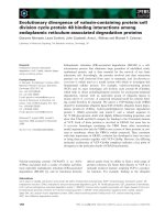
Fig. 6. Scheme demonstrating the ability of mature leaves of the DpsbE ,
DpsbF, DpsbL and DpsbJ mutants to form PSII–LHCII assemblies. In
wild-type thylakoid membranes, PSII core dimers together with
associated LHCII and OEC proteins form PSII–LHCII supercom-
plexes. In the absence of PsbJ, the PSII core dimers can harbor the
oxygen-evolving PsbO protein and also some PsbQ, but the LHCII
complexes remain completely detached. Lack of PsbL results in more
severe problems for the assembly of PSII: only PSII core monomers
can be assembled with labile association of CP43, and, of the oxygen-
evolving proteins, only PsbO is attached to the core monomer, pro-
vided that CP43 is also present. Mature leaves of the DpsbE and DpsbF
mutants do not accumulate any PSII core or OEC proteins but the
LHCII complexes remain free in the thylakoid membrane. Oxygen-
evolving proteins are shown as O (PsbO), P (PsbP) and Q (PsbQ). For
clarity, only the major subunits are included.
104 M. Suorsa et al.(Eur. J. Biochem. 271) Ó FEBS 2003
When assembly partners are not available, the chloro-
plast-encoded major PSII core proteins (D1, D2, CP47) are
typically down-regulated at the level of translation (for
reviews, see [9,10,54]). The regulation of the synthesis of
nuclear-encoded OEC proteins is still not understood, but
may occur at the level of transcription. According to our
results, it is likely that the regulation mechanisms for
chloroplast-encoded PSII core and nuclear-encoded OEC
proteins are different and independent of each other, as
suggested previously [54]. Another explanation for the
observed differences between young and mature leaves may
be the enhanced proteolytic activity in mature leaves
suffering from photo-oxidative stress because they either
lacked PSII (DpsbE and DpsbF) or had a defectively
assembled PSII (DpsbL and DpsbJ), as was discussed in
the recent report on the DpsbJ tobacco mutant with
dramatically reduced photosynthetic performance [29].
Lessons from
psbEFLJ
operon mutants on the role
of PsbW and PsbZ subunits
PSII assembly studies on psbEFLJ operon mutants also
provided some information on the two other small PSII
proteins, PsbW and PsbZ. Nuclear-encoded PsbW has been
found to accumulate in the thylakoid membranes of both
mature (Fig. 1A) and young [14] leaves of psbEFLJ operon
mutants, even in the complete absence of PSII complexes
(DpsbE and DpsbF). Similarly, PsbW was present, but at
reduced amounts, in tobacco DpsbA mutant with no PSII
assembly and activity [56]. Less stringent mutual regulation
of the accumulation of PsbW and the other PSII core
proteins was also evident in psbW antisense mutants of
Arabidopsis [8]. All this suggests that PsbW is not under the
same strict regulation and/or quality control as the other
PSII core proteins and the OEC proteins in mature leaves.
Recently characterized chloroplast-encoded PsbZ [38–
40], on the other hand, accumulated in mature leaves of
DpsbL and DpsbJ, in comparable amounts to assembled
PSII complexes, while being absent from DpsbE and DpsbF
(Fig. 1A). The presence of PsbZ even in DpsbL may suggest
the location of PsbZ in a very central core of PSII. Such a
central location in PSII, however, seems to contradict the
fact that PsbZ is not required for correct assembly of the
oxygen-evolving PSII complexes and photoautotrophic
growth of mutant plants [38–40].
Concluding remarks
The capacity of mature leaves of the psbEFLJ operon
mutants to assemble PSII (sub)complexes is schematically
presented in Fig. 6. When either PsbE or PsbF is missing,
the synthesis and accumulation of other PSII core proteins
and the OEC proteins are strictly prevented. Thus, in
contrast with young leaves [14], the absence of PSII
assembly partners in mature leaves either evokes a signal
to prevent the synthesis of other PSII proteins, of either
chloroplast or nuclear origin, or enhances the proteolytic
activity. Such control is, however, not exerted on the
synthesis and assembly of the nuclear-encoded LHCII
polypeptides or PSII LMM protein PsbW. Association of
PsbL with PSII subcomplexes, in particular, promotes the
stable and correct assembly of CP43 and thereby probably
also facilitates the dimerization of PSII. Assembly of PsbJ is
a subsequent step to the association of PsbL, and probably
occurs only after the assembly of the PSII core monomer, or
even the dimer, is accomplished. Of the OEC proteins, the
binding of PsbO is clearly dependent on the presence of
CP43 in the PSII core complex, whereas the correct
association of PsbP and PsbQ additionally requires the
presence of both the PsbL and PsbJ subunits. It remains to
be investigated whether the PsbL and/or PsbJ proteins offer
a direct docking site for PsbP and PsbQ or whether PsbL
and PsbJ only modulate the structure and mutual orienta-
tion of the PSII proteins on the lumenal side, making the
association of PsbP and PsbQ feasible. Finally, PsbJ is
clearly required for stable formation of PSII–LHCII
supercomplexes, thereby allowing greater organization of
PSII complexes in the thylakoid membrane.
Acknowledgements
Elena Baena-Gonzalez and Mika Kera
¨
nen are thanked for help with
the 77 K fluorescence measurements, and Drs Roberto Barbato, Toril
Hundal, Stefan Jansson, Wolfgang Schro
¨
der and Francis-Andre
Wollman for the gifts of antibodies. This work was supported by the
Academy of Finland, the Finnish Ministry of Agriculture and Forestry
(NKJ project), the German Research Foundation (SFB-TR1) and
Fonds der Chemischen Industrie.
References
1. Hankamer, B., Morris, E., Nield, J., Carne, A. & Barber, J. (2001)
Subunit positioning and transmembrane helix organisation in the
core dimer of photosystem II. FEBS Lett. 504, 142–152.
2. Nield, J., Orlova, E.V., Morris, E.P., Gowen, B., van Heel, M.
& Barber, J. (2000) 3D map of the plant photosystem II super-
comples obtained by cryoelectron microscopy and single particle
analysis. Nat. Struct. Biol. 1, 44–47.
3. Zouni, A., Witt, H T., Kern, J., Fromme, P., Krauss, N.,
Saenger, W. & Orth, P. (2001) Crystal structure of photosystem II
from Synechococcus elongatus at 3.8 A
˚
resolution. Nature 409,
739–743.
4. Kamiya, N. & Shen, J R. (2003) Crystal structure of oxygen-
evolving photosystem II from Thermosynechococcus vulcanus at
3.7-A
˚
resolution. Proc.NatlAcad.Sci.USA100, 98–103.
5. Hankamer, B., Nield, J., Zheleva, D., Boekema, E., Jansson, S. &
Barber, J. (1997) Isolation and biochemical characterisation of
monomeric and dimeric photosystem II complexes from spinach
and their relevance to the organisation of photosystem II in vivo.
Eur. J. Biochem. 243, 422–429.
6. Boekema, E.J., Roon, H. & Dekker, J.P. (1998) Specific associa-
tion of photosystem II and light-harvesting complex II in partially
solubilized photosystem II membranes. FEBS Lett. 424, 95–99.
7. Zheleva, D., Sharma, J., Panico, M., Morris, H.R. & Barber, J.
(1998) Isolation and characterization of monomeric and dimeric
CP47-reaction center photosystem II complexes. J. Biol. Chem.
273, 16122–16127.
8. Shi, L X., Lorkovic, J.L., Oelmuller, R. & Schro
¨
der, W.P. (2000)
The low molecular mass PsbW protein is involved in the stabili-
zation of the dimeric photosystem II complex in Arabidopsis
thaliana. J. Biol. Chem. 275, 37945–37950.
9. Baena-Gonzalez, E. & Aro, E M. (2002) Biogenesis, assembly
and turnover of photosystem II units. Philos. Trans. R. Soc. Lond.
B 357, 1451–1461.
10. Zerges, W. (2002) Does complexity constrain organelle evolution?
Trends Plant Sci. 7, 175–182.
Ó FEBS 2003 Low molecular mass proteins in the assembly of PSII (Eur. J. Biochem. 271) 105
11. Mu
¨
ller, B. & Eichacker, L.A. (1999) Assembly of the D1 precursor
in monomeric photosystem II reaction center precomplexes pre-
cedes chlorophyll a-triggered accumulation of reaction center II in
barley etioplasts. Plant Cell 11, 2365–2377.
12. Zhang, L., Paakkarinen, V., van Wijk, K.J. & Aro, E M. (1999)
Co-translational assembly of the D1 protein into photosystem II.
J. Biol. Chem. 274, 16062–16067.
13. Zhang, L., Paakkarinen, V., van Wijk, K.J. & Aro, E M. (2000)
Biogenesis of the chloroplast-encoded D1 protein: regulation of
translational elongation, insertion and assembly into photosystem
II. Plant Cell 12, 1769–1781.
14. Swiatek, M., Regel, R.E., Meurer, J., Wanner, G., Pakrasi, H.B.,
Ohad, I. & Herrmann, R.G. (2003) Effects of selective inactivation
of individual genes for low molecular mass subunits on the pho-
tosystem II assembly by chloroplast transformation: the psbEFLJ
operon in Nicotiana tabacum. Mol. General Genomics 268,
699–710.
15. Barber, J. & Nield, J. (2002) Organization of transmembrane
helices in photosystem II: comparison of plants and cyano-
bacteria. Philos. Trans. R. Soc. Lond. B 357, 1329–1337.
16. Mayes,S.R.,Dubbs,J.M.,Vass,I.,Hideh,E.,Nagy,L.&Barber,
J. (1993) Further characterization of the psbH locus of Syne-
chocystis sp. PCC6803: inactivation of psbH impairs Q
A
to
Q
B
electron transport in photosystem II. Biochemistry 32, 1454–
1465.
17. Summer, E.J., Schmid, V.H., Bruns, B.U. & Schmidt, G.W. (1997)
Requirement for the H phosphoprotein in photosystem II of
Chlamydomonas reinhardtii. Plant Physiol. 113, 1359–1368.
18. Ikeuchi, M., Shukla, V.K., Pakrasi, H.B. & Inoue, Y. (1995)
Directed inactivation of the psbI gene does not affect photosystem
II on the cyanobacterium Synechocystis sp. PCC 6803. Mol. Gen.
Genet. 249, 622–628.
19. Ku
¨
nstner, P., Guardiola, A., Takahashi, Y. & Rochaix, J D.
(1995) A mutant strain of Chlamydomonas reinhardtii lacking the
chloroplast photosystem II psbI gene grows photoautotrophically.
J. Biol. Chem. 270, 9651–9654.
20. Ikeuchi, M., Eggers, B., Shen, G.Z. & Webber, A., Yu, J.J.,
Hirano, A., Inoue, Y. & Vermass, W. (1991) Cloning of the psbK
gene from Synechocystis sp. PCC 6803 and characterization of
photosystem II in mutants lacking PSII-K. J. Biol. Chem. 266,
11111–11115.
21. Takahashi, Y., Matsumoto, H., Goldschmidt-Clermont, M.
& Rochaix, J D. (1994) Directed disruption of the Chlamydo-
monas chloroplast psbK gene destabilizes the photosystem II
reaction center complex. Plant Mol. Biol. 24, 779–788.
22. Morais, F., Barber, J. & Nixon, P.J. (1998) The chloroplast-
zencoded alpha subunit of cytochrome b-559 is required for
assembly of the photosystem two complex in both the light and
the dark in Chlamydomonas reinhardtii. J. Biol. Chem. 273,
29315–29320.
23. Pakrasi, H.B., Nyhus, K.J. & Granok, H. (1990) Targeted deletion
mutagenesis of the beta subunit of cytochrome b559 protein
destabilizes the reaction center of photosystem II. Z. Naturforsch.
45, 423–429.
24. Anbudurai, P.R. & Pakrasi, H.B. (1993) Mutational analysis of
the PsbL protein of photosystem II in the cyanobacterium
Synechocystis sp PCC 6803. Z. Naturforsch. 48, 267–274.
25. Kitamura, K., Ozawa, S., Shiina, T. & Toyoshima, Y. (1994)
L protein, encoded by psbL, restores normal functioning of the
primary quinone acceptor, Q
A
, in isolated D1/D2/CP47/Cytb-
559/I photosystem II reaction center core complex. FEBS Lett.
354, 113–116.
26. Ozawa, S., Kobayashi, T., Sugiyama, R., Hoshida, H., Shiina, T.
& Toyoshima, Y. (1997) Role of PSII-L protein (psbL gene
product) in the electron transfer in photosystem II complex 1.
Over-production of wild-type and mutant versions of PSII-L
protein and reconstitution into the PSII core complex. Plant Mol.
Biol. 34, 151–161.
27. Lind, L.K., Shukla, V.K., Nyhus, K.J. & Pakrasi, H.B. (1993)
Genetic and immunological analyses of the cyanobacterium
Synechocystis sp. PCC 6803 show that the protein encoded by the
psbJ gene regulates the number of photosystem II centers in thy-
lakoid membranes. J. Biol. Chem. 268, 1575–1579.
28. Regel, R.E., Ivleva, N.B., Zer, H., Meurer, J., Shestakov, S.V.,
Herrmann, R.G., Pakrasi, H.B. & Ohad, I. (2001) Deregulation of
electron flow within Photosystem II in the absence of the PsbJ
protein. J. Biol. Chem. 276, 41473–41478.
29.Hager,M.,Hermann,M.,Biehler,K.,Krieger-Liszkay,A.&
Bock, R. (2002) Lack of the small plastid-encoded PsbJ poly-
peptide results in a defective water-splitting apparatus of photo-
system II, reduced photosystem I levels, and hypersensitivity to
light. J. Biol. Chem. 277, 14031–14039.
30. Melis, A. (1999) Photosystem-II damage and repair cycle in
chloroplasts: what modulates the rate of photodamage in vivo?
Trends Plant. Sci. 4, 130–135.
31. De Santis-Maciossek, G., Kofer, W., Bock, A., Schoch, S., Maier,
R.M., Wanner, G., Rudiger, W., Koop, H U. & Herrmann, R.G.
(1999) Targeted disruption of the plastid RNA polymerase genes
rpoA, B and C1: molecular biology, biochemistry and ultra-
structure. Plant J. 18, 477–489.
32. Murashige, T. & Skoog, F.A. (1962) A revised medium for rapid
growth and bioassays with tobacco tissue cultures. Physiol. Plant
15, 473–479.
33. Porra, R.J., Thompson, W.A. & Kriedemann, P.E. (1989)
Determination of accurate extinction coefficients and simulta-
neous equations for assaying chlorophyll a and b with four dif-
ferent solvents: verification of the concentration of chlorophyll
by atomic absorption spectroscopy. Biochim. Biophys. Acta 975,
384–394.
34. Ku
¨
gler, M., Ja
¨
nsch, L., Kruft, V., Schmitz, U.K. & Braun, H P.
(1997) Analysis of the chloroplast protein complexes by blue-
native polyacrylamide gel electrophoresis (BN-PAGE). Photo-
synth. Res. 53, 35–44.
35. Laemmli, U.K. (1970) Clevage of structural proteins during
assembly of the head bacteriophage T4. Nature 227, 680–685.
36. Shevchenko, A., Wilm, M., Vorm, O. & Mann, M. (1996) Mass
spectrometric sequencing of proteins from silver-stained poly-
acrylamide gels. Anal. Biochem. 68, 850–858.
37. Kera
¨
nen, M., Aro, E M. & Tyystja
¨
rvi, E. (1999) Excitation-
emission map as a tool in studies of photosynthetic pigment-
protein complexes. Photosynthetica 37, 225–237.
38. Ruf, S., Biehler, K. & Bock, R. (2000) A small chloroplast-
encoded protein as a novel architectural component of the light-
harvesting antenna. J. Cell Biol. 149, 369–378.
39. Baena-Gonzalez, E., Gray, J.C., Tyystja
¨
rvi, E., Aro, E M.
&Ma
¨
enpa
¨
a
¨
, P. (2001) Abnormal regulation of photosynthetic
electron transport in a chloroplast ycf9 inactivation mutant.
J. Biol. Chem. 276, 20795–20802.
40. Swiatek, M., Kuras, R., Sokolenko, A., Higgs, D., Olive, J.,
Cinque, G., Muller, B., Eichacker, L.A., Stern, D.B., Bassi, R.,
Herrmann, R.G. & Wollman, F A. (2001) The chloroplast gene
ycf9 encodes a Photosystem II (PSII) core subunit, PsbZ. that
participates in Photosystem II supramolecular architecture. Plant
Cell 13, 1347–1367.
41. Yamamoto, Y. (2001) Quality control of Photosystem II. Plant
Cell Phys. 42, 121–128.
42. Krause, G.H. & Weis, E. (1991) Chlorophyll fluorescence and
photosynthesis: the basics. Annu. Rev. Plant Physiol. Plant Mol.
Biol. 42, 313–349.
43.Nield,J.,Kruse,O.,Ruprecht,J.,deFonseca,P.,Buchel,C.
& Barber, J. (2000) Three-dimensional structure of Chlamydo-
monas reinhardtii and Synechococcus elongatus photosystem II
106 M. Suorsa et al.(Eur. J. Biochem. 271) Ó FEBS 2003
complexes allows for comparison of their oxygen-evolving com-
plex organization. J. Biol. Chem. 275, 27940–27946.
44. Isogai, Y., Yamamoto, Y. & Nishimura, M. (1985) Association of
the 33-kDa polypeptide with the 43-kDa component in photo-
system II particles. FEBS Lett. 187, 240–244.
45. Yamamoto, Y. & Akasaka, T. (1995) Degradation of antenna
chlorophyll-binding protein CP43 during photoinhibition of
photosystem II. Biochemistry 34, 9038–9045.
46. Enami,I.,Tohri,A.,Kamo,M.,Ohta,H.&Shen,J R.(1997)
Identification of domains on the 43 kDa chlorophyll-carrying
protein (CP43) that are shielded from tryptic attack by binding
of the extrinsic 33 kDa protein with photosystem II complex.
Biochim. Biophys. Acta 1320, 17–26.
47. Henmi, T., Yamasaki, H., Sakuma, S., Tomokawa, Y., Tamura,
N.,Shen,J R.&Yamamoto,Y.(2003)Dynamicinteraction
between the D1 protein, CP43 and OEC33 at the lumenal side of
photosystem II in spinach chloroplasts: evidence from light-
induced cross-linking of the proteins in the donor-side photo-
inhibition. Plant Cell Phys. 44, 451–456.
48. Hashimoto, A., Ettinger, W.F., Yamamoto, Y. & Theg, S.M.
(1997) Assembly of newly imported oxygen-evolving complex
subunits in isolated chloroplasts: sites of assembly and mechanism
of binding. Plant Cell 9, 441–452.
49. Miyao, M. & Murata, N. (1989) The mode of binding of three
extrinsic proteins of 33 kDa, 23 kDa and 18 kDa in the Photo-
system II complex of spinach. Biochim. Biophys. Acta 977,
315–321.
50. Shen, J R., Burnap, R.L. & Inoue, Y. (1995) An independent role
of cytochrome c-550 in cyanobacterial photosystem II as revealed
by double-deletion mutagenesis of the psbO and psbV genes in
Synechocystis sp. PCC 6830. Biochemistry 34, 12661–12668.
51. Seidler, A. (1996) The extrinsic polypeptides of Photosystem II.
Biochim. Biophys. Acta 1277, 25–60.
52. Rova, M., Franzen, L G., Fredriksson, P O. & Styring, S.
(1994) Photosystem II in a mutant of Chlamydomonas reinhardtii
lacking the 23 kDa PsbP protein shows increased sensitivity to
photoinhibition in the absence of chloride. Photosynth. Res. 39,
75–83.
53. van Wijk, K.J., Roobol-Boza, M., Kettunen, R., Andersson, B. &
Aro, E M. (1997) Synthesis and assembly of the D1 protein into
photosystem II: processing of the C-terminus and identification of
the initial assembly partners and complexes during photosystem II
repair. Biochemistry 36, 6178–6186.
54. Wollman, F A., Minai, L. & Nechustai, R. (1999) The biogenesis
and assembly of photosynthetic proteins in thylakoid membranes.
Biochim. Biophys. Acta 1411, 21–85.
55. deVitry,C.,Olive,J.,Drapier,D.,Recouvreur,M.&Wollman,
F A. (1989) Posttranslational events leading to the assembly of
photosystem II protein complex: a study using photosynthesis
mutants of Chlamydomonas reinhardtii. J. Cell Biol. 109, 991–
1006.
56. Baena-Gonzalez, E., Allahverdiyeva, Y., Svab, Z., Maliga, P.,
Josse, E M., Kuntz, M., Ma
¨
enpa
¨
a
¨
,P.&Aro,E M.(2003)
Deletion of the tobacco plastid psbA gene triggers an up-
regulation of the thylakoid-associated NAD (P) H dehydrogenase
complex and the plastid terminal oxidase (PTOX). Plant J. 35,
704–716.
Ó FEBS 2003 Low molecular mass proteins in the assembly of PSII (Eur. J. Biochem. 271) 107