Báo cáo khóa học: Conformational changes of Newcastle disease virus envelope glycoproteins triggered by gangliosides pdf
Bạn đang xem bản rút gọn của tài liệu. Xem và tải ngay bản đầy đủ của tài liệu tại đây (282.75 KB, 8 trang )
Conformational changes of Newcastle disease virus envelope
glycoproteins triggered by gangliosides
Laura Ferreira, Enrique Villar and Isabel Mun
˜
oz-Barroso
Departamento de Bioquı
´
mica y Biologı
´
a Molecular, Universidad de Salamanca, Spain
We have investigated the conformational changes of New-
castle disease virus (NDV) glycoproteins in response to
receptor binding, using 1,1-bis(4-anilino)naphthalene-5,5-
disulfonic acid (bis-ANS) as a hydrophobicity-sensitive
probe. Temperature- and pH-dependent conformational
changes were detected in the presence of free bovine gan-
gliosides. The fluorescence of bis-ANS was maximal at
pH 5. The binding of bis-ANS to NDV was not affected
by chemicals that denature the fusion glycoprotein, such as
reducing agents, nor by the presence of neuraminidase
inhibitors such as N-acetyl neuramicic acid. Gangliosides
partially inhibited fusion and hemadsorption, but not
neuraminidase hemagglutinin-neuraminidase glycoprotein
(HN) activity. A conformational intermediate of HN, trig-
gered by the presence of gangliosides acting as receptor
mimics, was detected. Our results indicate that, upon binding
to free gangliosides, HN undergoes a certain conformational
change that does not affect the fusion glycoprotein.
Keywords: NDV; bis-ANS; conformational intermediates;
paramyxovirus receptors; gangliosides.
Newcastle disease virus (NDV) is an avian enveloped
virus belonging to the family of Paramyxoviridae, genus
Avulavirus. The membrane contains two transmembrane
glycoproteins, hemagglutin-neuraminidase (HN) and the
fusion (F) protein [1]. HN binds to sialic acid-containing
receptors at the cell surface through its hemagglutinating
activity (receptor-binding activity) and it also displays
neuraminidase or sialidase activity (receptor-destroying
activity), which probably prevents the aggregation of the
viral progeny. In addition, a third activity (the so-called
fusion promotion activity) has been proposed for the HN
protein [2–4]. The F protein is directly responsible for the
fusion between the viral envelope and the target mem-
brane. For paramyxoviruses, the fusion mechanism has
been proposed to occur at neutral pH; nevertheless, we
have previously shown that the fusion of NDV with
cultured cells is enhanced at acidic pH [5]. The F protein
is produced as a single inactive polypeptide, F
o
,which,
once cleaved by a cellular protease (reviewed in [6]),
becomes the active F
1
-F
2
form, with two peptides linked
by a disulfide bond [7]. To date, three domains of the F
1
polypeptide have been suggested to be involved in the
fusion mechanism of NDV. These are the N-terminal
fusion peptide [8] and two heptad repeat (HR) regions of
the ectodomain, one (HR1) located adjacent to the
fusion peptide, and the other (HR2) at the C-terminal
adjacent to the transmembrane domain [9–11]. Once
activated, the F protein is thought to undergo a series of
conformational changes that result in exposure of the
fusion peptide and interaction of the HR1 and HR2
domains. A six-helix bundle has emerged as the fusion
core structure of many viral fusion proteins, the
N-terminal HR forming the inner core, surrounded by
antiparallel C-terminal helices along the grooves located
between the helices of the central HR coiled-coil. The
formation of this structure is believed to pull the viral
and cell membranes into close proximity for merging.
The complete mechanism of NDV-induced membrane
fusion remains unknown. As with many other paramyxo-
viruses, NDV needs type-specific HN–F interactions that
must be present in the same bilayer to induce fusion
(reviewed in [12]). It has been proposed that the interaction
of HN with the cellular receptor induces conformational
changes in the HN protein that activates the F protein [12],
although the nature of such changes is obscure.
In the present study we analyzed the possible conform-
ational changes ocurring in NDV envelope glycoproteins
when interacting with free gangliosides as receptor mimics.
These changes were revealed through use of the fluorescent
probe 1,1-bis(4-anilino)naphthalene-5,5-disulfonic acid
(bis-ANS), which is nonfluorescent in aqueous solution
but increases its quantum yield when bound to hydrophobic
groups [13,14]. We observed that bis-ANS fluorescence was
maximal at 37 °C and at acidic pH. As reduction of the
disulfide bond of the F protein did not affect bis-ANS
Correspondence to I. Mun
˜
oz-Barroso and E. Villar, Departamento
de Bioquı
´
mica y Biologı
´
a Molecular, Universidad de Salamanca,
Edificio Departamental Laboratory 108, Plaza Doctores
de la Reina s/n, 37007 Salamanca, Spain.
Fax: + 34 923 294579, Tel.: + 34 923 294465,
E-mail: and
Abbreviations: bis-ANS, 1,1-bis(4-anilino)naphthalene-5,5-disulfonic
acid; DMEM, Dulbecco’s modified Eagle’s medium; F protein,
fusion glycoprotein; FDQ, fluorescence dequenching; HA,
influenza hemagglutinin; Had, hemadsorption; HN, hemagglutinin-
neuraminidase glycoprotein; HR, heptad repeat; KNP, 120 m
M
KCl,
30 m
M
NaCl, 10 m
M
sodium phosphate pH 7.4; NDV, Newcastle
disease virus; NeuAc, N-acetylneuraminic acid; p.f.u., plaque
formation units; R
18
, octadecylrhodamine B chloride.
(Received 7 November 2003, accepted 9 December 2003)
Eur. J. Biochem. 271, 581–588 (2004) Ó FEBS 2004 doi:10.1111/j.1432-1033.2003.03960.x
fluorescence, we suggest that the binding of NDV to free
gangliosides results in the formation of a conformational
intermediate of HN.
Materials and methods
Materials
Bis-ANS and octadecyl rhodamine B chloride (R
18
)were
from Molecular Probes Inc. (Junction City, OR, USA).
Bovine brain gangliosides, disialoganglioside GD1a, lacto-
cerebrosides, dithiothreitol, 2-mercaptoethanol, N-acetyl-
neuraminic acid and Triton X-100 were all from SIGMA
(St. Louis, MO, USA). Cell culture media were from BIO
Whittaker (Walkersvile, Maryland, USA). Fresh blood from
healthy donors (with their consent) was obtained from the
Blood Bank of the University Hospital in Salamanca (Spain).
Cells and viruses
NDV ÔClone 30Õ was grown and purified essentially as
described elsewhere [15]. COS-7, HeLa and Vero cells were
obtained from the American Type Culture Collection and
were maintained in Dulbecco’s Modified Eagle Medium
(DMEM) supplemented with
L
-glutamine (580 mgÆL
)1
),
penicillin/streptomycin (100 UÆmL
)1
/100 lgÆmL
)1
), and
heat-inactivated fetal bovine serum at 10% (v/v) for
COS-7 and HeLa cells and at 5% (v/v) for Vero cells.
For the fusion experiments, COS-7 cells grown in
monolayers were detached with trypsin/EDTA. Trypsin
was inactivated by the addition of DMEM. The cells were
washed twice with 15 m
M
Hepes buffer (130 m
M
NaCl,
5m
M
KCl, 2 m
M
CaCl
2
,1m
M
MgCl
2
,10m
M
glucose),
pH 7.4, and resuspended at 2.5 · 10
6
cells in 200 lLof
Hepes buffer, pH 7.4.
Ganglioside-induced conformational changes
Bis-ANSwasaddedto20lgofNDVat3l
M
in Hepes
buffer (final volume, 2 mL) under constant stirring in the
fluorimeter cuvette. Then, different concentrations of gan-
gliosides were added, and the fluorescence progress curve
was recorded for 2 min on a Hitachi F-4010 spectrofluo-
rimeter (excitation, 395 nm; emission, 500 nm; slit widths
5 and 10 nm for excitation and emission, respectively). The
background fluorescence resulting from gangliosides was
calculated.
To analyse the bis-ANS data, the relative fluorescence
(I
rel
) was calculated according to [16]:
I
relðtÞ
¼ðI
ðtÞ
À I
NDVþbis-ANS
Þ=ðI
maxBG
ÀI
bis-ANS
Þð1Þ
where I
(t)
is the fluorescence intensity at any given time,
I
NDV + bis-ANS
is the fluorescence intensity of bis-ANS in the
presence of the virus, I
maxBG
is the final fluorescence intensity
of bis-ANS in the presence of gangliosides, and I
bis-ANS
is the
fluorescence intensity of bis-ANS in aqueous solution.
R
18
fusion assays
Dequenching fusion assays were accomplished as described
previously [5]. Briefly, purified NDV was labeled with the
fluorescent probe octadecylrhodamine (R
18
)andafter
mixing with the target cells, the progress curve of R
18
fluorescence was spectrofluorimetrically monitored.
Neuraminidase assays
Neuraminidase activity was determined by a fluorimetric
procedure using 2¢-(4-methylumbelliferyl)a-
D
-N-acetyl-
neuraminic acid as substrate [17].
Hemadsorption assays
The hemadsorption (Had) activity of HN protein was
determined according to [18] with modifications. HeLa cells
were plated in 24-well plate 12 h before infection. Then, the
cell monolayers were infected with NDV at 1 multiplicity of
infection. At 24 h postinfection, the cells were washed twice
with NaCl/P
i
(10 m
M
KH
2
PO
4
,150m
M
NaCl, pH 7.2) and
incubated for 30 min at 4 °C with 2% of human erythro-
cytes. After washing twice with NaCl/P
i
, adsorbed erythro-
cytes were lysed in 50 m
M
NH
4
Cl for 5 min at 4 °C. The
lysates were clarified by centrifugation and Had activity was
quantified by measuring the absorbance at 540 nm and
subtracting the background absorbance obtained with
uninfected cells. To analyse the effect of gangliosides on
Had activity, the cells were incubated in the presence of
different concentrations of gangliosides for 10 min at 37 °C
before the addition of red blood cells.
Results and discussion
It has been shown previously that the exposure of hydro-
phobic regions of viral proteins as a consequence of
conformational changes can be analyzed by means of the
hydrophobic-sensitive probe bis-ANS [14,16,19]. This
water-soluble fluorophore undergoes a strong increase in
its quantum yield upon binding to hydrophobic sites [16]
and has been used to study protein structural changes
[14,16,19,20]. The exposure of hydrophobic segments of
NDV envelope proteins triggered by gangliosides was tested
by measuring binding to bis-ANS. Initially, the effect of
increasing concentrations of NDV or gangliosides on bis-
ANS fluorescence was studied. For these experiments, at
zerotimetheviruswasaddedto37°C-prewarmed Hepes
buffer at pH 7.4 in the fluorimeter cuvette, followed by the
addition of 3 l
M
of bis-ANS. The fluorescence emitted,
taken as I
NDV+bisANS
, became stablilized after a few
seconds. Then, gangliosides were added and the progress
curve of the fluorescence emission at 500 nm was recorded
for 2 min. To process the data, I
rel
was calculated according
to Eqn (1). I
NDV+bis-ANS
was subtracted from the intensity
of fluorescence observed at any given time and this was
relatedtothetermI
maxBG
–I
bis-ANS
, i.e. the maximal
fluorescence of gangliosides in the absence of virus (I
maxBG
)
after subtracting the fluorescence emission of the probe in
buffer (I
bis-ANS
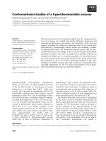
). Figure 1 shows the dose–response curves
of bis-ANS fluorescence in the presence of different
concentrations of NDV. As can be seen, after 20 lgof
NDV the fluorescence reached a plateau, suggesting
saturation. Similarly, different concentrations of bovine
brain gangliosides or of the disialoganglioside GD1a
582 L. Ferreira et al.(Eur. J. Biochem. 271) Ó FEBS 2004
(5–30 lg) were added to 20 lgofNDVinthepresenceof
bis-ANS (Fig. 2), and it was found that 10 lg of bovine
brain gangliosides and 5 lg of GD1a were sufficient to
observe saturation under the conditions of the experiment.
The observed saturation of the extent of bis-ANS fluores-
cence (Figs 1 and 2) may indicate that the conformational
change undergone by NDV glycoproteins in the presence of
gangliosides is limited (see below).
To assess the specific effect of gangliosides, 20 lg of NDV
was preincubated in the presence of 10 lgofbovine
gangliosides at 37 °C for 10 min. Then, an additional
10 lg was added and no increase in bis-ANS fluorescence
was detected above background (Table 1). We interpret
these results as pointing to the irreversibility of the
conformational change triggered by gangliosides. In addi-
tion, neutral glycolipids such as lactocerebrosides did not
lead to an increase in the fluorescence of bis-ANS (Table 1).
The temperature-dependence of bis-ANS fluorescence at
neutral pH in the presence of NDV and gangliosides was
analyzed (Fig. 3). The fluorescence of bis-ANS in the
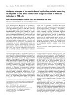
Fig. 1. Effect of NDV concentration on bis-ANS fluorescence. At zero time, 3 l
M
bis-ANSwasaddedto37°C prewarmed buffer containing
different concentrations of NDV, after which 25 lg of bovine gangliosides was added. Fluorescence was recorded continuously over 2 min at
excitation and emission wavelengths of 395 and 500 nm, respectively. The relative fluorescence, I
rel
, is shown (see Materials and methods). (A)
Kinetics of bis-ANS fluorescence at different NDV concentrations from a representative experiment. (B) Relative fluorescence of bis-ANS, I
rel
,at
90 s of reaction at the desired NDV concentration. Data taken from different experiments similar to that shown in (A). Data are means ± SE of at
least three independent experiments.
Fig. 2. Effect of ganglioside concentrations on bis-ANS fluorescence. At zero time, 3 l
M
bis-ANSwasaddedto37°C prewarmed buffer containing
20 lg of NDV, after which different concentrations of bovine brain gangliosides or GD1a were added. Fluorescence was recorded continuously for
2 min at excitation and emission wavelengths of 395 and 500 nm, respectively. The relative fluorescence, I
rel
, is shown (see Materials and methods).
(A) Kinetics of bis-ANS fluorescence at different ganglioside concentrations from a representative experiment. (B) Relative fluorescence of bis-
ANS, I
rel
, at 90 s of reaction at the desired ganglioside concentration; (d), bovine brain gangliosides; (m), GD1a. Data taken from different
experiments similar to that shown in (A). Data are means ± SE of at least two independent experiments.
Table 1. Effect of preincubation of NDV with different agents on the
fluorescence of bis-ANS. NDV (20 lg) was incubated in the presence of
10 lg of bovine brain gangliosides, 10 m
M
NeuAc or 50 m
M
NeuAc
for 10 min at 37 °C. Then, 3 l
M
bis-ANSwasaddedtoprewarmed
buffer at 37 °C containing 20 lg of treated virus, after which 10 lgof
bovine gangliosides or 10 m
M
NeuAcwereaddedfortriggeringthe
conformational change. Fluorescence was recorded continuously over
2 min at excitation and emission wavelengths of 395 and 500 nm,
respectively. I
rel
at90minofreactionareshown(seeMaterialsand
methods).
Preincubation
Trigger of the
conformational change I
rel
– Bovine gangliosides (10 lg) 6.98
– NeuAc (10 m
M
) 15.45
– Lactocerebrosides (10 lg) 0.85
Bovine gangliosides (10 lg) Bovine gangliosides (10 lg) 0.6
Bovine gangliosides (10 lg) NeuAc (10 m
M
) 12.71
NeuAc (10 m
M
) Bovine gangliosides (10 lg) 8.31
NeuAc (50 m
M
) NeuAc (10 m
M
) 1.97
Ó FEBS 2004 Conformational changes in NDV glycoproteins (Eur. J. Biochem. 271)583
presence of NDV but in the absence of gangliosides
[I
NDV+bisANS
from Eqn (1)], was independent of tempera-
ture (data not shown). No increase in fluorescence was
observed at 4 °C, whereas it increased gradually at 15 and
25 °C, showing a sharper increase after 30 °C. These data
are comparable to those of the temperature-dependence of
NDV fusion with cultured cells reported by us previously
[5]. In both cases, we failed to detect an increase in
fluorescence at 4 °C. Nevertheless, it has been established
that the HN protein of paramixoviruses can bind to the
sialoglycosides of the cell surface at 4 °C [21], suggesting
that NDV may interact with gangliosides at this tempera-
ture. However, this binding seems to be insufficient to
trigger any conformational change in NDV glycoproteins
detectable with the bis-ANS technique.
The pH-dependence of the fluorescence of bis-ANS in the
presence of NDV and gangliosides was also analyzed
(Fig. 4).Atzerotime,NDVwasaddedto37 °C-prewarmed
buffer at the desired pH, followed by the addition of 3 l
M
bis-ANS and then gangliosides; next I
rel
was calculated as
described above. Figure 4A shows the kinetics of bis-ANS
fluorescence in the presence of NDV and gangliosides at
different pH values; Fig. 4B depicts the final extent of bis-
ANS fluorescence after 90 s of virus–ganglioside contact.
The extent of bis-ANS fluorescence at the different
pH values assayed occurred in the following order:
pH5>pH5.5>pH6.5>pH7.Ithasbeenreported
that the increase in fluorescence at low pH can be partly
explained in terms of the protonation of negatively charged
groups, which facilitates the binding of bis-ANS [14]. In this
sense, we detected a slight increase in the fluorescence of
bis-ANS in the presence of NDV upon lowering the pH
[I
NDV+bis-ANS
in Eqn (1)], but these figures were subtracted
from the fluorescence intensity emitted in the presence of
gangliosides (Eqn 1). In Fig. 4B, a sharp increase in
fluorescence intensity at pH < 6.5 can be seen. The
fluorescence intensity observed at pH 5 was about twice
the value seen at pH 7.4. This difference is smaller than that
reported for viruses that show a pH-dependent entry
mechanism, since for influenza virus Korte and Herrman
(1994) [14] have reported that bis-ANS fluorescence is five
times higher at acidic than at neutral pH. Our data on the
Fig. 4. pH-dependence of the bis-ANS fluorescence. At zero time, 3 l
M
bis-ANSwasaddedto37 °C prewarmed buffer at the desired pH containing
20 lg of NDV, after which 15 lg of bovine gangliosides was added. Fluorescence was recorded continuously over 2 min at excitation and emission
wavelengths of 395 and 500 nm, respectively. The relative fluorescence, I
rel
, is shown (see Materials and methods). (A) Kinetics of bis-ANS
fluorescence at different pHs from a representative experiment (B) Relative fluorescence of bis-ANS, I
rel
, at 90 s of reaction at different pHs. Data
are means ± SE of two independent experiments.
Fig. 3. Temperature-dependence of bis-ANS fluorescence. At zero time, 3 l
M
bis-ANS was added to buffer prewarmed to the desired temperature
containing 25 lg of NDV, after which 10 lg of bovine gangliosides was added. Fluorescence was recorded continuously over 2 min at excitation
and emission wavelengths of 395 and 500 nm, respectively. The relative fluorescence, I
rel
, is shown (see Materials and methods). (A) Kinetics of bis-
ANS fluorescence at different temperatures from a representative experiment (B) Relative fluorescence of bis-ANS, I
rel
, at 90 s of reaction at the
desired temperature. Data are means ± SE of two independent experiments.
584 L. Ferreira et al.(Eur. J. Biochem. 271) Ó FEBS 2004
pH-dependence of bis-ANS fluorescence indicate that the
conformation of NDV proteins triggered at acidic pH
exposes a higher number of hydrophobic fluorophore-
binding sites. It is interesting to note the similarities between
the pH-dependence of NDV fusion activity reported
previously by us [5] and that of bis-ANS fluorescence,
pointing to the maximal extent of both fusion and bis-ANS
fluorescence at pH 5.0. We have previously hypothesized [5]
that NDV might use the endocytic pathway as a secondary
mechanism of entry. If the conformational change under-
gone by HN protein after receptor binding (see below) is
activated at acidic pH, as well as NDV fusion activity, the
present data confirm our hypothesis concerning the acidic
pH enhancement of NDV entry. Moreover, the pH-
dependence of viral entry seems debatable. In this sense,
Mothes et al. [22] have reported that the entry of the avian
leukosis virus, a retrovirus, into the host cell depends on a
low pH step that acts after receptor binding. For these
authors, partial conformational changes in env protein in
the presence of soluble receptors may be due to receptor
priming rather than complete activation. Additionally, it
has been recently reported [23] that the SER paramyxovirus
shows a low-pH-dependent fusion activity.
We performed a series of experiments to elucidate
whether the binding of bis-ANS to hydrophobic sites of
NDV glycoproteins was located in F and/or HN protein.
First, NDV was incubated in the presence of 10 m
M
2-mercaptoethanol or 2 m
M
dithiothreitol (agents that
reduce the disulfide bonds of the F protein) for 30 min at
37 °C before the addition of bis-ANS and gangliosides. As
deduced by PAGE analysis (data not shown), treatment of
viruses with 2-mercaptoethanol led to the loss of F
0
protein.
In another series of experiments, viruses were incubated in
the presence of 50 m
M
of N-acetylneuraminic sialic acid
(NeuAc)for30minat37°C. This compound is both a
product and an inhibitor of the neuraminidase activity of
the HN protein through binding to its active site [17].
Neither treatment affected the emission of bis-ANS fluor-
escence with respect to the control (NDV without treat-
ment) when gangliosides were added to treated viruses in the
bis-ANS assay (Table 1 and data not shown). To test the
possibility that bis-ANS might bind nonspecifically to
2-mercaptoethanol-treated-virus, we performed the follow-
ing experiment. Twenty micrograms of virus, both treated
and nontreated with the reducing agent, were preincubated
in the presence of 10 lg of bovine gangliosides for 10 min at
37 °C. Then, a further 10 lg of gangliosides was added
in the bis-ANS assay. In both cases, no increase in bis-ANS
fluorescence was detected above the background level,
unlike the findings on treated virus not preincubated in the
presence of gangliosides (data not shown). We therefore
assume that the fluorescence of bis-ANS of reduced virus in
the presence of gangliosides would not be due to the
nonspecific binding of the probe to 2-mercaptoethanol-
treated-NDV. Because viruses treated with these reducing
agents are fusion-deficient (data not shown), this seems to
indicate that the newly exposed hydrophobic binding sites
are not located within the F protein. On the other hand, the
increase in bis-ANS fluorescence did not vary after pre-
incubation with NeuAc, suggesting that the binding of
gangliosides, the putative agents of the conformational
change, did not compete with the neuraminidase inhibitor
NeuAc. To test this hypothesis, we performed a direct
binding assay between the sialic acid NeuAc and NDV
using the bis-ANS technique. Our data indicate that,
similarly to gangliosides, NeuAc leads to an increase in
the fluorescence of bis-ANS in the presence of NDV
(Table 1). We performed a series of experiments to analyze
the relationship between ganglioside and NeuAc binding
sites. NDV was incubated in the presence of 10 lg of bovine
brain gangliosides or 10 m
M
NeuAc for 10 min at 37 °C.
Then, 3 l
M
bis-ANS was added to prewarmed buffer at
37 °C containing 20 lg of treated virus, after which an
additional 10 lg of bovine gangliosides or 10 m
M
NeuAc
were added to trigger the conformational change. Our
results revealed that preincubation of NDV in the presence
of NeuAc did not abolish the increase in fluorescence when
gangliosides were added, but it did abolish it when
additional NeuAc was added. By contrast, preincubation
of NDV in the presence of gangliosides did not abolish the
increase in fluorescence when NeuAc was added, but it did
so when additional gangliosides were added (Table 1). Our
conclusion is that the binding sites for NeuAc do not
compete with the binding sites for gangliosides.
As mentioned above, we detected the exposure of hydro-
phobic binding sites of NDV proteins as measured by the
increase in bis-ANS emission intensity (Figs 1–4), triggered
by gangliosides. We assume that the new hydrophobic
binding sites must belong to the envelope glycoproteins of
NDV as the fluorophore shows a pronounced affinity for the
hydrophobic sites of proteins in comparison with its affinity
for lipids ([14] and references therein). The next step was to
investigate whether the presence of gangliosides might exert
some effect on NDV envelope glycoprotein activities. First,
the fusion of NDV with COS-7 cells was analyzed by
assaying the dequenching of the R
18
incorporated into the
viral membrane (see Materials and methods). For this, 20 lg
of R
18
-labeled NDV was incubated in the presence of 25 lg
of bovine gangliosides for 10 min at 37 °C. Then, 2.5 · 10
6
COS-7 cells were added and the dequenching of R
18
fluorescence was recorded for 30 min. Data from a typical
experiment are depicted in Fig. 5. As can be seen, fusion was
not abolished although it was partially inhibited, showing an
inhibition of the extent of fusion of about 27% as compared
with controls at 30 min of virus–cell contact. This reduction
was slightly lower if gangliosides were added to the virus–cell
mixture (at time zero) without preincubation (20% as
compared with control). As we observed that the denatur-
ation of F protein by the cleavage of disulfide bonds did not
exert any effect on bis-ANS fluorescence (data not shown),
we assume that the partial inhibition of fusion exerted by
gangliosides could be an indirect effect on fusion due to a
certain inhibition of the virus binding to cells in the presence
of gangliosides. As discussed below, gangliosides would bind
to HN, lowering its interaction with COS-7 cell receptors and
subsequently fusion of the virus with the cells. In addition, the
ability of the gangliosides to inhibit HN hemadsorption
activity was analyzed. NDV-infected HeLa cells were
incubated in the presence of different concentrations of
gangliosides for 1 h at 37 °C before the addition of red blood
cells. As shown in Fig. 6, the data indicated that the
inhibition of the Had activity of NDV HN protein exerted
by bovine brain gangliosides was dose-dependent. Taken
together, these results strongly suggest a specific interaction
Ó FEBS 2004 Conformational changes in NDV glycoproteins (Eur. J. Biochem. 271)585
of the viral proteins with gangliosides, which would act as
receptor mimics. In this sense, free gangliosides might
compete with the actual receptors of the cell surface,
inhibiting viral glycoproteins activities (Figs 5 and 6). Other
simple molecules have previously been used to trigger
conformational changes on viral receptors as soluble CD4
that induces certain conformational changes upon binding
to envelope glycoproteins of HIV and SIV [20,24].
Viral HN glycoprotein has three different biological
activities, sialidase or neuraminidase, hemagglutinating or
receptor-binding, and fusion promotion. Although there is a
considerable body of evidence both in favour of and against
the topological separation of the neuraminidase and recep-
tor-binding site ([18] and references therein), the crystal
structure of NDV HN protein [25] supports the notion of a
single site. Recently, on the basis of their crystallographic
data on the HN protein of NDV, Crennell et al. [25] have
proposed the existence of a single sialic acid recognition site
switchable between both activities: the binding site or
catalytic site. As summarized above, here we assayed (a) the
effect of neuraminidase inhibitors on bis-ANS fluorescence
and (b) the effect of gangliosides on neuraminidase and
hemagglutinating activities. The extent of bis-ANS fluores-
cence triggered by gangliosides was not affected by the
neuraminidase inhibitor NeuAc (Table 1). Moreover,
the presence of gangliosides did not exert any effect on the
neuraminidase activity of HN protein (data not shown),
although they did inhibit Had in a dose-dependent manner
(Fig. 6). In addition, gangliosides and NeuAc did not
compete for their binding sites in the bis-ANS assay
(Table 1). Taken together, these data suggest that ganglio-
sides bind to the receptor-binding site of HN protein and
that this binding is not altered by the presence of
neuraminidase inhibitors. Therefore, the data presented
here together with those from our previous work [19,26] fail
to account for the topological coincidence of both sites,
although they do not allow us to propose their separation.
In current models of membrane fusion induced by viral
proteins, exposure of the fusion peptide that triggers
membrane merging is a consequence of the conformational
change of the F protein that involves the two heptad repeat
regions (revised in [12]). The nature of these interactions and
changes is not completely understood, although it has been
established that for viruses that fuse with the target
membrane through a pH-independent mechanism, such as
most paramixoviruses and retroviruses, the conformational
change of the F protein must be triggered after receptor
binding. Upon comparing the 3D structure of HN, both
alone and in a complex with the neuraminidase substrate
2-deoxy-2,3-dehydro-N-acetylneuraminic acid, Takimoto
et al. [27] suggested that receptor binding induces a
structural change in the hydrophobic surface of the HN
protein that disrupts physical HN–F interactions, triggering
the activation of F protein to initiate membrane fusion.
Despite this, our results indicate that the binding alone of
simple molecules is insufficient to induce strong HN
conformational changes that would in turn affect the
F protein. In other words, the conformational changes
induced by free receptor mimic molecules are only partial.
As we have shown here, NDV glycoproteins undergo
conformational changes in the presence of gangliosides, as
indicated by the exposure of new hydrophobic binding sites
for the bis-ANS probe. Our data strongly support the idea
that binding to these receptor mimics induces a conform-
ational change in HN protein. Our observation that
inactivation of the F protein did not affect the extent of
bis-ANS fluorescence suggests that the fusion protein does
not undergo any conformational change in the presence
of gangliosides. Therefore, functional HN–F interactions
in vivo, i.e. interactions that drive fusion, may need a more
Fig. 5. Effect of bovine gangliosides on NDV fusion with COS-7 cells.
R
18
-labeled NDV (20 lg) was incubated in the presence of 25 lgof
bovine gangliosides for 10 min at 37 °C. Then, 2.5 · 10
5
COS-7 cells
were added and the sample was incubated at 37 °C for 30 min under
continuous stirring. Fusion was monitored continuously, as described
in Materials and methods, by measuring the dequenching of R
18
.(d)
Control; (m) virus and gangliosides with preincubation; (j)virusand
gangliosides without preincubation.
Fig. 6. Dose–response effect of ganglioside inhibition of hemadsorption.
HeLa cells were infected with NDV at 1 multiplicity of infection. At
24 h postinfection, the cells were incubated in the presence of different
concentrations of gangliosides for 10 min at 37 °C and then incubated
for 30 min at 4 °C with 2% of human erythrocytes. The rate of
hemadsorption in comparison with controls was calculated by meas-
uring the absorbance at 540 nm of the erythrocytes bound to NDV-
infected cells after lysing in 50 m
M
NH
4
Cl. Data are means ± SE of
two independent experiments.
586 L. Ferreira et al.(Eur. J. Biochem. 271) Ó FEBS 2004
complex environment than the presence alone of a putative
receptor, in this case gangliosides. In addition, the con-
formational change undergone by the HN protein after
binding to gangliosides can be completed in the presence of
the correct target, i.e. the cell membrane. The binding of
viruses to the host cell surface is a more complex phenom-
enon than a mere bimolecular interaction between a viral
protein and a cellular receptor. In this sense, viral binding
may occur through multiple interactions among several
viral and cellular molecules, accompanied by conforma-
tional changes in viral proteins. Therefore, a major task
would be to study the conformational changes of NDV
proteins in the presence of cells. Nevertheless, the high
extent of bis-ANS binding to hydrophobic sites of the cell
surface did not allow us to use this assay with intact cells as
targets (data not shown).
The existence of conformational intermediates for viral
proteins such as influenza HA [16], vesicular stomatitis
virus fusion protein [19,28] or HIV envelope glycoproteins
[20] has been reported. Additionally, the binding of bis-
ANS to different viral glycoproteins [14,16,19,20] has been
correlated with the fusion activation of the proteins.
Nevertheless, here we observed a conformational inter-
mediate of the HN protein prior to membrane merging,
confirming that changes leading to fusion might be slow
in the virus upon binding to the target membrane [16].
The newly exposed hydrophobic sequence of the HN
protein triggered by gangliosides is not clear. We suggest
several possibilities: (a) the HR stalk region, which has
been proposed to be responsible for HN–F interactions
[29]; (b) the interfaces of HN dimers, which presumably
dissociate after ganglioside binding [27]; or (c) sequential
conformational changes in HN protein, as proposed for
other viral proteins [16].
In summary, here we have demonstrated that ganglio-
sides bind to NDV, inducing the exposure of hydrophobic
binding sites for bis-ANS. We propose that the binding site
for gangliosides would be the receptor-binding site of HN
protein, triggering the conformational change detected here.
Our results indicate that the bis-ANS assay would also be
useful for studying conformational changes in viral proteins
that do not require an acidic pH to start fusion and that
simple molecules such as gangliosides can be used as
receptor mimics for triggering these changes.
Acknowledgements
This work was partially supported by the Spanish Fondo de
Investigaciones Sanitarias, FIS (PI021848) and Junta de Castilla y
Leo
´
n (SA 064/02) grants to E. V.; L. F. is a predoctoral fellowship
supported by the Ministerio de Ciencia y Tecnologı
´
a, Spain (Grant
DGES PM97-0160). We thank Drs E. Dı
´
ez Espada and J. A.
Rodrı
´
guez from Intervet Laboratories (Salamanca, Spain) for
providing the lentogenic ÔClone 30Õ strain of NDV. Thanks are
also due to N. Skinner for language corrections and proofreading
the manuscript.
References
1. Choppin, P.W. & Compans, R.W. (1975) Reproduction of para-
myxoviruses. In Comprehensive Virology (Fraenkel-Conrat,H.&
Wagner, R.R., eds), pp. 95–178. Plenum Press, New York.
2. Morrison, T., McQuain, C. & McGinnes, L. (1991) Com-
plementation between avirulent Newcastle disease virus and a
fusion protein gene expressed from a retrovirus vector: require-
ments for membrane fusion. J. Virol. 65, 813–822.
3. Horvath, C.M., Paterson, R.G., Shaughnessy, M.A., Wood, R. &
Lamb, R.A. (1992) Biological activity of paramyxovirus fusion
proteins: factors influencing formation of syncytia. J. Virol. 66,
4564–4569.
4. Sergel, T., McGinnes, L.W., Peeples, M.E. & Morrison, T.G.
(1993) The attachment function of the newcastle disease virus
hemagglutinin-neuraminidase protein can be separated from
fusion promotion by mutation. Virology 193, 717–726.
5. San Roma
´
n, K., Villar, E. & Mun
˜
oz-Barroso, I. (1999) Acidic pH
enhancement of the fusion of Newcastle disease virus with cul-
tured cells. Virology 260, 329–341.
6. Morrison, T.G. & Portner, A. (1991) Structure, function and
intracellular processing of the glycoproteins of Paramixoviridae.In
The Paramyxovirus (Fraenkel-Conrat, H. & Wagner, R.R., eds),
pp. 347–382. Plenum Press, New York.
7. Iwata, S., Schmidt, A.C., Titani, K., Suzuki, M., Kido, H., Gotoh,
B., Hamaguchi, M. & Nagai, Y. (1994) Assignment of disulfide
bridges in the fusion glycoprotein of Sendai virus. J. Virol. 68,
3200–3206.
8. Brasseur, R., Vandenbranden, M., Cornet, B., Burny, A. &
Ruysschaert, J.M. (1990) Orientation into the lipid bilayer of an
asymmetric amphipathic helical peptide located at the N-terminus
of viral fusion proteins. Biochim. Biophys. Acta 1029, 267–273.
9. Chambers, P., Pringle, C.R. & Easton, A.J. (1990) Heptad repeat
sequences are located adjacent to hydrophobic regions in several
types of virus fusion glycoproteins. J. General Virol. 71, 3075–
3080.
10. Buckland, R. & Wild, F. (1989) Leucine zipper motif extends.
Nature 338,547.
11. Sergel, T.A., McGinnes, L.W. & Morrison, T.G. (2000) A single
amino acid change in the Newcastle Disease Virus fusion protein
alters the requirement for HN protein in fusion. J. Virol. 74,
5101–5107.
12. Lamb, R.A. & Kolakofsky, D. (2001) Paramyxoviridae: the
viruses and their replication. In Fundamental Virology (Fields,
B.N., Knippe, D.M. & Kato, A., eds), pp. 1305–1340. Lippincot-
Raven, NY.
13. Rosen, C.G. & Weber, G. (1969) Dimer formation from 1-Anili-
no-8-naphthalenesulphonate catalyzed by bovine serum albumin.
A new fluorescent molecule with exceptional binding properties.
Biochemistry 8, 3915–3920.
14. Korte, T. & Herrmann, A. (1994) pH-dependent binding of the
fluorophore bis-ANS to influenza virus reflects the conformational
change of hemagglutinin. Eur Biophys. J. 23, 105–113.
15. Garcı
´
a-Sastre, A., Cabezas, J.A. & Villar, E. (1989) Proteins of
Newcastle disease virus envelope: Interaction between the outer
hemagglutinin-neuraminidase glycoprotein and the inner
non-glycosylated matrix protein. Biochim. Biophys. Acta 999, 171–
175.
16. Korte, T., Ludwig, K., Booy, F.P., Blumenthal, R. & Herrmann,
A. (1999) Conformational intermediates and fusion activity of
influenza virus hemagglutinin. J. Virol. 73, 4567–4574.
17. Garcı
´
a-Sastre, A., Cobaleda, C., Cabezas, J.A. & Villar, E. (1991)
On the inhibition mechanism of the sialidase activity from New-
castle Disease Virus. Biol. Chem. Hoppe-Seyler 372, 923–927.
18. Iorio, R.M., Field, G.M., Sauvron, J.M., Mirza, A.M., Deng, R.,
Mahon, P.J. & Langedijk, J.P. (2001) Structural and functional
relationship between the receptor recognition and neuraminidase
activities of the Newcastle disease virus hemagglutinin-neur-
aminidase protein: receptor recognition is dependent on neur-
aminidase activity. J. Virol. 75, 1918–1927.
Ó FEBS 2004 Conformational changes in NDV glycoproteins (Eur. J. Biochem. 271)587
19. Carneiro, F.A., Ferradosa, A.S. & Da Poian, A.T. (2001) Low
pH-induced conformational changes in vesicular stomatitis virus
glycoprotein involve dramatic structure reorganization. J. Biol.
Chem. 276, 62–67.
20. Jones, P.L., Korte, T. & Blumenthal, R. (1998) Conformational
changes in cell surface HIV-1 envelope glycoproteins are triggered
by cooperation between cell surface CD4 and co-receptors. J. Biol.
Chem. 273, 404–409.
21. Haywood, A.M. (1994) Virus receptors: binding, adhesion
strengthening, and changes in viral structure. J. Virol. 68,1–5.
22. Mothes, W., Boerger, A.L., Narayan, S., Cunningham, J.M. &
Young, J.A. (2000) Retroviral entry mediated by receptor priming
and low pH triggering of an envelope glycoprotein. Cell 103, 679–
689.
23. Seth, S., Vincent, A. & Compans, R.W. (2003) Activation of
fusion by the SER virus F protein: a low-pH-dependent
paramyxovirus entry process. J. Virol. 77, 6520–6527.
24. Sattentau, Q.J., Moore, J.P., Vignaux, F., Traincard, F. &
Poignard, P. (1993) Conformational changes induced in the
envelope glycoproteins of the human and simian immunodeficiency
viruses by soluble receptor binding. J. Virol. 67, 7383–7393.
25.Crennell,S.,Takimoto,T.,Portner,A.&Taylor,G.(2000)
Crystal structure of the multifunctional paramyxoviruses
hemagglutinin-neuraminidase. Nat. Struct. Biol. 7, 1068–1074.
26. Mun
˜
oz-Barroso, I., Cobaleda, C., Zhadan, G., Shnyrov, V. &
Villar, E. (1997) Dynamic properties of Newcastle Disease Virus
envelope and their relations with viral hemagglutinin-neur-
aminidase membrane glycoprotein. Biochim. Biophys. Acta 1327,
17–31.
27. Takimoto, T., Taylor, G.L., Connaris, H.C., Crennell, S.J. &
Portner, A. (2002) Role of the hemagglutinin protein in the
mechanism of paramyxovirus-cell membrane fusion. J. Virol. 76,
13028–13033.
28. Puri, A., Winick, J., Lowy, R.J., Covell, D., Eidelman, O., Walter,
A. & Blumenthal, R. (1988) Activation of vesicular stomatitis
virus fusion with cells by pretreatment at low pH. J. Biol. Chem.
263, 4749–4753.
29. Stone-Hulslander, J. & Morrison, T.G. (1999) Mutational analysis
of heptad repeats in the membrane proximal region of Newcastle
disease virus HN protein. J. Virol. 73, 3630–3637.
588 L. Ferreira et al.(Eur. J. Biochem. 271) Ó FEBS 2004