Báo cáo khóa học: Ouabain stimulates endothelin release and expression in human endothelial cells without inhibiting the sodium pump Robert Saunders and Georgios Scheiner-Bobis pptx
Bạn đang xem bản rút gọn của tài liệu. Xem và tải ngay bản đầy đủ của tài liệu tại đây (290.8 KB, 9 trang )
Ouabain stimulates endothelin release and expression in human
endothelial cells without inhibiting the sodium pump
Robert Saunders and Georgios Scheiner-Bobis
Institut fu
¨
r Biochemie und Endokrinologie, Fachbereich Veterina
¨
rmedizin, Justus-Liebig-Universita
¨
t Giessen, Germany
Ouabain, a sodium pump (Na
+
/K
+
-ATPase) inhibitor, has
been shown to act as a hormone and is possibly involved in
the pathogenesis of hypertension. The mechanism by which
ouabain may act was investigated using primary cultures of
human umbilical artery endothelial cells (HUAECs), which
are known to express and release the vasoconstrictive hor-
mone endothelin (ET-1). Five minutes after application, low
concentrations of ouabain induced Ca
2+
oscillations and
stimulated ET-1 release from endothelial cells into the
medium. To investigate whether the observed effects were
due to inhibition of the sodium pump, the effects of ouabain
on the uptake of
86
Rb
+
by HUAECs were examined.
Unexpectedly, ouabain concentrations below 10 n
M
stimu-
lated
86
Rb
+
uptake by 15–20%, and in some experiments
by 50%, results that are consistent with a stimulation of
the pump. Within the concentration range 1–10 n
M
, ouabain
induced a 2.5-fold stimulation (phosphorylation) of mito-
gen-activated protein kinase (MAP kinase). After incuba-
tion of HUAECs with ouabain for 12 h, the glycoside
stimulated cell growth by 49 ± 4%, as measured by cell
number, with a maximum response at 5 n
M
. At similar
concentrations, ouabain also increased ET-1 mRNA
1
abun-
dance by 19.5 ± 3.1%. The results indicate that, by influ-
encing ET-1 expression and release, ouabain may contribute
to the regulation of vascular tone. The data also confirm
that it is not a global inhibition of the sodium pump that
is involved in the mechanism of action of this cardiac
glycoside.
Keywords: endothelin; human umbilical cord endothelial cell
(HUAEC); mitogen-activated protein kinase (MAP kinase);
sodium pump; ouabain
2
.
Na
+
/K
+
-ATPase (the sodium pump) is a membrane-
embedded protein in all animal cells that couples ATP
hydrolysis to a vectorial transport of Na
+
and K
+
ions
against their electrochemical gradient. For each ATP
hydrolyzed, three Na
+
ions are moved out of the cytosol
and two K
+
ions are taken up from the environment,
resulting in the formation and maintenance of a negative
membrane potential.
The sodium pump is specifically inhibited by a series of
naturally occurring steroids, termed cardiac steroids or
cardiac glycosides, such as ouabain or digitalis glycosides
such as digoxin or digitoxin [1]. Inhibition of the sodium
pump by cardiac steroids is of clinical use, as application of
these substances, especially digitalis and its congeners, helps
to increase muscular contractility of the failing heart [2].
In recent years, various research groups succeeded not
only in isolating circulating factors that interact with the
sodium pump and inhibit
86
Rb
+
uptake (Rb
+
is a surrogate
for K
+
) but also in identifying several of them as ouabain or
its isomer [3–6] or as its congeners, such as digoxin [7,8],
proscillaridin A [9], 19-norbufalin [10] and marinobufagenin
[11]. In addition, evidence was provided in several investi-
gations that the level of so-called endogenous ouabain
increases in the plasma upon excessive work and is present
in higher concentrations in the serum of hypertensive
patients [12].
All of these data indicate that ouabain may be directly or
indirectly involved in the regulation of vascular tone and
possibly also in the pathogenesis of hypertension. To
address mechanisms that may be involved in the generation
of hypertension, we investigated the effects of ouabain on
human endothelial cells in culture.
Experimental procedures
Isolation and culture of human umbilical cord
endothelial cells (HUAECs)
Human umbilical cords were collected within 2 h of birth
and kept on ice in wash buffer [Hanks buffered salt
solution (HBSS) containing 20 m
M
Hepes] until ready for
cell isolation. An artery was cannulated, washed with the
above solution, filled with collagenase (CLS 2; Worthing-
ton) in Pucks saline solution (Seromed, Berlin, Germany)
containing 20 m
M
Hepes, and incubated at 37 °Cfor
20 min to detach the endothelium. The cells were washed
out using 20–50 mL wash buffer containing 10% fetal
bovine serum and centrifuged at 50 g,4°C for 10 min.
The cell pellet was suspended in 10 mL endothelial cell
Correspondence to G. Scheiner-Bobis, Institut fu
¨
r Biochemie und
Endokrinologie, Fachbereich Veterina
¨
rmedizin, Justus-Liebig-
Universita
¨
t Giessen, Frankfurter Str. 100, D-35392 Giessen,
Germany. Fax: + 49 641 9938189, Tel.: + 49 641 9938180,
E-mail:
Abbreviations: ET-1, endothelin; HUAEC, human umbilical cord
endothelial cell; HBSS, Hanks buffered salt solution; ECGM,
endothelial cell growth medium; MAP kinase, mitogen-activated
protein kinase.
(Received 17 October 2003, revised 16 January 2004,
accepted 26 January 2004)
Eur. J. Biochem. 271, 1054–1062 (2004) Ó FEBS 2004 doi:10.1111/j.1432-1033.2004.04012.x
growth medium (ECGM; Promocell, Heidelberg, Ger-
many) and transferred to a gelatin-coated, 94-mm cell
culture dish. After 4 h, the medium and any nonadherent
cells were aspirated and the medium replaced. During
culture, the medium was replaced every 48 h. After
the first passage, ECGM was mixed with M199
(Gibco, Eggenstein, Germany) to give a 2 : 1 ratio (v/v),
with an additional 2% fetal bovine serum supplement
(ECGM-2).
Measurement of Ca
2+
oscillations induced by ouabain
Relative changes in intracellular Ca
2+
concentration were
measured using the Ca
2+
-sensitive fluorescent dye, fura-2.
The cells were loaded as described below with the
membrane-permeable acetoxymethylester form of the dye
(fura-2 AM; Molecular Probes, Leiden, the Netherlands)
3
,
which is then converted into nonpermeable fura-2 by
intracellular esterase activity.
Glass coverslips of 18-mm diameter were coated with
0.1 gÆL
)1
poly(
L
-lysine) (Seromed) for 30 min at 4 °Cin
12-well plates before a wash with phosphate-buffered saline
(NaCl/P
i
). Thereafter, 1.5 · 10
5
HUAECs were pipetted on
to each coverslip in 300 lL ECGM-2 and allowed to adhere
for 1 h at 37 °C under 5% CO
2
before the addition of a
further 300 lL medium. The medium was changed every
48 h thereafter.
After 4 days of culture on the coverslips, the cells were
incubated for 1 h in 600 lL ECGM-2 containing 2.5 l
M
fura-2 AM, 0.01% (w/v) Pluronic F-127 (Molecular
Probes) at 37 °C, 5% CO
2
. Then the incubation medium
was carefully removed by a pipette and cells were washed
once with HBSS/20 m
M
Hepes. During microscopy, the
cells were maintained in fresh HBSS/20 m
M
Hepes.
Imaging was carried out on an inverted microscope
(Olympus IX-50) equipped with an epifluorescence set-up
and an image analysis system (Till Photonics, Martinsried,
Germany). The emission above 470 nm was measured from
several regions of interest, each approximately the size of
one cell. The cells were excited alternately at 340 nm and
380 nm, and the ratio of the emission signal at the two
excitation wavelengths was calculated.
86
Rb
+
uptake at various extracellular ouabain
concentrations
HUAECs were plated at a density of 2 · 10
4
cells per well
of a 12-well plate (Greiner, Frickenhausen, Germany)
precoated with 1% gelatin (Bio-Rad, Munich, Germany)
and grown to confluency. The cells were washed three times
in a potassium-free uptake medium containing (in m
M
)
NaCl 150, Hepes 10, glucose 10, RbCl 5.0, MgCl
2
5.0, and
CaCl
2
0.5, pH 7.0, and equilibrated in uptake medium with
various concentrations of ouabain for 30 min at 37 °C.
Then, 1 lCi
86
Rb was added to each well, and incubation
was continued for an additional 60 min. Afterwards, the
medium was aspirated and the cells washed three times with
ice-cold 0.1
M
MgCl
2
to stop pump activity and remove
excess
86
Rb
+
. The washed cells were then disrupted by
treatment with 10% (w/v) trichloroacetic acid at 4 °Cfor
1 h to release intracellular
86
Rb
+
, and the radioactivity in
the lysate was measured by liquid-scintillation counting.
The
86
Rb
+
activity was normalized against the amount of
protein [13] per well.
86
Rb
+
uptake was also investigated after preincubating
the cells for 15 min with 1 l
M
protein kinase C inhibitor
Ro-31-8425, 5 l
M
Na
+
-channel inhibitor tetrodotoxin,
500 l
M
mitochondrial ATP-sensitive K
+
[mitoK(ATP)]
channel inhibitor 5-hydroxydecanoate, or with 50 l
M
Na
+
/K
+
/2Cl
–
cotransporter inhibitor bumetanide.
Binding of ouabain to sodium pumps on the surface
of the HUAEC plasma membrane
This experiment was performed to investigate whether
ouabain treatment influences the sodium pump number on
the cell surface. Theexperimental conditions were the same as
for the Rb
+
-uptake experiment. After incubation for 30 min
in the uptake medium containing either no ouabain or 1 n
M
or 5 n
M
[
3
H]ouabain (6.7 ·
4
10
5
MBqÆmmol
)1
; Amersham-
Pharmacia, Freiburg, Germany), [
3
H]ouabain was added
instead of radioactive Rb
+
to a final concentration of 100 n
M
and incubation was continued for another 30 min. After-
wards, the medium was removed by aspiration, and cells were
washed twice in 500 lL ice-cold water. Then cells were
dissolved by incubation for 20 min at 70 °Cin500lL1
M
NaOH. Radioactivity was counted in a liquid-scintillation
counter after neutralizing 250 lL of the solution with 250 lL
1
M
HCl and adding 3 mL liquid-scintillation fluid.
Assay of mitogen-activated protein kinase (MAP kinase)
activation
HUAECs were plated at a density of 1.5 · 10
4
cells per well
of a 24-well plate and grown to confluency. The cells were
then serum-starved (i.e. 0.5% serum) in a 2 : 1
M
199/
ECGM (basal medium) mix (ECGM-SF) for 48 h before
the start of the experiment. Serum starvation was necessary
because otherwise serum components might induce MAP
kinase activation independently of ouabain.
Ouabain was added to the cells at various concentra-
tions in ECGM-SF, and the cells were further incubated
at 37 °C for 30 min. The cells were then lysed using a
commercially available cell lysis buffer (Cell Signaling
Technology, Frankfurt, Germany). The lysates were
screened for MAP kinase activation using SDS/PAGE
(10% acrylamide, 0.3% N,N¢-methylenebisacrylamide) [14]
followed by Western blotting with an antibody against
phospho-p44/42 (Cell Signaling Technology), and the
resulting signal was visualized using the luminescent
ECL system (Amersham Pharmacia). In all cases, the
protocols of the providers were followed. A positive
control derived from HEK cells after stimulation with
serum (Cell Signaling Technology) was also run in parallel.
Pre-stained proteins (Cell Signaling Technology) were used
as molecular mass markers. The bands were relatively
quantified using a digital documentation system (Biostep,
Jahnsdorf, Germany) and Phoretix TotalLab gel image
analysis software (Biostep).
To ensure that changes in phosphorylation are not due to
changes in the overall content of MAP kinase, parallel
samples were probed following the above protocols with the
only exception that in this case an antibody against the
overall (phosphorylated and nonphosphorylated) MAP
Ó FEBS 2004 Ouabain stimulation of ET-1 release and expression (Eur. J. Biochem. 271) 1055
kinase (Cell Signaling Technology) was used in place of the
antibody against the phosphorylated forms of MAP kinase.
The same cell extracts were also used here as a positive
control.
Effect of ouabain on cell number
HUAECs were plated at a density of 2 · 10
4
cells per well of
a 12-well plate precoated with 1% gelatin (Bio-Rad) and
grown to confluency. The cells were then incubated for 12 h
in a 2 : 1
M
199/ECGM mix with 0.5% fetal bovine serum
(ECGM-0.5) to minimize serum-related growth signals
before the start of the ouabain treatment. Ouabain was
added to the cells at various concentrations in fresh ECGM-
0.5, and the cells incubated for an additional 12 h. For
counting, cells were detached and dispersed in trypsin/
EDTA (Gibco). The trypsin was then neutralized using
2 : 1 M199/ECGM with 50% fetal bovine serum and
placed on ice. Suspended cells were counted using a
Neubauer haemocytometer.
Reverse transcription
Total cellular RNA was isolated from HUAECs using the
RNeasy kit (Qiagen, Hilden, Germany). Then, cDNA
synthesis with Moloney murine leukemia reverse transcrip-
tase was carried out by following the protocol of the enzyme
provider (Promega, Mannheim, Germany).
PCR amplification
Changes in the expression of endothelin (ET-1)mRNA
transcripts were measured using PCR. The 421-bp region
between bases 646 and 1067 of NM_001955.1 (GenBank;
NIH, Bethesda, MD, USA), the human prepro-endothelin-1
gene, was amplified with the primers: 5¢-GACCGTGA
GAATAGATGCCAATGTGCT-3¢ and 5¢-CTCCTGCT
CTGATCCCAGCCAG-3¢. The sequences of primers
used for the detection of the sodium pump a1-mRNA,
a2-mRNA and a3-mRNA have been published [15].
Normalization was performed by comparison with amplif-
icates of the housekeeper gene, glyceraldehyde-3-phosphate
dehydrogenase, which was amplified in parallel using the
primers 5¢-TGGGGAAGGTGAAGGTCGGAGTCAA-3¢
(ET-1 FORW) and 5¢-TAAGCAGTTGGTGGTGCAG
GAGGCA-3¢ (ET-1 REV) to amplify the 469-bp region
between bases 62 and 531 of NM_002046.1 (GenBank), the
human glyceraldehyde-3-phosphate dehydrogenase gene.
PCR amplification was performed in a gradient PCR
ÔMaster CyclerÕ (Eppendorf, Hamburg, Germany) following
published protocols [15]. After 24 amplification cycles, the
amplified DNA was separated by electrophoresis on a 1%
agarose gel in 40 m
M
Tris/acetate/2 m
M
EDTA buffer,
pH 8.5, visualized by ethidium bromide, and quantified
using a digital gel documentation system and the
PHORETIX
TOTALLAB
gel image analysis software.
Dot-blot measurement of ET-1 protein
A total of 300 lL cell culture supernatant was blotted under
vacuum through a dot-blot apparatus on to a nitrocellulose
membrane presoaked in Tris/NaCl. The membrane was
then blocked in Tris/NaCl containing 5% (w/v) skimmed
milk for 1 h before three washes in Tris/NaCl containing
0.1% (v/v) Tween 20. This was followed by incubation with
a mouse primary monoclonal antibody to ET-1 (MA3-005;
Dianova, Hamburg, Germany) diluted 1 : 2000 in Tris/
NaCl containing 0.1% (v/v) Tween 20 for 1 h before
another wash as above. The membrane was then incubated
with the secondary antibody, an anti-mouse IgG (NIF-825;
Amersham-Pharmacia) diluted 1 : 2500 in Tris/NaCl con-
taining 0.1% Tween 20. The membrane was washed four
times in the same buffer before luminescent detection of
ET-1 dots using the ECL system (Amersham-Pharmacia).
Dots were relatively quantified using the Phoretix TotalLab
array measurement.
Under the conditions used here, the signals follow a linear
dose/response relation in the range 0.1–32 ng ET-1 per vial.
This was established in experiments using ET-1 protein as
a standard (QBiogene-Alexis, Gru
¨
nberg, Germany), which
was dissolved in cell culture medium before being investi-
gated under otherwise identical conditions.
Statistical analysis
Statistical analysis of the results was carried out by an
unpaired, two-tailed t test. P < 0.05 indicates that results
are significantly different from each other.
Results
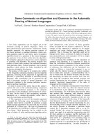
Immediate, nongenomic effects
Effect of ouabain on Ca
2
+
concentrations in endothelial
cells. As low concentrations of ouabain have been shown
to cause low frequency Ca
2+
oscillations in rat tubule
cells [16], we were interested in investigating the effects
of ouabain on cytosolic [Ca
2+
] of HUAECs. As shown in
Fig. 1B, ouabain at the low concentration of 1 n
M
induced
clear Ca
2+
oscillations in these cells with a period of about
4–8 min (Fig. 1B), sometimes even greater (Fig. 1C). First
oscillations were observed after 4 min (Fig. 1C). The cells
showing oscillations did not appear to be synchronized.
Nevertheless, not all cells investigated displayed such
oscillations. In the best case, four of the six cells observed
displayed Ca
2+
oscillations. The average over three inde-
pendent experiments was, however, about 38%. In the
absence of ouabain, however, control cells never showed any
calcium oscillations (Fig. 1A), indicating that the observed
slow Ca
2+
oscillations were specifically induced by ouabain.
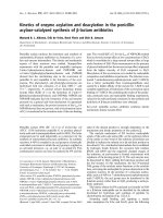
Effects of ouabain on ET-1 release. ET-1 is synthesized as
a prepro-hormone and is stored in vesicles. As vesicular
exocytosis is induced by a rise in intracellular [Ca
2+
], we
were interested in determining whether ouabain added to
endothelial cells in culture might induce such a response.
Various concentrations of ouabain were added to the cell
culture wells, and, after 10 min of incubation, the super-
natant was collected and analyzed for its ET-1 content using
the dot-blot method. The data in Fig. 2A show a stimula-
tion of ET-1 release even at 1 n
M
ouabain. The rise in ET-1
release follows a hyperbolic dose/response relation and
reaches a more than twofold stimulation at 50 n
M
ouabain
(Fig. 2A).
1056 R. Saunders and G. Scheiner-Bobis (Eur. J. Biochem. 271) Ó FEBS 2004
When extracellular Ca
2+
was omitted by replacing the
HBSS/20 m
M
Hepes with NaCl/P
i
, ET-1 in the supernatant
was considerably reduced (Fig. 2B). The same was observed
when the Ca
2+
channel blockers NiCl
2
(1 m
M
)andCdCl
2
(1 m
M
)andtheNa
+
/Ca
2+
-exchanger-specific inhibitor
2¢,4¢-dichlorobenzamil (0.1 m
M
) were included in HBSS/
20 m
M
Hepes and allowed to act on the HUAECs for
20 min before the addition of ouabain (Fig. 2B).
Under all these conditions, ET-1 in the supernatant was
about half of the amount detected in the controls and only
25% of the ET-1 secreted after ouabain stimulation.
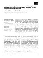
Effects of ouabain on
86
Rb
+
uptake. To investigate
whether the observations made thus far are based on a
global inhibition of the sodium pump, the uptake of
86
Rb
+
into endothelial cells was determined as a function of the
ouabain concentration. Rubidium is recognized by the
sodium pump and its uptake can easily be inhibited by
ouabain and several of its congeners. This experiment,
however, produced a most unexpected result. Ouabain at
low concentrations not only failed to inhibit
86
Rb
+
uptake
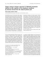
Fig. 1. Ouabain-induced Ca
2+
oscillations in HUAECs. (A) Without
ouabain, (B) with 1 n
M
ouabain (arrowhead), or (C) with 10 n
M
ouabain. Cells were cultured on coverslips and loaded with the Ca
2+
-
sensitive dye fura-2. Traces shown are for individual cells that were
representative of most of the cells observed in a field of view. Ouabain
or control treatment started at the arrowhead. See Experimental
procedures for details. (D) False-color image of endothelial cells loa-
ded with fura-2. The picture was taken at an excitation of 340 nm. The
scale on the right shows a relative loading condition with Ca
2+
.
Fig. 2. Effects of ouabain and extracellular Ca
2+
on ET-1 release from
HUAECs. (A) Accumulation of ET-1 in the medium of HUAECs was
measured by the dot-blot immunological method. Medium was col-
lected after 10 min of incubation with the various concentrations of
ouabain shown (bars represent ± SEM; n ¼ 6). (B) In the absence of
extracellular Ca
2+
, ET-1 in the supernatant is reduced by 50% when
compared with the control and is only 25% of the amount secreted
after stimulation by 10 n
M
ouabain. A mixture of the Ca
2+
channel
blockers NiCl
2
(1 m
M
), CdCl
2
(1 m
M
)andtheNa
+
/Ca
2+
-exchanger
inhibitor 2¢,4¢-dichlorobenzamil (0.1 m
M
) added before ouabain have a
similar effect.
Ó FEBS 2004 Ouabain stimulation of ET-1 release and expression (Eur. J. Biochem. 271) 1057
by the endothelial cells, but with all HUAEC preparations
from various umbilical cords a stimulation of
86
Rb
+
uptake
was observed at low ouabain concentrations. Although in
most cases the stimulation observed was 15–20% above the
control without ouabain, in a series of measurements with
HUAECs prepared from umbilical cord number 4, stimu-
lation of
86
Rb
+
uptake reached 50 ± 22% at 0.1 n
M
ouabain and 49 ± 2% at 1 n
M
ouabain over the control
without ouabain (Fig. 3A).
To investigate the mechanism for the observed stimula-
tion of
86
Rb
+
uptake by ouabain, the same experiment was
carried out after preincubation of the cells for 15 min with
various substances known to be specific inhibitors of cellular
components. Thus, tetrodotoxin, which specifically inhibits
Na
+
channels, was used to determine whether the observed
stimulation was due to a secondary stimulation of the
sodium pump by Na
+
cations that enter the cell via these
channels. The protein kinase C inhibitor Ro-31-8425 [17]
was used to see whether the observed stimulation of the
86
Rb
+
uptake was the result of Na
+
/K
+
-ATPase phos-
phorylation by this kinase, which in the past has been
repoted to activate or inactivate the sodium pump. The
5-hydroxydecanoate inhibitor of the mitochondrial ATP-
sensitive K
+
[mitoK(ATP)] channels [18] was used to
investigate whether the increased accumulation of
86
Rb
+
was the result of increased transportation of the cation into
the mitochondria, and finally, bumetanide, the specific
inhibitor of Na
+
/K
+
/2Cl
–
cotransporters [19], was used to
investigate whether the observed stimulation of Rb
+
uptake
was due to the stimulation of this uptake system. As shown
in Fig. 3B, however, none of these substances had any effect
on the stimulation of
86
Rb
+
uptake by ouabain when used
at concentrations reported to affect the various channels
and enzymes described above.
Finally, ouabain-binding experiments with whole cells
were carried out to investigate whether the observed stimu-
lation of
86
Rb
+
uptake is due to a translocation of sodium
pumps from cytosolic compartments to the surface of the
plasma membrane. The experiments, however, did not
indicate any differences in [
3
H]ouabain binding to the
membrane surface after the cells were preincubated with
either 1 or 5 n
M
[
3
H]ouabain. Whereas cell membranes that
were not preincubated with ouabain bound 685 ± 58
fmolÆmg
)1
protein, ouabain binding after preincubation with
either 1 or 5 n
M
ouabain was 638 ± 68 or 702 ± 54
fmolÆmg
)1
, respectively (all values are mean ± SEM; n ¼ 6).
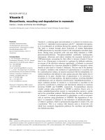
MAP kinase activation by ouabain
In rat cardiomyocytes, ouabain has been shown to
stimulate the MAP kinase reaction cascade [20,21]. To
investigate a similar mechanism in endothelial cells, MAP
kinase stimulation on ouabain exposure was investigated
by Western blotting using an antibody against the phos-
phorylated form of p44/p42 MAP kinase. Using this
method, MAP kinase activation was clearly detectable in
HUAECs after 30 min of stimulation (Fig. 4A). The
increase in the phosphorylated MAP kinase forms is not
due to an increase in overall MAP kinase in the various
preparations, as shown by an antibody to MAP kinase that
does not distinguish between phosphorylated and unphos-
phorylated forms of the enzyme (Fig. 4B). Notably, MAP
kinase stimulation was detectable at the low ouabain
concentration of 1 n
M
. Measured over the ouabain
concentration range 1 n
M
to 1 l
M
, stimulation was 2 to
2.5-fold over control (Fig. 4C).
Delayed effects
Cell proliferation in response to ouabain. Ouabain has
been described as a mitogen that induces cell proliferation in
the upper micromolar and low millimolar range. After 12 h
of incubation, the cell number of HUAECs increased
linearly with increasing ouabain concentrations (from 0.1 to
5n
M
), peaking at 5 n
M
(Fig. 5). At this concentration, the
relative increase was 49 ± 4% (P<0.05) above the
proliferation observed in the absence of the glycoside. Cell
Fig. 3. Stimulation of Rb
+
uptake by ouabain and effects of various
inhibitors. The accumulation of
86
Rb
+
during 1 h of treatment of
HUAECs was measured as described under Experimental procedures.
(A) Cells were treated during the
86
Rb
+
incubation with the indicated
concentrations of ouabain or control buffer. (B) Preincubation of
HUAECs with 5 l
M
tetrodotoxin (TTX), 1 l
M
Ro-31-8425, 500 l
M
5-hydroxydecanoate (5-HDA)
6
,or50l
M
bumetanide was carried out
for 15 min before addition of control buffer or 1 or 10 n
M
ouabain.
The two experiments (A and B) were carried out with primary cell
cultures prepared from two different umbilical cords. While the
stimulation of up to 50% above background was only observed in the
series of experiments shown here (A), most commonly the stimulation
of
86
Rb
+
uptake ranged between 15% and 20% above control, as
shown in B (bars represent ± SEM, n ¼ 3–6; *P <0.05).
1058 R. Saunders and G. Scheiner-Bobis (Eur. J. Biochem. 271) Ó FEBS 2004
numbers declined in response to higher ouabain concentra-
tions, such that 20 n
M
ouabain inhibited proliferation
relative to that of untreated cells (14%).
Effects of ouabain on ET-1 mRNA. The human ET-1 gene
promoter region contains two active transcription regula-
tory sites: an AP-1-binding site (TGACTAA) at )108 to
)102 bp from transcription initiation and a GATA-2-
binding site (TTATCT) at )136 to )131 bp [22]. As signal-
transduction pathways that activate MAP kinase also
activate various genes through these promoters, it was
important to address the question of whether ouabain could
cause a long-term upregulation in ET-1 mRNA biosyn-
thesis. As shown in Fig. 6, ouabain at 10 n
M
stimulated
ET-1 mRNA concentrations in HUAECs by 19.5 ± 3.1%
(mean ± SEM; n ¼ 8) after 12 h, as determined by
semiquantitative RT-PCR.
Discussion
Cardiac steroids have a positive inotropic effect on the heart
muscle. The mechanism is thought to involve inhibition of
the sodium pump, which results in a reduction in the sodium
gradient. This in turn has an impact on the transport activity
of the Na
+
/Ca
2+
exchanger, which does not transport
Ca
2+
ions out of the cytosol as effectively. Thus, the
resulting increased cellular concentration of Ca
2+
is thought
Fig. 5. Effect of ouabain on HUAEC cell number. Cells growing on
12-well plates were treated with the indicated concentrations of oua-
bainfor12h,afterwhichcellnumberwasassessedasdescribedunder
Experimental procedures (bars represent ± SEM; n ¼ 6).
Fig. 6. Effect of 12 h exposure to various ouabain concentrations on
ET-1 mRNA in HUAECs. HUAECs were incubated for 12 h with the
various concentrations of ouabain shown. Then, mRNA isolated from
HUAECs was transcribed into cDNA by a reverse transcriptase step,
and, by using ET-1-specific primers, the abundance of the latter was
analyzed by semiquantitative PCR (bars represent ± SEM; n ¼ 8;
*P<0.05).
Fig. 4. MAP kinase activation of endothelial cells by ouabain. (A) Cells
were incubated with the ouabain concentrations shown for 30 min as
described under Experimental procedures. Thereafter, 20 lg protein
was separated by SDS/PAGE and probed by an antibody to phospho-
p44/42. The resulting signal shown was obtained by the luminescent
ECL system. A positive control, commercially available phospho-p44/
42, was run in parallel (lane 5). Relative amounts of phosphoproteins
were analyzed with a digital documentation system and a gel image
analysis software. (B) The conditions were the same as in (A) except an
antibody to the total (nonphosphorylated) MAP kinase was used.
(C) Relative amounts of MAP kinase activated by 1 n
M
ouabain. Data
are derived from experiments similar to those described in (A) (bars
represent ± SEM; n ¼ 5).
Ó FEBS 2004 Ouabain stimulation of ET-1 release and expression (Eur. J. Biochem. 271) 1059
to stimulate the contractile elements of the heart or vascular
muscle and increase contractility.
Although thus far plausible, the model implies that the
increase in contractility by cardiac steroids is associated with
inhibition of the pump and that the rise in cytosolic Na
+
concentrations occurs before the rise in cytosolic Ca
2+
.
Several investigations, however, do not appear to support
this mechanism. Not only is inhibition of the pump not a
prerequisite for the positive inotropic effect [23], but
ouabain was shown to stimulate Ca
2+
transients in arterial
smooth muscle without raising cytosolic [Na
+
][24].
Our observations shown in Fig. 3 are consistent with
these results.
86
Rb
+
uptake by HUAECs was not inhibited
by ouabain, but at 1 n
M
or 10 n
M
concentrations of the
steroid, the uptake was stimulated by at least 15% (Fig. 3B)
and in some cases up to 50% over control uptake (Fig. 3A).
Although the molecular basis for this observation is not yet
clear, effects of ouabain at low concentrations that do not
correlate with sodium pump inhibition have been described
in several investigations, including induction of positive
inotropic effects [23] or cytosolic [Ca
2+
] elevation [24]. The
stimulation of Rb
+
uptake that we observed, however, was
not simply the result of cell swelling (data not shown) or
sodium pump recruitment to the plasma membrane
from cytosolic stores. It was also not affected by the
Na
+
/K
+
/2Cl
–
cotransporter inhibitor bumetanide or by the
mitoK(ATP) channel-specific blocker 5-hydroxydecanoate.
As these two major K
+
uptake systems are not involved in
the observed stimulation of Rb
+
uptake, the sodium pump
is the most likely route for
86
Rb
+
uptake. Stimulation of the
sodium pump by protein kinase C [25,26], however, can be
excluded, as no effect was observed in the presence of the
protein kinase C-specific inhibitor Ro-31-8425. As a direct
activation of the sodium pump at very low concentrations
of ouabain has been shown in a recent investigation [27], this
possibility is currently the most likely explanation for the
ability of ouabain to stimulate
86
Rb
+
uptake.
Several reports [20,21,28,29] show that ouabain induces
signaling cascades, resulting in both nongenomic and
genomic effects. The most apparent of the nongenomic
effects described thus far are the rise in cytosolic Ca
2+
concentration associated with slow Ca
2+
oscillations,
activation of NF-jB, activation of ERK1/2 (MAP kinase
p42/p44) by interactions of the sodium pump with the
epidermal growth factor (EGF) receptor, or the release of
reactive oxygen species from mitochondria [28].
Consistent with these results, we determined that ouabain
at low concentrations stimulates MAP kinase p42/p44
phosphorylation (activation) by 2 to 2.5-fold. Within the
same concentration and time range, it also induces Ca
2+
oscillations of an approximate frequency of 8 min and a
duration lasting for more than 40 min (until the experiment
was terminated; Fig. 1). This phenomenon, which was first
described for rat renal proximal tubule cells [16], is clearly
confirmed here for human endothelial cells. Whether such
Ca
2+
oscillations are also responsible for the induction of
the positive inotropic effect on the heart has still to be
investigated. In smooth muscle cells of rat mesenteric
arteries, Ca
2+
transients were induced at low (3–100 n
M
)
concentrations of ouabain [24]. Here, however, it was
thought that these effects are mediated by the a3 isoform of
the sodium pump a subunit, which binds ouabain with high
affinity. Nevertheless, a3 and a2 mRNA were not detectable
in HUAECs by RT-PCR (not shown), leading to the con-
clusion that in the experiments described here the abundant
a1 isoform is the most likely mediator of the effects of
ouabain at low concentrations.
HUAECs, like all endothelial cells, regulate the muscular
tone of the underlying arteries by releasing either the
vasoconstrictive ET-1 or the vasorelaxant NO. Taking into
consideration the fact that ouabain in animal models
applied over a long period of time causes hypertension
[30], we investigated here its effects on ET-1 release and
expression in HUAECs.
ET-1, the only member of the endothelin peptide family
produced by endothelial cells, is stored in vesicles before it is
released towards the underlying smooth muscle cells of the
artery. As this substance is a potent vasoconstrictor and
growth promoter of vascular smooth muscle cells via the
ET
A
receptor, and because vesicular fusion and release has
been shown in numerous cases to be triggered by Ca
2+
,we
were interested in investigating whether ET-1 release from
HUAECs to the medium might be stimulated by ouabain.
Indeed, a few minutes after application, ouabain causes
ET-1 release into the medium (Fig. 2A). This release was
depending on extracellular Ca
2+
, as its absence, or the
presence of Ca
2+
channel inhibitors such as Ni
2+
or Cd
2+
and the Na
+
/Ca
2+
-exchanger blocker 2¢,4¢-dichlorobenz-
amil considerably reduced ET-1 release (Fig. 2B). This
finding is in good agreement with earlier reports showing
that ET-1 release depends on extracellular Ca
2+
[31,32].
We do not know yet, however, whether or not ouabain
stimulates ET-1 secretion also in vivo. It is possible that
ouabain – should it indeed be an endogenously produced
hormone, as proposed by many – influences vascular tone
by such a mechanism and regulates blood pressure. Elevated
ET-1 production has been shown in human vessels subjec-
ted to increased pressure and shear stress [33], and increased
levels of circulating ET-1 are associated with pulmonary
and essential hypertension [34,35].
Besides these immediate, nongenomic effects, ouabain
has been shown previously to increase mitotic activity
[36–38], a result complemented by the newer findings of
ouabain-induced MAP kinase activation and induction of
Ca
2+
oscillations followed by the translocation of the
transcriptional factor NF-jB into the nucleus [16]. In
agreement with these results, we also showed that ouabain
at low concentrations stimulates the growth of HUAECs
by 49 ± 4% as measured by cell number (Fig. 5). This
stimulation was observed after 12 h of incubation. At the
same time, mRNA coding for ET-1 is also increased,
possibly because of ouabain stimulation of MAP kinase,
which is known to stimulate the Fos and Jun transcription
factors to form activator protein-1 (AP-1) heterodimers.
As the human ET-1 gene promoter region contains an
AP-1-binding site regulating transcription [22], this may be
responsible for the apparent increased ET-1 gene transcrip-
tion we observed. In addition, the bovine ET-1 gene
promoter region is also known to contain an NF-jB-
responsive region [39]. If this site were also to be present in
the human ET-1 promoter region, the Ca
2+
oscillations we
show to be induced by ouabain may also contribute to the
observed ET-1 upregulation. Although the increase in
ET-1 mRNA concentrations appears to be rather modest
1060 R. Saunders and G. Scheiner-Bobis (Eur. J. Biochem. 271) Ó FEBS 2004
(19.5 ± 3.1%), ET-1 in the serum of hypertensive patients
is no more than 13% above normal [40]. Thus, an increase
in ET-1 by this margin could be one of the reasons for the
observed induction of hypertension by ouabain in animal
experiments [30].
In conclusion, the results presented here clearly show that
ouabain can act in a hormone-like manner on endothelial
cells. It becomes effective at concentrations that are within
the same range as the effective concentrations of other
hormones, including the steroid hormones, and, like the
latter, it also induces both genomic and nongenomic effects
that are independent of its ability to inhibit the sodium
pump at higher concentrations. Assuming that ouabain
might be endogenously produced, as has been shown in
some instances [9,41], it could act as a hormone involved in
blood pressure regulation by fine-tuning and controlling
ET-1 release and expression.
Acknowledgements
R. S. was supported through the Giessen Graduiertenkolleg Molekulare
Veterina
¨
rmedizin; G. S. B. is supported through a grant from the
Deutsche Forschungsgemeinschaft (DFG), SCHE 307/5-1.
References
1. Hansen, O. (1984) Interaction of cardiac glycosides with
(Na
+
,K
+
)-activated ATPase. A biochemical link to digitalis-
induced inotropy. Pharmacol. Rev. 36, 143–163.
2. Schwinger, R.H., Bundgaard, H., Muller-Ehmsen, J. & Kjeldsen,
K. (2003) The Na,K-ATPase in the failing human heart. Cardio-
vasc. Res. 57, 913–920.
3. Mathews, W.R., DuCharme, D.W., Hamlyn, J.M., Harris, D.W.,
Mandel, F., Clark, M.A. & Ludens, J.H. (1991) Mass spectral
characterization of an endogenous digitalislike factor from human
plasma. Hypertension 17, 930–935.
4. Schneider, R., Wray, V., Nimtz, M., Lehmann, W.D., Kirch, U.,
Antolovic, R. & Schoner, W. (1998) Bovine adrenals contain, in
addition to ouabain, a second inhibitor of the sodium pump.
J. Biol. Chem. 273, 784–792.
5. Hamlyn, J.M., Blaustein, M.P., Bova, S., DuCharme, D.W.,
Harris, D.W., Mandel, F., Mathews, W.R. & Ludens, J.H. (1991)
Identification and characterization of a ouabain-like compound
from human plasma. Proc. Natl Acad. Sci. USA 88, 6259–6263.
6. Kawamura, A., Guo, J., Itagaki, Y., Bell, C., Wang, Y., Haupert,
G.T. Jr, Magil, S., Gallagher, R.T., Berova, N. & Nakanishi, K.
(1999) On the structure of endogenous ouabain. Proc.NatlAcad.
Sci. USA 96, 6654–6659.
7. Goto, A. & Yamada, K. (1998) Purification of endogenous digi-
talis-like factors from normal human urine. Clin.Exp.Hypertens.
20, 551–556.
8. Goto, A., Ishiguro, T., Yamada, K., Ishii, M., Yoshioka, M.,
Eguchi, C., Shimora, M. & Sugimoto, T. (1990) Isolation of
a urinary digitalis-like factor indistinguishable from digoxin.
Biochem. Biophys. Res. Commun. 173, 1093–1101.
9. Schneider, R., Antolovic, R., Kost, H., Sich, B., Kirch, U., Tepel,
M., Zidek, W. & Schoner, W. (1998) Proscillaridin A immuno-
reactivity: its purification, transport in blood by a specific binding
protein and its correlation with blood pressure. Clin. Exp.
Hypertens. 20, 593–599.
10. Lichtstein, D., Gati, I., Samuelov, S., Berson, D., Rozenman, Y.,
Landau, L. & Deutsch, J. (1993) Identification of digitalis-like
compounds in human cataractous lenses. Eur. J. Biochem. 216,
261–268.
11. Bagrov, A.Y., Fedorova, O.V., Dmitrieva, R.I., Howald, W.N.,
Hunter, A.P., Kuznetsova, E.A. & Shpen, V.M. (1998) Char-
acterization of a urinary bufodienolide Na
+
,K
+
-ATPase inhi-
bitor in patients after acute myocardial infarction. Hypertension
31, 1097–1103.
12. Schoner, W., Bauer, N., Mu
¨
ller-Ehmsen, J., Kra
¨
mer, U.,
Hambarchian, N., Schwinger, R., Mo
¨
ller, H., Kost, H., Weitk-
amp, C., Schweitzer, T., Kirch, U., Neu, H. & Gru
¨
nbaum, E H.
(2003) Ouabain as a mammalian hormone. Ann. NY Acad. Sci.
986, 1–7.
13. Lowry, O.H., Rosenbrough, N.J., Farr, A.L. & Randall, R.J.
(1951) Protein measurement with the folin phenol reagent. J. Biol.
Chem. 193, 265–275.
14. Laemmli, U.K. (1970) Cleavage of structural proteins during the
assembly of the head of bacteriophage T4. Nature 227, 680–685.
15. Jager, H., Wozniak, G., Akinturk, I.H., Hehrlein, F.W. & Schei-
ner-Bobis, G. (2001) Expression of sodium pump isoforms and
other sodium or calcium ion transporters in the heart of hyper-
tensive patients. Biochim. Biophys. Acta 1513, 149–159.
16. Aizman, O., Uhlen, P., Lal, M., Brismar, H. & Aperia, A. (2001)
Ouabain, a steroid hormone that signals with slow calcium oscil-
lations. Proc. Natl Acad. Sci. USA 98, 13420–13424.
17. Nixon, J.S., Bishop, J., Bradshaw, D., Davis, P.D., Hill, C.H.,
Elliott, L.H., Kumar, H., Lawton, G., Lewis, E.J., Mulqueen, M.,
5
Westmacott, D., Wadsworth, J. & Wilkinson, S.E. (1991) Novel,
potent and selective inhibitors of protein kinase C show oral anti-
inflammatory activity. Drugs Exp. Clin. Res. 17, 389–393.
18. Jaburek, M., Yarov-Yarovoy, V., Paucek, P. & Garlid, K.D.
(1998) State-dependent inhibition of the mitochondrial KATP
channel by glyburide and 5-hydroxydecanoate. J. Biol. Chem. 273,
13578–13582.
19. Haas, M. & Forbush, B. 3rd (1998) The Na-K-Cl cotransporters.
J. Bioenerg. Biomembr. 30, 161–172.
20. Kometiani, P., Li, J., Gnudi, L., Kahn, B.B., Askari, A. & Xie, Z.
(1998) Multiple signal transduction pathways link Na
+
/K
+
-
ATPase to growth-related genes in cardiac myocytes. The roles of
Ras and mitogen-activated protein kinases. J. Biol. Chem. 273,
15249–15256.
21. Scheiner-Bobis, G. & Schoner, W. (2001) A fresh facet for ouabain
action. Nat. Med. 7, 1288–1289.
22. Kawana,M.,Lee,M.E.,Quertermous,E.E.&Quertermous,T.
(1995) Cooperative interaction of GATA-2 and AP1 regulates
transcription of the endothelin-1 gene. Mol. Cell Biol. 15, 4225–
4231.
23. Godfraind, T. & Ghysel-Burton, J. (1980) Independence of the
positive inotropic effect of ouabain from the inhibition of the heart
Na
+
/K
+
pump. Proc.NatlAcad.Sci.USA77, 3067–3069.
24. Arnon, A., Hamlyn, J.M. & Blaustein, M.P. (2000) Ouabain
augments Ca
2+
transients in arterial smooth muscle without
raising cytosolic Na
+
. Am. J. Physiol. Heart Circ. Physiol. 279,
H679–H691.
25. Pedemonte, C.H., Pressley, T.A., Lokhandwala, M.F. & Cinelli,
A.R. (1997) Regulation of Na,K-ATPase transport activity by
protein kinase C. J. Membr. Biol. 155, 219–227.
26. Vasilets, L.A., Fotis, H. & Gartner, E.M. (1997) Regulatory
phosphorylation of the Na
+
/K
+
-ATPase from mammalian kid-
neys and Xenopus oocytes by protein kinases. Characterization
of the phosphorylation site for PKC. Ann. NY Acad. Sci. 834,
585–587.
27. Gao, J., Wymore, R.S., Wang, Y., Gaudette, G.R., Krukenkamp,
I.B., Cohen, I.S. & Mathias, R.T. (2002) Isoform-specific stimu-
lation of cardiac Na/K pumps by nanomolar concentrations of
glycosides. J. Gen. Physiol. 119, 297–312.
28. Xie, Z. & Askari, A. (2002) Na
+
/K
+
-ATPase as a signal trans-
ducer. Eur. J. Biochem. 269, 2434–2439.
Ó FEBS 2004 Ouabain stimulation of ET-1 release and expression (Eur. J. Biochem. 271) 1061
29. Nakagawa, Y., Rivera, V. & Larner, A.C. (1992) A role for the
Na/K-ATPase in the control of human c-fos and c-jun tran-
scription. J. Biol. Chem. 267, 8785–8788.
30. Manunta, P., Rogowski, A.C., Hamilton, B.P. & Hamlyn, J.M.
(1994) Ouabain-induced hypertension in the rat: relationships
among plasma and tissue ouabain and blood pressure.
J. Hypertens. 12, 549–560.
31. Brunner, F., Stessel, H. & Graier, W.F. (1995) The effects of
intracellular Ca
2+
concentration and hypoxia on basal endothe-
lin-1 secretion by cultured porcine aortic endothelial cells. Agents
Actions Supplement 45, 269–273.
32. Carlini, R.G., Gupta, A., Liapis, H. & Rothstein, M. (1995)
Endothelin-1 release by erythropoietin involves calcium signaling
in endothelial cells. J. Cardiovasc Pharmacol. 26, 889–892.
33. Gan, L.M., Selin-Sjogren, L., Doroudi, R. & Jern, S. (2000)
Temporal regulation of endothelial ET-1 and eNOS expression in
intact human conduit vessels exposed to different intraluminal
pressure levels at physiological shear stress. Cardiovasc. Res. 48,
168–177.
34. Li-Saw-Hee, F.L., Blann, A.D. & Lip, G.Y. (1997) The detection
of endothelial dysfunction in patients with essential hypertension.
Int. J. Cardiol. 61, 171–174.
35. Veyssier-Belot, C. & Cacoub, P. (1999) Role of endothelial
and smooth muscle cells in the physiopathology and treatment
management of pulmonary hypertension. Cardiovasc. Res. 44,
274–282.
36. Murata, Y., Matsuda, T., Tamada, K., Hosoi, R., Asano, S.,
Takuma, K., Tanaka, K. & Baba, A. (1996) Ouabain-induced cell
proliferationinculturedratastrocytes.Jpn. J. Pharmacol. 72, 347–
353.
37. Aydemir-Koksoy, A. & Allen, J.C. (2001) Low concentrations of
ouabain induce vascular smooth muscle cell proliferation. Cell
Mol. Biol. 47, 341–345.
38. Chueh, S.C., Guh, J.H., Chen, J., Lai, M.K. & Teng, C.M. (2001)
Dual effects of ouabain on the regulation of proliferation and
apoptosis in human prostatic smooth muscle cells. J. Urol. 166,
347–353.
39. Quehenberger, P., Bierhaus, A., Fasching, P., Muellner, C.,
Klevesath,M.,Hong,M.,Stier,G.,Sattler,M.,Schleicher,E.,
Speiser, W. & Nawroth, P.P. (2000) Endothelin 1 transcription is
controlled by nuclear factor-kappaB in AGE-stimulated cultured
endothelial cells. Diabetes 49, 1561–1570.
40. Ooi, B.S., Papademetrious, V. & Cohen, D.J. (1996) Demonstra-
tion of endothelial-activating properties of hypertensive sera. Am.
J. Hypertens. 9, 1232–1235.
41. Hamlyn, J.M., Lu, Z.R., Manunta, P., Ludens, J.H., Kimura, K.,
Shah, J.R., Laredo, J., Hamilton, J.P., Hamilton, M.J. &
Hamilton, B.P. (1998) Observations on the nature, biosynthesis,
secretion and significance of endogenous ouabain. Clin. Exp.
Hypertens. 20, 523–533.
1062 R. Saunders and G. Scheiner-Bobis (Eur. J. Biochem. 271) Ó FEBS 2004